Gastroenterology · Gastroenterology
Gastrointestinal Bleeding
Also known as GI bleed · Upper GI bleed · Lower GI bleed · Acute upper gastrointestinal haemorrhage · Melaena · Haematemesis · Variceal haemorrhage
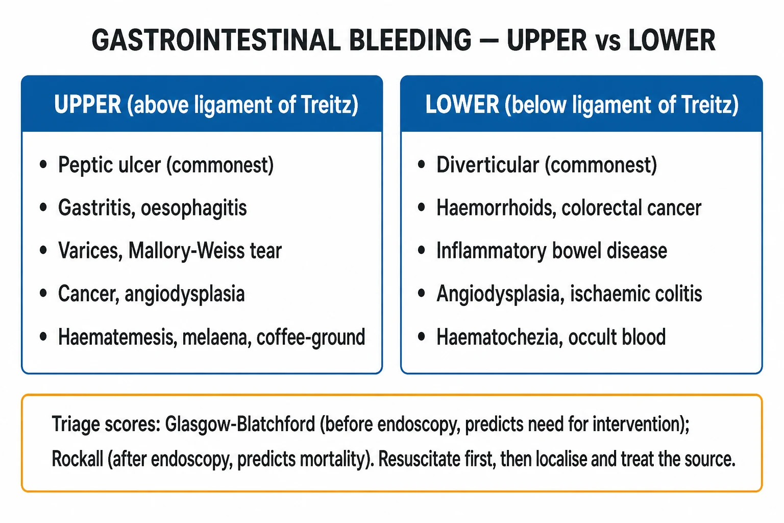
Gastrointestinal (GI) bleeding is any haemorrhage from the lumen of the gastrointestinal tract, divided anatomically at the ligament of Treitz into upper GI bleeding (UGIB) — haematemesis (fresh red blood), coffee-ground vomiting, and melaena (black tarry stool) — and lower GI bleeding (LGIB) — haematochezia (fresh red or maroon blood per rectum). The commonest UGIB cause is peptic ulcer disease (about a third to half of all cases), followed by erosive gastritis, oesophago-gastric varices, Mallory-Weiss tear, oesophagitis and malignancy; the commonest LGIB cause in older adults is diverticular bleeding, with colonic angiodysplasia, ischaemic colitis, colitis (inflammatory/infective), neoplasia and haemorrhoids following. Management is ABCDE resuscitation first — two large-bore cannulae, crossmatch, and a restrictive transfusion strategy targeting Hb at least 7 g/dL (Villanueva 2013). Risk-stratify UGIB with the Glasgow-Blatchford Score (pre-endoscopy) and Rockall (with endoscopy); perform oesophago-gastro-duodenoscopy (OGD) within 24 hours (within 12 hours for suspected variceal bleed). The high-risk bleeding ulcer receives adrenaline injection plus a mechanical/thermal method plus a high-dose intravenous proton-pump inhibitor infusion (omeprazole 80 mg IV bolus then 8 mg/hour for 72 hours). The variceal bundle is terlipressin 2 mg IV every 4 hours + broad-spectrum antibiotic (ceftriaxone 1 g IV daily) + endoscopic band ligation within 12 hours, with transjugular intrahepatic portosystemic shunt (TIPS) for failure to control bleeding. Lower GI bleeding is worked up with colonoscopy if stable and CT mesenteric angiography with superselective embolisation if massive or ongoing.
On this page & tools
Your progress
Saved locally on this device.
Practise this topic
Exam tags
Red flags

Overview & Definition
Gastrointestinal (GI) bleeding is defined as any loss of blood from the lumen of the gastrointestinal tract, clinically apparent either as overt bleeding (visible blood — haematemesis, coffee-ground vomit, melaena, haematochezia, red blood per rectum) or as occult bleeding (not visible to the patient — detected by a positive faecal occult blood test or by iron-deficiency anaemia).[1]
The single most important anatomical landmark in GI bleeding is the ligament of Treitz — the suspensory muscle of the duodenum that marks the duodenojejunal flexure, the junction of the fourth part of the duodenum with the jejunum. By convention: [1]
- Upper GI bleeding (UGIB) — bleeding above the ligament of Treitz (oesophagus, stomach, duodenum).
- Lower GI bleeding (LGIB) — bleeding below the ligament of Treitz (jejunum, ileum, colon, rectum, anus). [1]
A clinically useful third category is obscure GI bleeding — persistent or recurrent bleeding for which no source is identified on standard OGD and colonoscopy. It is subdivided into obscure-overt (visible bleeding) and obscure-occult (positive faecal occult blood or iron-deficiency anaemia), and the responsible lesion usually lies in the small bowel (evaluated by capsule endoscopy and device-assisted enteroscopy).[1]
The practical skill in GI bleeding is not recognising that blood is present (the patient tells you). It is, in order: (1) recognise and reverse shock; (2) localise the bleed to upper versus lower versus obscure; (3) risk-stratify to decide who needs inpatient care and how urgently; (4) deliver the source-specific haemostatic bundle; (5) prevent and treat rebleeding and complications. The single largest process lever in UGIB is early, structured endoscopy with appropriate vasoactive, antibiotic and PPI cover.[1]
Classification
GI bleeding is classified along four axes — anatomical (upper/lower/obscure), clinical (overt/occult), haemodynamic (stable/unstable), and aetiological. [1]
Anatomical classification — upper versus lower versus obscure
Upper GI bleed
- Above the **ligament of Treitz** (oesophagus, stomach, duodenum)
- Classical presentations: **haematemesis** (fresh red blood), **coffee-ground vomit**, **melaena** (black, tarry, foul-smelling stool)
- Commoner, higher mortality (overall 5 to 10%), more often life-threatening; commonest cause **peptic ulcer** (a third to a half of cases)
- Diagnosed by **OGD within 24 hours** (within 12 hours if variceal)
- Raised **urea-to-creatinine ratio** with normal creatinine (digested blood protein converted to urea) is a useful bedside pointer to UGIB
Lower GI bleed
- Below the **ligament of Treitz** (jejunum, ileum, colon, rectum, anus)
- Classical presentation: **haematochezia** (fresh red or maroon blood per rectum), sometimes with clots
- Less common, lower mortality (2 to 4%); commonest cause in older adults **diverticular bleeding**, then angiodysplasia, ischaemic colitis, colitis, neoplasia, haemorrhoids
- Stable patient: **colonoscopy** (often after bowel preparation within 24 to 48 hours); unstable or ongoing massive bleed: **CT mesenteric angiography** with superselective embolisation
- Beware: a brisk, massive UGIB with rapid transit can also present as haematochezia — never assume haematochezia is lower until a brisk UGIB is excluded in the unstable patient
Obscure GI bleed
- Bleeding that remains unlocalised after a **good-quality OGD and colonoscopy**; the source usually lies in the **small bowel**
- Subdivided into **obscure-overt** (visible, recurrent bleeding) and **obscure-occult** (iron-deficiency anaemia or positive faecal occult blood)
- Causes: angiodysplasia, small-bowel tumours (GIST, lymphoma, adenocarcinoma), Crohn disease, NSAID enteropathy, Meckel diverticulum, Dieulafoy lesion, hereditary haemorrhagic telangiectasia
- Work-up: **capsule endoscopy** first, then **device-assisted enteroscopy** (balloon-assisted) for diagnosis and therapy, with CT enterography as adjunct
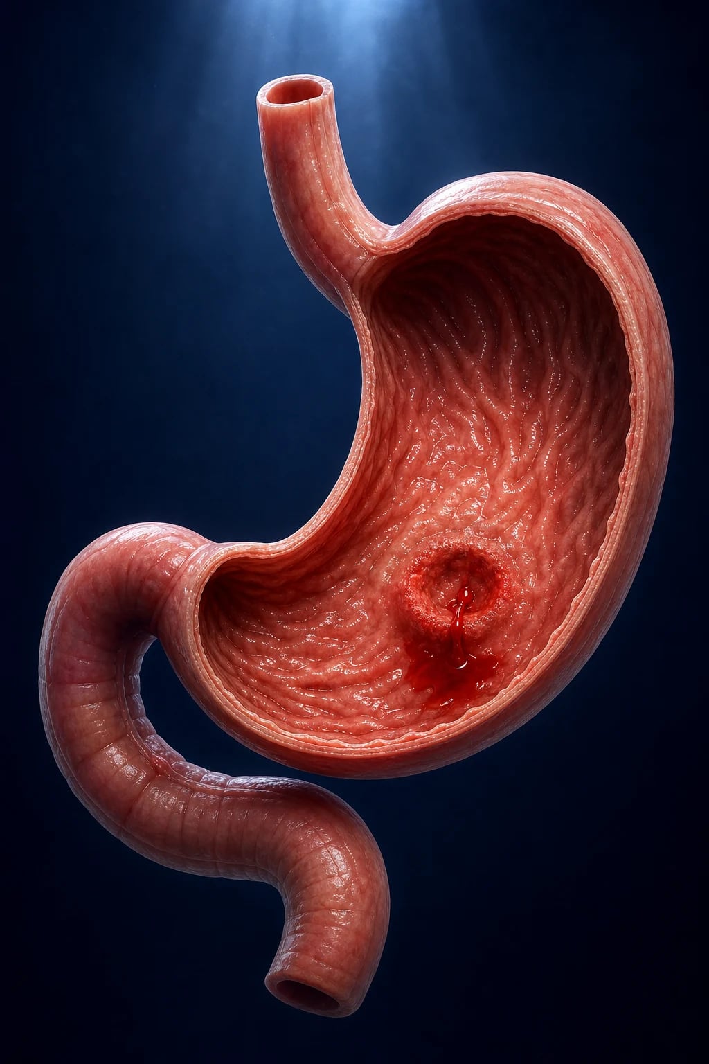
Clinical classification — overt versus occult
- Overt bleeding — visible blood loss (haematemesis, melaena, haematochezia, red blood per rectum). The scenario of acute presentation.
- Occult bleeding — non-visible blood loss detected by iron-deficiency anaemia (microcytic hypochromic, low ferritin) or a positive faecal immunochemical test (FIT) / faecal occult blood test. The scenario of outpatient work-up, where the mandate is to exclude colorectal cancer. [1]
Haemodynamic classification — stable versus unstable
This is the most important bedside axis because it determines the tempo of investigation and intervention: [1]
- Stable (no shock): normal pulse and blood pressure, no signs of hypoperfusion — proceed to urgent but scheduled endoscopy.
- Unstable (shock): tachycardia, hypotension, cold peripheries, oliguria, altered consciousness — resuscitate first, then urgent endoscopy (or angiography for LGIB) once perfusion is restored. [1]
Aetiological classification — reproduced in full in Pathophysiology and Differential Diagnosis
Causes are grouped as upper GI, lower GI, and obscure (small-bowel), and each is matched to its characteristic mechanism (erosion of a vessel, variceal rupture, vascular malformation, mucosal inflammation, neoplastic erosion, iatrogenic). [1]
Epidemiology & Risk Factors
UGIB is far commoner and deadlier than LGIB and dominates exam questions. [1]
GI bleeding — key numbers
Upper GI bleeding — relative frequency of causes (the order the examiner expects):[1]
- Peptic ulcer disease (gastric or duodenal) — about a third to a half of all UGIB. The single most testable cause.
- Erosive gastritis / gastropathy (NSAID, alcohol, stress-related mucosal disease in critical illness) — about 15 to 20%.
- Oesophago-gastric varices — about 10 to 20% overall but disproportionately deadly; the highest single-cause mortality.
- Mallory-Weiss tear — about 5 to 7%; retching-induced mucosal tear at the gastro-oesophageal junction.
- Erosive oesophagitis (reflux, infectious in the immunocompromised) — about 5%.
- Upper GI malignancy (gastric or oesophageal cancer) — about 1 to 5%; suspect with weight loss and dysphagia.
- Vascular lesions — angiodysplasia, gastric antral vascular ectasia (GAVE / watermelon stomach), Dieulafoy lesion, hereditary haemorrhagic telangiectasia (Osler-Weber-Rendu) — together under 5% but disproportionately cause massive bleeds.
- Iatrogenic / traumatic — post-mucosal biopsy, post-sphincterotomy (after ERCP), post-PEG, foreign-body ingestion.
- Aortoenteric fistula — rare (under 1%) but never miss: a prior aortic graft or aneurysm repair with any GI bleed is aortoenteric fistula until proven otherwise. [1]
Lower GI bleeding — relative frequency of causes:[2]
- Diverticular bleeding — the commonest cause of acute LGIB in older adults (about 30 to 40%); characteristically painless, often self-limiting.
- Colonic angiodysplasia — about 20%, right-sided predilection (caecum), elderly; associated with aortic stenosis (Heyde syndrome), CKD, HHT.
- Ischaemic colitis — typically a watershed-area insult after a low-flow state (hypotension, cardiac failure, surgery); left-sided predilection.
- Inflammatory colitis — ulcerative colitis, Crohn disease; chronic diarrhoea with mucus and pus.
- Infective colitis — Campylobacter, Salmonella, Shigella, EHEC (O157:H7), Clostridioides difficile, amoebic; acute diarrhoea with fever.
- Neoplasia — colorectal cancer, polyps; occult bleeding with iron-deficiency anaemia and change in bowel habit, or overt bleeding.
- Post-polypectomy bleeding — within 1 to 2 weeks of colonoscopy.
- Anorectal causes — haemorrhoids (painless, blood on the paper), anal fissure (painful defecation), radiation proctitis (after pelvic radiotherapy). [1]
Risk factors that predispose to GI bleeding (know the mechanism — examiners ask why):[1]
- Non-steroidal anti-inflammatory drugs (NSAIDs) and aspirin — inhibit cyclo-oxygenase, reducing mucosal prostaglandin and the protective mucus-bicarbonate barrier; the dominant iatrogenic cause.
- Antiplatelets (clopidogrel, prasugrel, ticagrelor) and anticoagulants (warfarin, heparin, direct oral anticoagulants — dabigatran, rivaroxaban, apixaban, edoxaban) — increase bleeding severity rather than causing it.
- Helicobacter pylori infection — chronic gastric inflammation and duodenal/gastric ulcer.
- Alcohol misuse — erosive gastritis, peptic ulcer, varices (via cirrhosis).
- Smoking — impairs ulcer healing.
- Critical illness / major trauma / severe burns (Curling ulcer) / head injury (Cushing ulcer) / mechanical ventilation over 48 hours — stress-related mucosal disease.
- Chronic liver disease and portal hypertension — variceal and portal-hypertensive gastropathy bleeding.
- Prior peptic ulcer, prior GI bleed — highest baseline risk.
- Increasing age and comorbidity — higher mortality, more aggressive resuscitation thresholds.
- Zollinger-Ellison syndrome (gastrinoma) — multiple/recurrent peptic ulcers.
- Aortic stenosis, CKD, hereditary haemorrhagic telangiectasia, von Willebrand disease — vascular-lesion bleeding. [1]
Pathophysiology
The mechanism differs by source. The student must be able to explain why each lesion bleeds — the molecular and mechanical detail is the pathophysiology the examiner wants. [1]
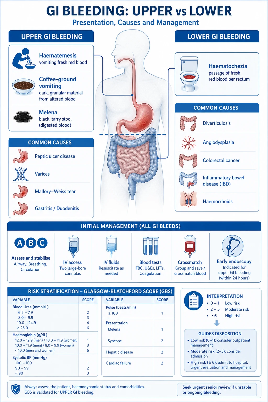
1. Peptic ulcer bleeding — the commonest and most testable mechanism
A peptic ulcer is a mucosal break penetrating the muscularis mucosae. Bleeding occurs when the ulcer erodes into a submucosal artery. Two lesions are high-yield: [1]
- Posterior duodenal ulcer erodes the gastroduodenal artery — a classic cause of massive UGIB.
- Posterior gastric wall (high lesser-curve) ulcer erodes the left gastric artery. [1]
The two dominant pathogenic insults — H. pylori and NSAIDs — converge on the same endpoint, impaired mucosal defence: [1]
- Helicobacter pylori colonises the gastric mucus layer, secretes urease (which hydrolyses urea to ammonia, raising local pH and enabling survival), and induces chronic neutrophilic gastritis with interleukin-8, TNF-alpha and reactive oxygen species. In the duodenum, gastric metaplasia of duodenal cap mucosa allows H. pylori colonisation and ulceration. The result is chronic ulceration that can erode a vessel.
- NSAIDs (and aspirin) inhibit cyclo-oxygenase — COX-1 constitutively produces gastric mucosal prostaglandins (PGE2, PGI2) that maintain the mucus-bicarbonate barrier, mucosal blood flow and epithelial restitution. Blocking COX-1 strips this defence, allowing acid back-diffusion, mucosal injury and ulceration. Selective COX-2 inhibitors spare gastric COX-1 (less ulcer risk) but carry cardiovascular risk.
- Stress-related mucosal disease in critical illness (severe burns = Curling ulcer; severe head injury = Cushing ulcer) arises from splanchnic hypoperfusion and acid back-diffusion. [1]
Why blood raises urea: digested blood protein is broken down to amino acids and absorbed, and the liver converts the amino nitrogen to urea. Hence a raised urea with a normal creatinine (a urea-to-creatinine ratio over 100:1 with normal Cr) is a bedside clue to UGIB. [1]
2. Variceal bleeding — the deadliest UGIB mechanism
Portal hypertension, defined by a hepatic venous pressure gradient (HVPG) over 5 mmHg, opens portosystemic collaterals. The collaterals at the gastro-oesophageal junction thin and dilate into varices. The risk thresholds (Baveno VII) are:[7]
- HVPG over 10 mmHg — clinically significant portal hypertension; varices form and may bleed.
- HVPG over 12 mmHg — the bleeding threshold for variceal haemorrhage.
- HVPG over 16 to 20 mmHg — high risk of failure to control bleeding and early rebleeding. [1]
Variceal bleeding is brisk and life-threatening because the thin-walled varix is under high pressure and lacks a muscular coat to constrict. The same portal hypertension produces portal-hypertensive gastropathy (a mosaic/granular mucosal pattern with chronic oozing) and gastric varices (treated differently — cyanoacrylate glue rather than band ligation).[7]
3. Mallory-Weiss tear
A longitudinal mucosal tear at the gastro-oesophageal junction (usually the posterolateral aspect), caused by a sudden rise in intra-gastric and intra-abdominal pressure during retching or vomiting — classically after alcohol binge, bulimia, hyperemesis gravidarum, or prolonged coughing. The sequence the examiner rewards is retching first, then haematemesis. The tear usually disrupts a submucosal artery; most bleeds are self-limiting but occasionally severe.[1]
4. Vascular lesions
- Angiodysplasia — ectatic, dilated, thin-walled submucosal vessels; classically the caecum and right colon in older adults (a chronic wall-tension hypothesis). Painless, recurrent, often small-volume but may be massive. Heyde syndrome is the association of aortic stenosis with gastrointestinal angiodysplasia and acquired von Willebrand disease (type 2A): high-shear flow across the stenotic valve cleaves the largest von Willebrand factor multimers (the ones needed for platelet adhesion in the high-shear microvasculature of angiodysplasia), producing both the lesions and a bleeding diathesis — valve replacement may resolve both. Other associations: CKD (uraemic platelet dysfunction), hereditary haemorrhagic telangiectasia (Osler-Weber-Rendu — autosomal dominant, ENG/ACVRL1).
- Gastric antral vascular ectasia (GAVE / watermelon stomach) — linear vascular streaks radiating from the pylorus in the gastric antrum; chronic occult iron-deficiency anaemia in older women; treated by argon plasma coagulation. Associated with cirrhosis, autoimmune disease, CKD.
- Dieulafoy lesion — an aberrant, large-calibre submucosal artery that erodes the overlying mucosa (usually the proximal stomach along the lesser curve), without an underlying ulcer. It causes painless, massive haematemesis in an otherwise fit patient; endoscopic clip or band ligation is definitive.
- Hereditary haemorrhagic telangiectasia (Osler-Weber-Rendu) — autosomal dominant; mucocutaneous telangiectases cause recurrent epistaxis and GI bleeding. [1]
5. Diverticular bleeding — the commonest LGIB mechanism
Colonic diverticula are pseudodiverticula (mucosa and submucosa herniating through the muscularis at points of vessel entry). A vasa recta artery running over the dome of the diverticulum is exposed at its neck and erodes into the lumen. Despite the left-sided predominance of diverticulosis in Western populations, right-sided diverticula bleed more often (their necks are wider and the vasa recta more exposed). Characteristically painless, haematochezia, with about 80% stopping spontaneously and about a quarter rebleeding.[2]
6. Ischaemic colitis and other lower-GI mechanisms
Ischaemic colitis follows a transient low-flow state (hypotension, cardiac failure, recent surgery, dehydration, dialysis, hypercoagulable state, vasculitis) affecting the watershed areas — the splenic flexure (Griffith point) and the rectosigmoid junction (Sudeck point) — because these segments sit at the junctions of vascular territories (superior and inferior mesenteric arteries, and inferior mesenteric and internal iliac). The injury ranges from reversible colopathy to gangrene. Infective and inflammatory colitis bleed because mucosal inflammation and ulceration erode submucosal vessels. Colorectal cancer bleeds from tumour neovascularity and surface ulceration. [1]
Why the bleeding presents the way it does (the gut handles blood differently by site)
- Haematemesis (fresh red blood) — blood in the stomach that has not been altered; indicates active, brisk UGIB.
- Coffee-ground vomit — blood altered by gastric acid to haematin (dark, granular); indicates a slower or recently stopped UGIB.
- Melaena (black, tarry, foul-smelling stool) — blood digested by gastric acid and bacterial action, producing haematin and the characteristic smell; requires as little as 50 to 100 mL of blood in the upper gut; indicates UGIB (or a right-colon/small-bowel bleed slow enough for digestion).
- Haematochezia (fresh red or maroon blood per rectum) — blood that has transited too quickly to be digested; usually LGIB, but a brisk massive UGIB with rapid transit can also present as haematochezia — never assume a bleeding source from the colour alone in an unstable patient. [1]
Clinical Presentation
Classic presentations of UGIB
- Haematemesis — vomiting fresh red blood; the cardinal sign of active UGIB.
- Coffee-ground vomit — vomiting dark, granular, altered blood.
- Melaena — black, tarry, foul-smelling stool; the other cardinal sign of UGIB.
- Symptoms of hypovolaemia — dizziness, presyncope, syncope, thirst, fatigue, dyspnoea, palpitations. [1]
Classic presentations of LGIB
- Haematochezia — fresh red or maroon blood per rectum, with or without clots.
- Abdominal cramping (especially in colitis, ischaemia, infection) or painless bleeding (diverticular, angiodysplasia, haemorrhoids).
- Symptoms of the underlying cause — diarrhoea and fever (colitis), change in bowel habit and weight loss (cancer), recent colonoscopy (post-polypectomy), pelvic radiotherapy (radiation proctitis). [1]
Bedside signs of shock — the ATLS classes of haemorrhagic shock (reproduced verbatim)
| Class | Blood loss (% of blood volume) | Approximate volume | Heart rate | Blood pressure | Respiratory rate | Urine output (mL/h) | Mental state |
|---|---|---|---|---|---|---|---|
| I | Up to 15% (under 750 mL) | under 750 mL | under 100 | normal | 14 to 20 | over 30 | anxious |
| II | 15 to 30% | 750 to 1500 mL | 100 to 120 | normal (pulse pressure narrows) | 20 to 30 | 20 to 30 | anxious |
| III | 30 to 40% | 1500 to 2000 mL | 120 to 140 | reduced | 30 to 40 | 5 to 15 | confused |
| IV | over 40% (over 2000 mL) | over 2000 mL | over 140 | markedly reduced | over 35 | negligible | lethargic |
Class III and IV are decompensated shock — the blood pressure has fallen. A normal blood pressure does NOT exclude major bleeding: healthy young adults compensate until 30% loss. Beta-blockers and pacemakers blunt the tachycardic response — do not be reassured by a "normal" heart rate. [1]
Atypical presentations (examiners love these)
- Elderly — may present with collapse, confusion, falls, angina, myocardial infarction, or acute kidney injury, with minimal or no abdominal symptoms; haemoglobin may be normal initially (haemodilution takes hours). Always examine the rectum and check the urea-to-creatinine ratio in any unexplained collapse or confusion in an older patient.
- Diabetic or autonomic neuropathy — blunted sympathetic response; shock may be silent.
- Pregnant — physiological plasma expansion (up to 50%) masks initial loss; variceal bleeding risk rises in known portal hypertension because blood volume and intra-abdominal pressure increase. The initial work-up is unchanged: ABC, restrictive transfusion, urgent OGD (safe in pregnancy with left lateral tilt and pulse oximetry).
- Immunocompromised — consider cytomegalovirus, herpes simplex, Candida oesophagitis, and atypical enteritides.
- Anticoagulated — a small lesion can produce a large bleed; assess and reverse per agent.
- Patient on beta-blockers — no tachycardia despite major loss; assess perfusion (capillary refill, lactate, urine output), not heart rate alone. [1]
Clues in the history and examination that point to the cause
- NSAID/aspirin use, dyspepsia, epigastric pain — peptic ulcer.
- Known chronic liver disease; spider naevi, palmar erythema, jaundice, ascites, splenomegaly, caput medusae — variceal/portal-hypertensive bleed.
- Retching before haematemesis, alcohol binge — Mallory-Weiss tear.
- Weight loss, dysphagia, cachexia, age over 50 — upper-GI malignancy.
- Prior abdominal aortic aneurysm repair or aortic graft — aortoenteric fistula (never miss).
- Aortic stenosis murmur, CKD, recurrent epistaxis, telangiectasia — angiodysplasia / HHT.
- Painless, self-limiting haematochezia in an older adult — diverticular bleed.
- Cramping abdominal pain, fever, diarrhoea, recent hypotension — colitis (ischaemic, infective, inflammatory).
- Painless bright red blood on the toilet paper, dripping into the bowl — haemorrhoids.
- Severe pain on defecation with a small amount of bright red blood — anal fissure.
- Recent colonoscopy with polypectomy — post-polypectomy bleed.
- Prior pelvic radiotherapy — radiation proctitis. [1]
Differential Diagnosis
The differential is organised by the bleeding site and the patient's presentation. For each, know the distinguishing feature that the examiner will probe. [1]
Causes of upper GI bleeding — distinguishing features
| Cause | Distinguishing features |
|---|---|
| Peptic ulcer (gastric/duodenal) | NSAID/aspirin use, H. pylori, epigastric pain (duodenal classically relieved by food, gastric worsened by food); OGD shows an ulcer with stigmata of recent haemorrhage (Forrest classification) |
| Erosive gastritis/gastropathy | NSAID, alcohol, or critical-illness history; multiple superficial erosions on OGD; no single dominant ulcer |
| Oesophago-gastric varices | Known or suspected chronic liver disease; stigmata (spider naevi, palmar erythema, jaundice, ascites, splenomegaly); thrombocytopenia; massive, brisk bleed; OGD shows varices |
| Mallory-Weiss tear | Retching precedes haematemesis; longitudinal mucosal tear at the gastro-oesophageal junction on OGD; usually self-limiting |
| Erosive oesophagitis | Reflux symptoms (heartburn, regurgitation), odynophagia; or infective (candida, CMV, HSV) in the immunocompromised |
| Upper-GI malignancy (gastric/oesophageal) | Weight loss, dysphagia, early satiety, epigastric mass, cachexia, age; OGD shows a malignant ulcer or mass |
| Dieulafoy lesion | Massive, painless haematemesis in an otherwise fit patient; aberrant large-calibre submucosal artery on OGD, usually proximal stomach along the lesser curve |
| Gastric antral vascular ectasia (GAVE) | Chronic occult iron-deficiency anaemia in older women; "watermelon" antral stripes on OGD |
| Angiodysplasia | Painless, recurrent UGIB; older adult; aortic stenosis (Heyde), CKD, HHT; ectatic vessels on OGD |
| Aortoenteric fistula | Prior aortic graft or aneurysm repair; "herald bleed" (small, self-limiting, followed days later by exsanguinating bleed); surgical emergency, NOT endoscopy first |
| Hereditary haemorrhagic telangiectasia | Family history, recurrent epistaxis since childhood, mucocutaneous telangiectases |
Causes of lower GI bleeding — distinguishing features
| Cause | Distinguishing features |
|---|---|
| Diverticular bleed | Older adult; painless; massive haematochezia; about 80% stop spontaneously; right colon bleeds more often despite left-sided predominance |
| Colonic angiodysplasia | Older adult; painless, recurrent; right colon; aortic stenosis/CKD/HHT |
| Ischaemic colitis | Recent low-flow state (hypotension, cardiac failure, surgery); pain out of proportion then bloody diarrhoea; watershed areas (splenic flexure, rectosigmoid); "thumbprinting" on imaging |
| Ulcerative colitis | Chronic bloody diarrhoea with mucus and pus, tenesmus, weight loss, extraintestinal features; continuous rectal involvement |
| Crohn disease | Chronic diarrhoea (may be bloody), perianal disease, fistulae, skip lesions, transmural inflammation |
| Infective colitis | Acute diarrhoea, fever, cramps, recent travel or food; Campylobacter, Salmonella, Shigella, EHEC (O157:H7 — risk of HUS), C. difficile, amoebic |
| Colorectal cancer / polyp | Change in bowel habit, weight loss, iron-deficiency anaemia; older adult; mass or polyp on colonoscopy |
| Post-polypectomy bleed | Within 1 to 2 weeks of colonoscopy |
| Haemorrhoids | Painless bright red blood on toilet paper, dripping into the bowl, prolapse |
| Anal fissure | Severe pain on defecation, small amount of bright red blood on paper, visible fissure |
| Radiation proctitis | Prior pelvic radiotherapy; tenesmus, rectal bleeding months to years later |
| Meckel diverticulum | Young adult or child; painless, maroon/red rectal bleeding; ectopic gastric mucosa (rule of 2s) |
The non-GI mimics (never be fooled)
- Haemoptysis — coughed-up frothy, bright red blood, alkaline, mixed with sputum; pulmonary source.
- Epistaxis / dental bleeding — swallowed blood can mimic haematemesis or melaena; inspect the nose and oropharynx.
- Black stool that is NOT melaena — iron supplements, bismuth (Pepto-Bismol), liquorice, beetroot, blueberries — none is foul-smelling or sticky like true melaena, and a faecal immunochemical test is negative. [1]
Clinical & Bedside Assessment
ABCDE first — every GI bleed is a resuscitation until proven otherwise
- A — Airway: protect from aspiration, especially in active haematemesis with reduced consciousness (alcohol, hepatic encephalopathy); intubate early if the airway is unprotected.
- B — Breathing: high-flow oxygen; assess respiratory rate and SpO2.
- C — Circulation: two large-bore (14 to 16 G) peripheral cannulae; take bloods including group and save/crossmatch; fluid resuscitate with a balanced crystalloid (Hartmann's or Plasma-Lyte) in aliquots of 500 mL; insert a urinary catheter in the unstable patient to monitor urine output; apply the restrictive transfusion strategy (see Resuscitation).
- D — Disability: Glasgow Coma Scale (eye opening, verbal response, motor response) for encephalopathy from hypoperfusion, or hepatic encephalopathy in a cirrhotic.
- E — Exposure: full examination including rectal examination (melaena, haematochezia, mass, haemorrhoids). [1]
Focused examination
- Vital signs and perfusion — heart rate, blood pressure, respiratory rate, SpO2, capillary refill, temperature, hourly urine output. Lactate (a perfusion marker — raised lactate signals tissue hypoperfusion even with a normal blood pressure).
- Abdominal examination — epigastric tenderness (peptic ulcer), hepatosplenomegaly and ascites (cirrhosis), abdominal mass (cancer), abdominal bruits (vascular), peritoneal signs (perforation, ischaemia).
- Rectal examination — melaena (black, tarry, foul-smelling stool) confirms UGIB even when haematemesis is absent; haematochezia suggests LGIB; palpable mass, haemorrhoids, fissure.
- Stigmata of chronic liver disease — spider naevi, palmar erythema, jaundice, parotid enlargement, gynaecomastia, ascites, splenomegaly, caput medusae, asterixis — flag a possible variceal bleed.
- Cardiac examination — ejection systolic murmur of aortic stenosis raises Heyde syndrome (angiodysplasia with acquired vWD).
- Skin — telangiectasia (HHT), petechiae and bruising (coagulopathy/thrombocytopenia), erythema nodosum/pyoderma gangrenosum (IBD). [1]
Investigations
Immediate blood panel
| Test | What it tells you |
|---|---|
| Full blood count | Haemoglobin — may be normal initially (haemodilution takes hours); trend serially. MCV low = chronic occult loss (iron deficiency). Platelets low = portal hypertension/coagulopathy; high = reactive. WCC high = infection/ischaemia |
| U&E (urea and creatinine) | Raised urea with normal creatinine (urea-to-creatinine ratio over 100, in SI units) points to UGIB (digested blood protein). Raised creatinine = AKI from hypovolaemia, or hepatorenal syndrome in cirrhosis |
| LFTs and albumin | Deranged LFTs, low albumin, low glucose = chronic liver disease (variceal risk) |
| Coagulation (PT/INR, APTT, fibrinogen) | Raised INR = cirrhosis (synthetic failure), warfarin, or DIC; guides reversal |
| Group and save / crossmatch | Crossmatch 2 to 4 units in the stable patient; 6 units or more in massive bleeding; emergency O-negative if no time to crossmatch |
| Venous blood gas and lactate | Acidosis and raised lactate = hypoperfusion; severity marker |
| Glucose, calcium, magnesium, lipase | Hypoglycaemia in cirrhosis; lipase if pancreatitis context |
| Troponin and ECG | Myocardial ischaemia/infarction from hypoperfusion in older adults |
| Drug levels | Aspirin/antiplatelet/anticoagulant history; specific levels where relevant (e.g. dabigatran) |
Risk-stratification scores — reproduced verbatim
Glasgow-Blatchford Score (GBS) — pre-endoscopy, predicts the need for intervention[3]
The GBS uses only pre-endoscopy variables (bloods, vital signs, history), which is why it is the score used at the front door to triage discharge versus admission. Score 0 = consider outpatient management. [1]
| Parameter | Points |
|---|---|
| Blood urea (mmol/L) | 6.5 to 7.9 = 2; 8.0 to 9.9 = 3; 10.0 to 24.9 = 4; 25 or more = 6 |
| Haemoglobin — men (g/dL) | 12.0 to 12.9 = 1; 10.0 to 11.9 = 3; under 10.0 = 6 |
| Haemoglobin — women (g/dL) | 10.0 to 11.9 = 1; under 10.0 = 6 |
| Systolic blood pressure (mmHg) | 100 to 109 = 1; 90 to 99 = 2; under 90 = 3 |
| Pulse (per minute) | 100 or more = 1 |
| Melaena | present = 1 |
| Syncope | present = 1 |
| Hepatic disease (history of cirrhosis or chronic liver disease) | present = 2 |
| Cardiac failure (history of) | present = 2 |
Interpretation: score range 0 to 23. Score 0 = low risk, consider outpatient management (with a low threshold for safety-net review). Score 1 or more = inpatient endoscopy. A score of 7 or more identifies high risk of needing intervention (transfusion, endoscopic therapy, surgery). The GBS is more sensitive than Rockall for identifying low-risk patients who can be safely discharged; its weakness is low specificity (many low-GBS patients are still admitted). [1]
Rockall Score — with endoscopy, predicts mortality and rebleeding[4]
The Rockall is calculated in two stages. The pre-endoscopy Rockall (0 to 4... actually a maximum of 7 once comorbidity is added) can be done at the front door; the full Rockall adds the endoscopic component. [1]
Pre-endoscopy Rockall: [1]
| Variable | 0 | 1 | 2 |
|---|---|---|---|
| Age (years) | under 60 | 60 to 79 | 80 or more |
| Shock | no shock (SBP at least 100, pulse under 100) | tachycardia (SBP at least 100, pulse 100 or more) | hypotension (SBP under 100) |
| Comorbidity | none | cardiac failure / ischaemic heart disease / any major comorbidity | renal failure / liver failure / disseminated malignancy |
Endoscopic additions (full Rockall): [1]
| Variable | 0 | 1 | 2 |
|---|---|---|---|
| Diagnosis | Mallory-Weiss tear or no lesion identified | all other diagnoses (including peptic ulcer, varices, oesophagitis, angiodysplasia) | upper-GI malignancy |
| Major stigmata of recent haemorrhage | none, or dark spot only | (no intermediate) | blood in the upper GI tract, adherent clot, visible vessel, or spurting vessel |
Interpretation: pre-endoscopy Rockall maximum 7; full Rockall maximum 11 (the full Rockall is often quoted as scoring out of 7 in older sources because the endoscopic component was originally weighted differently — use the modern reproduction above). A full Rockall of 0 carries a very low mortality and rebleeding risk; a full Rockall of 3 or more is high risk (rebleeding and mortality rise steeply). The Rockall is better at predicting death than the GBS but needs endoscopy and is less useful for triage. [1]
AIMS-65 — predicts in-hospital mortality in UGIB
Each component scores 1 (maximum 5): Albumin under 3.0 g/dL; INR over 1.5; altered Mental status (GCS under 14); Systolic blood pressure under 90 mmHg; age over 65 years. Higher scores predict in-hospital mortality. [1]
Forrest classification — endoscopic stigmata of recent haemorrhage in peptic ulcer bleeding (high-yield)
| Forrest class | Finding | Rebleeding risk | Action |
|---|---|---|---|
| Ia | Spurting arterial haemorrhage | very high (about 55 to 90%) | Endoscopic therapy + IV PPI |
| Ib | Oozing haemorrhage | high | Endoscopic therapy + IV PPI |
| IIa | Non-bleeding visible vessel | about 40 to 50% | Endoscopic therapy + IV PPI |
| IIb | Adherent clot | about 22 to 30% | Remove clot and treat underlying lesion; IV PPI |
| IIc | Dark spot / haematin | about 10% | IV PPI; no endoscopic therapy |
| III | Clean ulcer base | about 5% | No endoscopic therapy; oral PPI; can eat and discharge |
High-risk ulcers (Forrest Ia, Ib, IIa) receive endoscopic therapy and post-haemostasis IV PPI; low-risk ulcers (Forrest IIc, III) do not need endoscopic therapy and can be considered for early discharge on oral PPI. [1]
Endoscopy and radiology
- Oesophago-gastro-duodenoscopy (OGD) — the first-line test for UGIB, both diagnostic and therapeutic. Timing: within 24 hours of admission (within 12 hours if variceal is suspected); very urgent (within 12 hours) if uncontrolled bleeding. It identifies the source, provides stigmata (Forrest), and allows therapy (injection, clips, band ligation, thermal, glue).
- Prokinetic before OGD — erythromycin 250 mg IV 30 to 90 minutes before endoscopy clears intragastric blood and clots and improves the endoscopic view and diagnostic yield.
- Colonoscopy — first-line for the stable LGIB patient, usually after bowel preparation within 24 to 48 hours; diagnoses diverticular disease, cancer, polyps, colitis, and allows therapy (clipping, epinephrine injection, polypectomy).
- CT mesenteric angiography — the first-line test for the haemodynamically unstable or actively bleeding LGIB patient; localises the bleeding site (extravasation of contrast) and guides superselective embolisation.
- Tagged red-blood-cell scintigraphy — sensitive for slow or intermittent bleeding but poor localisation; largely superseded by CT angiography.
- Capsule endoscopy — first-line for obscure GI bleeding after a negative OGD and colonoscopy; images the small bowel.
- Device-assisted (balloon) enteroscopy — diagnostic and therapeutic for small-bowel lesions identified on capsule.
- CT enterography — evaluates small-bowel masses and inflammation. [1]
Management — Resuscitation
Airway and breathing
- Protect the airway — the patient with active haematemesis and reduced consciousness (alcohol intoxication, hepatic encephalopathy) is at high risk of aspiration; intubate early rather than wait for aspiration.
- High-flow oxygen; monitor SpO2 and respiratory rate. [1]
Circulation — fluids, blood, and the restrictive transfusion strategy
- Two large-bore (14 to 16 G) peripheral cannulae; take bloods including group and crossmatch.
- Fluid boluses with balanced crystalloid (Hartmann's or Plasma-Lyte; 0.9% saline acceptable), 500 mL aliquots, titrated to perfusion. Avoid excessive crystalloid — it dilutes clotting factors and can worsen bleeding, and in cirrhosis it raises portal pressure.
- Restrictive transfusion strategy (Villanueva 2013, NEJM) — in the haemodynamically stable UGIB patient, transfuse to a haemoglobin threshold of 7 g/dL (target 7 to 9 g/dL). A liberal strategy (threshold 9 g/dL) was associated with higher mortality and rebleeding, probably because a higher blood volume and viscosity raise portal and splanchnic pressure and impair coagulation.[5]
- Exceptions where a higher threshold is appropriate — massive ongoing bleeding, cardiovascular disease, elderly, acute coronary syndrome, and poor tissue perfusion — target Hb 8 to 9 g/dL.
- Massive transfusion protocol — for exsanguinating haemorrhage, deliver blood, fresh-frozen plasma, and platelets in a balanced ratio (approximately 1:1:1), with cryoprecipitate if fibrinogen is low; warm the blood; give calcium gluconate (citrate in stored blood chelates calcium) and tranexamic acid (antifibrinolytic — its routine use in GI bleeding is debated; the HALT-IT trial showed no mortality benefit but it is still given in many massive-transfusion protocols).
Correcting coagulopathy (by agent)
- Warfarin — vitamin K 5 to 10 mg IV (slowly) plus prothrombin complex concentrate (PCC) for rapid reversal in active bleeding (preferred over FFP); recheck INR.
- Dabigatran — idarucizumab (monoclonal Fab fragment, specific reversal).
- Apixaban, rivaroxaban, edoxaban (factor Xa inhibitors) — andexanet alfa; where unavailable, PCC is used.
- Heparin (UFH) — protamine sulfate (1 mg per 100 units heparin).
- Antiplatelets — hold; platelet transfusion rarely helpful unless on dual therapy with active bleeding; liaise with cardiology (aspirin in established coronary disease is often continued).
- Cirrhosis with raised INR — do not over-correct with FFP; the INR poorly reflects bleeding risk in cirrhosis, and volume loading raises portal pressure. Use vitamin K if deficient, and reserve PCC/FFP for active bleeding or procedures.
- Thrombocytopenia — consider platelet transfusion if under 50 x 10^9/L with active bleeding. [1]
Stop the culprit, start the cover
- Stop NSAIDs and aspirin (continue aspirin for established cardiovascular disease in consultation with cardiology — the risk of stopping after a bleed is balanced against stent thrombosis).
- Pre-endoscopy IV PPI — if endoscopy is likely to be delayed (over 24 hours); it downstages high-risk lesions and reduces the need for endoscopic therapy but does not improve mortality.[1]
- Vasoactive + antibiotic for suspected variceal bleed — start at first suspicion, do not wait for endoscopy to confirm varices (see Definitive Management).
Management — Definitive & Stepwise
Upper GI bleeding — definitive bundle
1. Endoscopic haemostasis for the high-risk bleeding ulcer (Forrest Ia/Ib/IIa): [1]
- Combination therapy — adrenaline (epinephrine) 1:10 000 injection in 4 quadrants around the bleeding point PLUS a mechanical or thermal method (through-the-scope clip, heater probe, bipolar electrocoagulation, or argon plasma coagulation). Adrenaline alone is inferior to combination therapy — monotherapy is associated with higher rebleeding.[1]
- Adherent clot (Forrest IIb) — consider cold-snare removal of the clot to expose the underlying lesion, then treat the visible vessel.
- Low-risk ulcers (Forrest IIc, III) — no endoscopic therapy; oral PPI.
2. Post-haemostasis IV proton-pump inhibitor: [1]
- Omeprazole, esomeprazole, or pantoprazole 80 mg IV bolus, then 8 mg/hour continuous infusion for 72 hours — reduces rebleeding, surgery, and (in some meta-analyses) mortality in high-risk ulcers after successful endoscopic haemostasis. The original Lau 2000 NEJM trial established this regimen with omeprazole.[6][8]
- After 72 hours, switch to oral PPI once daily for at least 4 weeks (continue until ulcer healing confirmed).
3. Variceal bleed bundle — start at first suspicion, do not wait for endoscopy: [1]
- Vasoactive drug — terlipressin 2 mg IV every 4 hours for 2 to 5 days (a vasopressin V1 agonist causing splanchnic vasoconstriction; the only vasoactive shown to reduce mortality in variceal bleed). Or octreotide 50 microgram IV bolus then 50 microgram/hour infusion; or somatostatin. Monitor for ischaemia and hyponatraemia with terlipressin.[7]
- Broad-spectrum antibiotic — ceftriaxone 1 g IV daily for up to 7 days (or oral ciprofloxacin/norfloxacin in low-risk patients). Antibiotics reduce spontaneous bacterial peritonitis, bacteraemia, rebleeding, and death in cirrhotics with GI bleeding — they are given to ALL cirrhotics with any GI bleed, not just proven variceal bleeds.[7]
- Endoscopic therapy — endoscopic band ligation (EBL) within 12 hours for oesophageal varices; cyanoacrylate (glue) injection for gastric varices (band ligation of gastric varices risks catastrophic bleeding).
- Restrictive transfusion — target Hb 7 to 8 g/dL; over-transfusion raises portal pressure and worsens bleeding.
- Rescue therapy — if bleeding is uncontrolled or recurs early: balloon tamponade (Sengstaken-Blakemore or Minnesota tube) or a self-expanding metal oesophageal stent as a temporary bridge (maximum 24 hours), and transjugular intrahepatic portosystemic shunt (TIPS) as the definitive rescue. Early (pre-emptive) TIPS (within 72 hours) is considered in high-risk cirrhotics (Child-Pugh B with active bleeding, or Child-Pugh C 7 to 13).[7]
4. Lesion-specific endoscopic therapy: [1]
- Mallory-Weiss tear — usually self-limiting; endoscopic clip or thermal therapy if actively bleeding.
- Dieulafoy lesion — endoscopic clip, band ligation, or thermal coagulation.
- Gastric antral vascular ectasia (GAVE) — argon plasma coagulation (often repeated); refractory cases use oral iron, thalidomide, or surgery.
- Angiodysplasia — argon plasma coagulation; refractory/recurrent cases consider octreotide, thalidomide, or hormonal therapy; treat the underlying trigger (aortic valve replacement in Heyde syndrome).
- Upper-GI malignancy — palliative endoscopic therapy (argon plasma coagulation, self-expanding metal stent, brachytherapy); definitive oncology/surgery.
- Aortoenteric fistula — surgical emergency, not endoscopy first; vascular surgery consult for graft repair/excision. [1]
5. After successful endoscopic haemostasis of peptic ulcer: [1]
- Test and treat Helicobacter pylori — urea breath test or stool antigen at 4 weeks (biopsy-based tests are unreliable in acute bleeding and on PPI); eradication with quadruple or triple therapy prevents rebleeding.
- Continue oral PPI for at least 4 weeks; long-term maintenance PPI if NSAIDs cannot be stopped.
- Address ongoing risks — stop NSAIDs, manage H. pylori, alcohol advice, anticoagulation review. [1]
6. What if endoscopy fails or rebleeds? [1]
- Repeat endoscopy (second-look) is reasonable for rebleeding.
- Surgical or radiological rescue — under-running of the bleeding vessel (gastroduodenal artery), partial gastrectomy, or mesenteric angiographic embolisation for refractory UGIB. Activate the surgical or interventional radiology pathway early; do not let the patient "bleed into the night". [1]
Lower GI bleeding — definitive bundle
- Stable patient (bleeding stopped or small-volume) — colonoscopy within 24 to 48 hours (after bowel preparation). Diagnosis and therapy (clipping, epinephrine injection, polypectomy, argon plasma for angiodysplasia).
- Unstable patient or ongoing massive bleed — CT mesenteric angiography first to localise the source; if extravasation is shown, proceed to superselective angiographic embolisation by interventional radiology.
- If angiography is negative or unavailable and bleeding is refractory — surgery (segmental colectomy after localisation); intra-operative on-table colonoscopy or enteroscopy may be needed.
- No role for routine PPI in LGIB.
- Obscure bleeding — capsule endoscopy then device-assisted enteroscopy. [1]
Specific Subtypes & Scenarios
Acute variceal haemorrhage
Variceal bleed bundle
VABE TIPS
terlipressin 2 mg IV every 4 hours (or octreotide 50 microgram bolus + 50 microgram/hour) for 2 to 5 days
ceftriaxone 1 g IV daily for up to 7 days — all cirrhotics with GI bleed
endoscopic band ligation of oesophageal varices within 12 hours; cyanoacrylate for gastric varices
OGD within 12 hours; intubate first if encephalopathic or uncontrolled
transjugular intrahepatic portosystemic shunt for failure to control bleeding or early rebleed; pre-emptive TIPS in high-risk Child-Pugh B/C
Peptic ulcer bleeding
The commonest UGIB cause. Combination endoscopic therapy (adrenaline plus clip/thermal) plus 72-hour IV PPI infusion. Test and treat H. pylori. Continue oral PPI. The posterior duodenal ulcer eroding the gastroduodenal artery is the classic massive-bleed scenario. [1]
Mallory-Weiss tear
Painless haematemesis preceded by retching (alcohol binge, bulimia, hyperemesis gravidarum). Usually self-limiting; endoscopic clip if actively bleeding. [1]
GAVE (watermelon stomach)
Chronic occult iron-deficiency anaemia in an older woman; antral vascular ectasia in linear watermelon stripes. Treated by repeated argon plasma coagulation; refractory cases use oral iron, thalidomide, or antrectomy. [1]
Dieulafoy lesion
An aberrant large-calibre submucosal artery in the proximal stomach (along the lesser curve) eroding the overlying mucosa — painless, massive haematemesis in an otherwise fit patient. Endoscopic clip, band ligation, or thermal coagulation. [1]
Angiodysplasia
Painless, recurrent bleeding from ectatic submucosal vessels, classically the right colon. Heyde syndrome — aortic stenosis with angiodysplasia and acquired von Willebrand disease (type 2A) (high-shear cleavage of large vWF multimers); valve replacement may resolve bleeding. Treated by argon plasma coagulation; refractory cases use octreotide or thalidomide. [1]
Diverticular bleeding
Painless haematochezia in an older adult; about 80% stop spontaneously, about 25% rebleed. Stable: colonoscopy (clip the bleeding diverticulum). Unstable or ongoing: CT mesenteric angiography and superselective embolisation; surgery (segmental colectomy) if refractory. [1]
Aortoenteric fistula
Prior aortic graft or aneurysm repair with any GI bleed, often a "herald" (small, self-limiting) bleed preceding an exsanguinating one. Surgical emergency — vascular surgery; do not rely on endoscopy (which may miss the fistula in the duodenum). [1]
Obscure GI bleeding
Bleeding unlocalised after good-quality OGD and colonoscopy. Work-up: capsule endoscopy first, then device-assisted (balloon) enteroscopy; CT enterography as adjunct. Causes: angiodysplasia, small-bowel tumours (GIST, lymphoma, adenocarcinoma, carcinoid), Crohn disease, NSAID enteropathy, Meckel diverticulum, Dieulafoy. [1]
Anticoagulated patient
Reverse per agent (warfarin — vitamin K + PCC; dabigatran — idarucizumab; Xa inhibitors — andexanet alfa or PCC; heparin — protamine). Balance bleeding against thrombotic risk; resume anticoagulation early after haemostasis (within 7 to 14 days for most). [1]
Pregnancy
Physiological plasma expansion masks early loss; variceal bleed risk rises in portal hypertension. Manage as for the non-pregnant patient: ABC, restrictive transfusion, urgent OGD (safe with left lateral tilt and pulse oximetry); prefer the left lateral position to avoid aortocaval compression. Avoid NSAIDs. [1]
Complications & Pitfalls
Immediate / haemodynamic
- **Hypovolaemic shock** and **cardiac arrest** from exsanguination
- **Aspiration pneumonia** — blood or active vomiting with reduced consciousness
- **Acute kidney injury** from hypovolaemia/prerenal azotaemia, or contrast nephropathy after angiography
- **Myocardial infarction, stroke, mesenteric ischaemia** from hypoperfusion in older adults
- **Coagulopathy and hypothermia** (the lethal triad) in massive transfusion
Rebleeding and procedure-related
- **Rebleeding** — highest risk in the first 72 hours; predicts mortality; indicates high-risk Forrest ulcers or uncontrolled portal pressure
- **Perforation** from endoscopic clips/thermal therapy or balloon tamponade
- **TIPS complications** — hepatic encephalopathy, heart failure, intraprocedural bleeding
- **Aspiration, oesophageal tear** from balloon tamponade (Sengstaken-Blakemore); never leave inflated over 24 hours
- **Contrast nephropathy** from mesenteric angiography
Late / systemic
- **Iron-deficiency anaemia** from chronic occult loss
- **Delirium, deconditioning, pressure injury, hospital-acquired infection** in older adults
- **Underlying malignancy missed** if the work-up is incomplete
- **Recurrence** if *H. pylori* not eradicated, NSAIDs not stopped, or PPI not continued
Classic pitfalls (high-yield viva)
- Assuming haematochezia is LGIB in an unstable patient — a brisk UGIB with rapid transit can present as haematochezia; exclude UGIB first if the patient is unstable.
- Over-transfusing a variceal bleed — a liberal strategy raises portal pressure and worsens bleeding; use a restrictive threshold (Hb 7 to 8 g/dL).
- Forgetting antibiotics in a cirrhotic with GI bleed — antibiotics reduce mortality; give them to all cirrhotics with GI bleeding.
- Using adrenaline alone for a bleeding ulcer — combination therapy (adrenaline plus clip/thermal) is superior.
- Forgetting pre-endoscopy erythromycin when the stomach is full of blood — improves the endoscopic view.
- Missing an aortoenteric fistula in the patient with a prior aortic graft and a "small" herald bleed.
- Mislabelling iron, bismuth, liquorice, or beetroot as melaena — true melaena is foul-smelling and sticky, and FIT-positive.
- Not resuscitating before endoscopy — a patient in shock is not a candidate for elective endoscopy; correct perfusion first.
- Forgetting to test and treat H. pylori after an ulcer bleed — eradication prevents recurrence.
- Ignoring rebleeding risk in the first 72 hours — keep the patient monitored, on IV PPI, and NPO until haemostasis is secure. [1]
Prognosis & Disposition
Overall UGIB mortality ~5–10%; variceal bleeds ~15–20% or higher. LGIB mortality is lower (~2–4%) and driven by age/comorbidity. Rebleeding is the strongest dynamic predictor of death; risk peaks in the first 72 hours.[1]
Glasgow-Blatchford Score (pre-endoscopy — reproduce components)
Points from: urea, haemoglobin, systolic BP, pulse, melaena, syncope, hepatic disease, cardiac failure.
- GBS 0 (strictly): very low risk — consider outpatient management with early OGD planned
- Rising scores → inpatient care; ≥7 commonly flags higher intervention need
Rockall score
- Pre-endoscopy Rockall: age, shock, comorbidity
- Full Rockall: adds diagnosis and endoscopic stigmata of recent haemorrhage
Full Rockall ≥3–5 range indicates rising rebleed/mortality risk (use as continuous risk, not a single magic cut).
Forrest classification (ulcer stigmata — reproduce)
| Forrest | Stigmata | Rebleed risk | Endoscopic therapy |
|---|---|---|---|
| Ia | Spurting | Very high | Yes |
| Ib | Oozing | High | Yes |
| IIa | Non-bleeding visible vessel | High | Yes |
| IIb | Adherent clot | Intermediate | Often after clot removal |
| IIc | Flat pigmented spot | Low | No routine |
| III | Clean base | Very low | No |
Disposition
- Monitored bed for haemodynamic instability, variceal suspicion, high GBS, massive transfusion
- Post-endoscopy: high-risk stigmata → 72 h high-dose PPI infusion strategies after dual endoscopic therapy; low-risk → early diet and discharge planning
Special Populations
Cirrhosis / variceal bleed
Resuscitate carefully (avoid prolonged over-transfusion — target Hb ~7–8 g/dL), antibiotics (e.g. ceftriaxone), vasoconstrictor (terlipressin or octreotide per region), urgent endoscopy for band ligation, rescue Sengstaken-Blakemore / Danis stent, then TIPS if refractory. Think Child-Pugh / MELD for prognosis and TIPS candidacy. [1]
Anticoagulated / antiplatelet patients
Reverse life-threatening bleed (PCC + vitamin K for warfarin; idarucizumab/andexanet or PCC pathways for DOACs). Hold antiplatelets with cardiology input if recent stent — do not reflexively stop all agents forever after ulcer bleed without balancing stent thrombosis risk. Restart timing individualised after haemostasis.
Elderly
Aortic stenosis + recurrent GI bleed → consider Heyde syndrome (acquired von Willebrand from shear). Diverticular LGIB common. Lower physiological reserve — early senior review.
Pregnancy
Resuscitate aggressively; endoscopy with obstetric support and fetal monitoring when viable; prefer drugs with pregnancy safety data; involve tertiary centres for variceal disease in pregnancy.
CKD / dialysis
Higher bleed risk from uraemic platelet dysfunction; time endoscopy with dialysis plan; desmopressin sometimes used peri-procedure in uraemic bleeding under specialist advice.
Evidence, Guidelines & Regional Differences
Landmark trials and scores
| Study / consensus | Year | Finding | |---|---| | Rockall TA et al. (Gut) | 1996 | Derived and validated the Rockall score for mortality and rebleeding after UGIB.[4] | | Blatchford et al. (Lancet) | 2000 | Derived the Glasgow-Blatchford Score, the pre-endoscopy score that identifies low-risk patients safe for outpatient care.[3] | | Lau JY et al. (NEJM) | 2000 | IV omeprazole 80 mg bolus then 8 mg/hour for 72 hours after endoscopic haemostasis of a bleeding ulcer reduced rebleeding and surgery.[6] | | Villanueva C et al. (NEJM) | 2013 | Restrictive transfusion (Hb threshold 7 g/dL) improved survival versus a liberal strategy (threshold 9 g/dL) in acute UGIB.[5] | | Baveno VII (J Hepatol) | 2022 | Modern consensus on portal hypertension: HVPG thresholds, NSBB (carvedilol), pre-emptive TIPS, and the variceal bleed bundle.[7] | | ACG Upper GI Bleed (Am J Gastroenterol) | 2021 | Risk-stratify, early OGD, combination endoscopic therapy plus IV PPI, restrict antibiotics to cirrhotics.[1] | | ACG Lower GI Bleed (Am J Gastroenterol) | 2023 | Colonoscopy for stable, CT mesenteric angiography for unstable LGIB; selective embolisation; surgery if refractory.[2] |
Regional differences
- United States (ACG) — guidelines as above; pre-endoscopy PPI may be used if endoscopy is delayed but is not routine; HALT-IT (tranexamic acid) showed no benefit and is not routine.
- United Kingdom (NICE CG141, BSG) — Glasgow-Blatchford for front-door risk; restrictive transfusion; no routine nasogastric tube; variceal bundle as above; no routine pre-endoscopy PPI unless delay over 24 hours.
- Europe (ESGE) — broadly aligned; emphasises combination endoscopic therapy and 72-hour IV PPI for high-risk ulcers; pre-emptive TIPS for high-risk cirrhotics.
- India — high H. pylori prevalence (and rising clarithromycin resistance — favour bismuth quadruple therapy); viral cirrhosis (HBV/HCV) common, so variceal bleeding is a leading UGIB cause; non-variceal UGIB from NSAIDs and stress ulcers also common; access to TIPS and interventional radiology is variable, so surgery remains a common rescue; generic DAAs have transformed HCV; widespread NSAID/over-the-counter analgesic use raises baseline risk; gastric cancer is more common than in the West. [1]
Controversies
- Pre-endoscopy PPI in all UGIB versus selective — it downstages lesions and reduces endoscopic therapy but does not improve mortality; most guidelines recommend it only if endoscopy is delayed over 24 hours.
- Restrictive versus liberal transfusion — restrictive (Hb 7 g/dL) is standard for stable UGIB since Villanueva 2013; debate persists for massive bleeding, elderly, and cardiac disease.
- Urgent (under 12 hours) versus early (under 24 hours) OGD — early within 24 hours is standard; very urgent within 12 hours for suspected variceal; over-urgent endoscopy in unstable, under-resuscitated patients may harm.
- Routine nasogastric tube — not recommended by NICE; can provoke bleeding and aspiration; an aspirate may help localise the source but does not change outcome.
- Tranexamic acid (HALT-IT 2020) — no mortality benefit in acute GI bleeding; not routine, but still used in some massive-transfusion protocols.
- Self-expanding metal stent versus balloon tamponade for refractory variceal bleed — stents are safer and as effective as a bridge to definitive therapy.
- Prophylactic haemoclip on a clean-base ulcer — generally not helpful; combination therapy is for high-risk stigmata only. [1]
UGIB Drug and Blood Product Exam Table
| Intervention | Exam dose / target | Role |
|---|---|---|
| Restrictive transfusion | Hb threshold 7 g/dL (70 g/L); 8–9 g/dL if cardiac disease / ongoing massive bleed | Villanueva NEJM — improves outcomes in UGIB |
| PPI post-endoscopic therapy | Omeprazole/pantoprazole 80 mg IV bolus then 8 mg/h for 72 h (or high-dose intermittent per updated protocols) | Reduces rebleed after ulcer haemostasis |
| Pre-endoscopy PPI | Often given if endoscopy delayed — improves stigmata appearance; does not replace endoscopy | NICE nuance if OGD >24 h |
| Terlipressin (variceal) | 2 mg IV q4h initially (then step down) — adjust for weight/ischaemic risk per product | Splanchnic vasoconstriction |
| Octreotide (alternative) | 50 mcg bolus then 50 mcg/h infusion | When terlipressin unavailable |
| Antibiotics (variceal/cirrhosis) | e.g. ceftriaxone 1 g IV daily (local protocol) | Reduces infection and rebleeding |
| Erythromycin prokinetic | 250 mg IV 30–120 min pre-OGD in selected cases | Improves view |
| TXA | Not routine for UGIB after HALT-IT (no benefit, more VTE) | Do not give reflexively |
Worked stem — variceal
Alcoholic cirrhosis, massive haematemesis, HR 120, BP 85/50. Two large-bore IVs, airway protection if encephalopathic, cautious crystalloid + blood to Hb ~7–8, terlipressin, antibiotic, correct coagulopathy carefully, ICU, OGD within 12 h for banding. If fails → balloon tamponade bridge → salvage TIPS.
Exam Pearls
- Haematemesis and melaena = UGIB; haematochezia = LGIB — but a brisk massive UGIB can also present as haematochezia; never assume in an unstable patient.
- The ligament of Treitz marks the upper-lower boundary.
- Peptic ulcer is the commonest UGIB cause (a third to a half); diverticular bleed is the commonest LGIB cause in older adults.
- Glasgow-Blatchford = pre-endoscopy, predicts need for intervention, 0 = consider discharge; Rockall = with endoscopy, predicts mortality.
- OGD within 24 hours (within 12 hours for variceal).
- Variceal bundle: terlipressin 2 mg IV every 4 hours + ceftriaxone 1 g IV daily + endoscopic band ligation within 12 hours + restrictive transfusion (Hb 7 to 8); TIPS if refractory.
- Antibiotics to ALL cirrhotics with GI bleed — reduces mortality.
- PPI infusion: omeprazole 80 mg IV bolus then 8 mg/hour for 72 hours for high-risk bleeding ulcers.
- Combination endoscopic therapy — adrenaline 1:10 000 PLUS clip/thermal; adrenaline alone is inferior.
- Villanueva 2013: restrictive transfusion (Hb threshold 7 g/dL) beats liberal in UGIB.
- Raised urea with normal creatinine (urea-to-creatinine ratio over 100) = UGIB (digested blood protein).
- Forrest classification: spurting (Ia), oozing (Ib), visible vessel (IIa), adherent clot (IIb), dark spot (IIc), clean base (III).
- Mallory-Weiss: retching then haematemesis; mucosal tear at the gastro-oesophageal junction.
- Dieulafoy: painless massive haematemesis, aberrant submucosal artery, proximal stomach.
- GAVE / watermelon stomach: antral vascular ectasia, older woman, iron deficiency; argon plasma coagulation.
- Heyde syndrome: aortic stenosis + angiodysplasia + acquired von Willebrand disease (type 2A).
- Aortoenteric fistula: prior aortic graft + herald bleed; surgical emergency.
- Posterior duodenal ulcer erodes the gastroduodenal artery — classic massive UGIB.
- Curling ulcer (severe burns) and Cushing ulcer (severe head injury) are stress-related mucosal disease.
- Meckel diverticulum: painless rectal bleed in a child or young adult (rule of 2s).
- ATLS shock classes: Class III and IV are decompensated (BP falls); a normal BP does NOT exclude major bleeding; beta-blockers mask tachycardia.
- Resuscitate first, endoscope second — ABC before OGD.
- Iron, bismuth, liquorice, beetroot can blacken stool but are NOT melaena (FIT-negative, not foul or sticky). [1]
Exam application bank (NEET-PG / INICET)
One-line answer
Gastrointestinal (GI) bleeding is any haemorrhage from the lumen of the gastrointestinal tract, divided anatomically at the ligament of Treitz into upper GI bleeding (UGIB) — haematemesis (fresh red blood), coffee-ground vomiting, and melaena (black tarry stool) — and lower GI bleeding (LGIB) — haematochezia (fresh red or maroon blood per rectum). The commonest UGIB cause is peptic ulcer disease (about a third to half of all cases), followed by erosive gastritis, oesophago-gastric varices, Mallory-Weiss tear, oesophagitis and malignancy; the commonest LGIB cause in older adults is diverticular bleeding, with colonic angiodysplasia, ischaemic colitis, colitis (inflammatory/infective), neoplasia and haemorrhoids following. Management is ABCDE resuscitation first — two large-bore cannulae, crossmatch, and a restrictive transfusion strategy targeting Hb at least 7 g/dL (Villanueva 20 [1]
Worked stems (answer without another resource)
Stem 1 — Classic presentation. Map symptoms to mechanism; name the first investigation and first treatment step with dose/route if drug therapy is standard. [1]
Stem 2 — Unstable / complicated. List red flags that force immediate resuscitation, theatre, ICU, antidote, or reperfusion — and what you do in the first 15 minutes. [1]
Stem 3 — Atypical group. Elderly, pregnancy, child, or immunocompromised: how presentation and thresholds change. [1]
Stem 4 — Differential trap. Name the three closest mimics and one discriminator for each. [1]
Stem 5 — Disposition. Who goes home with safety-netting, who is admitted, who needs HDU/ICU/theatre, and what follow-up is mandatory. [1]
Rapid viva checklist
- Definition + classification
- Pathophysiology chain
- Bedside signs / criteria
- Score with exact components (if any)
- Emergency bundle
- Definitive therapy with doses
- Complications of disease and of treatment
- Special populations
- Guideline/trial name if classic
- Three exam traps
Coverage self-check
If you cannot answer any stem above from this page alone, re-read the matching section — the page is intended to be self-sufficient for final-prof and NEET-PG/INICET questions on Gastrointestinal Bleeding.
References
- [1]Laine L, Barkun AN, Saltzman JR, et al. ACG Clinical Guideline: Upper Gastrointestinal and Ulcer Bleeding Am J Gastroenterol, 2021.PMID 33929377
- [2]Sengupta N, Feuerstein JD, Jairath V, et al. Management of Patients With Acute Lower Gastrointestinal Bleeding: An Updated ACG Guideline Am J Gastroenterol, 2023.PMID 36735555
- [3]Blatchford O, Murray WR, Blatchford S. A risk score to predict need for treatment for upper-gastrointestinal haemorrhage Lancet, 2000.PMID 11073021
- [4]Rockall TA, Logan RF, Devlin HB, Northfield TC. Risk assessment after acute upper gastrointestinal haemorrhage Gut, 1996.PMID 8675081
- [5]Villanueva C, Colomo A, Bosch A, et al. Transfusion strategies for acute upper gastrointestinal bleeding N Engl J Med, 2013.PMID 23281973
- [6]Lau JY, Sung JJ, Lee KK, et al. Effect of intravenous omeprazole on recurrent bleeding after endoscopic treatment of bleeding peptic ulcers N Engl J Med, 2000.PMID 10922420
- [7]de Franchis R, Bosch J, Garcia-Tsao G, et al. Baveno VII - Renewing consensus in portal hypertension J Hepatol, 2022.PMID 35120736
- [8]Sachar H, Vaidya K, Laine L, et al. Intravenous esomeprazole for prevention of recurrent peptic ulcer bleeding: a randomized trial Ann Intern Med, 2009.PMID 19221370