gastroenterology · gastroenterology
Hepatic Encephalopathy
Also known as Hepatic encephalopathy · HE · Portosystemic encephalopathy · Hepatic coma · Covert hepatic encephalopathy · Minimal hepatic encephalopathy · West Haven classification
Hepatic encephalopathy (HE) is a reversible syndrome of impaired brain function occurring in patients with advanced liver disease and/or portosystemic shunting, produced by gut-derived neurotoxins (predominantly ammonia) that the failing liver cannot clear. Classified by the underlying disease as Type A (acute liver failure), Type B (portosystemic shunt without intrinsic liver disease) or Type C (cirrhosis — the commonest). Severity by the West Haven classification: grade I mild confusion/sleep reversal, grade II lethargy and disorientation with asterixis, grade III somnolent but rousable with gross disorientation, grade IV coma (grades II–IV are overt). In Type C (cirrhotic) HE, an overt episode is almost always triggered by a precipitant — infection (SBP), GI bleed, constipation, sedatives, hyponatraemia, hypokalaemia, dehydration or TIPS. Diagnosis is clinical (altered mentation plus signs of chronic liver disease); serum ammonia supports but does not define the diagnosis. Management has four pillars: (1) recognise and grade, (2) hunt and treat the precipitant, (3) lower gut ammonia with lactulose (15–30 mL every 1–2 hours until 2–3 soft bowel motions daily, then titrate down) plus rifaximin 550 mg twice daily for overt/recurrent HE, and (4) supportive care — high protein intake 1.2–1.5 g/kg/day, avoid benzodiazepines, protect the airway in comatose patients. First overt HE episode carries a 1-year mortality approaching 40–50%; refer for liver transplant evaluation.
On this page & tools
Your progress
Saved locally on this device.
Exam tags
Red flags

Overview & Definition
Hepatic encephalopathy (HE) is defined as a reversible syndrome of impaired brain function occurring in patients with advanced liver disease and/or portosystemic shunting, in the absence of other identifiable neurological disease.[1] It manifests as a wide spectrum of neuropsychiatric abnormalities — from subtle cognitive impairment detectable only on psychometric testing (covert HE) through disorientation, somnolence and asterixis (overt HE) to deep coma.[6]
Three principles define the clinical problem and frame every answer: [1]
- It is a diagnosis of exclusion. A cirrhotic with new confusion has HE only after sepsis, hypoglycaemia, intracranial lesion, electrolyte disturbance, drug effect, Wernicke encephalopathy and delirium tremens have been considered and excluded.
- In cirrhosis (Type C), an overt episode is almost always precipitated. The single most important clinical act is to hunt and treat the precipitant — empirically, while the workup proceeds.[1]
- It is potentially reversible with treatment of the precipitant and ammonia-lowering therapy, but each episode signals decompensated cirrhosis with a poor prognosis (1-year mortality up to 40–50% after first overt HE) — referral for liver transplantation should be considered.[6]
Classification
HE is classified along three independent axes (the 1998 Vienna World Congress / 2014 EASL–AASLD Working Party classification): by underlying disease (Type A/B/C), by severity (covert vs overt, West Haven grade I–IV), and by time course and precipitants (episodic, recurrent, persistent).[1][6]
By underlying disease — Type A, B, C
Type A — Acute liver failure
- HE in **acute liver failure** (no pre-existing liver disease); encephalopathy + coagulopathy + INR over 1.5 within 26 weeks
- **Cerebral oedema risk is HIGH** — up to 75–80% in grade IV; the leading cause of death in ALF
- Separate management paradigm — see Acute Liver Failure topic; urgent King's College criteria, N-acetylcysteine, transplant listing
Type B — portosystemic shunt
- HE due to a **portosystemic shunt WITHOUT intrinsic liver disease** (congenital, surgical, or large iatrogenic shunt with normal synthetic function)
- Rare; treat the shunt — embolisation, surgical ligation, or progressive occlusion
Type C — cirrhosis (commonest)
- **Cirrhosis with portal hypertension and portosystemic shunting** — by far the commonest form (over 80% of all HE)
- Almost always **precipitated** (infection, GI bleed, constipation, sedatives, electrolytes, TIPS)
- Cerebral oedema risk **LOW** — brain adapts osmotically over time
By severity — covert vs overt (West Haven)
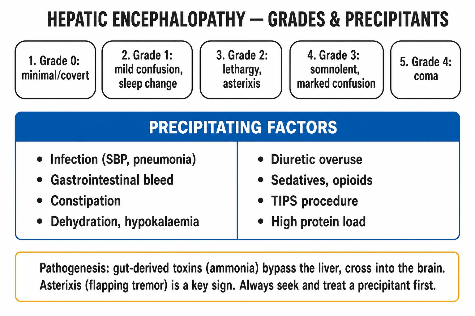
The West Haven classification (Conn and Lieberthal 1979) grades mental state from minimal cognitive change to deep coma. Grade 0 (minimal/covert HE) has no detectable clinical signs but impaired cognition on psychometric testing; grade I has mild, often missed, changes (sleep reversal, mild euphoria/anxiety, attention deficits). Grades II–IV are overt HE — the syndrome that prompts admission.[1]
| Grade | Mental state | Asterixis | Other features |
|---|---|---|---|
| 0 (minimal/covert) | No clinical signs; impairment only on psychometric testing (PHES) or specialist tests | Absent | Detectable only on testing; affects driving, quality of life |
| I — trivial | Mild confusion, euphoria or anxiety, shortened attention span, sleep reversal (awake at night, sleepy by day), impaired addition/subtraction | Subtle/intermittent | Often missed without a focused history |
| II — moderate | Lethargy, apathy, disorientation (time), personality change, inappropriate behaviour | Present (overt) | Dyspraxia (constructional apraxia — cannot draw a star), impaired calculation |
| III — severe | Somnolent but rousable, gross disorientation (time, place, person), incomprehensible speech, aggressive behaviour | Usually present but coarse | Marked confusion, requires stimulus to remain awake |
| IV — coma | Coma — unresponsive to verbal stimuli; may or may not respond to painful stimuli | Absent (patient cannot hold posture) | Decerebrate/decorticate posturing in deep coma |
Covert HE (CHE) = minimal HE (grade 0) plus grade I; overt HE (OHE) = grades II–IV. The covert/overt split matters because covert HE affects 30–80% of cirrhotics, impairs driving and quality of life, and predicts the first overt episode — yet is invisible at the bedside without formal testing.[3]
By time course
- Episodic — a single acute episode over hours–days; almost always precipitated.
- Recurrent — two or more episodes within 6 months without prophylaxis.
- Persistent — continuous cognitive/behavioural abnormality that fluctuates but never returns to baseline; seen in advanced cirrhosis and large spontaneous shunts. [1]

Epidemiology & Risk Factors
HE is a major complication of decompensated cirrhosis and a leading cause of hospital admission in cirrhosis.[6]
- Prevalence of overt HE at first decompensation — about 10–20% of cirrhotics.
- Lifetime cumulative risk of overt HE in cirrhosis — 50–70%.
- Covert (minimal) HE — present in 30–60% of cirrhotics with apparently normal mental state; high-yield exam point because it is invisible at the bedside and predicts the first overt episode.[3]
- Recurrence — about 40% at 1 year after a first overt episode despite standard therapy; this is the rationale for secondary prophylaxis with rifaximin.[1]
Mortality after first overt HE — 1-year mortality approaching 40–50%, 3-year mortality over 70%; the episode signals transition to a decompensated state and is an independent indication for transplant evaluation.[6]
Risk factors and precipitants (the high-yield mnemonic list): [1]
| Risk factor / precipitant | Mechanism by which it precipitates HE |
|---|---|
| Infection — especially SBP, pneumonia, UTI, bacteraemia | Sepsis increases gut permeability and ammonia production; systemic inflammation disrupts the BBB and sensitises the brain to ammonia |
| GI bleed (variceal, peptic ulcer) | Blood in the gut lumen is a high-protein load → digestion by bacteria releases large amounts of ammonia |
| Constipation | Increased contact time between gut flora and protein → more ammonia absorption |
| Excess dietary protein (rarely alone) | Substrate overload in marginal liver function |
| Sedatives — benzodiazepines, opioids, barbiturates | GABA-ergic and opioid receptor activity directly depresses a sensitised CNS |
| Hypokalaemia and alkalosis | Hypokalaemia promotes renal ammoniagenesis (renal glutaminase) and alkalosis favours NH3 (diffuses into brain) over NH4+ (trapped in tubule) |
| Hyponatraemia | The commonest electrolyte precipitant in cirrhosis; low serum osmolality causes astrocyte swelling and sensitises brain to ammonia |
| Over-diuresis / dehydration | Hypovolaemia → AKI → reduced hepatic and renal ammonia clearance; also electrolyte loss |
| TIPS (transjugular intrahepatic portosystemic shunt) | Iatrogenic large shunt → ammonia bypasses the liver; HE in 20–50% within the first year |
| Large spontaneous portosystemic shunts | Same mechanism as TIPS |
| Hepatocellular carcinoma, acute hepatitis flare, superimposed ACLF | Worsening hepatic synthetic function reduces ammonia clearance |
| Surgery / general anaesthesia | Catabolic stress + sedatives |
Pathophysiology
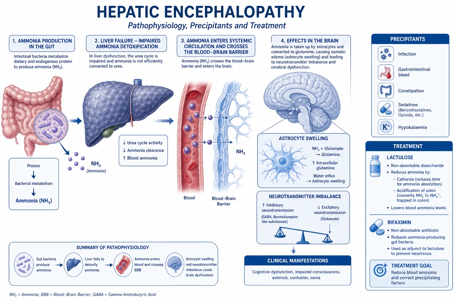
The core insight examiners want is: HE is a multifactorial syndrome of gut-derived neurotoxins (chiefly ammonia) acting on a brain made vulnerable by portosystemic shunting, inflammation and oxidative stress. No single toxin explains all of HE.[6]
Ammonia — the central toxin
Ammonia (NH3) is produced in the gut by:
- Bacterial degradation of dietary protein and urea (the urease-positive organisms — Klebsiella, Proteus, E. coli).
- Glutaminase in enterocytes (small intestinal mucosa) — small intestinal glutaminase is a major source. [1]
In health, portal venous ammonia is delivered to the liver where it is detoxified in the urea cycle (hepatocyte mitochondria → urea → kidney) and by glutamine synthetase in periportal hepatocytes (NH3 + glutamate → glutamine). In cirrhosis, both routes fail: reduced hepatocyte mass reduces urea cycle capacity, and portosystemic shunting diverts ammonia-rich portal blood straight into the systemic circulation.[6]
What ammonia does at the blood–brain barrier
Ammonia crosses the BBB (as NH3 gas, the uncharged form favoured by alkalosis) and is taken up almost exclusively by astrocytes, the only brain cell expressing glutamine synthetase. There, ammonia is converted to glutamine: [1]
NH3 + glutamate + ATP —(glutamine synthetase)→ glutamine + ADP + Pi [1]
Glutamine is osmotically active — it accumulates within the astrocyte, drawing in water, causing astrocyte swelling (the histological hallmark is the Alzheimer type II astrocyte — swollen nucleus, pale chromatin, marginal nucleus).[6]
The swollen astrocyte cannot:
- Regulate extracellular glutamate (astrocytes normally clear synaptic glutamate via EAAT-2 transporters) → extracellular glutamate accumulates → NMDA-receptor activation → excitotoxicity and oxidative stress.
- Maintain the GABA/glutamate balance → relative GABA-ergic tone increase (the basis of the GABA hypothesis and the reason benzodiazepines worsen HE).
- Regulate extracellular potassium and water → in severe cases (especially ALF) this progresses to cerebral oedema and intracranial hypertension. [1]
Other contributors (the synergistic toxin hypothesis)
- Systemic inflammation — sepsis/infection (precipitant) disrupts the BBB and sensitises the brain to ammonia; inflammation and ammonia act synergistically, not additively. This is why even a mild infection can trigger overt HE in a cirrhotic.[6]
- GABA and endogenous benzodiazepine-like substances — increased GABA-ergic tone produces the somnolence, ataxia and asterixis; flumazenil (a benzodiazepine antagonist) produces transient improvement in some patients, supporting this mechanism, but is not used clinically (low response rate, seizure risk).
- Manganese — deposited in the basal ganglia in cirrhotics with long-standing portosystemic shunting; produces extrapyramidal signs and the T1-weighted hyperintensity of the globus pallidus seen on MRI.
- Mercaptans (methanethiol, derived from methionine bacterial metabolism) — contribute to fetor hepaticus (the sweet musty breath) and were historically implicated in HE.
- False neurotransmitters (octopamine) and serotonergic/dopaminergic imbalance — older theories, now less central but still tested.
- Zinc deficiency — zinc is a cofactor for glutamate dehydrogenase and the urea cycle; cirrhotics (especially malnourished) are often zinc-deficient.
Clinical Presentation
The presentation depends on grade (severity) and on whether the episode is covert or overt. The classical teaching emphasises the progression: sleep reversal → mild confusion → asterixis and disorientation → somnolence → coma. Most patients present in the setting of known cirrhosis with a precipitant; rarely, HE may be the first manifestation of cirrhosis.[8]
Overt HE (grades II–IV) — the admission scenario
- Grade II — patient or family report daytime drowsiness, sleep reversal (insomnia at night, somnolence by day), irritability, personality change, disorientation for time, repetitive questioning. Examination shows asterixis (see below), constructional apraxia (cannot draw a five-pointed star / clock face), impaired subtraction of serial 7s.
- Grade III — somnolent but rousable, grossly disoriented for time and place, slurred/incomprehensible speech, may be aggressive or uncooperative, muscular rigidity, hyperreflexia, extensor plantar responses.
- Grade IV — coma; response only to painful stimuli (IVa) or no response (IVb); may have decerebrate posturing in deep coma. [1]
Bedside signs — named and reproduced
- Asterixis (flapping tremor) — ask the patient to hold arms outstretched with wrists dorsiflexed and fingers spread for 30 seconds; a rapid, irregular, non-rhythmic flexion–extension ("flap") at the wrist and metacarpophalangeal joints appears, with brief silent lapses of sustained posture. The sign of loss of sustained motor tone due to a metabolic encephalopathy. Bilateral in HE (unilateral asterixis suggests a focal structural lesion). Also seen in uraemia, hypercapnia (CO2 narcosis), and severe heart failure.[8]
- Constructional apraxia — patient cannot copy a five-pointed star, clock face, or intersecting pentagons (used in the Reitan trail / line drawing test). Sensitive for grade I–II HE.
- Fetor hepaticus — sweet, musty, faecal breath (volatile mercaptans — methanethiol) blown off from portosystemic shunting of gut-derived substances. High-yield exam pearl.
- Hyperreflexia, rigidity, clonus, extensor plantars in grade III.
- Signs of chronic liver disease — jaundice, spider naevi, palmar erythema, gynaecomastia, testicular atrophy, ascites, caput medusae, clubbing, leukonychia, asterixis, Dupuytren contracture, parotid enlargement, fetor hepaticus — establish the underlying cirrhosis and portal hypertension.
Covert (minimal) HE — grade 0
No detectable clinical signs at the bedside — by definition. Diagnosis requires psychometric testing (PHES battery — Psychometric Hepatic Encephalopathy Score) or specialist tools (critical flicker frequency, ICT, EncephalApp smartphone Stroop). Patients report impairment in driving, sleep, attention, work; family may note personality change. Affects 30–60% of cirrhotics and predicts the first overt episode.[3]
Atypical presentations
- Elderly cirrhotic — falls, functional decline, "off legs", urinary incontinence, quiet delirium rather than florid confusion; easily missed. Sleep reversal may be mistaken for dementia. Lower threshold to screen and admit.
- Post-TIPS patient — new confusion within days–weeks of shunt placement; consider shunt-related HE.
- Diabetic cirrhotic — hypoglycaemia is a powerful mimic and a co-precipitant; always check finger-prick glucose.
- Alcoholic cirrhotic — Wernicke encephalopathy (ataxia, ophthalmoplegia, confusion) and delirium tremens (hallucinations, autonomic hyperactivity) coexist and must be considered in parallel with HE; give parenteral thiamine empirically.
- Immunocompromised — infection may be occult; do not be reassured by a normal temperature. [1]
Differential Diagnosis
A cirrhotic with new confusion has HE only after other causes are considered and excluded. The differentials every student must be able to distinguish:[1]
Metabolic / electrolyte encephalopathy
- **Hyponatraemia** (Na under 125) — common in cirrhosis from diuretics and SIADH; confusion, seizures
- **Uraemia** — asterixis also present; check creatinine; may coexist with HRS
- **Hypoglycaemia** — sweating, tachycardia, rapid correction with IV dextrose; always check finger-prick glucose
- **Hypoxia / hypercapnia** — type 1 or type 2 respiratory failure
Sepsis-related encephalopathy
- Common in cirrhosis; fever, source (SBP, pneumonia, UTI), positive cultures
- Often coexists with HE — infection is the commonest precipitant, so look for both
Intracranial lesion
- **Subdural haematoma** — cirrhotics fall (ataxia, asterixis, alcohol) AND are coagulopathic (low platelets, raised INR); always consider and image if focal signs, seizures, or unwitnessed trauma
- **Intracerebral haemorrhage**, **cerebral infarct**, **brain abscess**, **meningitis** (especially cryptococcal in immunosuppressed)
Drug / toxin effect
- **Benzodiazepines, opioids, anticonvulsants, anticholinergics** — common in cirrhosis due to altered pharmacokinetics
- **Alcohol intoxication or withdrawal / delirium tremens** — autonomic hyperactivity, tremor, hallucinations distinguish DTs
- **Urine drug screen** if history unclear
Wernicke encephalopathy
- **Triad of confusion, ataxia, ophthalmoplegia (nystagmus, lateral rectus palsy, conjugate gaze palsy)**
- Give **parenteral thiamine (Pabrinex)** before any glucose in suspected alcohol misuse
- **MRI**: T2 hyperintensity of mammillary bodies, periaqueductal grey, thalamus
Other neurological disease in cirrhosis
- **Hepatic myelopathy** — spastic paraparesis without sensory loss, in long-standing cirrhosis
- **Acquired hepatocerebral degeneration** — chronic extrapyramidal and cognitive syndrome
- **Central pontine myelinolysis (osmotic demyelination)** — rapid Na correction
- **Post-ictal state** — subclinical seizures (more common in cirrhosis)
A practical rule: if the working diagnosis is HE but the patient is not improving after 48–72 hours of adequate lactulose and precipitant treatment, re-examine the diagnosis — obtain CT brain, EEG, repeat septic screen, and reconsider Wernicke and metabolic causes.[1]
Clinical & Bedside Assessment
Bedside assessment serves three purposes: (1) grade severity (West Haven), (2) identify the precipitant, (3) exclude mimics. [1]
Mental state and grade (West Haven)
- Orientation — time (day, date, month, year), place (which hospital, which city), person (name, DOB).
- Attention — serial subtraction of 7 from 100 (93, 86, 79, 72, 65…); digit span (repeat 7 digits forward, 5 backward); a normal person can do serial 7s in under 90 seconds with fewer than 4 errors. Errors mark grade I–II HE.
- Asterixis — see above; bilateral flap, hold posture for 30 seconds.
- Construction — copy a five-pointed star or draw a clock face with the time set at 11:10.
- Conscious level — GCS if grade III–IV or deteriorating. [1]
Hunt the precipitant — bedside examination
- Vitals — fever (infection), tachypnoea (acidosis, sepsis, aspiration), hypotension (sepsis, GI bleed), hypothermia (severe sepsis).
- Abdomen — ascites (tap if present — SBP), tenderness/peritonism (SBP, bacteraemia), hepatomegaly or hepatic mass (HCC).
- Rectal examination — melaena or bright blood (GI bleed), hard stool/loaded rectum (constipation).
- Signs of alcohol withdrawal — tremor, sweating, tachycardia, hallucinations.
- Hydration, signs of dehydration (over-diuresis).
- Neurological — focal deficit suggests structural lesion (image the brain).
- Drug chart — benzodiazepines, opioids, diuretics. [1]
Confirm underlying cirrhosis
Stigmata of chronic liver disease (jaundice, spider naevi, palmar erythema, gynaecomastia, ascites, caput medusae, clubbing, leukonychia, parotid enlargement) and portal hypertension (splenomegaly, ascites, caput) confirm that the underlying substrate is cirrhosis. [1]
Investigations
HE is a clinical diagnosis. Investigations support the diagnosis, identify the precipitant, exclude mimics and stage the underlying liver disease. [1]
First-line panel in every suspected HE
| Investigation | Why |
|---|---|
| Capillary glucose | Exclude hypoglycaemia (a common mimic and co-precipitant; finger-prick at bedside before further workup) |
| Full blood count | Anaemia (GI bleed), leucocytosis (infection), low platelets (hypersplenism, cirrhosis) |
| U&E, creatinine | Hyponatraemia (commonest electrolyte precipitant), hypokalaemia (renal ammoniagenesis), AKI/HRS |
| LFTs, albumin, INR, bilirubin | Stage liver disease and synthetic function; INR over 1.5 + encephalopathy = acute liver failure if no chronic disease |
| Venous blood gas — venous ammonia | Supports HE but not diagnostic; correlation with grade is poor; trend with treatment is more useful than a single value. Sample on ice, processed promptly (ammonia rises spuriously in delayed samples) |
| Septic screen — blood cultures, urine culture, chest X-ray | Identify infection — the commonest precipitant |
| Diagnostic paracentesis (if ascites) | SBP — ascitic PMN count over 250 cells/mm³ (the defining criterion); treat empirically |
| Calcium, magnesium, phosphate | Correct electrolyte abnormalities |
| Toxicology screen / drug levels | Exclude drug cause (lithium, benzodiazepines, opioids, alcohol) |
| CT brain (non-contrast) | Exclude subdural/ICH, mass lesion, hydrocephalus — mandatory if focal signs, seizures, head trauma, or failure to improve |
Serum ammonia — what it does and does not do
- Venous ammonia, sampled on ice and processed within 20 minutes, supports the diagnosis and helps distinguish HE from non-HE causes of encephalopathy (e.g. a normal ammonia in a confused cirrhotic argues against HE).
- Levels do not correlate well with West Haven grade — a patient may be in deep coma with a modestly raised ammonia, and vice versa.
- Levels correlate better with hepatic synthetic function than with neurological state.
- Arterial ammonia is not required routinely — venous suffices; arterial is reserved for research or specific situations.
- Falsely elevated by delayed processing, haemolysis, high-protein meal, exercise, smoking, muscle activity (tourniquet), valproate, carbamazepine.[3]
Confirming covert HE (minimal HE)
Covert HE has no bedside signs and requires formal testing:
- PHES (Psychometric Hepatic Encephalopathy Score) — five paper-and-pencil tests (number connection A and B, serial dotting, line tracing, digit symbol). Standardised; score of under −4 defines covert HE.
- Critical flicker frequency (CFF) — frequency at which a fused light appears to flicker; threshold below 39 Hz indicates covert HE.
- ICT (Inhibitory Control Test), EncephalApp (smartphone Stroop test) — bedside / smartphone-based screening tools. [1]
Neuroimaging
- CT brain (non-contrast) — first-line to exclude structural lesions (subdural, haemorrhage, mass).
- MRI brain — may show T1-weighted hyperintensity of the basal ganglia (globus pallidus) due to manganese deposition (a hallmark of chronic portosystemic shunting, not of acute HE); may show cortical oedema in ALF. MRI is not diagnostic of HE — it excludes other causes and shows supportive signs. [1]
Electroencephalogram (EEG)
- Classic finding: generalised slowing (theta and delta waves) with, in advanced cases, triphasic waves — high-amplitude (over 100 microvolts), bilaterally synchronous, symmetric, frontal-dominant waves at 1.5–3 Hz with a phase lag (frontal-to-occipital).
- Triphasic waves are NOT specific — also seen in uraemia, hyponatraemia, anoxia and other metabolic encephalopathies.
- EEG is rarely needed at the bedside but useful for research and to exclude non-convulsive status epilepticus in unexplained coma. [1]
Severity scoring — reproduced verbatim
West Haven (Conn–Lieberthal) classification (grades 0–IV) — reproduced above under Classification; this is the score reproduced in clinical and exam practice.[1]
Child–Pugh score stages the underlying cirrhosis and predicts mortality (one of its 5 components is encephalopathy: none = 1 point, grade I–II = 2 points, grade III–IV = 3 points). Other components: bilirubin, albumin, INR, ascites. Total 5–15 = classes A, B, C; class C cirrhosis has 1-year mortality around 45%. [1]
MELD / MELD-Na — used for transplant prioritisation; incorporates bilirubin, INR, creatinine and sodium. A rising MELD in a cirrhotic with new HE mandates transplant referral.[6]
Management — Resuscitation
ABCDE first. HE is rarely an immediate airway threat at grade I–II, but grade III–IV coma risks airway obstruction, aspiration and respiratory arrest.[1]
- Airway — protect the airway in grade III–IV; consider intubation if GCS under 8, inability to protect airway, or aspiration risk (especially if also vomiting from GI bleed). Lateral position + suction.
- Breathing — oxygen to keep SpO2 at 94–98% (88–92% if CO2 retainer). Treat hypoxaemia from aspiration pneumonia, sepsis, or hepatic hydrothorax.
- Circulation — IV access; treat hypovolaemia (GI bleed, over-diuresis, sepsis) with balanced crystalloids; transfuse for GI bleed per restrictive strategy (target haemoglobin 70–80 g/L in cirrhosis, 80 g/L in active bleeding). Avoid hyponatraemic fluids (5% dextrose worsens hyponatraemia) — use saline cautiously.
- Disability — finger-prick glucose immediately; if low give 50 mL of 50% IV dextrose (or 200 mL of 10% if central access). Give parenteral thiamine (Pabrinex — one pair of ampoules IV/IM OD for 3–5 days, then oral) before any glucose in suspected alcohol misuse to avoid precipitating Wernicke. Check GCS, pupils, focal signs.
- Exposure / examination — hunt the precipitant (see above). Empirically treat sepsis if any clue — blood cultures then broad-spectrum antibiotic (e.g. cefotaxime 2 g IV BD for SBP) after cultures. [1]
Do not delay precipitant workup and treatment while awaiting ammonia or neuroimaging. A septic cirrhotic with new confusion needs bloods, paracentesis, cultures, empiric antibiotics and lactulose started within the first hour of admission. [1]
Management — Definitive & Stepwise
The four pillars: (1) treat the precipitant, (2) lower gut ammonia, (3) prevent further episodes, (4) supportive care and nutrition. All four run in parallel from admission.[1][6]
Pillar 1 — Identify and treat the precipitant
| Precipitant | Specific treatment |
|---|---|
| SBP | Diagnostic paracentesis (PMN over 250); cefotaxime 2 g IV BD for 5 days (or ceftazidime/ceftriaxone); add IV albumin 1.5 g/kg day 1, then 1 g/kg day 3 to prevent HRS |
| GI bleed | Resuscitate, restrict transfusion (Hb 70–80), terlipressin + antibiotics (e.g. ceftriaxone 1 g IV OD), urgent endoscopy with band ligation; clear gut blood with lactulose (blood in gut = high ammonia load) |
| Constipation | Lactulose to achieve 2–3 soft stools/day; enema if impacted |
| Hypokalaemia / alkalosis | Replace potassium (oral and IV); correct alkalosis by treating cause |
| Hyponatraemia | Slow correction — water restrict, stop diuretics; avoid rapid Na correction (risk of osmotic demyelination); hypertonic saline only if severe symptoms/seizures |
| Sedatives / drugs | Stop the offending agent; consider reversal agents — flumazenil for benzodiazepines (only if severe, with monitoring — seizure risk) and naloxone for opioids |
| Infection (other than SBP) | Culture, source control, targeted antibiotics |
| TIPS-related HE | Optimise lactulose + rifaximin; if refractory, shunt embolisation / reduction |
| Hepatic decompensation / ACLF | Treat underlying cause; consider transplant evaluation |
Pillar 2 — Lower gut ammonia
HE — ammonia-lowering therapy at a glance
Lactulose (β-galactosidofructose) — non-absorbable synthetic disaccharide; first-line for overt HE.
- Mechanism (three actions):
- Catharsis — osmotic laxative → reduces gut transit time and contact of ammonia with mucosa.
- Acidification of colon — bacterial fermentation produces lactic acid and acetic acid, lowering colonic pH to under 5 → converts diffusible NH3 to non-absorbable NH4+ (ion trapping) → ammonia is trapped in the lumen and excreted in stool.
- Favours urease-negative flora — acidic pH suppresses urease-positive organisms (Klebsiella, Proteus).
- Dose — 15–30 mL (10–20 g) orally every 1–2 hours until two to three soft bowel motions daily, then titrate down to a maintenance dose (typically 15–30 mL two to four times daily). The endpoint is stool frequency/consistency, NOT a target ammonia level.
- In coma — give via nasogastric tube or as a retention enema (300 mL lactulose in 700 mL water, retained 1 hour, q4–6h) when NGT unsafe.
- Adverse effects — dehydration, hypernatraemia (from osmotic diarrhoea — which can paradoxically worsen HE), abdominal cramps, flatulence. Monitor U&E daily.
- Evidence — Als-Nielsen 2004 BMJ systematic review: non-absorbable disaccharides significantly improve HE versus placebo; no mortality benefit shown.[2]
Rifaximin — non-absorbable, gut-poorly-absorbed antibiotic (less than 0.4% systemic bioavailability) with broad-spectrum activity against ammonia-producing gut flora.
- Dose — 550 mg orally twice daily, added to lactulose (NOT as replacement).
- Evidence — Bass 2010 NEJM: in patients with at least two prior overt HE episodes, rifaximin 550 mg BD added to lactulose reduced the risk of breakthrough HE by 58% over 6 months and reduced HE-related hospitalisation. The benefit is additive to lactulose.[1]
- Cochrane 2023 — rifaximin probably reduces mortality and improves HE recovery versus non-absorbable disaccharides; quality of evidence moderate.[7]
- Adverse effects — well tolerated; rarely headache, nausea. Theoretical concern over Clostridioides difficile and antimicrobial resistance with long-term use (not yet clinically significant).
L-ornithine L-aspartate (LOLA) — provides substrates (ornithine, aspartate) that enhance residual urea cycle and glutamine synthetase activity in skeletal muscle; converts ammonia to urea and glutamine.
- Dose — IV 20–40 g/day in acute HE, oral 3–6 g three times daily as adjunct.
- Modest evidence of benefit; used in some countries (especially India/Germany) as adjunct where rifaximin is costly. [1]
Branched-chain amino acids (BCAA) — leucine, isoleucine, valine; compete with aromatic amino acids for BBB transport and promote muscle glutamine synthesis (ammonia disposal in skeletal muscle).
- May reduce HE recurrence and improve nitrogen balance; not first-line. [1]
Other agents (with limited evidence, exam-known):
- Polyethylene glycol (PEG) — some RCTs show faster resolution of acute overt HE than lactulose; not standard.
- Probiotics — modest benefit in covert HE; not for acute overt HE.
- Zinc — zinc sulphate 600 mg/day; zinc is a cofactor for urea cycle enzymes; useful in zinc-deficient malnourished cirrhotics.
- L-carnitine — limited evidence in HE due to valproate.
- Shunt embolisation — for refractory Type B / large spontaneous portosystemic shunt HE. [1]
Agents that DO NOT work and should NOT be used:
- Branched-chain ketoanalogues — not beneficial.
- Dopamine agonists (bromocriptine) — not beneficial for HE.
- Flumazenil — transient improvement only; not used clinically (seizure risk).
- Sorbitol / other osmotic laxatives — no ammonia-lowering beyond catharsis; lactulose preferred (acidification). [1]
Pillar 3 — Prevent recurrence (secondary prophylaxis)
After a first overt HE episode, recurrence is around 40% at 1 year without prophylaxis. Standard secondary prophylaxis:
- Continue lactulose titrated to 2–3 soft stools/day indefinitely.
- Add rifaximin 550 mg BD indefinitely — reduces breakthrough HE and HE-related hospitalisation (Bass 2010 NEJM).[1]
- Treat and prevent precipitants — avoid constipation, treat infections early, avoid sedatives.
- Refer for liver transplant evaluation — first overt HE episode is a transplant indication (1-year mortality 40–50%).[6]
Pillar 4 — Supportive care and nutrition
Nutrition — the single most under-prescribed intervention in HE. [1]
- Do NOT restrict dietary protein. The older dogma of protein restriction is wrong and harmful — it precipitates sarcopenia, and skeletal muscle is the major extrahepatic site of ammonia disposal. The Córdoba 2004 RCT showed a normal-protein diet (1.2 g/kg/day) is safe and superior to protein restriction in episodic HE.[4]
- Recommended protein intake — 1.2–1.5 g/kg/day in cirrhosis (higher than in healthy adults because cirrhosis is a catabolic state). vegetable/dairy protein preferred over meat (better branched-chain:aromatic ratio).
- Late evening snack — 50 g carbohydrate at night reduces overnight catabolism.
- Vitamin supplementation — thiamine (alcoholics), folate, vitamin D, zinc.
- Avoid benzodiazepines — they are GABA-ergic and worsen HE. If unavoidable (alcohol withdrawal, seizures) use a short-acting agent (lorazepam, oxazepam) at the lowest effective dose. Use propofol (not midazolam) for ICU sedation in HE.[1]
- Avoid opioids; if required use at reduced dose.
- Aspiration precautions in coma — head-up 30 degrees, nasogastric tube if ileus or to give lactulose.
- DVT prophylaxis (cirrhotics are procoagulant-balanced but bed-bound).
- Pressure-area care.
- Stool chart — essential to titrate lactulose.
Specific Subtypes & Scenarios
Covert (minimal) HE
- Affects 30–60% of cirrhotics; impairs driving, work and quality of life; predicts overt HE.
- Diagnose with PHES, CFF (under 39 Hz), EncephalApp.
- Treatment: lactulose (titrated to 2 soft stools/day) and/or rifaximin 550 mg BD improve cognition; probiotics modest benefit; counsel about driving/operating machinery. [1]
Post-TIPS HE
- New or worsening HE in 20–50% of patients within the first year after TIPS, especially in those with prior HE, older age, large shunt, or sarcopenia.
- Most episodes responds to lactulose + rifaximin; refractory cases need shunt embolisation or reduction.
- Pre-TIPS prophylaxis with rifaximin reduces incidence. [1]
Recurrent and persistent HE
- Recurrent — two or more episodes within 6 months; add rifaximin to lactulose, search for occult large spontaneous shunt.
- Persistent — continuous cognitive impairment; consider large portosystemic shunt embolisation, transplant evaluation. [1]
Type A HE (acute liver failure)
- High cerebral oedema risk — leading cause of death in ALF.
- Different management paradigm: head-of-bed 30 degrees, hypertonic saline to keep Na 145–155 mmol/L, mannitol for raised ICP, N-acetylcysteine regardless of cause, urgent King's College criteria and transplant listing. See Acute Liver Failure topic. [1]
ACLF-related HE
- Acute decompensation of cirrhosis with extrahepatic organ failure; high short-term mortality. Treat HE precipitant and ACLF in parallel; consider NAC and transplant. [1]
Complications & Pitfalls
Complications of HE itself:
- Aspiration of gastric contents / secretions in coma → aspiration pneumonia.
- Falls and trauma — confusion + ataxia → subdural haematoma in coagulopathic patient.
- Pressure sores, decubitus ulcers, DVT/PE in immobile patients.
- Cerebral oedema — rare in Type C, common in Type A (ALF); presents with hypertension, bradycardia, abnormal pupillary reflexes, decerebrate posturing.
- Progression to coma and death if untreated.
- Recurrence — 40% at 1 year after first episode.
- Hepatic myelopathy and acquired hepatocerebral degeneration — chronic irreversible neurological complications of long-standing portosystemic shunting. [1]
Complications of treatment:
- Hypernatraemia / dehydration / AKI from over-aggressive lactulose catharsis — titrate carefully, monitor U&E daily.
- Worsening HE from protein restriction — a now-discarded but still-encountered error.
- Wernicke encephalopathy precipitated by IV glucose without thiamine in alcoholic cirrhotics.
- Osmotic demyelination (central pontine myelinolysis) from rapid correction of hyponatraemia — limit Na rise to under 8 mmol/L in 24 hours.
- Benzodiazepine-induced worsening — a classic pitfall; lorazepam/oxazepam only if essential. [1]
Classic pitfalls (exam favourites):
- Treating the ammonia without finding the precipitant — SBP missed, patient deteriorates.
- Restricting protein — causes sarcopenia, worsens ammonia disposal.
- Attributing confusion to HE in an alcoholic without excluding Wernicke — give thiamine.
- Giving IV dextrose before thiamine — precipitates Wernicke.
- Rapidly correcting Na — osmotic demyelination.
- Using midazolam for ICU sedation — worsens HE; use propofol.
- Confusing HE with delirium tremens — DTs has autonomic hyperactivity, tremor, hallucinations.
- Forgetting to look for a subdural in a cirrhotic with falls and confusion.
- Stopping rifaximin on discharge — breakthrough HE; secondary prophylaxis is indefinite. [1]
Prognosis & Disposition
- Recovery from each episode — usually within 3–7 days of appropriate treatment; failure to improve by 72 hours mandates re-evaluation of the diagnosis (CT brain, EEG, repeat septic screen).
- 1-year mortality after first overt HE — 40–50%; 3-year mortality over 70%.[6]
- Predictors of poor outcome — higher MELD, low albumin, persistent precipitant (uncontrolled sepsis), older age, sarcopenia, ACLF, need for ICU.
- Recurrence — 40% at 1 year despite standard therapy.
- Discharge criteria — back to baseline mental state, precipitant treated, tolerating oral intake, lactulose titrated, rifaximin prescribed, transplant referral made, safe home/social situation.
- Disposition — first overt HE episode is an indication for liver transplant evaluation; ambulatory care for covert HE with driving advice.
Special Populations
- Elderly — atypical (quiet delirium, falls); lower threshold to admit and image; multiple comorbidities; avoid benzodiazepines (excess sensitivity); watch for cumulative drug effects.
- Pregnancy with cirrhosis — rare; coordinate with obstetric hepatology; intrahepatic cholestasis and HELLP can mimic; manage at a transplant centre.
- Children — urea cycle disorders, biliary atresia post-Kasai; present with vomiting, ataxia, school decline, reversible dementia. Treat precipitants; liver transplant for inborn errors.
- Post-liver-transplant patients — HE usually resolves rapidly with restoration of hepatic function; persistent cognitive deficits reflect pre-transplant brain injury.
- Immunocompromised — occult infection; lower threshold to start empiric broad-spectrum antibiotics.
- Anticoagulated cirrhotic — INR often already prolonged; balance bleeding risk (do not routinely transfuse FFP for INR correction unless bleeding).
- Alcoholic cirrhotic — give parenteral thiamine before any glucose; screen for Wernicke and DTs; treat alcohol withdrawal concomitantly with short-acting benzodiazepines at lowest effective dose. [1]
Evidence, Guidelines & Regional Differences
Landmark trials
- Bass 2010 NEJM (RFHEPS) — rifaximin 550 mg BD added to lactulose reduced breakthrough HE by 58% over 6 months in patients with at least two prior HE episodes; established rifaximin as standard secondary prophylaxis.[1]
- Als-Nielsen 2004 BMJ — Cochrane systematic review of non-absorbable disaccharides; significant improvement in HE versus placebo/comparator, no mortality benefit.[2]
- Córdoba 2004 J Hepatol — RCT showed normal-protein diet (1.2 g/kg/day) is safe and superior to protein restriction; killed the protein-restriction dogma.[4]
- Cochrane 2023 (Zacharias et al.) — rifaximin probably reduces mortality and improves HE recovery versus non-absorbable disaccharides (moderate-quality evidence).[7]
Current guidelines
- EASL–AASLD 2014 Practice Guideline (Vilstrup) — the international standard; introduced the Type A/B/C and covert/overt classification.[1]
- Rose 2020 (ISHEN/WCOG consensus) — novel pathophysiology, classification and therapy.[6]
Regional differences
[1] [1] [1]Controversies
- Whether serum ammonia should be measured routinely — most guidelines: useful for trend and to support diagnosis, not diagnostic, not predictive of grade.
- Whether non-absorbable disaccharides reduce mortality — no trial has demonstrated a mortality benefit; benefit is symptomatic.
- Long-term safety of rifaximin — no signal yet for clinically significant resistance or C. difficile, but surveillance continues.
- Role of embolising large spontaneous shunts in refractory HE — emerging evidence.
- Whether albumin infusion benefits HE specifically (beyond SBP/HRS) — under investigation. [1]
Exam Pearls
HE precipitants — SHIP-GO mnemonic
SHIP
the commonest precipitant — culture and tap the ascites; PMN over 250 = SBP
blood in gut = high ammonia load; clear with lactulose
benzodiazepines, opioids, post-TIPS shunt
hypokalaemia → renal ammoniagenesis; rarely pure protein load
HE treatment ladder — LARI mnemonic
LARI
15–30 mL q1–2h to 2–3 soft stools/day, then titrate down — endpoint is stool frequency, not ammonia
additive to lactulose for overt/recurrent HE; reduces breakthrough HE by 58% (Bass 2010 NEJM)
the single most important act — treat SBP, GI bleed, constipation, hypokalaemia, hyponatraemia; stop sedatives
do NOT restrict protein (Córdoba 2004); avoid benzodiazepines; protect airway
High-yield exam pearls: [1]
- Definition: reversible syndrome of impaired brain function in advanced liver disease/portosystemic shunting; multifactorial, ammonia central.
- Classification: Type A (Acute liver failure), Type B (portosystemic shunt without intrinsic liver disease), Type C (Cirrhosis — commonest, over 80%).
- West Haven grades: 0 covert/minimal (only psychometric testing); I trivial (sleep reversal, mild confusion); II lethargy + disorientation + asterixis; III somnolent but rousable + gross disorientation; IV coma.
- Asterixis = flapping tremor = loss of sustained motor tone; bilateral in HE; unilateral suggests structural lesion; also in uraemia, hypercapnia, severe cardiac failure.
- Fetor hepaticus = sweet musty breath = mercaptans (methanethiol).
- Triphasic EEG waves — classic, not specific; also in uraemia, hyponatraemia, anoxia.
- T1 hyperintensity of globus pallidus on MRI = manganese deposition in chronic portosystemic shunting.
- Astrocyte swelling = Alzheimer type II astrocyte — the histological hallmark.
- Ammonia detoxified in brain by glutamine synthetase (only in astrocytes) → glutamine accumulates → osmotic swelling.
- Ammonia detoxified in liver by urea cycle (mitochondrial) and glutamine synthetase in periportal hepatocytes.
- Ammonia detoxified in skeletal muscle by glutamine synthetase — basis for high-protein diet (preserve muscle mass).
- Lactulose endpoint = 2–3 soft stools/day, NOT a target ammonia.
- Lactulose mechanism = catharsis + colonic acidification (NH3 → NH4+ ion trapping) + favour urease-negative flora.
- Rifaximin = non-absorbable antibiotic, 550 mg BD, added (not replacing) to lactulose — reduces breakthrough HE 58%.
- Do NOT restrict protein — 1.2–1.5 g/kg/day (Córdoba 2004 RCT).
- Avoid benzodiazepines — GABA-ergic, worsen HE; if essential use lorazepam/oxazepam (short-acting).
- Always check glucose first and give thiamine before any glucose in alcoholic cirrhotic.
- First overt HE = transplant referral (1-year mortality 40–50%).
- Covert (minimal) HE affects 30–60% of cirrhotics; impairs driving; predicts overt HE.
- Commonest precipitant = infection — especially SBP (ascitic PMN over 250 cells/mm³).
- Cerebral oedema is rare in Type C, common (up to 80%) in Type A (ALF).
- Hyponatraemia = the commonest electrolyte precipitant; correct slowly (under 8 mmol/L per 24 h).
- Wernicke triad — confusion, ataxia, ophthalmoplegia — give parenteral thiamine (Pabrinex) empirically.
- Always image the brain (CT) if focal signs, seizures, head trauma, or failure to improve.
- Propofol (not midazolam) for ICU sedation in HE. [1]
Exam application bank (NEET-PG / INICET)
One-line answer
Hepatic encephalopathy (HE) is a reversible syndrome of impaired brain function occurring in patients with advanced liver disease and/or portosystemic shunting, produced by gut-derived neurotoxins (predominantly ammonia) that the failing liver cannot clear. Classified by the underlying disease as Type A (acute liver failure), Type B (portosystemic shunt without intrinsic liver disease) or Type C (cirrhosis — the commonest). Severity by the West Haven classification: grade I mild confusion/sleep reversal, grade II lethargy and disorientation with asterixis, grade III somnolent but rousable with gross disorientation, grade IV coma (grades II–IV are overt). In Type C (cirrhotic) HE, an overt episode is almost always triggered by a precipitant — infection (SBP), GI bleed, constipation, sedatives, hyponatraemia, hypokalaemia, dehydration or TIPS. Diagnosis is clinical (altered mentation plus
Worked stems (answer without another resource)
Stem 1 — Classic presentation. Map symptoms to mechanism; name the first investigation and first treatment step with dose/route if drug therapy is standard. [1]
Stem 2 — Unstable / complicated. List red flags that force immediate resuscitation, theatre, ICU, antidote, or reperfusion — and what you do in the first 15 minutes. [1]
Stem 3 — Atypical group. Elderly, pregnancy, child, or immunocompromised: how presentation and thresholds change. [1]
Stem 4 — Differential trap. Name the three closest mimics and one discriminator for each. [1]
Stem 5 — Disposition. Who goes home with safety-netting, who is admitted, who needs HDU/ICU/theatre, and what follow-up is mandatory. [1]
Rapid viva checklist
- Definition + classification
- Pathophysiology chain
- Bedside signs / criteria
- Score with exact components (if any)
- Emergency bundle
- Definitive therapy with doses
- Complications of disease and of treatment
- Special populations
- Guideline/trial name if classic
- Three exam traps
Coverage self-check
If you cannot answer any stem above from this page alone, re-read the matching section — the page is intended to be self-sufficient for final-prof and NEET-PG/INICET questions on Hepatic Encephalopathy.
References
- [1]Vilstrup H, Amodio P, Bajaj J, Cordoba J, Ferenci P, Mullen KD, Weissenborn K, Wong P Hepatic encephalopathy in chronic liver disease: 2014 Practice Guideline by the American Association for the Study of Liver Diseases and the European Association for the Study of the Liver Hepatology, 2014.PMID 25042402
- [2]Als-Nielsen B, Gluud LL, Gluud C Non-absorbable disaccharides for hepatic encephalopathy: systematic review of randomised trials BMJ, 2004.PMID 15054035
- [3]Nabi E, Bajaj JS Useful tests for hepatic encephalopathy in clinical practice Curr Gastroenterol Rep, 2014.PMID 24357348
- [4]Córdoba J, López-Hellín J, Planas M, et al. Normal protein diet for episodic hepatic encephalopathy: results of a randomized study J Hepatol, 2004.PMID 15246205
- [5]Ferenci P Hepatic encephalopathy Gastroenterol Rep (Oxf), 2017.PMID 28533911
- [6]Rose CF, Amodio P, Bajaj JS, et al. Hepatic encephalopathy: Novel insights into classification, pathophysiology and therapy J Hepatol, 2020.PMID 33097308
- [7]Zacharias HD, Kamel F, Tan J, et al. Rifaximin for prevention and treatment of hepatic encephalopathy in people with cirrhosis Cochrane Database Syst Rev, 2023.PMID 37467180
- [8]Dellatore P, Cheung M, Mahpour NY, et al. Clinical Manifestations of Hepatic Encephalopathy Clin Liver Dis, 2020.PMID 32245526