Gastroenterology · Gastroenterology
Inflammatory Bowel Disease
Also known as IBD · Crohn disease · Ulcerative colitis · Crohn's disease
Inflammatory bowel disease (IBD) is a group of chronic relapsing immune-mediated disorders of the gastrointestinal tract comprising Crohn disease (CD) — transmural inflammation in a skip-lesion distribution anywhere from mouth to anus, frequently involving terminal ileum, with fistula, stricture, abscess and perianal disease — and ulcerative colitis (UC) — diffuse mucosal inflammation continuous from the rectum proximally, presenting with bloody diarrhoea, urgency and tenesmus. A third working group, IBD-unclassified (IBDU) or colonic IBD type-unclassified, is used when differentiation is not possible on initial work-up. Incidence is highest in the second and third decades (a smaller second peak in the seventh decade) in populations of North European, North American, Australasian and now increasingly South Asian and East Asian descent; the global burden has risen dramatically as newly industrialised regions adopt a Western diet and lifestyle. Pathology is driven by an inappropriate mucosal immune response against luminal antigens — particularly the gut microbiome — in a genetically susceptible host (NOD2/CARD15, IL23R, ATG16L1, ECM1, HLA), with impaired mucosal barrier function, dysbiosis (reduced Faecalibacterium prausnitzii, increased adherent-invasive Escherichia coli), and a T-cell cytokine pattern dominated by Th1 and Th17 cells in CD and a Th2-like (non-classical Th2 / natural killer T) cytokine pattern in UC. Drug therapy is stepped from 5-aminosalicylates (mesalazine 2.4 to 4.8 g daily PO or 1 g PR nightly) and corticosteroids (prednisolone 40 to 60 mg daily then taper, or budesonide 9 mg daily for ileocaecal CD) through immunomodulators (azathioprine 2 to 2.5 mg/kg daily, mercaptopurine 1 to 1.5 mg/kg daily, methotrexate 25 mg SC/IM weekly then 15 mg weekly), to biologics (infliximab 5 mg/kg IV at weeks 0, 2, 6 then every 8 weeks, adalimumab 160/80/40 mg SC then 40 mg every 2 weeks, vedolizumab 300 mg IV at weeks 0, 2, 6 then every 8 weeks, ustekinumab 6 mg/kg IV then 90 mg SC every 8 weeks) and small molecules (tofacitinib 10 mg twice daily for acute UC then 5 mg twice daily maintenance) under a treat-to-target strategy (clinical remission, biochemical remission with CRP and faecal calprotectin under 150 to 250 µg/g, and endoscopic remission per Mayo endoscopic subscore 0 to 1 or SES-CD under 3; STRIDE-II). Acute severe ulcerative colitis (ASUC) — Truelove and Witts criteria, more than six bloody stools daily with systemic toxicity — is a gastrointestinal emergency managed with IV hydrocortisone 100 mg every 6 hours, intravenous fluid and electrolyte resuscitation, broad-spectrum antibiotics if infection suspected, exclusion of cytomegalovirus by colonic biopsy PCR, venous thromboembolism prophylaxis, and rescue therapy with infliximab 5 mg/kg (or ciclosporin 2 mg/kg/day continuous infusion where available) on day 3 if no response to steroids; failure of rescue mandates subtotal colectomy with end ileostomy (curative for UC, but a life-changing procedure that the patient must consent to in advance). In CD, surgical resection is never curative — recurrence at and proximal to the anastomosis is the rule (scored endoscopically as Rutgeerts i0 to i4 at the first post-operative colonoscopy at 6 to 12 months). Complications include toxic megacolon, perforation, fistula-in-ano, intra-abdominal abscess, fibrotic stricture, colorectal cancer (8 percent at 30 years in extensive UC), primary sclerosing cholangitis, osteopenia and osteoporosis, venous thromboembolism, malnutrition, and extraintestinal manifestations (erythema nodosum, pyoderma gangrenosum, episcleritis, uveitis, seronegative arthritis, axial spondyloarthritis, aphthous stomatitis).
On this page & tools
Your progress
Saved locally on this device.
Practise this topic
Exam tags
Red flags

Overview & Definition
Inflammatory bowel disease (IBD) is an umbrella term for two principal chronic, relapsing, immune-mediated disorders of the gastrointestinal tract — Crohn disease (CD) and ulcerative colitis (UC) — and a smaller group labelled IBD-unclassified (IBDU) when the type cannot be determined on initial work-up (10 to 15 percent of cases, particularly with early-onset severe colitis). Both diseases are characterised by uncontrolled activation of mucosal effector T cells in response to luminal antigens (chiefly the gut microbiota) in a genetically susceptible host, producing relapsing inflammation, structural damage, and a substantially increased lifetime risk of colorectal cancer, hospitalisation, surgery and disability.[1][2]
The clinical burden is enormous. IBD prevalence is now above 0.3 percent in North America, Northern Europe and Australasia, and is rising sharply in newly industrialised regions (South Asia, East Asia, the Middle East, South America) as populations adopt a Western diet and urban lifestyle — the so-called "second wave" of the global IBD epidemic. The typical onset is in the second to third decade, with a smaller second peak in the seventh decade. CD presents with transmural inflammation in a skip-lesion distribution anywhere from mouth to anus (most often terminal ileum and ileocolonic), with fistula, stricture, abscess and perianal disease as the structural hallmarks. UC presents with diffuse mucosal inflammation, continuous from the rectum proximally for a variable distance, with bloody diarrhoea, urgency and tenesmus as the cardinal symptoms, and toxic megacolon, perforation, colorectal cancer and primary sclerosing cholangitis as the most feared complications. Both diseases are associated with systemic extraintestinal manifestations in up to a quarter of patients — musculoskeletal (seronegative peripheral arthritis, axial spondyloarthritis, sacroiliitis), cutaneous (erythema nodosum, pyoderma gangrenosum), ocular (uveitis, episcleritis), hepatobiliary (primary sclerosing cholangitis) and oral cavity (aphthous stomatitis, orofacial granulomatosis in CD). [1]
Diagnosis is established by correlating clinical features with endoscopic findings, histology, cross-sectional imaging and laboratory biomarkers — and by the exclusion of infection. Modern management has shifted from symptom control alone to objective treat-to-target (STRIDE-II) — clinical remission + biochemical remission (CRP, faecal calprotectin) + endoscopic remission (Mayo endoscopic subscore 0 to 1, SES-CD under 3) — and from a uniform step-up sequence to individualised risk stratification with top-down biologic therapy in high-risk CD and early biologic rescue in acute severe UC. [1]
Classification
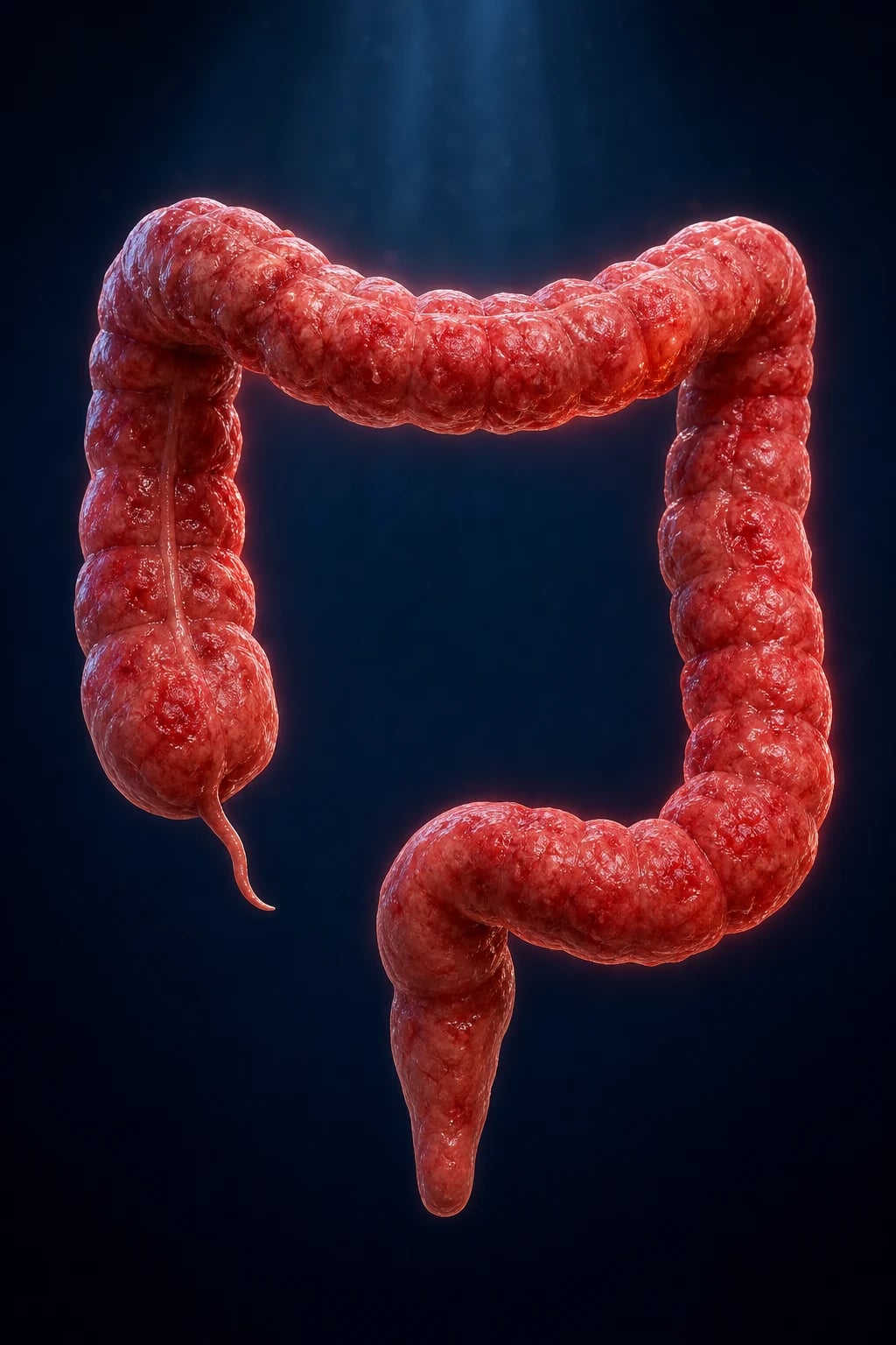
IBD is classified along three clinical axes — disease type, anatomic extent and phenotype (Montreal classification), and activity (Truelove and Witts for acute severe UC; Mayo score; Crohn's Disease Activity Index CDAI; SES-CD endoscopic score; Rutgeerts post-operative score).[1][2][3]
1. Disease type — Crohn vs UC vs IBD-unclassified: [1]
Crohn disease (CD)
- Transmural inflammation — fibrosis, fistula, abscess, stricture
- Skip lesions — diseased segments separated by normal mucosa
- Anywhere mouth to anus — mouth, oesophagus, stomach, small bowel (terminal ileum in 80 percent), colon, perianal (30 percent at presentation)
- Cobblestone mucosa, fissuring ulcers, granulomas (non-caseating, in 30 to 50 percent of biopsies)
- Perianal disease (skin tags, fissures, fistula-in-ano, abscess) in up to a third at presentation
- Surgery not curative — recurrence at anastomosis is the rule (Rutgeerts i0 to i4)
Ulcerative colitis (UC)
- Mucosal and submucosal inflammation only — no transmural disease, no granulomas
- Continuous from rectum proximally — no skip lesions, no perianal fistulae
- Limited to colon — rectum always involved; extension to distal sigmoid (proctitis), left colon (left-sided), or beyond hepatic flexure (pancolitis / extensive)
- Continuous erythema, granularity, friability, ulceration; pseudopolyps in chronic disease
- Lead-pipe colon (loss of haustration) on barium in long-standing disease
- Colectomy is curative for the colonic disease (ileal pouch-anal anastomosis or end ileostomy)
2. Montreal classification (CD and UC): [1]
| Feature | Crohn disease (CD) | Ulcerative colitis (UC) |
|---|---|---|
| Age at diagnosis | A1 under 16, A2 17 to 40, A3 above 40 years | Same |
| Location | L1 ileal, L2 colonic, L3 ileocolonic, L4 upper GI (modifier) | E1 ulcerative proctitis, E2 left-sided (distal to splenic flexure), E3 extensive / pancolitis |
| Behaviour | B1 inflammatory, B2 stricturing, B3 penetrating; perianal disease modifier (p) | n/a |
| Severity | n/a | S0 remission, S1 mild, S2 moderate, S3 severe (per Truelove and Witts) |
3. Activity scoring systems (high yield): [1]
- Truelove and Witts criteria for acute severe UC (ASUC): more than 6 bloody stools per day plus at least one systemic feature — fever above 37.8 °C, pulse above 90/min, anaemia (haemoglobin below 105 g/L) or ESR above 30 mm/hour. Hospitalise for IV steroids.[1][3]
- Mayo score (UC): 0 to 12 sum of (a) stool frequency, (b) rectal bleeding, (c) endoscopic findings (Mayo endoscopic subscore MES 0 = normal, 1 = erythema / reduced vascular pattern, 2 = marked erythema / erosions / no spontaneous bleeding, 3 = spontaneous bleeding / ulceration), (d) physician global assessment. Clinical remission = Mayo 0 to 2 with no subscore above 1; endoscopic remission = MES 0.
- Crohn's Disease Activity Index (CDAI): composite of 8 items (stool count, abdominal pain, general wellbeing, complications, abdominal mass, haematocrit, weight, antidiarrhoeal use). Remission under 150, mild 150 to 220, moderate 220 to 450, severe above 450. Now used principally in trials; in the clinic, CRP and faecal calprotectin are easier.
- SES-CD (Simple Endoscopic Score for Crohn's Disease): 0 to 60 sum of ulcer size, ulcerated surface, affected surface and stenosis across 5 ileocolonic segments. Remission under 3; mild 3 to 6; moderate 7 to 15; severe above 15.[4]
- Rutgeerts post-operative endoscopic score (CD after ileocolonic resection): i0 no lesions, i1 up to 5 aphthous ulcers, i2 more than 5 aphthous ulcers with normal intervening mucosa, i3 diffuse aphthous ileitis with diffusely inflamed mucosa, i4 diffuse ileitis with larger ulcers, nodules and/or stricture. i0 to i1 = remission; i2 to i4 = recurrence and an indication to start or escalate prophylaxis.[2]
Epidemiology & Risk Factors
Incidence and prevalence. IBD is a global disease with the highest age-standardised incidence in Northern Europe (Finland, Sweden, Norway — 30 to 40 per 100,000 person-years for UC; 10 to 20 per 100,000 for CD), North America and the UK; intermediate rates in Southern and Eastern Europe, Australasia and Canada; and historically low but rapidly rising rates in South Asia (India, Bangladesh, Sri Lanka), East Asia (China, Japan, Korea), the Middle East and South America. The global prevalence is now estimated at above 7 million people, with the highest absolute burden in industrialised nations. The age distribution is bimodal — a major peak in the second and third decades and a smaller second peak in the seventh decade (more often CD). Sex distribution is roughly equal, with slight male predominance in CD and slight female predominance in UC.[2][3]
Established risk factors: [1]
- Family history. First-degree relatives of an IBD patient have a 10- to 15-fold increased risk of IBD; concordance in monozygotic twins is approximately 50 percent for CD and 15 percent for UC. Multiple IBD susceptibility loci — NOD2/CARD15 (ileal CD, stricturing phenotype), IL23R, ATG16L1 (paneth cell function, ileal CD), ECM1, IRGM, HLA-DRB1*0103 (UC), TNFSF15 (Asian CD) — account for approximately 25 percent of heritability.[2]
- Smoking. Paradoxically divergent: smoking is protective against UC (current smokers have about half the risk; quitting raises risk) but strongly promotes CD (worse disease course, more surgery, more recurrence after surgery). A central exam fact.
- Appendectomy. Performed for true appendicitis before age 20 — protective against UC (about 50 percent risk reduction); appendectomy in CD is associated with more severe ileal disease (causality debated).
- Diet and microbiome. High intake of animal protein, sugar, ultra-processed food, emulsifiers and a Western-style low-fibre diet is associated with higher incidence — plausibly via dysbiosis (loss of Faecalibacterium prausnitzii, Bifidobacterium, expansion of adherent-invasive E. coli in CD and Ruminococcus gnavus in UC). Breastfeeding is protective.
- Geographic gradient. Highest at higher latitudes; vitamin D deficiency is implicated in the latitude gradient and in disease severity.
- Age. Bimodal — peak onset 15 to 30 years, second peak above 60 years (more CD).
- Stress, sleep deprivation, antibiotics in childhood, oral contraceptive pills (modest associations, particularly for CD).
- Perinatal factors — caesarean delivery, urban birth — all very modestly increase risk.
Protective factors: breastfeeding, high dietary fibre and fruit, appendectomy in young adulthood (for UC), physical activity, possibly high vitamin D status. [1]
Pathophysiology
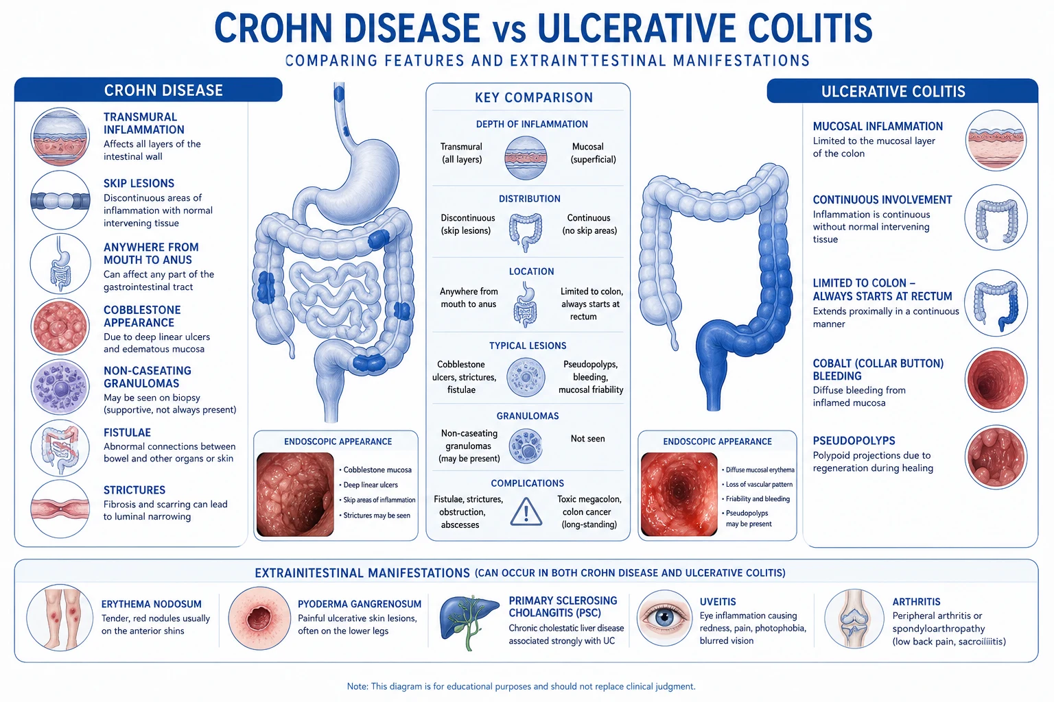
IBD is the prototype complex polygenic disease with a major environmental contribution — the clinical phenotype emerges when genetic susceptibility, an aberrant gut microbiome, a leaky mucosal barrier and a dysregulated immune response conspire in a susceptible host.[1][2]
1. Genetic susceptibility. Genome-wide association studies have identified above 240 risk loci for IBD, the majority shared between CD and UC, accounting for approximately 25 percent of disease heritability. Key loci and their functional effects: [1]
Crohn disease loci
- NOD2/CARD15 (chromosome 16) — defective recognition of bacterial muramyl dipeptide; ileal CD; stricturing phenotype; fistulae
- ATG16L1 and IRGM — autophagy of intracellular bacteria; ileal CD; Paneth cell dysfunction
- IL23R — altered Th17 signalling; both CD and UC; target of ustekinumab and risankizumab
- TNFSF15 (TL1A) — Asian CD
- ECM1, ADAM30, PTPN22, MST1
Ulcerative colitis loci
- HLA-DRB1*0103 — extensive / severe UC; primary sclerosing cholangitis overlap
- IL23R, IL10, NKX2-3, IL1R2, FCGR2A
- PTPN2, HNF4A, lamin B — epithelial barrier function
- Larger genetic load from mucosal barrier genes than from immune genes
2. Microbiome (dysbiosis). Patients with IBD show reduced microbial diversity, reduced abundance of Faecalibacterium prausnitzii (a major butyrate producer that promotes Treg differentiation and barrier integrity), reduced Bifidobacterium and expansion of pro-inflammatory species — particularly adherent-invasive E. coli (AIEC) in ileal CD, Ruminococcus gnavus in UC, and Mucispirillum in several models. Dysbiosis is thought to be both a cause and a consequence of inflammation.[2]
3. Barrier dysfunction. The intestinal epithelium, the mucus layer (particularly MUC2 in colonic goblet cells) and the tight-junction apparatus (claudins, occludin, ZO-1) are structurally and functionally impaired in IBD, allowing translocation of luminal antigens to the lamina propria. Smoking, NSAIDs, diet and dysbiosis aggravate the defect. [1]
4. Immune dysregulation — Th1/Th17 in CD, atypical Th2 in UC. In CD, lamina propria CD4+ T cells polarise to a Th1 and Th17 phenotype, producing interferon-γ, IL-17, IL-22 and TNF-α under the influence of IL-12 and IL-23 from dendritic cells (which signal via their heterodimeric receptors on the T cell — the rationale for ustekinumab and risankizumab). In UC, the cytokine pattern is less classically Th2 — it shows natural killer T-cell-derived IL-13, IL-5 and increased IL-23/IL-17 signalling with an atypical Th2 signature. TNF-α is the final common effector in both diseases — the rationale for anti-TNF therapy (infliximab, adalimumab, golimumab, certolizumab). Naïve T-cell differentiation into regulatory T cells (Tregs) is impaired, removing an important brake on inflammation. [1]
The result is a chronic, non-resolving transmural (CD) or mucosal (UC) inflammation, with progressive structural damage — fibrosis, stricture, fistula, dysplastic change — that explains the natural history of untreated disease and motivates treat-to-target. [1]
Clinical Presentation
The presentation differs fundamentally between CD and UC. [1]
Crohn disease — variable, segmental, often insidious. [1]
- Abdominal pain — typically right-lower-quadrant (terminal ileal disease), crampy, worse after meals.
- Chronic diarrhoea — non-bloody in pure small-bowel disease; bloody only when colon is involved.
- Weight loss, anorexia, fatigue — driven by malabsorption, anorexia, cytokine-mediated cachexia.
- Perianal disease in up to a third at presentation — skin tags, fissures, fistula-in-ano, perianal abscess.
- Fever — low-grade in uncomplicated disease; high and swinging with intra-abdominal or pelvic abscess.
- Palpable right-lower-quadrant mass — thickened terminal ileum ± mesentery.
- Aphthous oral ulcers, ocular involvement (uveitis, episcleritis), skin (erythema nodosum, pyoderma gangrenosum); seronegative peripheral arthritis (type 2 — pauciarticular, parallel to disease activity) and axial spondyloarthritis (sacroiliitis, ankylosing spondylitis — type 1, independent of disease activity).[2]
Ulcerative colitis — bloody diarrhoea, urgency, tenesmus. [1]
- Bloody diarrhoea is the cardinal presenting symptom, with urgency (inability to defer defecation) and tenesmus (sensation of incomplete evacuation).
- Abdominal cramping in the left lower quadrant and rectal pain with proctitis.
- Systemic features — fever, tachycardia, weight loss — escalate with severity.
- Toxic megacolon — severe systemic toxicity with radiographically dilated colon (above 6 cm transverse diameter) and absent haustration; a surgical emergency.
- Extraintestinal manifestations — primary sclerosing cholangitis, pyoderma gangrenosum (often pathergy), erythema nodosum, uveitis, seronegative arthritis; ankylosing spondylitis in 2 to 6 percent; osteoporosis from chronic inflammation and steroid use.[1][3]
Differential Diagnosis
The differential diagnosis of IBD is broad — both CD and UC mimic infective, ischaemic, drug-induced, radiation and other immune-mediated colitides. The following must be excluded before a diagnosis of IBD is accepted:[1][2][3]
Infective mimics
- *Salmonella*, *Shigella*, *Campylobacter*, *Yersinia enterocolitica* (mimics ileal CD), *E. coli* — stool culture and PCR
- *Clostridioides difficile* — toxin PCR; can trigger and mimic a UC flare; test in every acute severe colitis
- *Entamoeba histolytica* — stool PCR and serology; in returning travellers and migrants
- Cytomegalovirus (CMV) — biopsy immunohistochemistry / PCR of colonic biopsies in severe or steroid-refractory UC
- Mycobacterium tuberculosis — ileocaecal TB mimics ileocaecal CD; biopsy for caseating granulomas and acid-fast bacilli, Quantiferon / IGRA, chest radiograph
- HIV, *Schistosoma mansoni* (chronic schistosomiasis), *Strongyloides* in the immunocompromised
Non-infective mimics
- Drug-induced colitis — NSAIDs (most common), mycophenolate, immune checkpoint inhibitors, ipilimumab / nivolumab
- Radiation colitis / proctitis — pelvic radiotherapy for cervical, prostate, rectal, bladder cancer; can present years later
- Ischaemic colitis — watershed areas (splenic flexure, rectosigmoid junction); elderly, atherosclerotic; thumbprinting on imaging
- Microscopic colitis (lymphocytic and collagenous) — chronic watery diarrhoea, normal endoscopy, biopsy diagnosis; more common in older women and with certain drugs (PPIs, NSAIDs, SSRIs)
- Diverticular disease–associated colitis — segmental involvement of sigmoid with diverticula; biopsy helps
- Behcet disease — oro-genital ulcers, uveitis, pathergy, ileocaecal ulceration mimicking CD
- Graft-versus-host disease (GVHD) — post allogeneic stem cell transplant; skin and gut involvement
- Solitary rectal ulcer syndrome, portal-hypertensive colopathy, eosinophilic colitis (infants, peripheral eosinophilia), endometriosis
- Colorectal cancer and lymphoma — must be excluded in older patients, in those with alarm features, and in long-standing IBD with dysplasia surveillance
Clinical & Bedside Assessment
The bedside evaluation of a patient with suspected or established IBD follows a structured sequence — assessment of current disease activity, screening for complications, and a comprehensive search for extraintestinal disease.[1][2][3]
1. History. [1]
- Bowel habit — stool frequency, consistency, the presence of blood, mucus or pus; urgency and tenesmus; nocturnal symptoms; incontinence; perianal pain, swelling, discharge.
- Pain — site, character, radiation, relation to meals and defecation, nocturnal wakening.
- Systemic — fever, rigors (abscess), weight loss, anorexia, fatigue, menstrual disturbance (anaemia, hypoalbuminaemia).
- Drug and family history — current medications (especially NSAIDs, antibiotics, contraceptives), family history of IBD, colorectal cancer, primary sclerosing cholangitis, autoimmune disease. Smoking in CD is a key driver; in UC it is curiously protective.
- Past medical history — appendectomy, abdominal surgery, perinatal and childhood exposures, recent infections, recent antibiotics, travel.
- Diet — fibre intake, dairy (secondary lactose intolerance in CD), red meat and ultra-processed food, alcohol.
- Vaccination status — immunosuppression will be anticipated: varicella, hepatitis B, MMR, HPV, pneumococcal and influenza vaccines should be verified and updated before starting immunomodulator or biologic therapy.
- Pregnancy and contraception plan in women of reproductive age. [1]
2. General examination. [1]
- Vital signs — temperature, pulse, blood pressure, respiratory rate, weight, BMI, fluid balance.
- Pallor (iron deficiency, B12 deficiency in ileal CD, anaemia of chronic inflammation); clubbing (a marker of severe or active IBD, particularly CD); oedema (hypoalbuminaemia from protein-losing enteropathy or malnutrition).
- Skin and mucosal — erythema nodosum (tender red nodules on shins, parallels disease activity), pyoderma gangrenosum (violaceous undermined ulcers, often pathergic), aphthous oral ulcers (CD), peristomal skin breakdown, finger clubbing, pale conjunctivae and koilonychia in chronic iron deficiency. [1]
3. Abdominal examination. [1]
- Inspection — surgical scars (resection, stoma), abdominal distension (megacolon, obstruction, ileus), visible abdominal wall fistulae.
- Palpation — right-lower-quadrant mass (thickened terminal ileum), diffuse tenderness, hepatomegaly (PSC, fatty liver), splenomegaly.
- Percussion — tympany over megacolon or obstruction.
- Auscultation — high-pitched tinkling bowel sounds in obstruction; silent abdomen in ileus/perforation. [1]
4. Perianal examination (essential in every patient). [1]
- Inspection at rest and with gentle lateral traction — skin tags (typical or "elephant ears" in CD), fissures (atypical, broad-based, multiple), anorectal stricture, fistula openings, abscess, ulceration, excoriation.
- Digital rectal examination — tone, mucosal feel, intrinsic mass, blood on glove.
- Anoscopy — anterior anal canal fistula / abscess or distal rectal disease. [1]
5. Joints and axial skeleton. [1]
- Peripheral joints — swollen, tender joints (especially ankles, knees, wrists); dactylitis; enquire about back pain, stiffness and improvement with exercise (inflammatory axial disease). [1]
6. Ocular examination. [1]
- Visual acuity, redness, photophobia, pain on accommodation — refer to ophthalmology urgently if suspected uveitis (pain, photophobia, blurred vision) or scleritis. [1]
7. Nutritional assessment. [1]
- BMI, mid-arm circumference, hand-grip strength; serum albumin, prealbumin, ferritin, B12, folate, 25-OH vitamin D, zinc; dietetic referral for moderate to severe malnutrition. [1]
8. Mental-health screen. [1]
- Anxiety, depression, fatigue, sleep disturbance, fear of incontinence, work impairment, sexual dysfunction, body-image concerns (stoma, fistulae, weight loss). IBD carries a substantial psychological burden. [1]
Investigations
Investigation in IBD is layered — establish the diagnosis, define extent and severity, screen for complications, and exclude infection before any immunosuppression.[1][2][3]
1. Initial laboratory work-up. [1]
- Full blood count, differential, blood film — anaemia (iron, B12, folate, anaemia of chronic inflammation), leucocytosis (abscess, severe flare), thrombocytosis (inflammation, iron deficiency).
- CRP and ESR — markers of systemic inflammation; CRP is more responsive in CD than ESR.
- Urea, electrolytes, creatinine, eGFR — baseline renal function before imaging and immunosuppression.
- Liver biochemistry (bilirubin, ALT, ALP, GGT) — cholestatic pattern suggests PSC (MRCP).
- Albumin — marker of severity and malnutrition; low albumin in acute severe UC predicts steroid failure.
- Iron studies, ferritin, transferrin saturation, B12, folate, 25-OH vitamin D — micronutrient deficiencies.
- TPMT activity (or NUDT15 genotyping in East Asian patients) — before azathioprine / mercaptopurine.
- Stool microbiology — C. difficile toxin / PCR; stool culture and PCR for Salmonella, Shigella, Campylobacter, Yersinia, enteric viruses; stool parasites for travellers; Entamoeba histolytica PCR in migrants.
- Faecal calprotectin — a calcium- and zinc-binding protein released by neutrophils; above 150 to 250 µg/g strongly suggests active intestinal inflammation in the right clinical setting (a normal calprotectin has a high negative predictive value for IBD in primary care).
- Serology — perinuclear antineutrophil cytoplasmic antibodies (p-ANCA) are positive in about 60 to 70 percent of UC and anti-Saccharomyces cerevisiae antibodies (ASCA) in about 50 to 70 percent of CD. Their main role is in the differentiation of indeterminate colitis (p-ANCA positive / ASCA negative favours UC; the reverse favours CD).
- Hepatitis B, hepatitis C, HIV, varicella, MMR, EBV — pre-immunosuppression.
- Quantiferon / IGRA and chest radiograph — exclude TB before anti-TNF therapy, particularly in endemic regions. [1]
2. Endoscopy. [1]
- Ileocolonoscopy with segmental biopsies — the diagnostic cornerstone. Multiple biopsies from the terminal ileum, every colonic segment, and rectum (minimum 2 per segment; 5 or more to maximise granuloma yield in CD); macroscopic appearance, extent, disease pattern, biopsies for histology and CMV PCR in severe disease.
- Upper endoscopy with duodenal biopsies in CD when upper GI symptoms are present, in high-risk phenotypes and in suspected monogenic IBD.
- Wired or video capsule endoscopy for suspected small-bowel CD when MRE is normal or inconclusive — risk of retention in stricturing disease; perform a patency capsule first. [1]
3. Cross-sectional imaging. [1]
- Magnetic resonance enterography (MRE) — preferred for small-bowel assessment in CD; identifies mural inflammation, strictures, fistula, abscess, mesenteric inflammation (comb sign) without ionising radiation; safe in young patients.
- CT enterography — equivalent accuracy to MRE, faster, ionising radiation; use in emergencies when MRE is unavailable.
- Contrast-enhanced abdominopelvic CT — for the acute presentation: rule out abscess, perforation, obstruction.
- Pelvic MRI — perianal and pelvic fistulising disease, the gold standard for mapping complex perianal anatomy.
- Plain abdominal radiograph — toxic megacolon (transverse colon diameter above 6 cm, loss of haustration), obstruction, perforation. [1]
4. CMV PCR on colonic biopsies — in acute severe UC (or flares on immunosuppression) before rescue, because CMV can drive steroid-refractory disease. [1]
5. Stool studies for infection — C. difficile toxin / PCR, enteric pathogens, TB (when relevant) — mandatory before any escalation of immunosuppression. [1]
Inflammatory Bowel Disease — key numbers
Management — Resuscitation
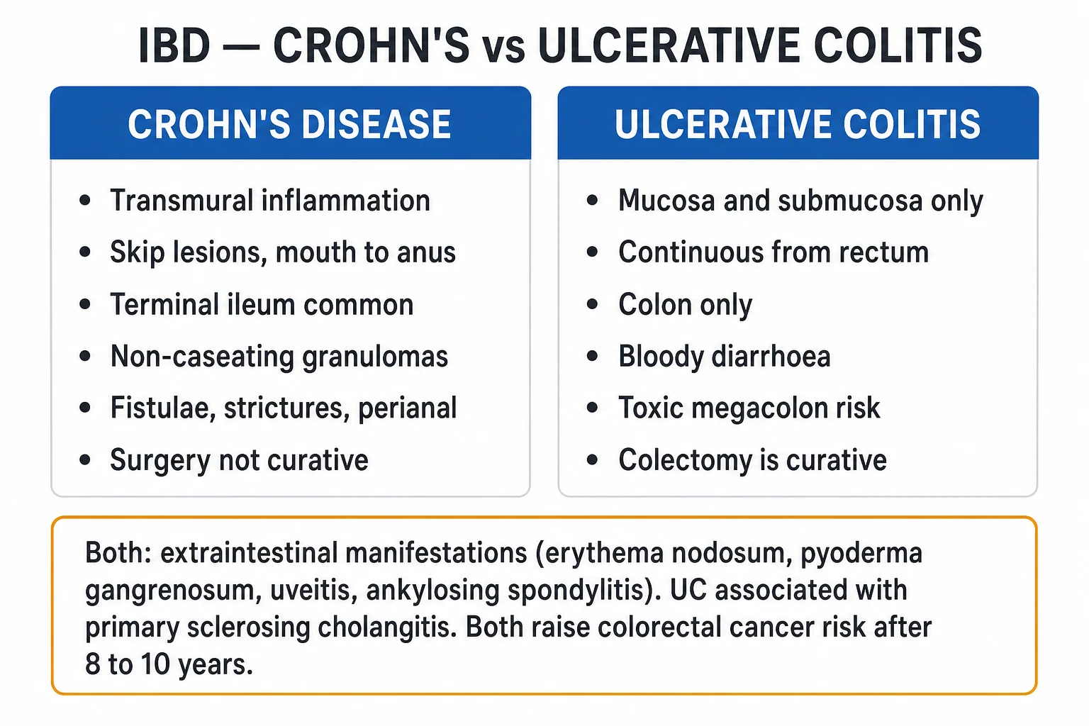
The resuscitation problem in IBD is acute severe ulcerative colitis (ASUC) — a gastrointestinal emergency with a mortality of 1 to 3 percent even in expert centres. The diagnostic threshold is the Truelove and Witts criteria: more than 6 bloody stools per day plus at least one of fever above 37.8 °C, tachycardia above 90/min, anaemia (Hb below 105 g/L) or ESR above 30 mm/hour. Acute severe CD with obstruction, abscess or perforation is similarly managed on a surgical-medical pathway.[1][3]
1. Resuscitate the patient. [1]
- Two large-bore IV cannulae; FBC, U&E, CRP, LFTs, albumin, magnesium, blood cultures; group and save / crossmatch (colectomy is never off the table in this population).
- IV crystalloid resuscitation with electrolyte replacement — particularly potassium and magnesium (hypokalaemia and hypomagnesaemia worsen disease activity and prolong the QT).
- Transfuse if Hb below 70 to 80 g/L or symptomatic anaemia; aim for Hb above 100 g/L in ASUC (per Oxford criteria, transfusion at Hb under 100 may reduce colectomy).
- VTE prophylaxis — all admitted IBD patients should receive prophylactic low-molecular-weight heparin (e.g., enoxaparin 40 mg SC daily) unless actively bleeding; IBD flares raise VTE risk 3- to 6-fold.
- IV hydrocortisone 100 mg every 6 hours (or methylprednisolone 60 mg IV daily) — first-line; do not delay for endoscopy if the diagnosis is established and C. difficile is excluded.
- Broad-spectrum IV antibiotics (e.g., ceftriaxone 1 g IV daily + metronidazole 500 mg IV every 8 hours) — if perforation, abscess or systemic sepsis is suspected.
- Exclude CMV (colonic biopsy PCR / immunohistochemistry) and C. difficile (stool PCR) in every patient — both are modifiable causes of steroid-refractory disease. [1]
2. Reassess on day 3. Approximately 70 percent of ASUC patients respond to IV steroids by day 3. The validated Travis index (CRP above 45 mg/L and stool frequency above 8/day on day 3) predicts steroid failure with about 85 percent specificity. Patients meeting the Travis criteria should proceed to rescue therapy that same day.[3]
3. Rescue therapy — decision. [1]
- Infliximab 5 mg/kg IV — a single induction dose in ASUC; ACCELERATE (2024) supports intensified induction (10 mg/kg) in selected patients with very high CRP and severe disease, but standard practice in most centres remains 5 mg/kg, then reassess.
- Ciclosporin 2 mg/kg/day continuous IV infusion (target trough 150 to 250 ng/mL) — equivalent efficacy to infliximab in trials (CONSTRUCT), reserved for non-naïve centres; nephrotoxic; requires IV then oral bridging to thiopurine.
- Choice between infliximab and ciclosporin depends on local expertise, prior biologic exposure (ciclosporin not used after anti-TNF failure), and renal function. [1]
4. Failure of rescue therapy on day 5 to 7. The patient, the gastroenterologist and the colorectal surgeon must agree on the next step now. Subtotal colectomy with end ileostomy (Hartmann) is the life-saving operation; it is never an admission of failure. Delaying surgery in fulminant disease is the commonest cause of preventable mortality. [1]
5. Surgical emergencies. Perforation, uncontrolled haemorrhage, toxic megacolon unresponsive to medical therapy, and refractory obstruction are immediate indications for laparotomy. [1]
Management — Definitive & Stepwise
The definitive medical management of UC and CD is stepped, individualised, and now oriented to objective treat-to-target rather than symptoms alone (STRIDE-II).[1][3][4][6]
5-aminosalicylates (5-ASA, mesalazine). [1]
- First-line for mild-moderate UC and maintenance after remission. Mesalazine 2.4 to 4.8 g daily PO (active UC), 2 g daily PO (maintenance); mesalazine 1 g PR (suppository) nightly for proctitis, mesalazine 2 g PR (enema) nightly for left-sided disease. Combination PO + PR is more effective than either alone.
- Sulphasalazine 1 g two to four times daily — cheaper but sulfa-related side-effects (rash, haemolysis, oligospermia) limit use; the active moiety is 5-ASA delivered by bacterial cleavage of the sulfa bond.
- 5-ASA is largely ineffective in CD (the Cochrane review shows no benefit for maintenance); budesonide 9 mg PO daily for 8 to 12 weeks is the preferred first-line for mild-moderate ileocaecal CD. [1]
Corticosteroids. [1]
- Induction only — never maintenance (toxicity outweighs benefit beyond short courses).
- Prednisolone 40 to 60 mg PO daily for 1 to 2 weeks, then taper by 5 mg weekly (or equivalent) for active flares.
- Budesonide 9 mg PO daily for mild-moderate ileocaecal CD — high first-pass hepatic metabolism limits systemic toxicity.
- IV hydrocortisone 100 mg every 6 hours for ASUC.
- Never re-treat chronic steroid-dependent disease with long steroids — add a steroid-sparing immunomodulator. [1]
Thiopurines (azathioprine, mercaptopurine). [1]
- Azathioprine 2 to 2.5 mg/kg PO daily (or mercaptopurine 1 to 1.5 mg/kg daily) — for maintenance of steroid-induced remission in UC and CD, steroid sparing, post-operative Crohn prophylaxis (Rutgeerts i2 or higher). Onset of action is 6 to 12 weeks (overlap with steroids during this window).
- Check TPMT activity before starting (low activity → fatal myelosuppression); NUDT15 genotyping in East Asian patients; baseline FBC, LFTs and weekly FBC for 4 weeks, then monthly.
- Toxicity — bone marrow suppression, hepatotoxicity, pancreatitis (idiosyncratic, usually early), lymphoproliferative disorders after years of use, rare but life-threatening post-mononucleosis EBV-driven lymphoma (consider EBV serology before starting). [1]
Methotrexate. [1]
- Subcutaneous or intramuscular — 25 mg weekly for induction, then 15 mg weekly (oral bioavailability is suboptimal). Add folic acid 5 mg weekly on a non-methotrexate day.
- Maintenance of remission in CD (stronger evidence than thiopurines in CD; not supported in UC).
- Teratogenic — strict contraception in women of reproductive age (and men) for at least 3 months after stopping. [1]
Anti-TNF agents (infliximab, adalimumab, golimumab, certolizumab). [1]
- Infliximab 5 mg/kg IV at weeks 0, 2, 6 then every 8 weeks — for moderate-severe CD or UC refractory or intolerant to steroids and immunomodulators, and for rescue in ASUC. Combination with azathioprine is more effective than either alone for induction of steroid-free remission in moderate-severe disease (SONIC and UC SUCCESS), but with higher infection risk in older patients (when to add an immunomodulator vs. monotherapy is now individualised).[5]
- Adalimumab 160 mg SC at week 0, 80 mg at week 2, then 40 mg every 2 weeks — subcutaneous, home-administered; effective in CD and UC. Certolizumab pegol is efficacious in CD; golimumab in UC.[2][3]
- Loss of response — check drug levels and anti-drug antibodies; intensify dosing (10 mg/kg or shorten interval), add immunomodulator to reduce immunogenicity, or switch class.
- Pre-treatment — exclude latent TB (Quantiferon / IGRA + chest radiograph), hepatitis B and HIV; vaccinate before starting; consider varicella and HPV if not already immune.
Anti-integrin (vedolizumab) and anti-IL12/23 (ustekinumab) and anti-IL23 (risankizumab). [1]
- Vedolizumab 300 mg IV at weeks 0, 2, 6 then every 8 weeks — gut-selective anti-α4β7 integrin; excellent safety profile (no PML risk) in long-term use; first-line biologic in older or infection-prone patients.
- Ustekinumab 6 mg/kg IV loading, then 90 mg SC every 8 weeks — anti-IL12/23 (p40); effective in moderate-severe CD and UC.
- Risankizumab 600 mg IV at weeks 0, 4, 8 then 360 mg SC every 8 weeks — anti-IL23 (p19); more selective than ustekinumab; effective in moderate-severe CD (ADVANCE, MOTIVATE, FORTIFY) and increasingly in UC. [1]
Small molecules (JAK inhibitors, S1P modulators). [1]
- Tofacitinib 10 mg PO twice daily for 8 weeks (acute UC) then 5 mg twice daily (maintenance) — oral JAK inhibitor for moderate-severe UC; rapid onset; boxed warning for VTE, major adverse cardiac events and malignancy in patients above 50 with cardiovascular risk factors — risk-benefit must be discussed before initiation.
- Upadacitinib 45 mg PO daily for 8 weeks (induction) then 15 to 30 mg daily (maintenance) — selective JAK1 inhibitor; effective in moderate-severe UC and now CD.
- Ozanimod 1 mg PO daily — S1P receptor modulator for moderate-severe UC; monitor FBC and LFTs. [1]
Antibiotics and probiotics. Metronidazole 400 to 500 mg PO three times daily and ciprofloxacin 500 mg twice daily have a role in perianal CD, intra-abdominal abscess (with drainage), pouchitis, and post-operative prophylaxis — but not as maintenance therapy for active luminal IBD. [1]
Surgical management. [1]
- UC — restorative proctocolectomy with ileal pouch-anal anastomosis (IPAA) — usually two or three stages (subtotal colectomy with end ileostomy, then pouch construction, then ileostomy closure); curative of the colonic disease, but chronic pouchitis in 30 to 50 percent and pouch failure in 5 to 10 percent; high fertility costs (consider staged procedure in young women). End ileostomy alone is the safer alternative for frail or steroid-dependent patients.
- CD — segmental resection of the diseased segment (ileocolonic, segmental colonic, or strictureplasty for short jejunal/ileal strictures to spare bowel); perianal disease — seton drainage, fistulotomy, LIFT, advancement flap, stem-cell therapy in selected centres. Recurrence at the anastomosis is the rule (Rutgeerts i0 to i4) and is the rationale for post-operative prophylaxis with thiopurine, anti-TNF, or 5-ASA in CD. [1]
Stepwise Management
The 5-ASA → corticosteroids → immunomodulator → biologic sequence is now modified by disease risk stratification at diagnosis (top-down vs step-up) and the STRIDE-II treat-to-target framework of clinical, biochemical and endoscopic remission.[4][6]
Stepwise induction of remission in UC by severity (per ACG / ECCO):[1][3]
- Mild proctitis — top rectal 5-ASA (suppository 1 g nightly) ± oral 5-ASA 2 to 4 g daily.
- Mild-moderate left-sided UC — oral 5-ASA ≥ 2.4 g/day + rectal 5-ASA enema ± oral corticosteroid (budesonide MMX 9 mg/day or prednisolone 40 mg with taper).
- Mild-moderate extensive UC — oral 5-ASA ≥ 4.8 g/day ± rectal 5-ASA; escalate to oral prednisolone or biologic if inadequate.
- Moderate-severe UC refractory to 5-ASA / steroids — biologic (anti-TNF, vedolizumab, ustekinumab) or JAK inhibitor (tofacitinib, upadacitinib) for induction and maintenance; immunomodulator (thiopurine or methotrexate) as steroid-sparing adjunct.
- ASUC — IV steroids → infliximab or ciclosporin rescue → colectomy if rescue fails. [1]
Stepwise in CD by severity and phenotype (per ECCO):[2]
- Mild ileocaecal — budesonide 9 mg daily or 5-ASA; glucocorticoid-sparing thiopurine if steroid-dependent.
- Moderate CD — systemic corticosteroids (prednisolone 40 to 60 mg with taper) plus thiopurine or methotrexate for maintenance; early biologic (anti-TNF, vedolizumab, ustekinumab, risankizumab) for steroid-sparing and better outcomes.
- Severe CD (high-risk phenotype — stricturing, penetrating, perianal, extensive small-bowel, deep ulcers on endoscopy) — top-down anti-TNF or anti-IL23 biologic with immunomodulator for induction and maintenance (PROFILE-style strategy). The historical step-up sequence is now overruled by the prognostic argument: early biologic in high-risk disease reduces hospitalisation, surgery and permanent structural damage.
- Stricturing CD with obstructive symptoms — endoscopic balloon dilatation of short anastomotic strictures; resection or strictureplasty for symptomatic strictures refractory to medical therapy.
- Penetrating / fistulising CD — seton drainage and biologic (anti-TNF is best evidenced) with MRI pelvis follow-up.
- Perianal CD — combined surgical and anti-TNF. [1]
Treat-to-target monitoring schedule (STRIDE-II): [1]
- Week 0 — baseline clinical activity, CRP, faecal calprotectin, endoscopic activity (Mayo UC, SES-CD Crohn); set the target.
- Week 10 to 14 — reassess clinical and biochemical response (CRP / calprotectin).
- Months 6 to 12 — endoscopic re-assessment (the new standard). Target: Mayo endoscopic subscore 0 to 1 with MES 0 ideal in UC; SES-CD under 3 in CD.
- Long-term — endoscopic surveillance for dysplasia in UC; MR enterography every 6 to 12 months in CD small-bowel disease; ileocolonoscopy at 6 to 12 months after CD resection (Rutgeerts) with step-up of prophylaxis if i2 to i4. [1]
Specific Subtypes & Scenarios
Crohn disease subtypes: [1]
- Ileal (Montreal L1) — most common; terminal ileum the commonest site; presents with right-lower-quadrant pain, obstructive episodes, B12 deficiency; high stricturing rate.
- Ileocolonic (L3) — ileum and colon; mixed inflammatory and stricturing behaviour.
- Colonic (L2) — bloody diarrhoea, urgency (mimics UC); tends to be more inflammatory, less penetrating than ileal.
- Upper GI (L4 modifier) — mouth, oesophagus, stomach, duodenum, proximal small bowel; aggressive, often with strictures.
- Perianal (p) — skin tags (often the sentinel lesion), fissures (broad-based, painless), anorectal stricture, fistula-in-ano, anorectal abscess; MRI pelvis to map fistula tracts; combined surgical drainage and biologic induction.
- Stricturing (B2) — fibrotic narrowing; obstructive episodes; endoscopic balloon dilatation, strictureplasty, segmental resection; medical therapy alone cannot reverse established fibrosis.
- Penetrating (B3) — intra-abdominal abscess, fistula, free perforation; abscess drainage first, then biologic.
- Fistulising — enteric, enterovesical, enterocutaneous, enteroenteric, perianal. [1]
UC subtypes: [1]
- Ulcerative proctitis (E1) — rectum only; tenesmus, fresh blood; PR 5-ASA is the cornerstone.
- Left-sided (E2) — distal to splenic flexure; combined PO + PR 5-ASA.
- Extensive / pancolitis (E3) — beyond hepatic flexure; higher risk of severe flare, CRC and PSC; surveillance colonoscopy from 8 years of disease.
- Acute severe UC (ASUC) — Truelove and Witts; IV steroids, rescue with infliximab or ciclosporin, early surgery.
- Pouchitis — inflammation in an ileal pouch after IPAA for UC; presents with increased stool frequency, urgency, pelvic cramping; metronidazole 400 mg three times daily for 2 weeks, or ciprofloxacin, or oral budesonide; chronic pouchitis may respond to adalimumab or ustekinumab. [1]
Extraintestinal manifestations (EIMs): [1]
- Musculoskeletal — type 1 (peripheral pauciarticular) — parallels disease activity, lower-limb joints; type 2 (polyarticular) — independent of disease activity; axial spondyloarthritis (sacroiliitis, ankylosing spondylitis) — independent course.
- Skin — erythema nodosum (parallel, resolves with disease control), pyoderma gangrenosum (independent, often pathergy, needs systemic treatment — corticosteroids, cyclosporine, anti-TNF).
- Ocular — episcleritis (parallel), uveitis (independent — pain, photophobia, blurred vision; ophthalmology referral within 24 hours; topical and systemic steroid, anti-TNF).
- Hepatobiliary — primary sclerosing cholangitis (PSC) — cholestatic LFTs, MRCP; 2 to 7 percent of UC, 1 to 3 percent of CD; lifetime risk of cholangiocarcinoma; colon-CRC risk is substantially higher in PSC-IBD.
- Oral — aphthous stomatitis (CD); orofacial granulomatosis. [1]
Complications
Acute / structural
- Toxic megacolon (UC) — transverse colon above 6 cm, systemic toxicity — NBM, IV fluids, IV steroids, broad-spectrum antibiotics, urgent surgical review
- Perforation — free air, peritonitis — emergency laparotomy
- Intra-abdominal and pelvic abscess — IV antibiotics, percutaneous or surgical drainage, then biologic induction
- Fistula-in-ano, enteric, enterovesical, enterocutaneous, rectovaginal — seton drainage for perianal, anti-TNF first line
- Fibrotic stricture — endoscopic balloon dilatation, strictureplasty, resection
- Acute severe colitis — emergency
Chronic / systemic
- Colorectal cancer — 0.5 to 1 percent per year after 8 to 10 years of extensive UC, cumulative 8 percent at 30 years — surveillance colonoscopy with chromoendoscopy from year 8
- Primary sclerosing cholangitis and cholangiocarcinoma — lifetime risk
- Osteopenia and osteoporosis — chronic inflammation, corticosteroids, malabsorption; DEXA at diagnosis and after prolonged steroids
- Venous thromboembolism — 3- to 6-fold higher in IBD flares; VTE prophylaxis during hospital admission
- Malnutrition, micronutrient deficiency (iron, B12, folate, vitamin D, zinc, selenium)
- Amyloidosis (AA — chronic inflammatory burden)
- Pyoderma gangrenosum — independent course, often pathergy; systemic anti-TNF or ciclosporin
- Infertility (in women after IPAA pouch surgery) and sexual dysfunction
- Pouchitis (30 to 50 percent after IPAA), pouch failure (5 to 10 percent)
Prognosis & Disposition
Prognosis. IBD is a lifelong disease with relapsing and remitting behaviour. Outcomes depend on age at onset, disease extent, phenotype (stricturing / penetrating in CD; pancolitis and acute severe in UC), treatment adherence, and time to effective therapy. The cumulative lifetime risk of surgery is approximately 50 percent in CD and 10 to 30 percent in UC (UC colectomy has fallen in the biologic era). CRC risk is most strongly linked to duration and extent of UC, concomitant PSC, prior dysplasia, and family history of CRC — the rationale for surveillance colonoscopy with chromoendoscopy from 8 to 10 years of extensive colitis. Mortality is modestly increased over the background population, principally from CRC, cholangiocarcinoma, VTE, and treatment-related complications (immunosuppression, infection, malignancy).[1][2][3]
Disposition and follow-up cadence: [1]
- Stable mild-moderate UC in remission — 6 monthly review with FBC, CRP, faecal calprotectin, LFTs; annual surveillance colonoscopy after 8 to 10 years of extensive disease.
- CD in clinical and biochemical remission — 3 to 6 monthly review; MR enterography every 6 to 12 months in small-bowel disease; ileocolonoscopy at 6 to 12 months after resection (Rutgeerts).
- Biologic or immunomodulator therapy — clinic review every 3 to 6 months; trough drug levels where relevant; vaccinations annually (influenza) and as per schedule; latent TB and hepatitis B reactivation screening before each biologic escalation.
- Pregnancy, paediatric and elderly subgroups — joint clinic with obstetrics, paediatrics and geriatrics respectively. [1]
Counselling at diagnosis — provide written information, IBD-nurse contact, dietary advice, mental-health support, fertility preservation where relevant, and signpost to patient organisations (Crohn's and Colitis UK, Crohn's and Colitis Foundation of America, IOIBD). [1]
Special Populations
Paediatric IBD. [1]
- Phenotype is more severe at onset than adult disease — extensive UC, ileocolonic CD with upper GI involvement, rapid progression to structuring or penetrating disease. Consider monogenic IBD (IL10, IL10R, XIAP, CYBB, NCF4 mutations) in infantile-onset or refractory disease with extraimmune features.
- First presentation is often growth failure and delayed puberty; chronic inflammation, corticosteroid use and reduced intake all contribute. Exclusive enteral nutrition (EEN) for 6 to 8 weeks is as effective as corticosteroids for inducing remission in mild-moderate paediatric CD.
- PUCAI / PCDAI scoring; colonoscopic and small-bowel imaging; paediatric psychological support.
- Drug dosing is weight-based; biologic approvals are wider for paediatric disease (infliximab, adalimumab); transition to adult services is a high-risk period. [1]
Pregnancy. [1]
- Pregnancy outcome is best when IBD is in remission at conception and throughout pregnancy. Active disease, malnutrition, anaemia and corticosteroid exposure all increase the risk of preterm birth, low birth weight and postpartum flare.
- Maintenance therapy should generally continue through pregnancy — 5-ASA, azathioprine and infliximab (with last dose at 16 to 20 weeks to minimise neonatal exposure) are acceptable; methotrexate is teratogenic and must be stopped before conception (and in men before conception); tofacitinib, upadacitinib are contraindicated.
- Vaginal delivery is acceptable except — absolute contraindication: active perianal disease; relative: ileal pouch-anal anastomosis (high pouch-vaginal fistula risk).
- Mode of delivery and surgical decisions should be discussed in a multidisciplinary clinic with obstetricians. [1]
Elderly. [1]
- CD rather than UC, less severe phenotype, more left-sided or distal disease; more comorbidities (cardiovascular, renal); higher rates of infection on immunomodulators and biologics.
- Vedolizumab favoured by virtue of its gut-selective action and excellent safety profile.
- Polypharmacy, falls risk, and colorectal cancer screening require specific attention. [1]
Post-operative CD. [1]
- Recurrence is the rule — 70 to 90 percent endoscopic recurrence within 1 year, 30 percent clinical recurrence within 3 years.
- Risk stratification — high-risk disease (smoking, penetrating disease, prior resection, two or more postoperative predictors, fistulising or stricturing behaviour) → biologic prophylaxis (anti-TNF or anti-IL23), ideally within 4 weeks of surgery.
- Lower-risk patients — endoscopic surveillance at 6 to 12 months; start or escalate prophylaxis if Rutgeerts i2 to i4.[2]
Evidence & Guidelines
The contemporary evidence base for IBD is dominated by well-conducted randomised controlled trials, large registries and continuous guideline updates. The exam candidate must know the principal guidelines and the few landmark trials.[1][2][3][4][5][6]
Guidelines: [1]
- ACG Clinical Guideline: Ulcerative Colitis in Adults (Rubin et al., 2019) — PMID 30840605 — outlines initial evaluation, severity stratification by Truelove and Witts and Mayo, the 5-ASA and steroid induction sequence, and the role of biologics and small molecules.
- ECCO Guidelines on Therapeutics in Crohn's Disease: Medical Treatment (Torres et al., 2020) — PMID 31711158 — European framework for induction and maintenance of CD by phenotype and severity, with risk-stratified top-down vs step-up.
- ECCO Guidelines on Therapeutics in Ulcerative Colitis: Medical Treatment (Raine et al., 2022) — PMID 34635919 — analogous European framework for UC, including ASUC rescue algorithms and drug-specific positioning.
- STRIDE-II (Turner et al., 2021) — PMID 33359090 — update of the original STRIDE (Peyrin-Biroulet 2015 — PMID 26303131) for treat-to-target — clinical, biochemical (CRP, faecal calprotectin) and endoscopic targets with explicit time-points.
- BSG guidelines — UK-specific consensus on 5-ASA dosing, thiopurines, biologics, ASUC rescue and post-operative CD prophylaxis.
- Toronto Consensus Statements on the management of CD and UC, ECCO-ESPGHAN paediatric IBD guidelines. [1]
Landmark trials: [1]
- Targan SR et al., NEJM 1997 — PMID 9321530 — the original infliximab induction trial in moderate-severe CD — established anti-TNF as a therapy for the first time.
- SONIC — combination infliximab plus azathioprine superior to either alone for steroid-free remission in moderate CD (raises infection risk in older patients).
- UC SUCCESS — infliximab plus azathioprine superior to either alone for steroid-free remission in moderate-severe UC.
- ACCENT I and II — maintenance infliximab in CD and fistulising CD.
- UNITI / IM-UNITI — ustekinumab induction and maintenance in moderate-severe CD.
- OCTAVE Induction and Sustain — tofacitinib in moderate-severe UC.
- VARSITY — vedolizumab superior to adalimumab for endoscopic remission in moderate-severe UC.
- ADVANCE / MOTIVATE / FORTIFY — risankizumab induction and maintenance in moderate-severe CD.
- CONSTRUCT — infliximab and ciclosporin equivalent rescue in ASUC, with different cost and safety profiles. [1]
Exam Pearls
Crohn vs UC — mnemonic CROSUL
CROHNS
Cobblestone mucosa, fissuring ulcers, mural transmural inflammation, granulomas in 30 to 50 percent
Skip lesions, rectum spared in CD (involved in UC)
Mouth to anus, transmural with strictures and fistulae
Surgery not curative; Rutgeerts scoring for post-op recurrence
Granulomas on biopsy; NOD2 ileal stricturing; smoking a strong promoter of CD
Smoking is protective in UC but worsens CD
Continuous from rectum; bloody diarrhoea; pseudopolyps; lead-pipe colon
- Crohn disease = transmural, skip lesions, anywhere from mouth to anus, fistulae / strictures / abscesses, granulomas, surgery is not curative.
- UC = mucosal, continuous from rectum, bloody diarrhoea, urgency, tenesmus, pseudopolyps, lead-pipe colon on barium, colectomy is curative.
- Smoking — protective in UC (paradoxical), strongly promotes CD — an exam favourite.
- Appendectomy for true appendicitis before 20 — protective against UC.
- Treatment ladder — 5-ASA (UC), budesonide (mild-moderate ileocaecal CD), systemic steroids (flares), thiopurine / methotrexate (maintenance and steroid-sparing), biologic (anti-TNF, anti-integrin, anti-IL12/23, anti-IL23), small molecule (JAK, S1P).
- Acute severe UC — Truelove and Witts (>6 bloody stools/day + systemic toxicity); IV hydrocortisone 100 mg every 6 hours; Travis index on day 3 predicts steroid failure; rescue with infliximab 5 mg/kg IV or ciclosporin 2 mg/kg/day IV; failure = surgery.
- CMV — biopsy PCR in severe UC; HIV/HBV/TB screening before biologic.
- STRIDE-II — treat to clinical + biochemical (CRP / calprotectin) + endoscopic remission (Mayo MES 0 to 1, SES-CD less than 3).
- Rutgeerts post-operative CD score at 6- to 12-month ileocolonoscopy; i2+ drives biologic prophylaxis.
- PSC — cholestatic LFTs; MRCP; lifetime cholangiocarcinoma risk; raised CRC risk in PSC-UC.
- CRC surveillance — chromoendoscopy from year 8 to 10 of extensive UC, more often if PSC, prior dysplasia, or family history of CRC.
- VTE prophylaxis in every admitted flare.
- EIMs — type 1 (parallel) and type 2 (independent) arthritis; uveitis is independent and an ophthalmic emergency.
- Pregnancy — keep disease in remission; methotrexate contraindicated; infliximab last dose 16 to 20 weeks; vaginal delivery not safe with active perianal disease. [1]
Exam application bank (NEET-PG / INICET)
One-line answer
Inflammatory bowel disease (IBD) is a group of chronic relapsing immune-mediated disorders of the gastrointestinal tract comprising Crohn disease (CD) — transmural inflammation in a skip-lesion distribution anywhere from mouth to anus, frequently involving terminal ileum, with fistula, stricture, abscess and perianal disease — and ulcerative colitis (UC) — diffuse mucosal inflammation continuous from the rectum proximally, presenting with bloody diarrhoea, urgency and tenesmus. A third working group, IBD-unclassified (IBDU) or colonic IBD type-unclassified, is used when differentiation is not possible on initial work-up. Incidence is highest in the second and third decades (a smaller second peak in the seventh decade) in populations of North European, North American, Australasian and now increasingly South Asian and East Asian descent; the global burden has risen dramatically as newly in
Worked stems (answer without another resource)
Stem 1 — Classic presentation. Map symptoms to mechanism; name the first investigation and first treatment step with dose/route if drug therapy is standard. [1]
Stem 2 — Unstable / complicated. List red flags that force immediate resuscitation, theatre, ICU, antidote, or reperfusion — and what you do in the first 15 minutes. [1]
Stem 3 — Atypical group. Elderly, pregnancy, child, or immunocompromised: how presentation and thresholds change. [1]
Stem 4 — Differential trap. Name the three closest mimics and one discriminator for each. [1]
Stem 5 — Disposition. Who goes home with safety-netting, who is admitted, who needs HDU/ICU/theatre, and what follow-up is mandatory. [1]
Rapid viva checklist
- Definition + classification
- Pathophysiology chain
- Bedside signs / criteria
- Score with exact components (if any)
- Emergency bundle
- Definitive therapy with doses
- Complications of disease and of treatment
- Special populations
- Guideline/trial name if classic
- Three exam traps
Coverage self-check
If you cannot answer any stem above from this page alone, re-read the matching section — the page is intended to be self-sufficient for final-prof and NEET-PG/INICET questions on Inflammatory Bowel Disease.
References
- [1]Rubin DT, Ananthakrishnan AN, Siegel CA, et al. ACG Clinical Guideline: Ulcerative Colitis in Adults Am J Gastroenterol, 2019.PMID 30840605
- [2]Torres J, Mehandru S, Colombel JF, et al. ECCO Guidelines on Therapeutics in Crohn's Disease: Medical Treatment J Crohns Colitis, 2020.PMID 31711158
- [3]Raine T, Bonovas S, Burisch J, et al. ECCO Guidelines on Therapeutics in Ulcerative Colitis: Medical Treatment J Crohns Colitis, 2022.PMID 34635919
- [4]Turner D, Ricciuto A, Lewis A, et al. STRIDE-II: An Update on the Selecting Therapeutic Targets in Inflammatory Bowel Disease (STRIDE) Initiative of the International Organization for the Study of IBD (IOIBD): Determining Therapeutic Goals for Treat-to-Target strategies in IBD Gastroenterology, 2021.PMID 33359090
- [5]Targan SR, Hanauer SB, van Deventer SJ, et al. A short-term study of chimeric monoclonal antibody cA2 to tumor necrosis factor alpha for Crohn's disease. Crohn's Disease cA2 Study Group N Engl J Med, 1997.PMID 9321530
- [6]Peyrin-Biroulet L, Sandborn W, Sands BE, et al. Selecting Therapeutic Targets in Inflammatory Bowel Disease (STRIDE): Determining Therapeutic Goals for Treat-to-Target Am J Gastroenterol, 2015.PMID 26303131