Gastroenterology · General Surgery / General Medicine
Oesophageal & Gastric Cancer
Also known as Oesophageal cancer · Gastric cancer · Stomach cancer · Oesophagogastric cancer · Gastro-oesophageal junction cancer · Gastric carcinoma
Oesophageal and gastric cancer are the two principal upper-gastrointestinal malignancies. Oesophageal cancer has two distinct histologies: adenocarcinoma arising in the lower oesophagus and gastro-oesophageal junction (GOJ) from Barrett's intestinal metaplasia (driven by obesity and chronic acid reflux), and squamous cell carcinoma of the upper and middle oesophagus (driven by alcohol, smoking, very hot beverages, achalasia and nitrosamine-rich diets). Gastric adenocarcinoma is driven by Helicobacter pylori (a Class I carcinogen), salted and smoked diets, smoking, blood group A, family history and pernicious anaemia; the Correa cascade (chronic gastritis through atrophic gastritis, intestinal metaplasia and dysplasia to invasive carcinoma) describes the intestinal-type sequence. Both diseases present late with progressive dysphagia, weight loss, dyspepsia, early satiety, nausea and iron-deficiency anaemia, and gastric cancer may produce haematemesis and a palpable Virchow (supraclavicular) lymph node. Diagnosis is by oesophagogastroduodenoscopy (OGD) with biopsy, with multidisciplinary staging by CT, endoscopic ultrasound (EUS), PET-CT (oesophageal) and staging laparoscopy (gastric/GOJ). Surgery is the only cure — oesophagectomy (after neoadjuvant chemoradiotherapy (CROSS) or chemotherapy) for oesophageal cancer, and gastrectomy with D2 lymphadenectomy plus perioperative FLOT chemotherapy for gastric cancer. H. pylori eradication is a rare, proven population-level cancer-prevention intervention.
On this page & tools
Your progress
Saved locally on this device.
Exam tags
Red flags

Overview & Definition
Oesophageal cancer and gastric cancer are the two principal malignancies of the upper gastrointestinal tract. Although anatomically distinct, they are grouped together in modern oncology because they share a diagnostic pathway (endoscopy with biopsy), a multidisciplinary staging algorithm (CT, EUS, laparoscopy), a curative reliance on radical surgery with perioperative systemic therapy, and — at the gastro-oesophageal junction (GOJ) — an overlapping biology.[1][2]
Oesophageal cancer comprises two entirely different diseases that happen to occupy the same tube: [1]
- Oesophageal adenocarcinoma (OAC) arises in the distal oesophagus and GOJ from Barrett's intestinal metaplasia, a columnar reprogramming of the squamous mucosa in response to chronic acid reflux. Its incidence has risen more than six-fold in Western populations over four decades, tracking the epidemics of obesity and gastro-oesophageal reflux disease (GORD), and it predominates in overweight white males in their sixth to seventh decade.[1]
- Oesophageal squamous cell carcinoma (OSCC) arises in the upper and middle thirds of the oesophagus and is driven by chronic epithelial injury from alcohol, tobacco, very hot beverages, achalasia, caustic injury and nitrosamine-rich diets. OSCC is the dominant subtype globally and is concentrated in the "oesophageal cancer belt" stretching across Central and Eastern Asia, Eastern Africa and parts of South America and the Caspian region.[1][3]
Gastric cancer is overwhelmingly an adenocarcinoma (about 95 percent), with the remainder being lymphomas (mostly MALT), gastrointestinal stromal tumours (GIST) and neuroendocrine tumours. By the Lauren classification, gastric adenocarcinoma splits into an intestinal type (gland-forming, environment-driven, following the Correa cascade — older males, better prognosis) and a diffuse type (poorly cohesive signet-ring cells infiltrating the wall, driven by CDH1 / E-cadherin loss, producing the rigid "leather-bottle stomach" (linitis plastica), worse prognosis).[2]
The clinical flavour of both cancers is stealthy: they grow through the submucosa and muscularis before causing obstructive (dysphagia, vomiting), bleeding (anaemia, haematemesis) or systemic (weight loss, cachexia) symptoms. By the time progressive dysphagia or iron-deficiency anaemia declares itself, the tumour is usually transmural or metastatic. The two highest-yield clinical skills are therefore (i) recognising red-flag dyspepsia and dysphagia for urgent OGD, and (ii) multidisciplinary staging to find the resectable minority for whom surgery with perioperative therapy is curative.[1][2]
Classification
Upper-GI cancer is classified on three axes — histology, anatomical location (Siewert for the GOJ), and Lauren subtype (for gastric adenocarcinoma). [1]

The Siewert classification of GOJ adenocarcinoma (high-yield)[1]
| Type | Tumour centre | Origin | Typical surgery |
|---|---|---|---|
| I | Distal oesophagus, 1 to 5 cm above the GOJ | Barrett's / distal oesophagus growing downward | Ivor Lewis oesophagectomy (treated as oesophageal cancer; neoadjuvant CROSS) |
| II | At the GOJ, 1 cm above to 2 cm below | True cardia / junctional | Total gastrectomy or extended gastrectomy (treated as either; perioperative FLOT) |
| III | Subcardial, 2 to 5 cm below the GOJ | Proximal stomach growing upward | Total gastrectomy (treated as gastric cancer; perioperative FLOT) |
Why it matters: the Siewert type drives the surgical approach and the choice of neoadjuvant strategy (CROSS chemoradiotherapy favours Siewert I; perioperative chemotherapy favours II/III).[1]
Lauren classification of gastric adenocarcinoma[2]
| Feature | Intestinal type | Diffuse type |
|---|---|---|
| Histology | Gland-forming, cohesive | Poorly cohesive signet-ring cells |
| Epidemiology | Older males, high-incidence regions | Younger, female, any region |
| Aetiology | Environmental (H. pylori, diet, salt), Correa cascade | Genetic (CDH1 / E-cadherin loss), hereditary diffuse gastric cancer |
| Macroscopy | Ulcerated, polypoid mass | Linitis plastica (rigid, thickened "leather-bottle" wall) |
| Prognosis | Better (more resectable) | Worse (submucosal spread, early metastasis) |
WHO histological classification of gastric carcinoma[2]
Tubular, papillary, mucinous, poorly cohesive (including signet-ring cell), mixed, and other rare variants (hepatoid, medullary, adenosquamous, neuroendocrine). The poorly cohesive/signet-ring pattern overlaps with Lauren diffuse and carries the poorest prognosis. [1]
Other upper-GI malignancies — distinguish from adenocarcinoma
- Oesophageal — small-cell (neuroendocrine) carcinoma, melanoma, GIST, lymphoma; the cervical oesophagus also harbours head-and-neck-type SCC.
- Gastric — MALT lymphoma (H. pylori-driven, antibiotics-responsive), diffuse large B-cell lymphoma, GIST (KIT/PDGFRA mutation), neuroendocrine tumours (gastrinoma, type II in MEN-1/Zollinger-Ellison). [1]
Oesophageal adenocarcinoma (OAC)
Lower oesophagus / GOJ
- Rising in the West; tracks obesity, GORD and Barrett's metaplasia
- Overweight white males, 6th to 7th decade
- Driven by the metaplasia-dysplasia-carcinoma (Barrett's) sequence
- CROSS neoadjuvant chemoradiotherapy then Ivor Lewis oesophagectomy
- HER2 testing and PD-L1 CPS relevant for advanced disease
Oesophageal SCC (OSCC)
Upper / middle oesophagus
- Dominant subtype globally; the oesophageal cancer belt (Central/East Asia, East Africa)
- Alcohol, tobacco, very hot drinks, achalasia, caustic injury, nitrosamines, tylosis, Plummer-Vinson
- Cervical lesions treated with definitive chemoradiotherapy for organ preservation
- Neoadjuvant chemoradiotherapy (CROSS) or chemotherapy (JCOG9907, cisplatin/5-FU) then surgery
Gastric adenocarcinoma — intestinal (Lauren)
Correa cascade
- H. pylori-driven (Class I carcinogen), salted/smoked diet, blood group A, older males
- Chronic gastritis -> atrophy -> intestinal metaplasia -> dysplasia -> invasive carcinoma
- Gland-forming; more often resectable; better prognosis
- Total/subtotal gastrectomy with D2 lymphadenectomy + perioperative FLOT
Gastric adenocarcinoma — diffuse (Lauren)
Signet-ring / linitis plastica
- CDH1 (E-cadherin) loss; hereditary diffuse gastric cancer; younger, female
- Signet-ring cells infiltrate the wall; rigid 'leather-bottle stomach' (linitis plastica)
- Early submucosal and peritoneal spread; worse prognosis
- Often presents advanced; palliative chemotherapy if unresectable
Epidemiology & Risk Factors
Oesophageal cancer is the eighth most commonly diagnosed cancer and the sixth leading cause of cancer death worldwide (GLOBOCAN 2020). Gastric cancer is the fifth most commonly diagnosed and the fourth leading cause of cancer death. Together they cause more than 1.6 million new cases and 1.2 million deaths each year.[3]
Oesophageal & gastric cancer — the numbers that matter
Risk factors — oesophageal adenocarcinoma[1]
- Barrett's oesophagus (intestinal metaplasia) — a 30- to 60-fold increased risk of OAC; risk concentrated with long-segment disease and dysplasia.
- Chronic GORD — symptomatic reflux confers a 5- to 7-fold risk; duration and severity matter.
- Obesity (especially central/visceral) — increases reflux and is independent of reflux via adipokine-driven inflammation.
- Male sex, white ethnicity, age over 50, smoking.
- Tylosis (Howel-Evans syndrome, autosomal dominant) — nearly certain oesophageal SCC by middle age (parenthetically an SCC, not adenocarcinoma, risk). [1]
Risk factors — oesophageal squamous cell carcinoma[1][3]
- Alcohol and tobacco (synergistic; 5- to 10-fold together).
- Very hot beverages (above about 65 deg C) — IARC Group 2A carcinogen (mate, hot tea).
- Achalasia (stasis, fermentation, chronic irritation — up to 30-fold lifetime risk).
- Caustic injury / lye stricture — cancer 15 to 40 years later.
- Plummer-Vinson syndrome (dysphagia, glossitis, iron deficiency, post-cricoid web).
- Tylosis (Howel-Evans) — palmoplantar keratoderma, TOC gene / RHBDF2, near-complete lifetime risk.
- Nitrosamine-rich diet, HPV infection, low socioeconomic status, prior radiation, coeliac disease, head-and-neck cancer (field change). [1]
Risk factors — gastric adenocarcinoma (recall "HABITS + BAD")[2]
- H. pylori — IARC Group 1 (Class I) carcinogen; attributable fraction roughly 89 percent of non-cardia gastric cancers. The CagA-positive strain is most virulent.
- Atrophic gastritis and intestinal metaplasia (Correa cascade, often autoimmune pernicious anaemia or chronic H. pylori).
- Blood group A — modest increased risk (receptor for H. pylori adherence / altered inflammation).
- Diet — salted, smoked, pickled foods (N-nitroso compounds); low intake of fresh fruit and vegetables (low ascorbate).
- Smoking — roughly 1.5- to 2-fold risk.
- Family history and hereditary syndromes — hereditary diffuse gastric cancer (CDH1/E-cadherin), Lynch syndrome (MLH1/MSH2/MSH6/PMS2), familial adenomatous polyposis (APC), Peutz-Jeghers (STK11/LKB1), BRCA2, Li-Fraumeni.
- Partial gastrectomy (15 to 20 years later — bile reflux, bacterial overgrowth, nitrosamines).
- Ménétrier's disease, pernicious anaemia (autoimmune gastritis), chronic atrophic gastritis. [1]
Hereditary diffuse gastric cancer (CDH1)[2]
Germline CDH1 mutation (E-cadherin) confers a 70 to 80 percent lifetime risk of diffuse gastric cancer and a high risk of lobular breast cancer. Prophylactic total gastrectomy is offered to asymptomatic adult carriers (typically late teens to 20s/30s) — the paradigm of precision cancer prevention. [1]
Indian epidemiology[3]
Gastric cancer concentrates in the north-east (especially the Kashmir valley, where dietary nitrosamines in sun-dried and smoked foods are extreme) and parts of southern India; oesophageal SCC is common in the north-eastern corridor (tobacco, betel quid, very hot beverages). Late presentation dominates because of limited endoscopic access; H. pylori eradication, tobacco and alcohol control, and lowering dietary salt are the most practical prevention levers. [1]
Pathophysiology
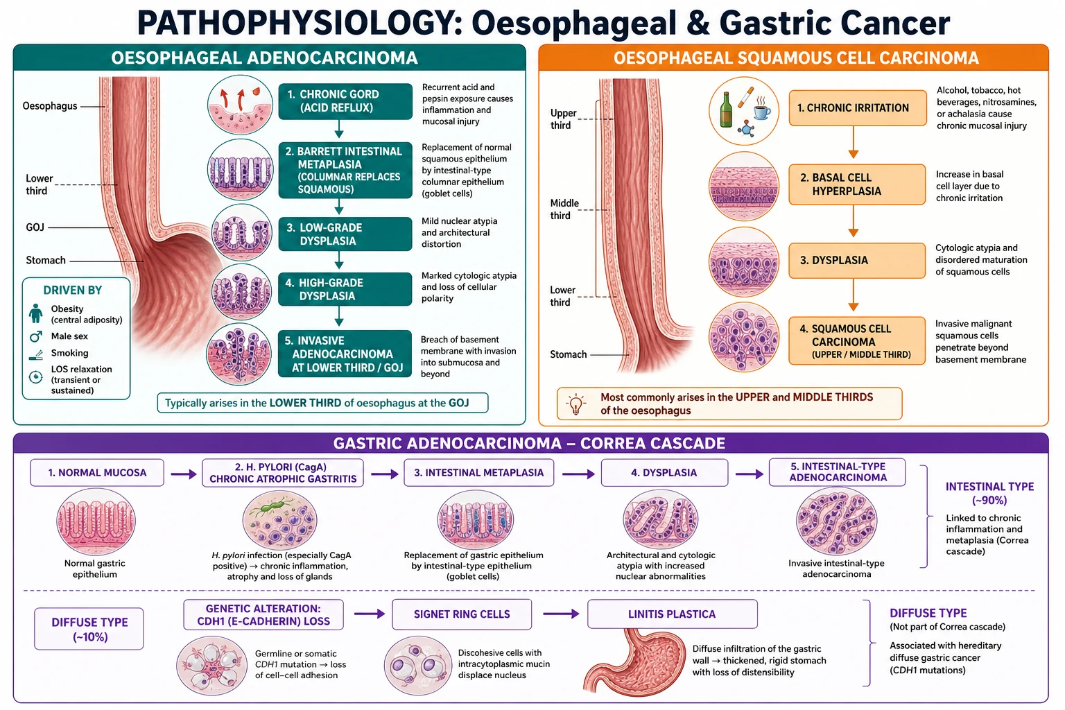
The Barrett's metaplasia-dysplasia-adenocarcinoma cascade (OAC)[1]
In response to chronic acid and bile reflux, the stratified squamous epithelium of the distal oesophagus is replaced by specialised intestinal metaplasia (columnar cells with goblet cells) — Barrett's oesophagus. The molecular switch is driven by CDX1 and CDX2 transcription factors (intestine-determining genes aberrantly activated by bile-acid signalling), which reprogramme the squamous epithelium. This metaplastic mucosa accumulates TP53 mutations, chromosomal instability, LOH, and DNA methylation, progressing through low-grade dysplasia -> high-grade dysplasia -> invasive adenocarcinoma. The risk of progression from non-dysplastic Barrett's is roughly 0.3 to 0.5 percent per year, but high-grade dysplasia carries a yearly risk of 6 percent or more — the rationale for endoscopic surveillance and endoscopic eradication therapy (EMR + radiofrequency ablation).[1]
The Correa cascade — intestinal-type gastric adenocarcinoma[2]
The classic multistep sequence, over decades: [1]
Normal mucosa -> chronic (superficial) gastritis -> atrophic gastritis -> intestinal metaplasia -> dysplasia -> invasive adenocarcinoma. [1]
- H. pylori (especially CagA-positive) injects CagA into epithelial cells via a type IV secretion system, activating SHP-2, ERK/MAPK and NF-kB; its urease generates ammonia, raising local pH; neutrophil-derived reactive oxygen species (ROS) cause DNA damage; chronic inflammation drives epithelial turnover, mutation and atrophy.
- Loss of acid-secreting parietal cells (atrophy) raises gastric pH, permitting bacterial overgrowth that converts dietary nitrate to N-nitroso compounds (potent carcinogens).
- Intestinal metaplasia (CDX2-driven, like Barrett's) replaces gastric mucosa with intestinal-type glands; TP53, APC and CDH1 mutations accumulate.
- Dysplasia is the penultimate, potentially reversible step; invasive carcinoma follows basement-membrane breach. [1]
Clinical significance: the Correa cascade makes gastric cancer a largely preventable malignancy — H. pylori eradication interrupts the cascade and reduces incident cancer, the basis for screen-and-treat programmes in high-incidence countries (Japan, Korea, China).[2]
Diffuse gastric adenocarcinoma and CDH1E-cadherin[2]
In the diffuse (Lauren) type, the molecular driver is loss of E-cadherin (CDH1 on 16q22) — either germline (hereditary diffuse gastric cancer) or somatic. E-cadherin is the transmembrane adhesion molecule of the epithelial adherens junction; its loss dissolves cell-cell adhesion, freeing discohesive, mucin-laden signet-ring cells that infiltrate singly through the gastric wall. Submucosal infiltration with desmoplastic fibrosis produces a rigid, thickened, non-distensible stomach — linitis plastica ("leather-bottle stomach") — which presents as early satiety despite a small tumour burden. [1]
Squamous oesophageal carcinoma — mechanism[1]
Chronic injury from ethanol (metabolised by CYP2E1 and alcohol dehydrogenase to acetaldehyde, a DNA-crosslinking carcinogen), tobacco (N-nitrosamines, polycyclic aromatics) and very hot beverages drives basal-cell hyperplasia, dysplasia and carcinoma. East-Asian populations carrying the ALDH2*2 variant (low acetaldehyde dehydrogenase activity) accumulate acetaldehyde and have a markedly raised SCC risk with alcohol — a classic gene-environment interaction. In achalasia, stasis causes fermentation and chronic chemical irritation of retained food, predisposing the dilated segment to SCC. [1]
Molecular subtypes of gastric cancer (TCGA)[2]
The Cancer Genome Atlas divides gastric adenocarcinoma into four molecular subtypes with prognostic and therapeutic implications: [1]
- EBV-positive (~9%) — PIK3CA mutations, PD-L1/PD-L2 overexpression, CDKN2A silencing; best prognosis, highly immunotherapy-responsive.
- Microsatellite-instability-high (MSI-H, ~22%) — defective mismatch repair, hypermutation; good prognosis, responsive to pembrolizumab.
- Genomically stable (~20%) — enriched for diffuse histology and CDH1/RHOA mutations; poor prognosis.
- Chromosomal instability (~50%) — HER2 amplification, receptor tyrosine kinase amplifications; HER2-targeted therapy relevant. [1]
Routes of spread[1][2]
- Direct — submucosal extension (skip lesions via the rich submucosal lymphatic plexus — explains the need for wide margins in oesophagectomy), through the muscularis and serosa into adjacent organs (trachea, aorta, pericardium, pancreas, liver, transverse colon).
- Lymphatic — the named eponymous metastases:
- Virchow's node (left supraclavicular) — Troisier's sign — via the thoracic duct; the classic sign of advanced gastric cancer.
- Irish node (left axillary).
- Sister Mary Joseph nodule (umbilical) — peritoneal spread.
- Blumer's shelf (palpable rectal shelf) — pelvic peritoneal deposit.
- Krukenberg tumour (ovarian) — haematogenous or transcoelomic signet-ring metastasis.
- Haematogenous — liver, lung (cannonball), bone, brain.
- Transcoelomic — peritoneal seeding, ascites, ovarian (Krukenberg). [1]
HER2 and PD-L1 — biology that changes therapy[2][8][9]
HER2 (ERBB2) amplification occurs in roughly 15 to 20 percent of gastric/GOJ adenocarcinomas (more common in intestinal and GOJ tumours); it is the target of trastuzumab (ToGA trial). PD-L1 expression (measured by Combined Positive Score, CPS) predicts response to immune checkpoint inhibitors (nivolumab, pembrolizumab) — benefit is greatest in CPS at least 5 (CheckMate 649).[8][9]
Clinical Presentation
The dominant presentation is late: symptoms arise only when the tumour obstructs, bleeds or metastasises. This is the central reason for the poor overall prognosis and the rationale for urgent endoscopy on red-flag symptoms.[1][2]
Oesophageal cancer
- Progressive dysphagia — the hallmark: solids then soft foods then liquids ("mechanical" pattern, unlike the liquids-and-solids-together pattern of motility disorders like achalasia). It indicates a luminal diameter reduced by roughly two-thirds — a late event.
- Weight loss — often rapid and profound; reflects both obstruction and cachexia (TNF-alpha, IL-6).
- Odynophagia (painful swallowing), retrosternal chest pain, regurgitation of undigested food (risk of aspiration pneumonia).
- Chronic reflux / heartburn (often long-standing in OAC; paradoxically may improve as the tumour scars the GOJ).
- Hoarseness — recurrent laryngeal nerve involvement (advanced).
- Cough on swallowing — tracheo-oesophageal fistula (advanced SCC). [1]
Gastric cancer
- Dyspepsia that is new, progressive, or changed in character; unresponsive to proton-pump inhibitors or H. pylori eradication — the cardinal red flag.
- Early satiety — a small volume of food produces fullness; the hallmark of linitis plastica (non-distensible wall).
- Weight loss, anorexia, fatigue.
- Nausea and vomiting — particularly with antral/outlet obstruction (undigested food, succussion splash) or gastric stasis.
- Iron-deficiency anaemia — occult bleeding from the tumour surface; presents as fatigue, pallor, dyspnoea, occasionally angina.
- Haematemesis or melaena — overt upper-GI bleeding (less common than in peptic ulcer disease, but a recognised presentation).
- Epigastric mass — advanced disease. [1]
The named metastatic signs (must know)[2]
| Sign / eponym | Site | Significance |
|---|---|---|
| Virchow's node (Troisier's sign) | Left supraclavicular fossa | Metastatic gastric (or other abdominal) cancer via the thoracic duct |
| Sister Mary Joseph nodule | Umbilicus | Peritoneal/lymphatic spread |
| Krukenberg tumour | Ovary | Signet-ring cell metastasis (haematogenous/transcoelomic) |
| Blumer's shelf | Rectal shelf on PR exam | Pelvic peritoneal drop metastasis |
| Irish node | Left axilla | Lymphatic spread |
Paraneoplastic phenomena[2]
- Acanthosis nigricans (and tripe palms, sign of Leser-Trélat — sudden eruption of seborrhoeic keratoses) — classic paraneoplastic sign of gastric cancer.
- Trousseau's syndrome (migratory thrombophlebitis) — hypercoagulability.
- Membranous nephropathy (nephrotic syndrome).
- Polyarteritis nodosa (rare association).
- Microcytic anaemia, polymyositis, cerebellar degeneration (rare). [1]
Atypical presentations[1][2]
- Elderly — dysphagia dismissed as "stroke" or "dementia"; anaemia and weight loss attributed to "old age"; dyspepsia treated empirically without endoscopy.
- Diabetic / neuropathic — silent, painless dyspepsia; delayed gastric emptying confounds the picture.
- Young adults — consider hereditary diffuse gastric cancer (CDH1) or Lynch syndrome; diffuse-type, aggressive.
- Immunocompromised — atypical and more rapid course.
- Pregnancy — rare; symptoms (dyspepsia, reflux) overlap with normal pregnancy and delay diagnosis. [1]
Differential Diagnosis
A complete differential, with distinguishing features:[1][2]
The top differentials for the two cardinal presentations are: [1]
- Progressive dysphagia — oesophageal/GOJ cancer vs peptic stricture (long-standing GORD, solids only) vs achalasia (liquids and solids together, bird's-beak) vs eosinophilic oesophagitis (atopy, rings/furrows) vs Schatzki ring / Plummer-Vinson web vs extrinsic compression (goitre, left atrium) vs oesophageal motility disorder.
- Dyspepsia / epigastric pain — gastric cancer vs peptic ulcer disease vs GORD vs functional dyspepsia vs pancreatic cancer (back pain, jaundice) vs biliary colic vs angina/MI.
- Gastric mass on imaging — gastric adenocarcinoma vs GIST (submucosal, CD117/DOG1+) vs gastric lymphoma (MALT/DLBCL) vs neuroendocrine tumour vs metastasis (breast, melanoma) vs Ménétrier's / inflammatory.
- Iron-deficiency anaemia in an adult — upper-GI malignancy (oesophageal/gastric) vs colorectal cancer vs peptic ulcer vs coeliac disease vs angiodysplasia vs menorrhagia.
- Supraclavicular lymph node — reactive vs tuberculosis vs lymphoma vs metastatic (lung, gastric, breast, melanoma). [1]
Differential of progressive dysphagia
| Differential | Key distinguishing features |
|---|---|
| Oesophageal / GOJ cancer | Progressive solids-then-liquids, weight loss, older patient, smoking/alcohol/obesity; OGD + biopsy shows a tumour |
| Peptic stricture | Long-standing GORD, solids only (intermittent), no weight loss, endoscopy shows a distal stricture with erosive oesophagitis |
| Achalasia | Liquids and solids together from onset, regurgitation of undigested food, no weight loss initially; bird's-beak on barium swallow; absence of peristalsis and failure of LOS relaxation on manometry |
| Eosinophilic oesophagitis | Young adults, atopy, solid-food impaction, rings/furrows/strictures on OGD, 15+ eosinophils/HPF on biopsy |
| Schatzki ring / oesophageal web | Intermittent solid-food impaction ("steakhouse syndrome"); Plummer-Vinson for post-cricoid webs (iron deficiency) |
| Extrinsic compression | Goitre, mediastinal mass, left atrial enlargement (mitral stenosis), thoracic aneurysm |
| Oesophageal motility | Liquids and solids together; no weight loss; diagnosed on high-resolution manometry |
Differential of dyspepsia / epigastric pain
| Differential | Key distinguishing features |
|---|---|
| Gastric cancer | New/progressive dyspepsia over 55, weight loss, early satiety, IDA, Virchow node; OGD shows tumour |
| Peptic ulcer disease | Epigastric pain relieved by food (duodenal) or worsened (gastric), H. pylori/NSAID, no weight loss; OGD shows ulcer (biopsy to exclude malignancy in gastric ulcers) |
| GORD | Heartburn, regurgitation, postprandial, no weight loss; OGD shows oesophagitis |
| Functional dyspepsia | Chronic (>6 months), no alarm features, normal OGD; a diagnosis of exclusion |
| Pancreatic cancer | Epigastric pain radiating to back, painless jaundice (head), new diabetes, weight loss; CT shows mass |
| Gallstone disease | Right upper quadrant pain, fatty-food trigger, Murphy's sign, US shows stones |
| Angina / MI | Exertional, radiation to arm/jaw, ECG changes; no relationship to food |
Differential of a gastric mass
| Differential | Key distinguishing features |
|---|---|
| Gastric adenocarcinoma | Irregular ulcerated mass, Virchow node, weight loss; biopsy confirms |
| GIST | Submucosal, smooth surface, exophytic, KIT (CD117)/DOG1 positive; may bleed |
| Gastric lymphoma (MALT / DLBCL) | Diffuse thickening/folds, H. pylori association (MALT), CD20+; MALT may regress with eradication alone |
| Neuroendocrine tumour | Often type II in Zollinger-Ellison/MEN-1; chromogranin/synaptophysin+ |
| Metastasis | Breast (signet-ring), melanoma ("target lesions"), lung |
| Inflammatory / benign | Ménétrier's (thickened folds, protein loss), peptic ulcer |
Differentiating a benign from a malignant gastric ulcer
Endoscopic features favouring malignancy: irregular, heaped-up edges, friable base with necrotic slough, asymmetric/folded surrounding mucosa, location on the greater curvature. Benign: clean, punched-out base, straight edges, pyloric channel or lesser-curve. Every gastric ulcer must be biopsied (a minimum of six biopsies from the rim and base), and a follow-up OGD at 8 to 12 weeks must document complete healing — failure to heal raises malignancy until proven otherwise.[2]
Clinical & Bedside Assessment
Examination of the patient with suspected upper-GI malignancy[1][2]
- General: cachexia, muscle wasting, pallor (anaemia), jaundice (liver metastases), acanthosis nigricans (axillae, neck), lymphadenopathy.
- Hands: clubbing, Koilonychia (iron deficiency), Trousseau's migratory thrombophlebitis.
- Neck: palpate the left supraclavicular fossa for Virchow's node (a hard, fixed node = Troisier's sign); examine the thyroid and cervical nodes.
- Abdomen: epigastric mass (advanced gastric cancer), hepatomegaly (metastases), ascites (peritoneal disease), succussion splash (gastric outlet obstruction), epigastric tenderness.
- Umbilicus: Sister Mary Joseph nodule.
- Pelvis / PR exam: Blumer's shelf (rectal shelf), ovarian mass (Krukenberg), bleeding (sync malignancy).
- Respiratory: pleural effusion, aspiration pneumonia, cannonball metastases.
- Bedside nutritional and performance assessment: weight, BMI, recent weight loss, hand-grip strength, ECOG performance status (0 = fully active, 4 = bedridden) or Karnofsky score — these drive treatment eligibility for surgery and chemotherapy. [1]
Bedside assessment of dysphagia (the structured history)
Define onset, progression (solids then liquids), rate of progression, regurgitation, odynophagia, weight loss, reflux, aspiration, alcohol/tobacco, prior stricture or achalasia. A solids-then-liquids progression over weeks to months is mechanical (tumour); a liquids-and-solids-together pattern from onset is neuromuscular (achalasia, motility).[1]
Investigations
Diagnostic: OGD with biopsy (gold standard)[1][2]
Oesophagogastroduodenoscopy with targeted biopsy is the diagnostic test for both cancers. For an oesophageal tumour, six or more biopsies from the tumour and the mucosal margin are taken. For a gastric ulcer, a minimum of six biopsies (four from the rim, two from the base) is mandatory; repeat OGD at 8 to 12 weeks confirms healing (or unmasks malignancy). Narrow-band imaging (NBI) and chromoendoscopy (acetic acid, Lugol's iodine for SCC) enhance lesion detection. Endoscopic ultrasound (EUS) at the same setting assesses depth (T-stage) and regional nodes. [1]
Staging investigations — each has a specific job[1][2]
| Test | What it answers |
|---|---|
| CT chest/abdomen/pelvis (with IV contrast) | Distant metastases (liver, lung), local invasion, enlarged nodes, M-staging |
| Endoscopic ultrasound (EUS) | T-stage (depth of wall invasion) and regional nodes (N-stage) — critical before deciding neoadjuvant therapy |
| PET-CT (18-FDG) | Oesophageal cancer — occult distant metastases, response assessment; increasingly used in gastric/GOJ |
| Staging laparoscopy + peritoneal washout | Gastric and GOJ adenocarcinoma — detects occult peritoneal disease missed on CT in 10 to 30 percent; cytology positive = M1 |
| Diagnostic laparoscopic ultrasound | Combines laparoscopy and liver assessment |
Why staging laparoscopy is non-negotiable for gastric cancer[2]
CT misses peritoneal disease in 10 to 30 percent of patients with apparently resectable gastric/GOJ cancer. Staging laparoscopy with peritoneal washings for cytology detects these occult deposits; positive cytology upstages to M1, avoids a futile laparotomy, and re-routes the patient to systemic therapy. It is standard of care before gastrectomy in fit patients. [1]
AJCC 8th edition TNM staging (summary)[1][2]
Oesophageal cancer (T definitions shared conceptually):
- T1 = lamina propria / muscularis mucosae / submucosa; T2 = muscularis propria; T3 = adventitia; T4 = adjacent structures (T4a resectable — pleura, pericardium, azygos vein, diaphragm; T4b unresectable — aorta, vertebral body, trachea).
- N0-N3 by number of involved regional nodes; M1 = distant metastasis (non-regional nodes or organ). [1]
Gastric cancer:
- T1 = lamina propria/submucosa; T2 = muscularis propria; T3 = subserosa; T4a = serosa (visceral peritoneum); T4b = adjacent organs.
- N0-N3b by number of nodes; M1 = distant metastasis (including positive peritoneal cytology). [1]
Stage grouping (conceptual): Stage I = localised (T1-2, N0, M0) — surgical cure; Stage II-III = locally advanced (deeper wall and/or nodes, M0) — multimodality therapy (neoadjuvant/perioperative + surgery); Stage IV = metastatic (M1) — palliative systemic therapy. [1]
Molecular and tumour-marker testing[2][8][9]
- HER2 (ERBB2) by immunohistochemistry (IHC) and in-situ hybridisation (ISH) — for all metastatic/recurrent gastric and GOJ adenocarcinomas; IHC 3+ or ISH+ defines HER2-positivity for trastuzumab.
- PD-L1 Combined Positive Score (CPS) — guides immune checkpoint inhibitor use (nivolumab/pembrolizumab), benefit greatest at CPS at least 5.
- MSI / MMR testing — MSI-H tumours respond to pembrolizumab and carry a better prognosis.
- Tumour markers — CA 19-9 and CEA are not diagnostic but useful for monitoring response and recurrence. [1]
Baseline bloods and fitness
Full blood count (anaemia, microcytosis), iron studies (ferritin, transferrin saturation), U&E (dehydration, renal impairment), LFT (liver metastases, albumin as a nutritional/prognostic marker), clotting, calcium, glucose, ECG and cardiorespiratory assessment (fitness for major surgery), nutritional assessment by a dietitian. [1]
Barium swallow[1]
Largely superseded by OGD, but still useful for mapping a stricture, characterising achalasia (bird's-beak), rings/webs, or when OGD is contraindicated. A malignant oesophageal tumour gives an "apple-core" or "shouldered" stricture; achalasia gives a smooth, tapered narrowing. [1]
NICE NG12 / NG83 — 2-week-wait referral thresholds (UK)[1]
Urgent (2-week-wait) upper-GI endoscopy for: dysphagia at any age; dyspepsia with any of weight loss, vomiting, dysphagia, anaemia, or a palpable mass; new dyspepsia at age 55 or over with progressive symptoms; unexplained iron-deficiency anaemia (men of any age, non-menstruating women). In India and most settings, any progressive dysphagia warrants urgent OGD regardless of age. [1]
Management — Resuscitation
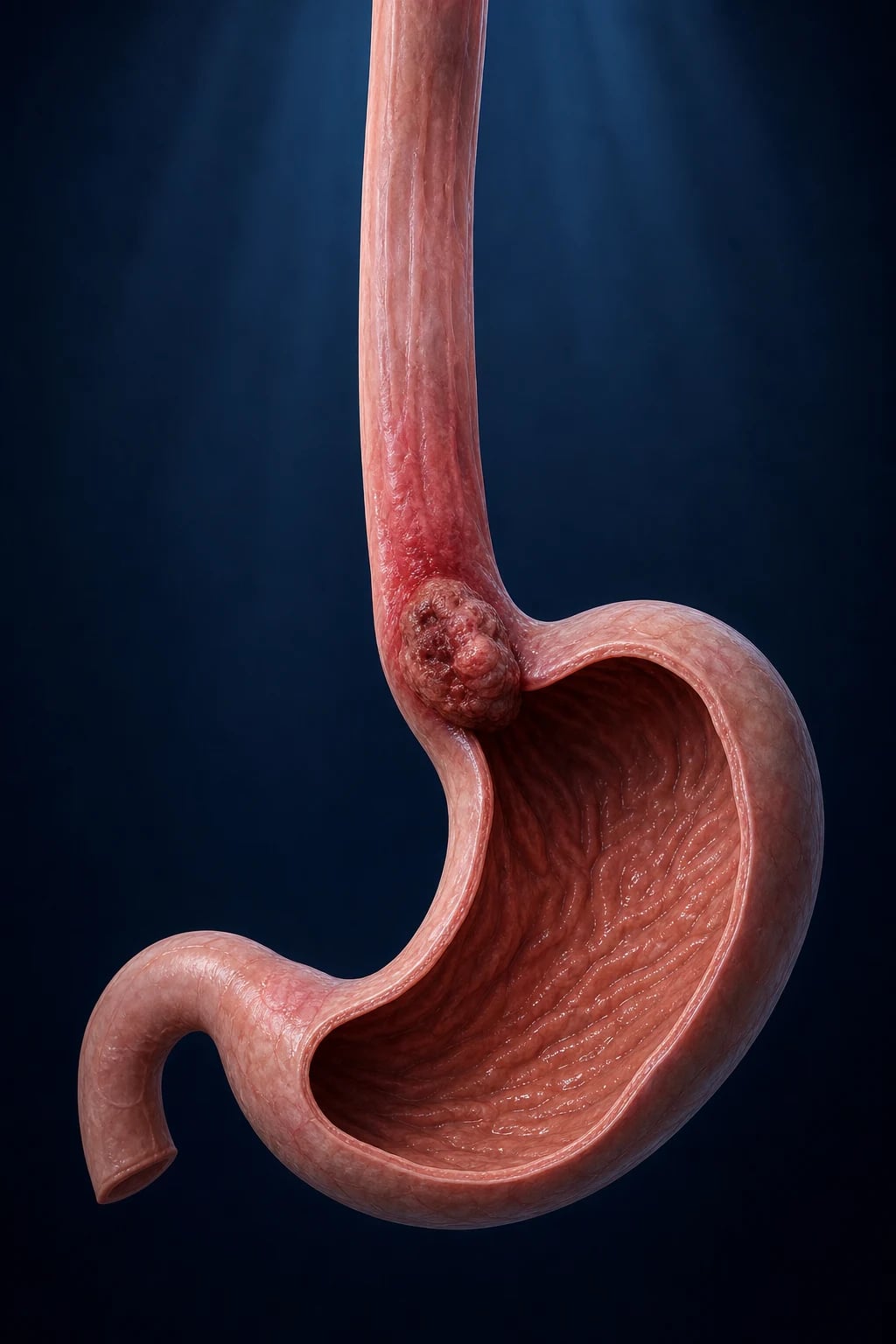
Although most upper-GI cancers present electively, several time-critical scenarios demand immediate management.[1][2]
Acute upper-GI haemorrhage from a tumour
- ABCDE; two large-bore cannulae; cross-match; IV fluid resuscitation; transfuse to Hb 70 to 80 g/L (higher targets if symptomatic, cardiovascular disease, or active bleeding).
- IV proton-pump inhibitor infusion (pantoprazole 80 mg IV bolus then 8 mg/hour, or omeprazole equivalent) — reduces rebleeding in peptic causes; of limited benefit for tumour bleeding but standard practice.
- Urgent OGD for diagnosis and haemostasis — endoscopic clips, argon plasma coagulation, epinephrine injection; if uncontrolled, interventional radiology embolisation (e.g. left gastric artery); surgery rarely for refractory tumour bleeding.
- Correct coagulopathy (vitamin K, FFP, platelets) and stop anticoagulants (reverse as appropriate). [1]
Acute dysphagia with dehydration or aspiration
- IV hydration, correct electrolytes; assess aspiration risk (bedside swallow, chest X-ray).
- Nutritional support: nasogastric / nasojejunal tube if feasible, radiologically-inserted gastrostomy/jejunostomy if obstruction, or total parenteral nutrition short-term.
- Urgent oesophageal self-expanding metal stent (SEMS) for complete malignant obstruction to restore oral intake and relieve aspiration. [1]
Severe anaemia from a bleeding tumour
- Transfuse to symptomatic relief / safe threshold; IV iron (ferric carboxymaltose) for iron deficiency once haemodynamically stable; investigate and treat the source. [1]
Malignant gastric outlet obstruction
- NG decompression, correct dehydration and electrolytes; endoscopic enteral stent for palliation; surgical gastrojejunostomy (bypass) in fit patients with longer prognosis. [1]
Pre-operative nutritional optimisation
Major upper-GI surgery carries high morbidity in malnourished patients. A dietitian-led plan — oral nutritional supplements, jejunostomy feeding during neoadjuvant therapy, TPN if enteral feeding impossible — for 7 to 14 days before surgery reduces complications. Preoperative carbohydrate loading and ERAS (enhanced recovery after surgery) protocols are standard. [1]
Management — Definitive & Stepwise
The overarching principle: surgery is the only cure, but only the resectable minority (Stage I-III, M0) benefit. Multidisciplinary (MDT) staging decides who is resectable; perioperative systemic therapy improves survival in locally advanced disease; advanced/metastatic (M1) disease is palliative.[1][2]
Early-stage disease: endoscopic resection (organ-preserving)[1][2]
Endoscopic mucosal resection (EMR) and endoscopic submucosal dissection (ESD) are curative for mucosal (T1a) disease with low risk of nodal metastasis: [1]
- Barrett's high-grade dysplasia and T1a adenocarcinoma — EMR of visible lesions + radiofrequency ablation (RFA) of remaining Barrett's; 5-year survival over 95 percent.
- Early gastric cancer (intestinal type, well-differentiated, under 2 cm, no ulceration, T1a, no lymphovascular invasion) — ESD; Japanese standard.
- Strict pathological criteria confirm en-bloc, R0 resection; deep submucosal invasion, LVI, or poor differentiation triggers surgical gastrectomy/oesophagectomy with lymphadenectomy. [1]
Resectable oesophageal cancer (Stage II-III, M0)[1][6][7]
Neoadjuvant therapy then oesophagectomy is the standard. Two evidence-based options: [1]
- Neoadjuvant chemoradiotherapy (the CROSS regimen) — carboplatin (AUC 2) + paclitaxel (50 mg/m²) weekly for 5 weeks with concurrent radiotherapy 41.4 Gy in 23 fractions, followed by oesophagectomy at 6 to 8 weeks. The CROSS trial (van Hagen, NEJM 2012) showed improved R0 resection (92 percent vs 69 percent) and overall survival (median 49 months vs 24 months); benefit in both SCC and adenocarcinoma.[6]
- Neoadjuvant chemotherapy (the JCOG9907 / OE02 approach) — cisplatin + 5-fluorouracil (CF) for two cycles, then surgery. The JCOG9907 trial (Ando, Ann Surg Oncol 2012) established neoadjuvant CF as standard for Japanese SCC (better survival than adjuvant CF); the OE02 (MRC) trial supported neoadjuvant CF for adenocarcinoma.[7]
Choice: CROSS is widely preferred in Europe and for Siewert I / adenocarcinoma; perioperative FLOT is increasingly used for Siewert II/III and GOJ adenocarcinoma. Definitive chemoradiotherapy (without surgery) is preferred for cervical oesophageal SCC (organ preservation) and unfit patients. [1]
The oesophagectomy — approaches: [1]
- Ivor Lewis (two-phase, right thoracotomy + laparotomy) — the standard for mid/lower oesophagus and GOJ; gastric conduit pulled up and anastomosed in the right chest.
- McKeown / three-stage (cervical anastomosis) — for upper thoracic tumours; allows cervical lymphadenectomy.
- Transhiatal — abdominal + cervical approach (no thoracotomy); lower respiratory morbidity, more anastomotic leaks.
- Minimally invasive (thoracoscopic/laparoscopic / robotic) — less morbidity, equivalent oncology; now standard in many centres. [1]
Resectable gastric and GOJ adenocarcinoma (Stage II-III, M0)[2][4][5]
Surgery with D2 lymphadenectomy plus perioperative (neoadjuvant + adjuvant) chemotherapy is the standard. [1]
Perioperative FLOT chemotherapy (current global standard):
- FLOT = 5-Fluorouracil + Leucovorin (folinic acid) + Oxaliplatin + Docetaxel, given four cycles pre-operatively and four cycles post-operatively (each cycle every 2 weeks).
- The FLOT4 trial (Al-Batran, Lancet 2019) compared FLOT with the previous anthracycline-based ECF/ECX (epirubicin, cisplatin, capecitabine/5-FU) regimen and showed improved median overall survival (50 vs 35 months) and 5-year survival (45 percent vs 36 percent); FLOT replaced ECF/ECX as standard.[5]
- The earlier MAGIC trial (Cunningham, NEJM 2006) established the principle of perioperative chemotherapy (ECF) improving 5-year survival from 23 percent to 36 percent.[4]
Adjuvant chemoradiotherapy (the Macdonald/Intergroup 0116 approach) — used mainly in North America where D2 uptake was historically low: 5-FU + leucovorin with 45 Gy of radiotherapy after D0/D1 gastrectomy. The Intergroup 0116 trial (Macdonald, NEJM 2001) showed improved survival with adjuvant chemoradiotherapy after gastrectomy.[10] In D2-operated patients (Asian/European practice), adjuvant chemotherapy (e.g. S-1 or XELOX in the ARTIST/CLASSIC trials) is preferred over chemoradiotherapy.
The gastrectomy and D2 lymphadenectomy:[2]
- Total gastrectomy — for proximal, diffuse, or linitis plastica tumours; Roux-en-Y oesophagojejunostomy reconstruction.
- Subtotal (distal) gastrectomy — for distal tumours with a 5 cm proximal margin; Billroth II or Roux-en-Y reconstruction.
- D2 lymphadenectomy = D1 (perigastric nodes) plus nodes along the left gastric, common hepatic, splenic, coeliac and (for some) hepatoduodenal ligament arteries — at least 15 nodes examined. The Dutch D1D2 trial (Songun et al., 15-year follow-up) showed lower gastric-cancer-related death and recurrence with D2, establishing it as standard; spleen-preserving D2 has lower morbidity than historical D2 with distal pancreateatosplenectomy.
- Multivisceral resection (spleen, distal pancreas, transverse colon) only for direct invasion (R0) — never as a routine. [1]
Advanced / metastatic (Stage IV) disease — palliative systemic therapy[2][8][9]
Goals are symptom control, quality of life and survival extension, not cure. Choice is guided by performance status, HER2 status and PD-L1 CPS: [1]
- First-line chemotherapy: fluoropyrimidine (5-FU or capecitabine) + platinum (cisplatin or oxaliplatin) +/- a taxane (FLOT, FOLFOX, CAPOX, ECF) — typically 2-year treatment duration or until progression/toxicity.
- HER2-positive (IHC 3+ or ISH+): add trastuzumab (8 mg/kg loading then 6 mg/kg IV every 3 weeks) to first-line chemotherapy — ToGA trial (Bang, Lancet 2010) improved median OS from 11.1 to 13.8 months.[8]
- PD-L1 CPS at least 5 (or MSI-H): add nivolumab (360 mg/m² every 3 weeks) or pembrolizumab to first-line chemotherapy — CheckMate 649 (Janjigian, Lancet 2021) showed improved OS with nivolumab + chemo, benefit greatest at CPS at least 5.[9]
- Second line: ramucirumab (anti-VEGFR2, REGARD/RAINBOW trials) +/- paclitaxel; irinotecan; pembrolizumab for MSI-H.
- Third line and beyond: ** trifluridine/tipiracil (TAS-102)**, best supportive care.
Advanced oesophageal SCC: cisplatin + 5-FU (CF) or paclitaxel; immune checkpoint inhibitors (nivolumab, pembrolizumab) for CPS at least 10 or after chemotherapy; palliative radiotherapy for dysphagia, bleeding or bone pain; oesophageal stent for obstruction. [1]
Palliative and supportive care[1][2]
- Oesophageal self-expanding metal stent (SEMS) — rapid relief of malignant dysphagia; risks of perforation, bleeding, migration, reflux, food impaction.
- Brachytherapy / single-fraction external beam radiotherapy — slower but more durable relief than stent; can be combined.
- Laser recanalisation / argon plasma coagulation — for exophytic tumour bleeding/obstruction.
- Pain management — WHO analgesic ladder; opioids, nerve blocks (coeliac plexus for gastric pain).
- Nutrition — oral supplements, PEG/RIG for feeding (caution in oesophageal obstruction; risk of tumour seeding on the tract), TPN selectively.
- Ascites — paracentesis, diuretics (spironolactone).
- Psychological and palliative-care support, advanced care planning. [1]
When surgery is NOT offered
- Metastatic (M1) disease — including positive peritoneal cytology — palliative only.
- Locally advanced unresectable (T4b invading aorta/trachea/vertebra) — definitive chemoradiotherapy (oesophageal) or systemic therapy.
- Poor performance status (ECOG 3-4) or severe comorbidity precluding major surgery. [1]
Specific Subtypes & Scenarios
Siewert I, II, III gastro-oesophageal junction adenocarcinoma[1]
- Siewert I — treated as oesophageal cancer: neoadjuvant CROSS chemoradiotherapy then Ivor Lewis oesophagectomy.
- Siewert II/III — treated as gastric cancer: perioperative FLOT then total gastrectomy (or extended total gastrectomy) with D2 lymphadenectomy; staging laparoscopy mandatory.
- The modern ESOPEC and NEO-AEGIS trials refine the choice of perioperative FLOT versus neoadjuvant CROSS for GOJ adenocarcinoma — both are acceptable; FLOT increasingly preferred for II/III. [1]
Oesophageal squamous cell carcinoma[1][7]
- Resectable (thoracic): neoadjuvant chemoradiotherapy (CROSS) or neoadjuvant chemotherapy (cisplatin/5-FU, JCOG9907) then oesophagectomy.
- Cervical oesophagus: definitive chemoradiotherapy (organ-preserving — avoid pharyngo-laryngo-oesophagectomy where possible).
- Unfit for surgery: definitive chemoradiotherapy (cisplatin/5-FU + 50.4 Gy) — long-term survival in a subset.
- SCC tends to metastasise earlier to lymph nodes and shows a strong response to chemoradiotherapy; recurrence is often systemic. [1]
Linitis plastica (diffuse / signet-ring)[2]
Submucosal infiltration makes the stomach rigid and non-distensible; often presents with early satiety and weight loss with a deceptively normal-looking mucosa on early OGD (multiple deep biopsies required). Laparoscopy frequently reveals peritoneal disease; resectability is low; palliative chemotherapy (FLOT if fit) is the mainstay. Prognosis is poor. [1]
HER2-positive metastatic gastric/GOJ cancer[8]
Trastuzumab + chemotherapy (5-FU/capecitabine + cisplatin) first line; median OS improved from 11.1 to 13.8 months (ToGA). Newer HER2-directed agents (trastuzumab deruxtecan, fam-trastuzumab deruxtecan) extend options after progression. [1]
EBV-positive and MSI-high gastric cancer[2]
Both have an excellent prognosis and are highly responsive to immune checkpoint inhibitors; pembrolizumab is first-line for MSI-H disease. These molecular subtypes (TCGA) increasingly guide therapy. [1]
Gastric MALT lymphoma[2]
Extranodal marginal-zone B-cell lymphoma of mucosa-associated lymphoid tissue, driven by H. pylori in about 90 percent. Localised (stage I-II), low-grade MALT often regresses completely with H. pylori eradication alone (amoxicillin + clarithromycin + PPI); refractory or high-grade disease gets radiotherapy or rituximab/chemotherapy. Surgery has no role. [1]
Hereditary diffuse gastric cancer (CDH1)[2]
Germline CDH1 mutation; 70 to 80 percent lifetime risk of diffuse gastric cancer and high risk of lobular breast cancer. Prophylactic total gastrectomy is offered to asymptomatic adult carriers (typically late teens to 30s) after counselling — the clearest example of precision cancer prevention. Endoscopic surveillance is insufficient because diffuse cancer is submucosal and patchy. [1]
Barrett's high-grade dysplasia and early adenocarcinoma[1]
Endoscopic resection (EMR/ESD) of visible lesions + radiofrequency ablation (RFA) of the residual Barrett's segment; surveillance endoscopy thereafter (Prague C&M criteria document the circumferential and maximal extent). This achieves over 95 percent 5-year survival and obviates oesophagectomy for most mucosal disease. [1]
Complications & Pitfalls
Post-oesophagectomy complications[1]
- Anastomotic leak (5 to 15 percent) — managed with drainage, antibiotics, stenting; cervical anastomoses leak more but are better tolerated.
- Anastomotic stricture — endoscopic dilatation.
- Gastric conduit necrosis — catastrophic; requires re-operation.
- Chylothorax — thoracic duct injury; medium-chain triglyceride diet, octreotide, surgical ligation if persistent.
- Recurrent laryngeal nerve palsy — hoarseness, aspiration risk.
- Atrial fibrillation (common post-op), respiratory complications (atelectasis, pneumonia — commonest cause of early mortality).
- Reflux, dumping, early satiety (conduit denervation and loss of the lower oesophageal sphincter). [1]
Post-gastrectomy complications[2]
- Dumping syndrome — early (15 to 30 min post-prandial: vasomotor — palpitations, sweating, dizziness, diarrhoea — from rapid osmotic fluid shift) and late (1 to 3 h: hypoglycaemia from exaggerated insulin response to the glucose load). Manage with small, frequent, low-carbohydrate, high-protein meals, separating fluids and solids, octreotide for refractory cases.
- Postgastrectomy syndrome, afferent/efferent loop syndrome (Billroth II), alkaline reflux gastritis (bile).
- Anaemia: B12 deficiency (loss of intrinsic factor — lifelong IM hydroxocobalamin), iron deficiency (loss of acid, rapid transit).
- Calcium and vitamin D malabsorption -> osteoporosis and osteomalacia (bone densitometry, supplementation).
- Internal hernia (after Roux-en-Y), marginal ulcer, gastric remnant cancer (15 to 20 years later).
- Postgastrectomy weight loss and malnutrition. [1]
Chemotherapy and chemoradiotherapy toxicities[4][5][6]
- FLOT — neutropenia and febrile neutropenia (G-CSF prophylaxis often used), diarrhoea, mucositis, peripheral neuropathy (oxaliplatin and docetaxel), alopecia, nausea/vomiting, hypersensitivity.
- CROSS — oesophagitis, radiation pneumonitis, cardiac toxicity, myelosuppression, neuropathy.
- Trastuzumab — cardiac dysfunction (LVEF decline), infusion reactions.
- Immune checkpoint inhibitors — immune-related adverse events (colitis, pneumonitis, hepatitis, endocrinopathies — thyroiditis, hypophysitis, type 1 diabetes, adrenal insufficiency).
- Thromboembolism — high risk in gastric/pancreatic cancer; consider prophylactic LMWH (CASSINI/AVERT trials). [1]
Tumour complications[1][2]
Perforation, tracheo-oesophageal fistula, haemorrhage, malignant obstruction, malnutrition, cachexia, and metastatic events (cord compression, pathological fracture, malignant ascites). [1]
Classic pitfalls
- Chalking progressive dyspepsia up to GORD/PUD in a patient over 55 with weight loss — urgent OGD.
- Failing to biopsy a gastric ulcer (malignant until proven otherwise) and failing to document healing at 8 to 12 weeks.
- Omitting staging laparoscopy before gastrectomy — missing occult peritoneal disease.
- Operating on metastatic (M1) disease — confirm with laparoscopy/PET first.
- Forgetting lifelong B12 after total gastrectomy.
- Mislabelling achalasia as cancer (or vice versa) — a careful solids-then-liquids vs liquids-and-solids history plus manometry resolves most cases.
- Underestimating the young patient — consider CDH1 hereditary diffuse gastric cancer or Lynch. [1]
Prognosis & Disposition
Five-year survival by stage (overall poor)[1][2]
- Oesophageal cancer — overall 5-year survival roughly 15 to 20 percent; localised (Stage I) about 45 to 50 percent; regional (Stage II-III with nodes) about 25 percent; distant (Stage IV) under 5 percent.
- Gastric cancer — overall 5-year survival roughly 25 to 30 percent; localised about 65 to 70 percent; regional about 30 percent; distant under 6 percent. In Japan/Korea, where endoscopic screening finds early disease, overall 5-year survival exceeds 60 to 70 percent. [1]
Predictors of poor outcome
Advanced stage, signet-ring / diffuse histology, linitis plastica, peritoneal disease, R1/R2 resection, low albumin / cachexia, poor performance status (ECOG 2+), high PD-L1 CPS paradoxically predicts better immunotherapy response. [1]
Benefit of modern perioperative therapy
- CROSS (oesophageal): median OS 49 vs 24 months.[6]
- FLOT (gastric/GOJ): median OS 50 vs 35 months; 5-year survival 45 percent vs 36 percent.[5]
- Trastuzumab (HER2+ metastatic): median OS 13.8 vs 11.1 months.[8]
- Nivolumab + chemo (CPS at least 5, metastatic): median OS roughly 14 vs 11 months.[9]
Surveillance after curative treatment[1][2]
History, examination, bloods (FBC, LFT, iron studies, B12, tumour markers), and CT at intervals (e.g. every 6 months for 2 years, then annually for up to 5 years); OGD to monitor the anastomosis and gastric remnant (cancer risk after partial gastrectomy). Nutrition, B12/iron/calcium/vitamin D supplementation, bone densitometry after gastrectomy. [1]
Disposition
All patients are discussed in a multimodality upper-GI cancer MDT; surgery is performed in specialist upper-GI cancer centres (volume-outcome relationship); palliative care is involved early in advanced disease; clinical trial enrolment is encouraged. [1]
Special Populations
- Young patients (under 40) with gastric cancer — suspect hereditary diffuse gastric cancer (CDH1) or Lynch syndrome; refer for genetic counselling and testing.[2]
- Elderly / frail — comorbidity and performance status (not age alone) drive treatment; less extensive surgery (subtotal over total where possible), dose-reduced or single-agent chemotherapy, definitive chemoradiotherapy for oesophageal cancer in those unfit for surgery, and early palliative-care involvement.
- Barrett's oesophagus surveillance — Prague C&M criteria document circumferential (C) and maximal (M) extent; non-dysplastic Barrett's surveillance endoscopy every 3 to 5 years; indefinite for dysplasia at 6 months on high-dose PPI; low-grade dysplasia at 6 to 12 months (consider RFA); high-grade dysplasia / T1a — EMR + RFA.[1]
- Previous partial gastrectomy — cancer risk 15 to 20 years later (bile reflux, bacterial overgrowth, nitrosamines); investigate new upper-GI symptoms.
- Pernicious anaemia / autoimmune gastritis — surveillance for gastric cancer and type I gastric neuroendocrine tumours.
- H. pylori eradication as cancer prevention — population-level strategy in high-incidence regions; screen-and-treat reduces incident gastric cancer.
- Immunocompromised (HIV, post-transplant) — higher incidence of certain subtypes (e.g. EBV-positive, lymphoma); manage with multidisciplinary input.
- Pregnancy — rare; MRI and ultrasound for staging (avoid CT radiation where possible); weigh maternal treatment timing against foetal safety; involve the MDT, obstetric and neonatal teams.[1][2]
Evidence, Guidelines & Regional Differences
Landmark trials (know these by name)[4][5][6][7][8][9][10]
International guidelines — NCCN (US) 2024 and ESMO 2024 for oesophageal and gastric cancer broadly converge on: perioperative FLOT for resectable gastric/GOJ adenocarcinoma; neoadjuvant CROSS chemoradiotherapy for oesophageal cancer; trastuzumab for HER2-positive and nivolumab (CPS at least 5) for metastatic disease; D2 gastrectomy as the surgical standard.
- MAGIC (Cunningham, NEJM 2006, PMID 16822992) — perioperative ECF chemotherapy vs surgery alone for resectable lower-oesophageal/gastric/GOJ adenocarcinoma; improved 5-year survival 23 percent to 36 percent. Established the principle of perioperative chemotherapy.[4]
- FLOT4 (Al-Batran, Lancet 2019, PMID 30982686) — perioperative FLOT vs ECF/ECX; median OS 50 vs 35 months; FLOT is now the global standard.[5]
- CROSS (van Hagen, NEJM 2012, PMID 22646630) — neoadjuvant chemoradiotherapy (carboplatin/paclitaxel + 41.4 Gy) vs surgery alone for oesophageal/GOJ cancer; R0 resection improved (92 percent vs 69 percent) and median OS 49 vs 24 months; benefit in both histologies.[6]
- JCOG9907 (Ando, Ann Surg Oncol 2012, PMID 21879261) — neoadjuvant cisplatin/5-FU vs adjuvant for thoracic SCC; established neoadjuvant CF as standard in Japan.[7]
- ToGA (Bang, Lancet 2010, PMID 20728210) — trastuzumab + chemotherapy vs chemotherapy alone for HER2-positive metastatic gastric/GOJ cancer; median OS 13.8 vs 11.1 months — first targeted-therapy success in gastric cancer.[8]
- CheckMate 649 (Janjigian, Lancet 2021, PMID 34102137) — nivolumab + chemotherapy vs chemotherapy first-line for advanced gastric/GOJ/oesophageal adenocarcinoma; improved OS, benefit greatest at PD-L1 CPS at least 5.[9]
- Intergroup 0116 (Macdonald, NEJM 2001, PMID 11547741) — adjuvant chemoradiotherapy (5-FU/leucovorin + 45 Gy) vs surgery alone for resected gastric/GOJ adenocarcinoma; improved survival — the basis of US adjuvant chemoradiotherapy (mainly after D0/D1 resection).[10]
- Dutch D1D2 (Songun et al., 15-year follow-up, 2010) — D2 vs D1 gastrectomy; higher early morbidity/mortality historically (with distal pancreateatosplenectomy) but lower gastric-cancer death and recurrence at 15 years; established D2 as standard with spleen-preserving technique.
Controversies
- Oesophagectomy approach — transthoracic Ivor Lewis vs transhiatal; minimally invasive/robotic vs open; ongoing individualisation.
- Neoadjuvant chemo vs chemoradiotherapy for adenocarcinoma — ESOPEC and NEO-AEGIS trials comparing FLOT vs CROSS; both acceptable.
- Role of radiation in gastric cancer — ARTIST/ARTIST-II trials refine adjuvant chemoradiotherapy after D2; benefit confined to node-positive disease.
- Immunotherapy in early (perioperative) disease — ongoing (e.g. KEYNOTE-585, DANTE, MATTERHORN).
- Screening — cost-effectiveness of population endoscopy outside high-incidence regions; H. pylori vaccine still in development.
- Prophylactic gastrectomy in CDH1 carriers — timing and extent; surveillance alternatives. [1]
Exam Pearls
Risk factors — oesophageal cancer by histology
OAC vs OSCC
Lower oesophagus / GOJ; white males; rising in the West
Upper/middle oesophagus; synergistic
OSCC risks: very hot beverages, lye stricture, achalasia stasis
OSCC syndromes: iron-deficiency web; Howel-Evans palmoplantar keratoderma (near-certain SCC)
Gastric cancer risk factors — recall 'HABITS + BAD'
HABITS
Class I carcinogen (IARC); CagA-positive most virulent; ~89 percent of non-cardia gastric cancers
Correa cascade; intestinal metaplasia
Modest increased risk
CDH1 (hereditary diffuse gastric cancer), Lynch, FAP, Peutz-Jeghers
1.5- to 2-fold risk
N-nitroso compounds; low ascorbate
Long-term risks after partial gastrectomy
Named metastatic signs — recall the 'big five'
VKSB-I
Left supraclavicular node via the thoracic duct — classic gastric cancer
Ovarian signet-ring metastasis
Umbilical peritoneal deposit
Palpable rectal shelf on PR exam — pelvic drop metastasis
Left axillary lymph node
The chemotherapy regimens by name
FLOT-ECF-CF-CROSS
5-Fluorouracil, Leucovorin, Oxaliplatin, Docetaxel (Taxotere); 4 cycles pre + 4 post
Epirubicin, Cisplatin, 5-FU (or Capecitabine); historical standard, superseded by FLOT
Cisplatin + 5-Fluorouracil; also definitive chemo for oesophageal SCC
Carboplatin (AUC 2) + Paclitaxel (50 mg/m²) weekly x 5 + 41.4 Gy in 23 fractions
- The Correa cascade, verbatim: normal mucosa -> chronic (superficial) gastritis -> atrophic gastritis -> intestinal metaplasia -> dysplasia -> invasive adenocarcinoma.[2]
- Siewert classification: I (above GOJ, oesophagectomy), II (at GOJ, gastrectomy/extended), III (below GOJ, gastrectomy).
- Lauren: intestinal (glands, Correa, better prognosis) vs diffuse (signet-ring, CDH1, linitis plastica, worse).
- After total gastrectomy = lifelong IM hydroxocobalamin (B12) — the loss of intrinsic factor.
- H. pylori is the only bacterium classified as a Class I (definite) human carcinogen — eradication is a population-level cancer-prevention intervention.[2]
- Dysphagia: solids then liquids = mechanical (tumour/stricture); liquids and solids together from onset = neuromuscular (achalasia).
- Every gastric ulcer is malignant until proven otherwise — six biopsies, repeat OGD at 8 to 12 weeks to confirm healing.
- Staging laparoscopy with peritoneal washout is non-negotiable before gastrectomy — picks up 10 to 30 percent of CT-occult peritoneal disease.[2]
- Acanthosis nigricans / sign of Leser-Trélat in an older patient = suspect gastric cancer (paraneoplastic).
- CROSS, FLOT, JCOG9907, MAGIC, ToGA, CheckMate 649, Intergroup 0116 — be able to match each trial to its management decision.[4][5][6][7][8][9][10]
Exam application bank (NEET-PG / INICET)
One-line answer
Oesophageal and gastric cancer are the two principal upper-gastrointestinal malignancies. Oesophageal cancer has two distinct histologies: adenocarcinoma arising in the lower oesophagus and gastro-oesophageal junction (GOJ) from Barrett's intestinal metaplasia (driven by obesity and chronic acid reflux), and squamous cell carcinoma of the upper and middle oesophagus (driven by alcohol, smoking, very hot beverages, achalasia and nitrosamine-rich diets). Gastric adenocarcinoma is driven by Helicobacter pylori (a Class I carcinogen), salted and smoked diets, smoking, blood group A, family history and pernicious anaemia; the Correa cascade (chronic gastritis through atrophic gastritis, intestinal metaplasia and dysplasia to invasive carcinoma) describes the intestinal-type sequence. Both diseases present late with progressive dysphagia, weight loss, dyspepsia, early satiety, nausea and iron-
Worked stems (answer without another resource)
Stem 1 — Classic presentation. Map symptoms to mechanism; name the first investigation and first treatment step with dose/route if drug therapy is standard. [1]
Stem 2 — Unstable / complicated. List red flags that force immediate resuscitation, theatre, ICU, antidote, or reperfusion — and what you do in the first 15 minutes. [1]
Stem 3 — Atypical group. Elderly, pregnancy, child, or immunocompromised: how presentation and thresholds change. [1]
Stem 4 — Differential trap. Name the three closest mimics and one discriminator for each. [1]
Stem 5 — Disposition. Who goes home with safety-netting, who is admitted, who needs HDU/ICU/theatre, and what follow-up is mandatory. [1]
Rapid viva checklist
- Definition + classification
- Pathophysiology chain
- Bedside signs / criteria
- Score with exact components (if any)
- Emergency bundle
- Definitive therapy with doses
- Complications of disease and of treatment
- Special populations
- Guideline/trial name if classic
- Three exam traps
Coverage self-check
If you cannot answer any stem above from this page alone, re-read the matching section — the page is intended to be self-sufficient for final-prof and NEET-PG/INICET questions on Oesophageal & Gastric Cancer.
References
- [1]Smyth EC, Lagergren J, Fitzgerald RC, et al. Oesophageal cancer Nat Rev Dis Primers, 2017.PMID 28748917
- [2]Ajani JA, Lee J, Sano T, et al. Gastric adenocarcinoma Nat Rev Dis Primers, 2017.PMID 28569272
- [3]Sung H, Ferlay J, Siegel RL, et al. Global Cancer Statistics 2020: GLOBOCAN Estimates of Incidence and Mortality Worldwide for 36 Cancers in 185 Countries CA Cancer J Clin, 2021.PMID 33538338
- [4]Cunningham D, Allum WH, Stenning SP, et al. (MAGIC). Perioperative chemotherapy versus surgery alone for resectable gastroesophageal cancer N Engl J Med, 2006.PMID 16822992
- [5]Al-Batran SE, Homann N, Pauligk C, et al. (FLOT4). Perioperative chemotherapy with fluorouracil plus leucovorin, oxaliplatin, and docetaxel versus fluorouracil or capecitabine plus cisplatin and epirubicin for locally advanced, resectable gastric or gastro-oesophageal junction adenocarcinoma (FLOT4): a randomised, phase 2/3 trial Lancet, 2019.PMID 30982686
- [6]van Hagen P, Hulshof MCCM, van Lanschot JJB, et al. (CROSS). Preoperative chemoradiotherapy for esophageal or junctional cancer N Engl J Med, 2012.PMID 22646630
- [7]Ando N, Kato H, Igaki H, et al. (JCOG9907). A randomized trial comparing postoperative adjuvant chemotherapy with cisplatin and 5-fluorouracil versus preoperative chemotherapy for localized advanced squamous cell carcinoma of the thoracic esophagus (JCOG9907) Ann Surg Oncol, 2012.PMID 21879261
- [8]Bang YJ, Van Cutsem E, Feyereislova A, et al. (ToGA). Trastuzumab in combination with chemotherapy versus chemotherapy alone for treatment of HER2-positive advanced gastric or gastro-oesophageal junction cancer (ToGA): a phase 3, open-label, randomised controlled trial Lancet, 2010.PMID 20728210
- [9]Janjigian YY, Shitara K, Moehler M, et al. (CheckMate 649). First-line nivolumab plus chemotherapy versus chemotherapy alone for advanced gastric, gastro-oesophageal junction, and oesophageal adenocarcinoma (CheckMate 649): a randomised, open-label, phase 3 trial Lancet, 2021.PMID 34102137
- [10]Macdonald JS, Smalley SR, Benedetti J, et al. (Intergroup 0116). Chemoradiotherapy after surgery compared with surgery alone for adenocarcinoma of the stomach or gastroesophageal junction N Engl J Med, 2001.PMID 11547741