Gastroenterology · General Medicine
Peptic Ulcer Disease
Also known as Peptic ulcer disease · PUD · Gastric ulcer · Duodenal ulcer · H. pylori ulcer
Peptic ulcer disease (PUD) is a break in the gastric or duodenal mucosa extending through the muscularis mucosae, caused by an imbalance between aggressive acid-peptic factors and mucosal defence. The two dominant causes are Helicobacter pylori (about 90 percent of duodenal ulcers) and NSAIDs/aspirin; less common are Zollinger-Ellison syndrome (gastrinoma), stress ulcers (ICU; Curling/Cushing), and malignant ulcers. It presents with epigastric burning or gnawing pain (duodenal eased by food and waking at night; gastric worsened by food) with dyspepsia; complications are bleeding (haematemesis/melaena), perforation (peritonitis, free gas), penetration, and gastric outlet obstruction. Oesophagogastroduodenoscopy (OGD) diagnoses and biopsies (H. pylori and to exclude cancer); non-invasive urea breath test or stool antigen confirm active H. pylori (stop PPIs 2 weeks first). Treat with H. pylori eradication (NICE triple therapy 7 days where clarithromycin resistance is low; bismuth quadruple or vonoprazan-amoxicillin dual for 14 days where it is high) and test of cure, a PPI for 4 to 8 weeks, and gastroprotection of NSAIDs; complications are managed endoscopically (haemostasis) or surgically (perforation).
On this page & tools
Your progress
Saved locally on this device.
Exam tags
Red flags
Overview & Definition
Peptic ulcer disease (PUD) is a break in the mucosal lining of the stomach (gastric ulcer, GU) or duodenum (duodenal ulcer, DU) that extends through the muscularis mucosae into the submucosa or deeper. It is distinguished from an erosion, which is a shallower break confined to the epithelium and which heals without scarring.[4]
PUD arises when aggressive factors (acid, pepsin, Helicobacter pylori, NSAIDs) overwhelm mucosal defence (the mucus-bicarbonate barrier, mucosal blood flow, prostaglandins, and rapid epithelial cell turnover). The two dominant causes — H. pylori and NSAIDs/aspirin — together account for the great majority of ulcers; the remainder are due to Zollinger-Ellison syndrome, stress ulcers in the critically ill, and malignancy.[1][4]
The clinical task in PUD is not simply to heal the ulcer (a PPI does that in weeks) but to find and remove the cause — eradicate H. pylori and review NSAIDs — because ulcer recurrence is driven by the cause, not by acid suppression alone. After successful H. pylori eradication, ulcer recurrence falls from roughly 60 percent to under 10 percent; if only acid suppression is given, the ulcer recurs in most patients within a year.[1]
Classification
By site — the distinction that drives symptoms, cancer risk, and follow-up:[4]
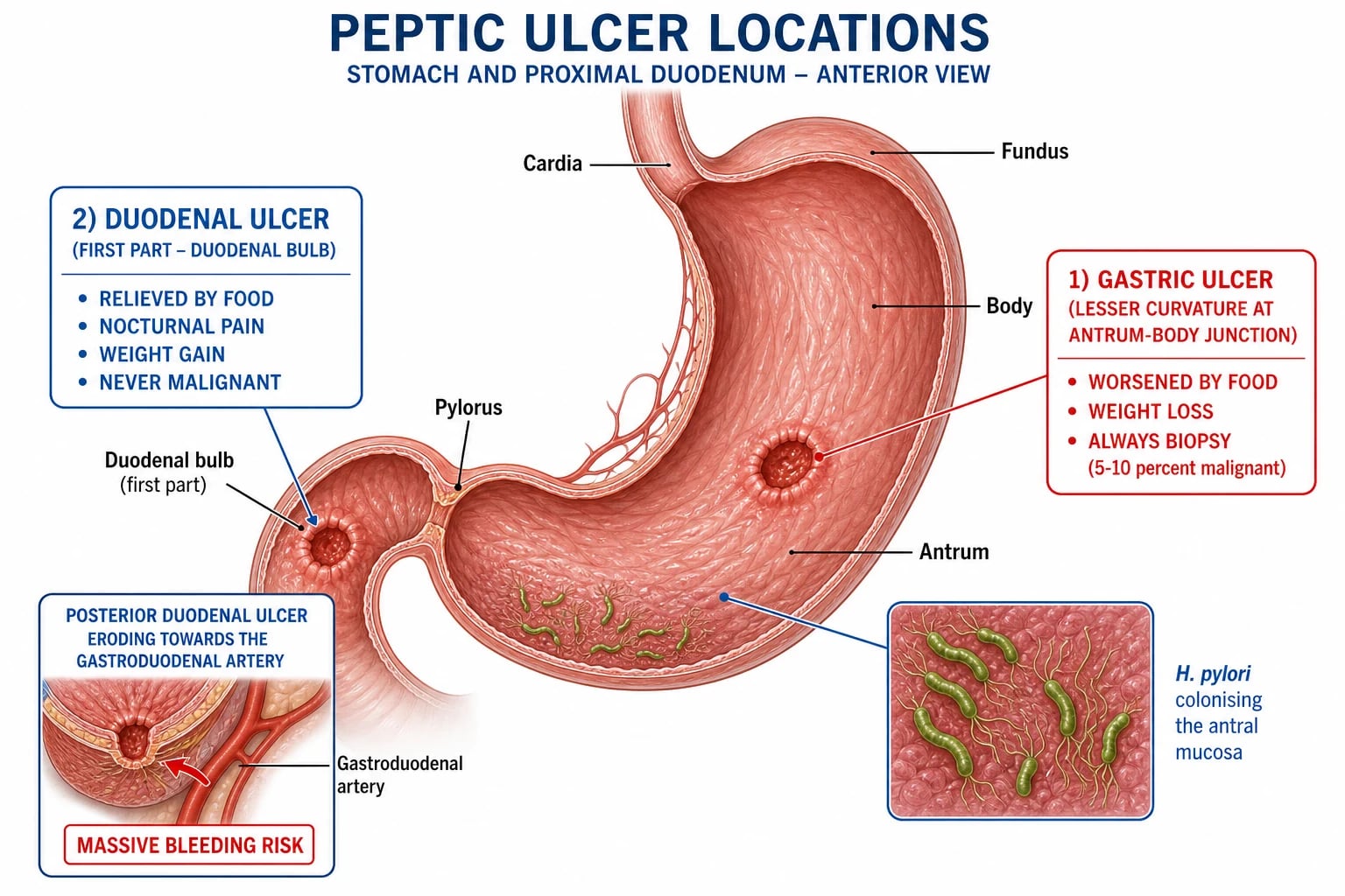
- Duodenal ulcer (DU) — in the first part of the duodenum (duodenal bulb); almost always benign; strongly linked to H. pylori (about 90 percent) and acid hypersecretion. Does not require routine repeat OGD.
- Gastric ulcer (GU) — anywhere in the stomach (Johnson type I body/antrum, type II combined with DU, type III prepyloric, type IV near the cardia); carries a malignant potential, so every gastric ulcer must be biopsied and re-endoscoped at 8 to 12 weeks to confirm healing and exclude cancer. [1]
By aetiology — the classification that guides treatment:[1][3]
- H. pylori — commonest cause worldwide.
- NSAIDs/aspirin — second commonest; the leading cause of bleeding ulcers where H. pylori is declining.
- Zollinger-Ellison syndrome (gastrinoma) — multiple, distal (jejunal), or refractory ulcers with acid hypersecretion and diarrhoea.
- Stress ulcers — ICU (Curling = burns, Cushing = head injury); ischaemic mucosa.
- Malignancy — gastric adenocarcinoma, lymphoma; biopsy mandatory.
- Other — Crohn disease, CMV/HSV (immunocompromised), radiation, cocaine ("crack stomach"), idiopathic. [1]
Duodenal ulcer
- **Site**: first part of duodenum (bulb)
- **H. pylori**: about 90 percent
- **Acid**: hypersecretion (antral-predominant gastritis → hypergastrinaemia)
- **Pain**: relieved by food/antacid; 2 to 5 h after meals; nocturnal (wakes at night)
- **Weight**: may increase
- **Malignancy**: essentially never — biopsy for H. pylori only
- **Healing**: PPI 4 weeks; NO routine repeat OGD
Gastric ulcer
- **Site**: body/antrum (Johnson I), prepyloric (III), cardia (IV); combined with DU = II
- **H. pylori**: about 60 to 70 percent
- **Acid**: normal or low (corpus atrophic gastritis)
- **Pain**: worsened by food; nausea, early satiety, weight loss
- **Weight**: tends to fall
- **Malignancy**: must exclude — BIOPSY every GU
- **Healing**: PPI 6 to 8 weeks; repeat OGD at 8 to 12 wk to confirm healing
Epidemiology & Risk Factors
PUD is common worldwide. The lifetime prevalence is about 10 percent, though it has fallen in developed countries as H. pylori carriage declines and acid suppression has improved. H. pylori infects roughly half the world's population, acquired in childhood via the faecal-oral/oral-oral route; prevalence is far higher in developing regions (over 80 percent in some Indian and African cohorts) and falls with improving sanitation.[3][4]
H. pylori causes about 90 percent of duodenal ulcers and about 60 to 70 percent of gastric ulcers; NSAIDs are the second commonest cause and the leading cause of bleeding ulcers wherever H. pylori is declining. About 20 to 30 percent of chronic NSAID users develop endoscopic ulcers, though most are silent.[8]
Peptic ulcer disease — the numbers that decide the answer
Risk factors for an ulcer, and — more importantly — for an ulcer complication (bleeding, perforation):[4][8]
- H. pylori infection and NSAID/aspirin use (dose-dependent and synergistic with H. pylori).
- Age over 60, prior ulcer or bleed, smoking (impairs healing, doubles recurrence), alcohol excess, and stress.
- Comorbidity: chronic lung disease, cardiovascular disease, chronic kidney disease, cirrhosis.
- Concurrent drugs: anticoagulants, antiplatelets (clopidogrel), SSRIs, corticosteroids (synergistic with NSAIDs), spironolactone. [1]
The single most dangerous combination is an NSAID plus H. pylori in an older patient on an anticoagulant — this is the patient who presents with massive haematemesis.[8]
Pathophysiology
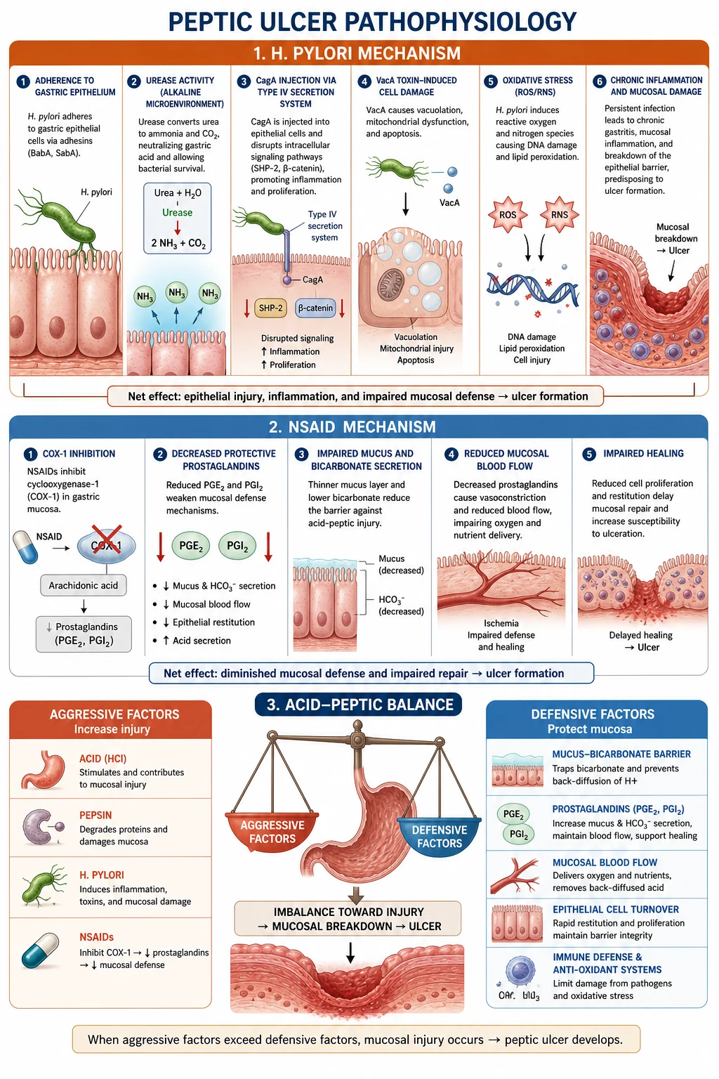
Normal mucosal defence depends on a mucus-bicarbonate layer (maintaining a near-neutral pH at the epithelial surface), adequate mucosal blood flow, prostaglandins (PGE2, PGI2) that stimulate mucus and bicarbonate secretion and maintain blood flow, and rapid epithelial renewal from gastric stem cells. PUD results when this balance is tipped towards acid-peptic injury.[4]
The pathogenesis differs between gastric and duodenal ulcer, which is an examiner favourite. [1]
Gastric ulcer — impaired mucosal defence
A gastric ulcer forms because mucosal defence is weakened, often with normal or reduced acid. The archetypal mechanism is H. pylori-induced corpus-predominant (multifocal) atrophic gastritis: parietal cell mass falls, acid output falls, and the damaged mucosa is injured even by modest acid. NSAIDs reproduce the same picture by depleting prostaglandins. Causes:[3][4]
- H. pylori — the leading cause (about 60 to 70 percent).
- NSAIDs/aspirin — COX-1 inhibition depletes prostaglandins (see below).
- Smoking, stress, alcohol — cofactors that impair healing.
- Zollinger-Ellison — gastrinoma drives acid hypersecretion (a hyper-acid gastric ulcer, an exception to the "weak defence" rule).
- Malignancy — the ulcer is the tumour (adenocarcinoma, lymphoma). [1]
Duodenal ulcer — acid hypersecretion with H. pylori
A duodenal ulcer forms because of acid hypersecretion combined with H. pylori. The typical pattern is antral-predominant gastritis: somatostatin-secreting D cells are damaged, gastrin release is unchecked, and acid output rises. The excess acid load reaches the duodenum, the duodenal mucosa undergoes gastric metaplasia, and H. pylori colonises that metaplastic epithelium, causing ulceration. H. pylori causes about 90 percent of duodenal ulcers; NSAIDs cause most of the remainder.[3][4]
H. pylori virulence — the molecules examiners name
Helicobacter pylori is a spiral, microaerophilic, urease-producing Gram-negative bacterium that colonises the gastric mucus layer. Its virulence factors are high-yield:[3]
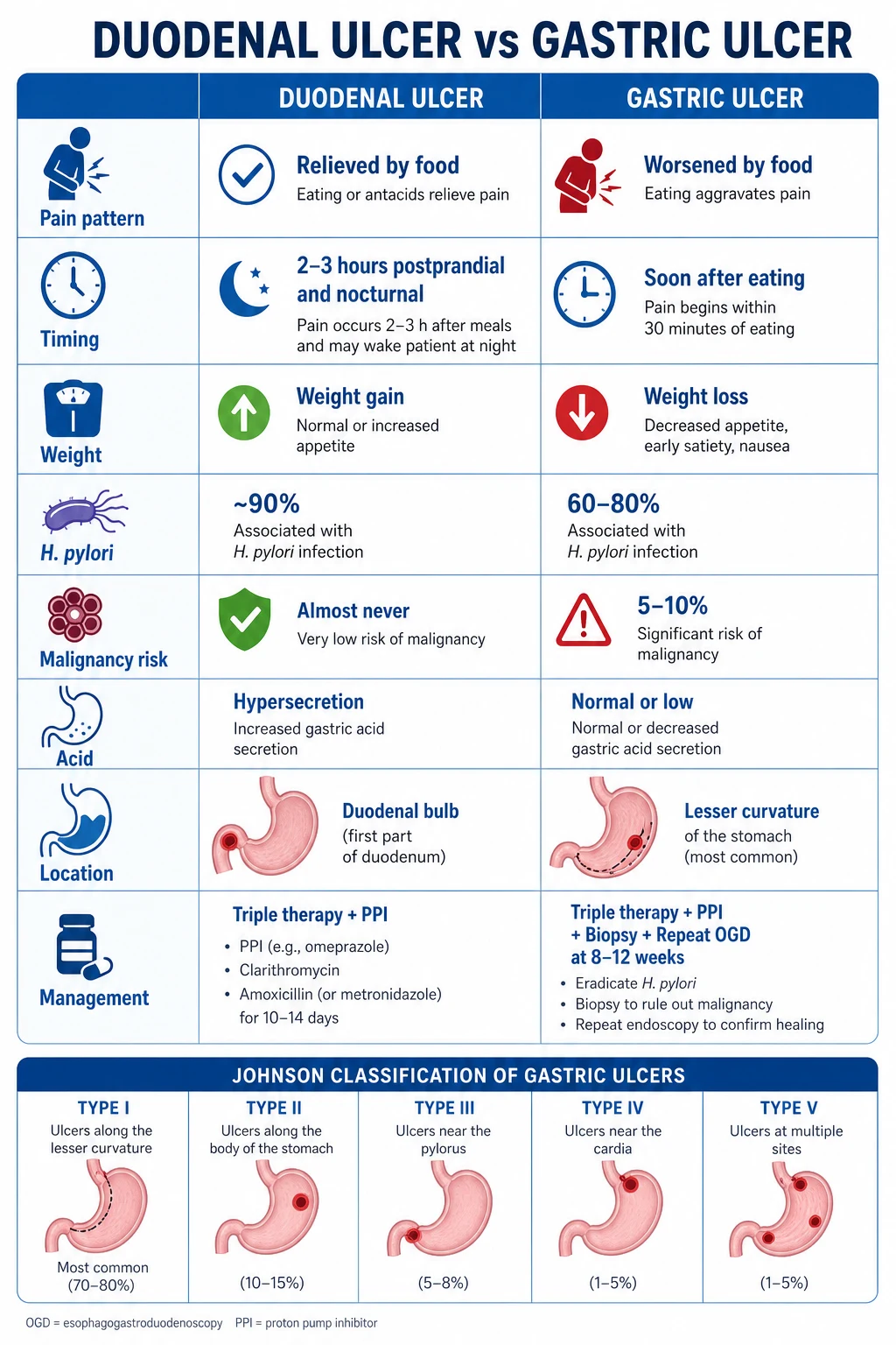
- Urease — hydrolyses urea to ammonia, alkalinising the microenvironment and protecting the organism from gastric acid. Ammonia and bacterial products directly injure epithelial cells. (This is the basis of the rapid urease / CLO test and the urea breath test.)
- CagA (cytotoxin-associated gene A) — injected into epithelial cells via a type IV secretion system; activates host signalling, drives inflammation and ulcer risk, and is strongly linked to gastric adenocarcinoma. Strains carrying the cag pathogenicity island are more virulent.
- VacA (vacuolating cytotoxin A) — forms pores in epithelial cell membranes, induces apoptosis and vacuolation; contributes to mucosal damage.
- BabA (blood-group antigen-binding adhesin) — binds the Lewis-b blood-group antigen on gastric epithelium, enabling tight adherence and more severe disease. [1]
Why the same organism causes different diseases: the pattern of gastritis decides the outcome. Antral-predominant gastritis → hypergastrinaemia, acid hypersecretion, duodenal ulcer. Corpus-predominant (multifocal) atrophic gastritis → hypochlorhydria, gastric ulcer and gastric adenocarcinoma. This is why H. pylori is a class I gastric carcinogen (IARC).[3][4]
NSAID mechanism — COX inhibition
NSAIDs inhibit cyclo-oxygenase (COX). Inhibition of the constitutive COX-1 reduces mucosal prostaglandin (PGE2, PGI2) synthesis, so mucus and bicarbonate secretion fall, mucosal blood flow drops, and epithelial turnover slows — the mucosa is stripped of its defences and becomes vulnerable to acid. NSAIDs that are weak acids (e.g. aspirin) also cause direct topical injury in an acidic stomach. Selective COX-2 inhibitors (celecoxib, etoricoxib) spare COX-1 and reduce (but do not abolish) GI toxicity, at the cost of increased cardiovascular risk.[4][8]
Zollinger-Ellison and stress ulcers
Zollinger-Ellison syndrome (ZES) — a gastrin-secreting tumour (usually pancreatic or duodenal, part of MEN-1 in a quarter of cases) drives massive acid hypersecretion, producing multiple, recurrent, or distal (jejunal) ulcers that are refractory to standard PPI doses, with diarrhoea and a markedly raised fasting serum gastrin (check while off PPI). Stress ulcers in the critically ill arise from splanchnic hypoperfusion and mucosal ischaemia rather than acid — hence prophylaxis is targeted at the ICU patient with specific risk factors.[4]
Clinical Presentation
Classic symptom — epigastric pain, described as burning or gnawing, that is episodic and periodic (clusters over days to weeks, then remits). The relationship to food is the discriminator examiners test:[4]
- Duodenal ulcer — pain is relieved by food (and by antacids or milk), occurs between meals (2 to 5 hours after eating), and classically wakes the patient at night (typically 1 to 2 a.m.). Weight may increase because eating relieves pain.
- Gastric ulcer — pain is worsened by food, occurs soon after eating, and is accompanied by nausea, early satiety, and weight loss. [1]
Associated dyspepsia: bloating, belching, fullness, early satiety, nausea, and water brash. Many ulcers are silent — especially in the elderly and in chronic NSAID users, who may present directly with a complication (bleeding or perforation) without any preceding pain.[4]
Atypical presentation in the elderly: an elderly patient on NSAIDs may have a painless ulcer presenting as melaena, haematemesis, sudden perforation, or unexplained iron-deficiency anaemia — a low threshold for OGD is essential. Up to half of NSAID-related bleeds occur with no warning symptoms.[8]
Complication presentations (the way many ulcers declare themselves):[4][5]
- Bleeding — haematemesis (vomiting blood; "coffee-ground" if altered) and/or melaena (black, tarry, foul-smelling stool); syncope and postural dizziness with significant volume loss.
- Perforation — sudden, severe, generalised abdominal pain with peritonism (rigidity, guarding, rebound, absent bowel sounds) and free gas under the diaphragm on an erect chest X-ray.
- Penetration (posterior DU into the pancreas) — severe boring back pain that does not respond to antacids, with raised amylase/lipase.
- Gastric outlet obstruction — non-bilious projectile vomiting of undigested food, early satiety, a succussion splash, and visible peristalsis. [1]
Alarm features — the trigger for urgent OGD
These features, applied to any patient with dyspepsia, mandate urgent oesophagogastroduodenoscopy to exclude malignancy and serious disease:[1]
[1]Differential Diagnosis
Recurrent or acute epigastric pain is not always a peptic ulcer. The key mimics:[4]
- GORD — retrosternal burning, regurgitation, worse on lying or after large meals; no ulcer on OGD.
- Functional (non-ulcer) dyspepsia — dyspepsia with a normal OGD (no ulcer, no erosion, no malignancy); the commonest cause of dyspepsia after H. pylori is excluded.
- Gastric cancer — weight loss, dysphagia, early satiety, anaemia, palpable mass; biopsy every gastric ulcer.
- Biliary colic / cholecystitis — right upper quadrant pain, worse after fatty food, Murphy sign.
- Acute pancreatitis — severe epigastric pain radiating to the back, relieved by sitting forward; raised lipase.
- Myocardial infarction / inferior MI — epigastric pain in the elderly or diabetic; do an ECG.
- Mesenteric ischaemia — pain out of proportion to examination, postprandial. [1]
Always look for the alarm features above; their presence moves the patient straight to urgent OGD and away from an empirical trial of acid suppression.[1]
Clinical & Bedside Assessment
In uncomplicated PUD, examination is often unremarkable apart from epigastric tenderness. The examination must instead seek the complications:[4][5]
- Peritonism (rigidity, guarding, rebound, absent bowel sounds) — perforation until proven otherwise.
- Signs of volume loss (tachycardia, hypotension, postural drop, cool peripheries, oliguria) — bleeding.
- Melaena on rectal examination; a palpable succussion splash and visible gastric peristalsis — gastric outlet obstruction.
- Pallor of anaemia; signs of chronic liver disease (varices rather than PUD may bleed).
- Epigastric mass or Virchow's (left supraclavicular) node — gastric cancer. [1]
Bedside observations (HR, BP, RR, SpO2, temperature, urine output, GCS) define severity in bleeding or complicated PUD and drive resuscitation. A postural drop in systolic BP (over 20 mmHg) or rise in pulse (over 30 bpm on standing) suggests significant volume loss (over 1 litre).[5]
Investigations
Gold standard — oesophagogastroduodenoscopy (OGD)
OGD localises the ulcer, identifies stigmata of recent haemorrhage (spurting vessel, non-bleeding visible vessel, adherent clot, oozing, clean base), treats bleeding (injection, thermal, clips), and biopsies — for H. pylori and to exclude malignancy in every gastric ulcer. Gastric ulcers are re-endoscoped at 8 to 12 weeks to confirm healing and exclude cancer. A duodenal ulcer is essentially always benign and does not mandate biopsy for malignancy, though biopsies are taken for H. pylori.[4][5]
Tests for H. pylori — a critical exam area
The tests divide into invasive (require biopsy at OGD) and non-invasive.[1][3]
Invasive (biopsy at OGD)
- **Rapid urease test (CLO test)** — biopsy placed in urea-containing agar; *H. pylori* urease turns it red (pH change). Fast (hours), cheap, ~90 percent sensitive
- **Histology** — direct visualisation on Giemsa/HE stain; gold standard for presence and gastritis pattern; sensitivity ~95 percent
- **Culture** — for resistance testing; reserved for treatment failure
- All three need the patient OFF PPIs for 2 weeks and antibiotics for 4 weeks or false negatives occur
Non-invasive
- **Urea breath test (UBT)** — patient ingests 13C- or 14C-labelled urea; *H. pylori* urease splits it, releasing labelled CO2 measured in breath. ~90 to 95 percent accurate; confirms ACTIVE infection; preferred test of cure
- **Stool antigen test** — comparable accuracy to UBT; also detects active infection; the cheap outpatient option
- **Serology (IgG)** — detects past OR present infection; CANNOT distinguish current from resolved; NOT a test of cure; useful only in epidemiology or in a never-treated patient
Crucial pre-test rule: stop PPIs for at least 2 weeks and antibiotics for at least 4 weeks before a urea breath test, stool antigen, or biopsy-based urease test, because these agents suppress H. pylori and cause false negatives. (Serology is unaffected.)[1][3]
Bloods and other tests
- FBC — anaemia from chronic bleeding; MCV/ferritin for iron deficiency.
- U&E, LFTs, coagulation, group and crossmatch — in bleeding; raised urea with normal creatinine is a bedside pointer to upper-GI bleeding (digested blood protein converted to urea).
- Fasting serum gastrin — off PPI — if Zollinger-Ellison is suspected (multiple/jejunal/refractory ulcers with diarrhoea).
- Erect chest X-ray or CT abdomen — free intra-abdominal gas indicates perforation. [1]
Risk-stratification scores in upper GI bleeding
Glasgow-Blatchford score (GBS) — a pre-endoscopy score predicting the need for intervention (transfusion, endoscopic or surgical treatment). Components: blood urea, systolic blood pressure, pulse (heart rate), haoglobin, presence of melaena, syncope, and major cardiac or hepatic disease. A score of 0 identifies low-risk patients suitable for outpatient management; higher scores predict severe bleeding.[5]
Rockall score — a post-endoscopy score predicting rebleeding and mortality. The full score combines pre-endoscopy components with endoscopic findings:[5]
| Component | 0 | 1 | 2 | 3 |
|---|---|---|---|---|
| Age | under 60 | 60 to 79 | 80 or over | — |
| Shock | no shock (SBP at least 100, pulse under 100) | tachycardia (SBP at least 100, pulse 100 or over) | hypotension (SBP under 100) | — |
| Comorbidity | none | — | cardiac failure, ischaemic heart disease, any major comorbidity | renal failure, liver failure, disseminated malignancy |
| Diagnosis (endoscopic) | Mallory-Weiss tear, no lesion | all other diagnoses | malignancy of upper GI tract | — |
| Stigmata of recent haemorrhage | none, or dark spot only | — | blood in upper GI tract, adherent clot, visible or spurting vessel | — |
A total Rockall score of 0 to 2 carries a low risk of rebleeding and death; 3 or more indicates high risk. The pre-endoscopy Rockall (age + shock + comorbidity, maximum 7) is used before OGD.[5]
Management — Resuscitation
For the bleeding peptic ulcer (the commonest life-threatening presentation), resuscitation precedes definitive treatment:[5]
ABCDE and access
Airway (intubate if encephalopathic or unprotected), high-flow oxygen, two large-bore (14 to 16 G) IV cannulae
Fluid resuscitation
Balanced crystalloid boluses; group-and-crossmatch 2 to 4 units (more if massive)
Restrictive transfusion
Transfuse to Hb about 70 to 80 g/L (80 to 90 g/L if elderly/cardiac); restrictive in cirrhosis. Avoid over-transfusion — it raises portal pressure and rebleeding
Correct coagulopathy
Stop NSAIDs; reverse anticoagulants per agent (vitamin K, PCC, idarucizumab, andexanet); platelets if thrombocytopenic and bleeding
IV PPI
Omeprazole 80 mg IV bolus then 8 mg/hour infusion for 72 hours AFTER endoscopic haemostasis (Lau 2000)
Urgent OGD
Within 24 hours (within 12 hours if variceal or unstable); endoscopic haemostasis for high-risk stigmata
Rebleed or failed haemostasis
Repeat endoscopy, then angiographic embolisation or surgery (under-running vessel, partial gastrectomy)
Management — Definitive & Stepwise
The definitive ladder addresses the cause, heals the ulcer, and prevents recurrence. [1]
1. H. pylori eradication
The cure for H. pylori-associated ulcers. The choice of first-line regimen is regional, driven by local clarithromycin resistance:[1][3][1]
Maastricht VI/Florence (2022) selects the regimen by local clarithromycin resistance: if resistance is over 15 percent, use bismuth quadruple (preferred) or vonoprazan-amoxicillin dual; if under 15 percent, clarithromycin triple remains an option. Vonoprazan-amoxicillin dual (10 to 14 days) is non-inferior to bismuth quadruple with fewer adverse events (Yan 2024).[2][3]
2. Test of cure — mandatory
Confirm eradication with a urea breath test or stool antigen at least 4 weeks after completing antibiotics and at least 2 weeks after stopping the PPI. (Serology cannot be used for cure — it stays positive.) Test of cure is essential after every eradication course because resistance-driven failure is common.[1][3]
3. Acid suppression for healing
A PPI for 4 weeks (duodenal ulcer) to 6 to 8 weeks (gastric ulcer): omeprazole 20 to 40 mg once daily, or pantoprazole 40 mg, lansoprazole 30 mg, esomeprazole 20 to 40 mg, rabeprazole 20 mg. If a PPI is unavailable or contraindicated, an H2-receptor antagonist (e.g. famotidine 40 mg at night) or a mucosal protectant (sucralfate 1 g QDS) is an alternative.[1][4]
4. NSAID-induced ulcer
Stop the NSAID where possible (switch to paracetamol, up to 1 g QDS). Give a PPI — omeprazole 20 to 40 mg once daily for 4 to 8 weeks — to heal the ulcer. Misoprostol 200 micrograms QDS is a prostaglandin analogue that restores defence and is an alternative gastroprotective agent (abortifacient — contraindicated in pregnancy). Test and eradicate H. pylori if co-existent, as the combination markedly raises risk.[4][8]
5. Confirm gastric ulcer healing
Gastric ulcers must be re-endoscoped at 6 to 12 weeks to confirm healing and exclude malignancy; a non-healing ulcer demands re-biopsy and surgical referral. Duodenal ulcers are benign and need no routine repeat OGD once H. pylori is eradicated.[4]
Management sequence — bleeding peptic ulcer
Specific Subtypes & Scenarios
- Stress ulcer prophylaxis (ICU) — indicated for patients on mechanical ventilation for over 48 hours, coagulopathy, shock/sepsis, major burns (Curling ulcer), and severe head injury (Cushing ulcer). Use an enteral PPI (or an H2-blocker such as famotidine IV 20 mg BD); re-evaluate and stop when the patient resumes oral intake and risk factors resolve. Do not prescribe prophylaxis to every ward patient — it raises C. difficile and pneumonia risk.[4]
- Zollinger-Ellison syndrome — suspect with multiple, jejunal, or refractory ulcers with diarrhoea and acid hypersecretion. Confirm with a raised fasting serum gastrin (off PPI) and a positive secretin stimulation test (gastrin paradoxically rises); localise the gastrinoma with CT, MRI, and endoscopic ultrasound / somatostatin receptor scintigraphy (Ga-68 DOTATATE PET). Treat with a high-dose PPI (omeprazole 60 to 80 mg/day or more) for acid control and surgical resection of the tumour if localised; metastatic disease uses somatostatin analogues (octreotide/lanreotide), everolimus, or sunitinib.
- Curling ulcer — acute stress ulceration within 72 hours of major burns (over 30 percent body surface area), from hypovolaemia and splanchnic hypoperfusion; presents with bleeding. Prevent with early enteral feeding and acid suppression.[4]
- Cushing ulcer — stress ulcer after severe head injury or raised intracranial pressure, from direct vagal stimulation causing massive acid hypersecretion; high risk of perforation.
- Marginal (anastomotic/gastrojejunostomy) ulcer — occurs at a gastrojejunostomy after partial gastrectomy or Roux-en-Y; driven by acid exposure of jejunal mucosa and H. pylori; smoking and NSAIDs are cofactors.
- Refractory ulcer — failure to heal after 8 (DU) to 12 (GU) weeks of PPI. Reconsider: ongoing NSAID use, persistent H. pylori (check adherence/resistance), Zollinger-Ellison, malignancy (re-biopsy), Crohn disease, cocaine, ischaemia, and non-adherence to the PPI.
Complications & Pitfalls
The four complications (and a fifth for gastric ulcers):[4][5]
- Bleeding — the commonest complication; haematemesis/melaena; managed by endoscopic therapy plus high-dose IV PPI infusion. The leading cause of non-variceal upper GI bleeding.
- Perforation — sudden severe peritonitis with free gas; emergency surgical repair (Graham omental patch for DU; resection for GU).
- Penetration — posterior DU penetrates into the pancreas (back pain, raised amylase/lipase) or, less often, the liver or biliary tree; no free gas, so it is often missed.
- Gastric outlet obstruction — from oedema and scarring of a pyloric-channel or DU; non-bilious projectile vomiting, succussion splash; decompress with NG tube, correct hypokalaemic hypochloraemic metabolic alkalosis (from vomiting), then endoscopic balloon dilatation or surgery.
- Malignant transformation (gastric ulcer only) — the reason every GU is biopsied and re-endoscoped. [1]
Classic pitfalls: failing to test and eradicate H. pylori; not performing a test of cure; relying on serology to confirm active infection; giving a PPI before a urea breath test (false negative); not biopsying a gastric ulcer; not re-endoscoping a gastric ulcer at 8 to 12 weeks to confirm healing; and missing perforation in an elderly, painless NSAID ulcer presenting as vague sepsis. A further trap: over-transfusing a bleeding patient raises splanchnic venous pressure and precipitates rebleeding — stay restrictive.[1][4][5]
Prognosis & Disposition
With successful H. pylori eradication, ulcer recurrence falls dramatically — from roughly 60 percent to under 10 percent — so eradication is the definitive cure for the H. pylori-associated ulcer. NSAID ulcers recur if NSAIDs are resumed without gastroprotection.[1]
Prognostic factors in bleeding peptic ulcer: older age, major comorbidity, shock at presentation, and high-risk stigmata (spurting vessel, visible vessel) on OGD predict rebleeding and death. Mortality of upper GI bleeding overall is about 5 to 10 percent, higher in the elderly and those with comorbidity. Gastric outlet obstruction from malignancy has a worse prognosis than that from benign scarring.[5]
Follow-up: duodenal ulcers are benign and need no routine repeat OGD once H. pylori is eradicated and cure confirmed; gastric ulcers must be re-endoscoped at 8 to 12 weeks to confirm healing and exclude cancer. Any patient bleeding on an NSAID should leave hospital on a PPI with advice to avoid NSAIDs.[4]
Special Populations
- NSAID/aspirin users — give gastroprotection with a PPI; test and eradicate H. pylori before starting long-term NSAID therapy. Aspirin for secondary cardiovascular prevention is usually continued through a bleed once haemostasis is secure (stop during active bleeding, restart early — within 7 days — to avoid thrombotic events).[5][8]
- The elderly — present atypically (painless bleed or perforation), have more comorbidity, and bleed more often on NSAIDs; lower threshold for OGD and PPI prophylaxis.
- Anticoagulated patients — bleeding ulcers are commoner and more severe; reverse anticoagulation per agent and re-start anticoagulation early (within 7 days) once haemostasis is secured, balancing thrombotic against rebleeding risk.
- Pregnancy — PUD is uncommon; aluminium/magnesium antacids and sucralfate are first-line (safe); H2-blockers (except near term) and PPIs (omeprazole — the most data, generally safe) are second-line; avoid bismuth and tetracycline (tetracycline is teratogenic; bismuth is absorbed). Eradication is deferred until after delivery unless bleeding.
- Immunocompromised — consider CMV (giant ulcers, intranuclear inclusions on biopsy) and HSV ulcers; treat the underlying virus.
- Chronic kidney disease — higher ulcer and bleed risk; dose-adjust PPI; avoid NSAIDs.
Prevention
Prevention targets the two dominant causes and the high-risk patient before ulcer or complication occurs.[4][8]
- Search-and-treat for H. pylori — screen and eradicate H. pylori in patients before starting long-term NSAID/aspirin or low-dose aspirin, in those with a prior ulcer or bleed, and in first-degree relatives of gastric-cancer patients. Population-level screen-and-treat in high-prevalence regions reduces gastric-cancer incidence.[3]
- Gastroprotect NSAIDs — for any patient needing a chronic NSAID, co-prescribe a PPI (e.g. omeprazole 20 mg OD); use the lowest effective dose for the shortest time; avoid combining NSAIDs; prefer a COX-2 selective NSAID (celecoxib) only when cardiovascular risk is low. Misoprostol 200 micrograms QDS is a prostaglandin-replacement alternative (abortifacient — avoid in pregnancy).
- Lifestyle — smoking cessation (smoking impairs healing and roughly doubles recurrence), moderate alcohol, and stress management. Dietary advice (avoid excessive caffeine, alcohol, spicy food during symptoms) has modest symptomatic benefit but does not heal ulcers.
- Stress ulcer prophylaxis in ICU — restrict to genuine indications (mechanical ventilation over 48 hours, coagulopathy, shock/sepsis, major burns, severe head injury); stop on ward transfer or resumption of oral intake to limit C. difficile and pneumonia risk.[4]
- Aspirin — for secondary cardiovascular prevention, continue aspirin with PPI cover; do not stop a statin or antihypertensive. Counsel the patient that aspirin and NSAIDs are the commonest cause of a painless bleeding ulcer.
Evidence, Guidelines & Regional Differences
The three guideline pillars — NICE (UK), ACG (US), and Maastricht VI/Florence (Europe) — agree on the principles (eradicate H. pylori, heal with PPI, gastroprotect NSAIDs, treat complications endoscopically) but differ on the first-line eradication regimen and duration, driven by clarithromycin resistance:[1][3][1]
- NICE (UK) — 7-day clarithromycin-based triple therapy (PPI + amoxicillin + clarithromycin) first-line; bismuth quadruple second-line.
- ACG (US, Chey 2017) and Maastricht VI (2022) — 14-day course; bismuth quadruple or vonoprazan-amoxicillin dual preferred where clarithromycin resistance exceeds 15 percent (now most regions).
- Duration: 14 days consistently outperforms 7 to 10 days (higher eradication); NICE retains 7 days on cost and adherence grounds. [1]
Landmark trials: [1]
Lau et al., NEJM 2000 — IV omeprazole after endoscopic haemostasis
In bleeding peptic ulcers with endoscopically controlled bleeding, an omeprazole 80 mg IV bolus then 8 mg/hour infusion for 72 hours significantly reduced rebleeding, surgery, and repeat endoscopy versus placebo. This is the basis of the post-endoscopy IV PPI infusion that is now universal practice.[6]
Yan et al., AJG 2024 — vonoprazan-amoxicillin dual
A multicentre randomised trial showed a 10-day vonoprazan 20 mg BD + amoxicillin 1 g BD regimen was non-inferior to 14-day bismuth quadruple for first-line eradication (over 90 percent per-protocol), with significantly fewer adverse events. Vonoprazan (a potassium-competitive acid blocker) may displace PPI-based regimens.[2]
Ng et al., Ann Surg 2000 — eradication at perforation
After simple closure of a perforated DU, H. pylori eradication prevented ulcer recurrence at one year versus acid suppression alone — supporting eradication (rather than acid-suppressive long-term therapy or definitive acid-reducing surgery) as the standard after patch closure.[7]
Regional deltas and controversies: [1]
- Clarithromycin resistance is the single biggest driver of regimen choice. In much of India and southern Europe resistance is high, so bismuth quadruple (or vonoprazan-amoxicillin) is first-line; ICMR/NCDC antibiograms should guide local practice. In low-resistance regions, clarithromycin triple therapy remains valid.[3]
- PPI–clopidogrel interaction: omeprazole inhibits CYP2C19 and may reduce clopidogrel activation — prefer pantoprazole in patients on dual antiplatelet therapy.
- Histological legacy: historically PUD was treated with truncal or highly selective vagotomy and Billroth I/II partial gastrectomy; these are now rarely used for uncomplicated PUD since eradication and PPIs, but appear in exam vivas (vagotomy denervates parietal cells; Billroth II carries dumping syndrome, afferent loop syndrome, stump carcinoma, and marginal ulcer risk).
- Routine PPI prophylaxis on every ward patient is discouraged — it raises C. difficile and pneumonia risk; reserve for genuine ICU indications.
Exam Pearls
H. pylori virulence factors — U-C-V-B
UCVB
hydrolyses urea to ammonia → survives acid; basis of CLO/urea breath test
injected into epithelium via type IV secretion; inflammation, ulcer, GASTRIC CANCER (cag PAI strains)
vacuolating cytotoxin — pores, apoptosis, mucosal injury
blood-group antigen-binding adhesin — binds Lewis-b, enables tight adherence
Alarm features in dyspepsia — the trigger for urgent OGD
WD-VAMP-55
unintentional
difficulty swallowing
persistent — gastric outlet obstruction
iron-deficiency from chronic bleeding
GI bleeding
epigastric mass or Virchow node
new-onset, persistent, unexplained dyspepsia
Causes of PUD — 'H-N-Z-S-O'
HNZSO
commonest — 90 percent DU, 60 to 70 percent GU
second; COX-1 inhibition → prostaglandin depletion; leading cause of BLEEDING ulcers
gastrinoma — multiple/jejunal/refractory ulcers + diarrhoea
Curling (burns), Cushing (head injury); splanchnic hypoperfusion
malignancy, Crohn, CMV/HSV, radiation, cocaine, idiopathic
- DU = relieved by food, night pain, H. pylori about 90 percent; GU = worsened by food, weight loss, biopsy to exclude cancer.[4]
- Always biopsy every gastric ulcer and re-endoscope at 8 to 12 weeks; duodenal ulcers are benign and need no repeat OGD.[4]
- Urea breath test and stool antigen detect ACTIVE infection; serology cannot distinguish current from past — never use it as a test of cure.[1][3]
- Stop the PPI 2 weeks and antibiotics 4 weeks before a urea breath test, stool antigen, or CLO test — otherwise false negatives.[1]
- NICE triple = PPI BD + amoxicillin 1 g BD + clarithromycin 500 mg BD for 7 days; bismuth quadruple 14 days where resistance is high; TEST FOR CURE.[3][1]
- Bleeding: Glasgow-Blatchford at the front door; dual endoscopic haemostasis (adrenaline + thermal/clip) then IV PPI infusion 80 mg bolus + 8 mg/h for 72 h (Lau).[5][6]
- Perforation: free gas, emergency surgery (Graham patch for DU); eradicate H. pylori afterwards (Ng).[7]
- Gastric outlet obstruction: decompress, correct hypokalaemic hypochloraemic alkalosis, then dilatation or surgery.[4]
Exam application bank (NEET-PG / INICET)
One-line answer
Peptic ulcer disease (PUD) is a break in the gastric or duodenal mucosa extending through the muscularis mucosae, caused by an imbalance between aggressive acid-peptic factors and mucosal defence. The two dominant causes are Helicobacter pylori (about 90 percent of duodenal ulcers) and NSAIDs/aspirin; less common are Zollinger-Ellison syndrome (gastrinoma), stress ulcers (ICU; Curling/Cushing), and malignant ulcers. It presents with epigastric burning or gnawing pain (duodenal eased by food and waking at night; gastric worsened by food) with dyspepsia; complications are bleeding (haematemesis/melaena), perforation (peritonitis, free gas), penetration, and gastric outlet obstruction. Oesophagogastroduodenoscopy (OGD) diagnoses and biopsies (H. pylori and to exclude cancer); non-invasive urea breath test or stool antigen confirm active H. pylori (stop PPIs 2 weeks first). Treat with H. pyl
Worked stems (answer without another resource)
Stem 1 — Classic presentation. Map symptoms to mechanism; name the first investigation and first treatment step with dose/route if drug therapy is standard. [1]
Stem 2 — Unstable / complicated. List red flags that force immediate resuscitation, theatre, ICU, antidote, or reperfusion — and what you do in the first 15 minutes. [1]
Stem 3 — Atypical group. Elderly, pregnancy, child, or immunocompromised: how presentation and thresholds change. [1]
Stem 4 — Differential trap. Name the three closest mimics and one discriminator for each. [1]
Stem 5 — Disposition. Who goes home with safety-netting, who is admitted, who needs HDU/ICU/theatre, and what follow-up is mandatory. [1]
Rapid viva checklist
- Definition + classification
- Pathophysiology chain
- Bedside signs / criteria
- Score with exact components (if any)
- Emergency bundle
- Definitive therapy with doses
- Complications of disease and of treatment
- Special populations
- Guideline/trial name if classic
- Three exam traps
Coverage self-check
If you cannot answer any stem above from this page alone, re-read the matching section — the page is intended to be self-sufficient for final-prof and NEET-PG/INICET questions on Peptic Ulcer Disease.
References
- [1]Chey WD, Leontiadis GI, Howden CW, et al. ACG Clinical Guideline: Treatment of Helicobacter pylori Infection Am J Gastroenterol, 2017.PMID 28071659
- [2]Yan TL, Wang JH, He XJ, et al. Ten-Day Vonoprazan-Amoxicillin Dual Therapy vs Standard 14-Day Bismuth-Based Quadruple Therapy for First-Line Helicobacter pylori Eradication: A Multicenter Randomized Clinical Trial Am J Gastroenterol, 2024.PMID 37975609
- [3]Malfertheiner P, Megraud F, Rokkas T, et al. Management of Helicobacter pylori infection: the Maastricht VI/Florence consensus report Gut, 2022.PMID 35944925
- [4]Lanas A, Chan FKL. Peptic ulcer disease Lancet, 2017.PMID 28242110
- [5]Laine L, Barkun AN, Saltzman JR, et al. ACG Clinical Guideline: Upper Gastrointestinal and Ulcer Bleeding Am J Gastroenterol, 2021.PMID 33929377
- [6]Lau JY, Sung J, Hill C, et al. Effect of intravenous omeprazole on recurrent bleeding after endoscopic treatment of bleeding peptic ulcers N Engl J Med, 2000.PMID 10922420
- [7]Ng EK, Lam YH, Sung JJ, et al. Eradication of Helicobacter pylori prevents recurrence of ulcer after simple closure of duodenal ulcer perforation: randomized controlled trial Ann Surg, 2000.PMID 10674604
- [8]Sostres C, Carrera-Lasfuentes P, Lanas A. Non-steroidal anti-inflammatory drug related upper gastrointestinal bleeding: types of drug use and patient profiles in real clinical practice Curr Med Res Opin, 2017.PMID 28569554