Gastroenterology · General Medicine
Variceal Haemorrhage
Also known as Variceal bleeding · Bleeding oesophageal varices · Bleeding gastric varices · Portal hypertensive upper GI bleed
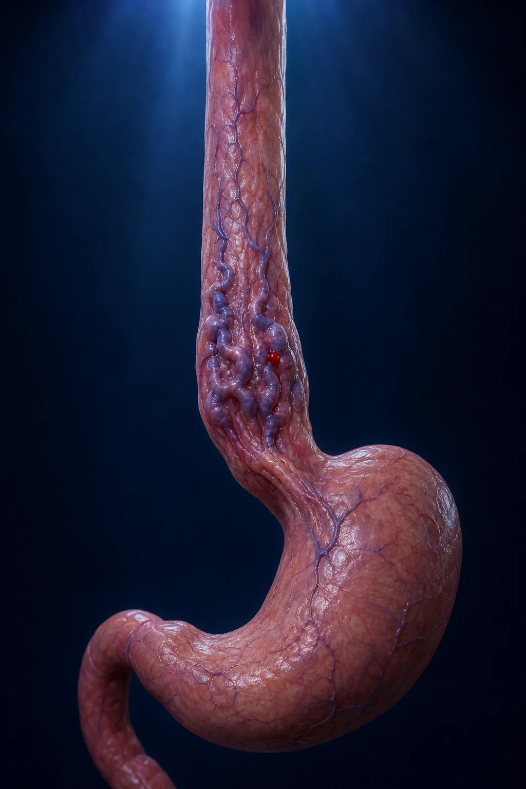
Variceal haemorrhage is massive upper gastrointestinal bleeding from ruptured oesophageal or gastric varices — dilated submucosal portosystemic collaterals that form when portal venous pressure rises. It is defined by a hepatic venous pressure gradient (HVPG) over 12 mmHg, and almost always occurs in cirrhosis. Each bleed carries 15 to 25 percent 6-week mortality. Acute management follows a four-step bundle: resuscitation with a restrictive transfusion strategy (Hb target 70 to 80), vasoactive drug (terlipressin or octreotide) plus prophylactic ceftriaxone (both proven to reduce mortality), urgent endoscopy within 12 hours for band ligation (or cyanoacrylate for gastric varices), and rescue or pre-emptive TIPS for failure or high-risk patients. Prevention is primary (non-selective beta-blocker or endoscopic band ligation for medium or large varices) and secondary (beta-blocker plus serial band ligation), all aimed at lowering portal pressure.
On this page & tools
Your progress
Saved locally on this device.
Exam tags
Red flags
Overview & Definition
Varices are dilated, tortuous submucosal veins that develop as portosystemic collaterals when portal venous pressure rises. They are the most dramatic and lethal consequence of portal hypertension. The pressure that drives them is measured as the hepatic venous pressure gradient (HVPG) — the difference between the wedged (occluded) hepatic venous pressure and the free hepatic venous pressure, which estimates the pressure gradient across the hepatic sinusoids.[1][8]
Three HVPG thresholds govern the entire disease: [1]
- HVPG 5 mmHg or higher — portal hypertension is present (normal is 3 to 5 mmHg).
- HVPG 10 mmHg or higher — clinically significant portal hypertension (CSPH); varices and ascites begin to appear.
- HVPG 12 mmHg or higher — the bleeding threshold; varices are at risk of rupture. [1]
Varices are distinguished by location. Oesophageal varices arise in the distal 5 cm of the oesophagus, supplied mainly by the left gastric (coronary) vein, and are the commonest and most likely to bleed. Gastric varices lie in the fundus (supplied by the short gastric veins) and, although they bleed less often, bleed far more torrentially when they do.[7]
Because each variceal bleed carries 15 to 25 percent 6-week mortality, the entire clinical effort is built around four ideas: resuscitate carefully, combine drugs with endoscopy, escalate to TIPS early in the high-risk, and prevent the first and subsequent bleeds by lowering portal pressure.[1]
Classification

By anatomical site (the classification that drives therapy): [1]
- Oesophageal varices — within and just above the gastro-oesophageal junction; treated by endoscopic band ligation (EBL).
- Gastric varices — sub-classified by the Sarin system:[7]
- GOV1 — varices continuous with oesophageal varices along the lesser curve (commonest, behaves like oesophageal).
- GOV2 — continuous along the greater curve into the fundus (higher rebleed risk).
- IGV1 — isolated gastric fundal varices (no oesophageal component); high bleeding severity.
- IGV2 — isolated varices elsewhere in the stomach (rare).
- Ectopic varices — duodenal, jejunal, stomal, rectal — rare, often non-cirrhotic.
By varix size (North Italian Endoscopic Club, drives primary prevention): [1]
- Small — minimally elevated above the mucosa.
- Medium — tortuous, occupying less than one-third of the lumen.
- Large — occupying more than one-third of the lumen.
- Red signs (red wale marks, cherry-red spots) on the varix surface indicate imminent rupture risk. [1]
By aetiology of the portal hypertension: [1]
- Pre-hepatic — portal vein thrombosis, congenital atresia, splenic vein thrombosis (isolated gastric fundal varices).
- Intrahepatic presinusoidal — schistosomiasis (a leading cause worldwide), idiopathic portal hypertension.
- Intrahepatic sinusoidal — cirrhosis (the commonest cause globally; alcohol, hepatitis B and C, NAFLD/MASLD).
- Intrahepatic postsinusoidal — veno-occlusive disease.
- Post-hepatic — right heart failure, constrictive pericarditis, Budd-Chiari syndrome. [1]
By clinical phase (drives the prevention strategy): [1]
- Never bled — primary prophylaxis.
- Acute bleed / acute bleed survivor — acute treatment then secondary prophylaxis. [1]
Epidemiology & Risk Factors
Portal hypertension is a near-universal late complication of cirrhosis. Among patients with cirrhosis, approximately 50 to 60 percent develop gastro-oesophageal varices, and the annual incidence of new varix formation is 5 to 8 percent. Once varices are present, the annual risk of a first bleed is 5 to 15 percent in those with medium or large varices who receive no prophylaxis.[8]
Risk of a first bleed is highest in those with:[8]
- Large varix size (the single strongest predictor).
- Child-Pugh class B or C (decompensated liver disease).
- Red wale signs or cherry-red spots on endoscopy.
- Continued alcohol intake.
- HVPG over 16 mmHg (steep rise in bleeding risk above this). [1]
Underlying causes by region (regional deltas that examiners reward): [1]
- Worldwide — alcohol-related cirrhosis and chronic hepatitis C dominate in the West; hepatitis B is the leading cause across South and East Asia and sub-Saharan Africa.
- India — hepatitis B, alcohol, and a large cryptogenic / NAFLD-cirrhosis cohort; extrahepatic portal vein obstruction is a notably common cause in the young.
- Middle East, Africa, parts of South America — schistosomiasis (presinusoidal portal hypertension; preserved liver synthetic function).
- Rising globally — NAFLD/MASLD-related cirrhosis, now rivalling alcohol and viral hepatitis. [1]
Predisposing clinical scenarios: any decompensated cirrhosis, hepatocellular carcinoma (tumour thrombus in portal vein), acute alcoholic hepatitis on top of cirrhosis, and any patient with known varices who develops infection (SBP, pneumonia, bacteraemia) — infection both precipitates bleeding and worsens its outcome.[3]
Pathophysiology
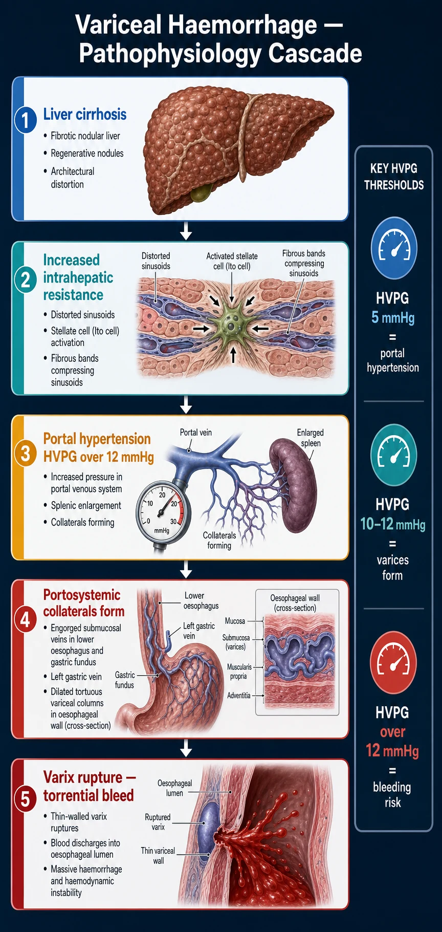
Portal hypertension is fundamentally a balance between resistance to portal blood flow and portal blood inflow. In cirrhosis, both arms of the balance are abnormal.[1]
1. Increased intrahepatic vascular resistance. Cirrhosis distorts the hepatic microcirculation in two ways. The structural component is the fixed fibrosis — regenerative nodules and fibrous septae physically compress the sinusoids. The dynamic component is the activated hepatic stellate cell (Ito cell). In health a quiescent lipid-storing perisinusoidal cell, in injury it transforms into a myofibroblast that contracts around the sinusoid and lays down collagen. This contractile tone is mediated by endothelin-1, angiotensin II and sympathetic signalling, and is partly reversible — which is why splanchnic vasoconstrictors (terlipressin, somatostatin analogues) lower portal pressure acutely. [1]
2. Increased portal venous inflow. As portal pressure rises, a reflex splanchnic vasodilatation develops, driven by nitric oxide (NO), glucagon, carbon monoxide and endocannabinoids. Vasodilatation increases the volume of blood delivered to the splanchnic bed, raising portal inflow and completing a vicious circle: high resistance raises pressure, vasodilatation raises inflow, pressure climbs higher still. [1]
3. Collateral formation and varix development. Sustained portal hypertension reopens the vestigial embryonic portosystemic channels. The clinically important channels are at the gastro-oesophageal junction, where the left gastric (coronary) vein and the short gastric veins drain into the azygos/systemic circulation through the submucosa of the distal oesophagus and gastric fundus. As flow rises, these submucosal veins dilate, elongate and become tortuous — varices. They sit just beneath a thin mucosa in a region of high pressure and are therefore mechanically vulnerable. [1]
4. Why varices bleed — Laplace's law. Variceal rupture is a mechanical failure governed by wall tension: [1]
T = (P × r) / w — wall tension (T) = transmural pressure (P) × varix radius (r) ÷ wall thickness (w). [1]
As a varix enlarges (r rises) and its wall thins (w falls) while pressure (P) remains high, wall tension climbs steeply. Rupture occurs when wall stress exceeds the tensile strength of the wall — empirically at an HVPG above 12 mmHg. This is the single most examinable number in the topic. [1]
5. The pressure thresholds. The three HVPG landmarks recapitulate the natural history: at 5 mmHg the system is hypertensive but clinically silent; at 10 to 12 mmHg varices form; above 12 mmHg they bleed. Conversely, reducing HVPG to below 12 mmHg, or by at least 20 percent from baseline (a haemodynamic response), virtually abolishes the risk of bleeding and is the goal of pharmacological prevention. [1]
6. The role of infection. Bacterial infection (SBP, bacteraemia) both precipitates variceal bleeding and worsens its outcome. The mechanism is a combination of endotoxaemia-driven splanchnic vasodilatation (raising portal inflow), systemic inflammation, and coagulopathy. This is why prophylactic antibiotics are not just a nice-to-have but a mortality-reducing intervention.[3]
Clinical Presentation
A variceal bleed is, at the bedside, a massive upper gastrointestinal haemorrhage with stigmata of chronic liver disease. The presentation has two faces: the acute bleed and the underlying portal hypertension.[1]
Acute bleeding: [1]
- Haematemesis — the cardinal feature. May be fresh bright-red blood (active, brisk bleeding) or coffee-ground (slower, partially digested). Volumes can be enormous.
- Melaena — black, tarry, foul-smelling stool. Indicates at least 60 mL of blood has entered the gut; may be the only sign if bleeding is distal or slow.
- Haematochezia (maroon or red stool) — a sign of massive, rapid upper GI bleeding with rapid gut transit; a marker of severity.
- Shock — tachycardia, hypotension, cool clammy peripheries, oliguria, weak thready pulse, altered sensorium. Indicates at least 20 to 30 percent of circulating volume lost. [1]
Stigmata of chronic liver disease and portal hypertension (the clues that point to varices as the source): [1]
- Jaundice, spider naevi, palmar erythema, gynaecomastia, testicular atrophy (oestrogen excess).
- Ascites, abdominal wall collaterals (caput medusae), splenomegaly (the bedside hallmark of portal hypertension).
- Muscle wasting, parotid enlargement, Dupuytren contracture (alcoholic cirrhosis).
- Asterixis and drowsiness — hepatic encephalopathy, either precipitated by the bleed or pre-existing. [1]
Atypical presentations (the dimension examiners test): [1]
- Elderly — may present in shock without haematemesis, with confusion or delirium as the first sign, or with painless melaena and falls. Comorbidity (cardiac, renal) and anticoagulants complicate the course.
- Diabetic — autonomic neuropathy blunts the tachycardic response; silent, painless large bleeds are common.
- Immunocompromised — may lack fever or signs of infection that would otherwise point to a precipitating SBP.
- Pregnant — variceal bleed risk is highest in the second trimester, when the gravid uterus compresses the IVC and increases splanchnic venous pressure; pregnant cirrhotics have a markedly higher bleed risk than non-pregnant.
- Anticoagulated — warfarin, DOACs, antiplatelets may convert a minor bleed into a torrential one and obscure the underlying source. [1]
Differential Diagnosis
Not every upper GI bleed in a cirrhotic is variceal — up to one-third of upper GI bleeds in cirrhosis are non-variceal. The differentials, with their distinguishing features:[2]
Peptic ulcer disease
Commonest non-variceal UGIB
- Epigastric pain, NSAID use, H. pylori
- Often less massive than variceal; may have a prodrome of dyspepsia
- Endoscopy: clean ulcer base or visible vessel, no varix
Mallory-Weiss tear
Mucosal tear at GO junction
- Preceded by **forceful retching or vomiting** before the haematemesis
- Self-limiting in most; bleeding stops spontaneously
- Endoscopy: longitudinal mucosal tear at the gastro-oesophageal junction
Gastritis / gastropathy
Alcohol, NSAID, stress
- Recent alcohol binge or NSAID course
- Coffee-ground vomiting more than frank haematemesis
- Endoscopy: diffuse erosions, no discrete ulcer or varix
Upper GI malignancy
Gastric or oesophageal cancer
- Weight loss, dysphagia, anorexia, lymphadenopathy
- Often low-grade chronic bleeding; iron-deficiency anaemia
- Endoscopy: mass, ulcerated lesion; biopsy confirms
Dieulafoy lesion
Aberrant submucosal artery
- Painless, massive, recurrent bleed
- No mucosal ulcer — a small punctum bleeding briskly
- Easy to miss at endoscopy; requires careful repeat look
Aortoenteric fistula
Surgical emergency
- Prior **aortic graft** or AAA repair; classic herald bleed then massive bleed
- Endoscopy often normal; CT angiography diagnostic
- Needs urgent vascular surgery
Always consider: a known varix does not exclude another source — endoscopy is what assigns the cause. In a cirrhotic with a haematemesis, assume variceal until endoscopy proves otherwise, but be ready for a non-variceal cause. [1]
Clinical & Bedside Assessment
Primary survey (ABCDE): [1]
- A — Airway. Protect the airway in any patient with massive haematemesis, reduced GCS, or active encephalopathy. Aspiration of blood is a leading cause of death. Intubate early before endoscopy if the airway is not safe.
- B — Breathing. High-flow oxygen by mask; assess for aspiration.
- C — Circulation. Two large-bore cannulae (14G or 16G); take blood for group and crossmatch (4 to 6 units), FBC, U&E, LFT, coagulation, glucose, lactate. Resuscitate with balanced crystalloid. Apply the restrictive transfusion strategy (below).
- D — Disability. Assess GCS and hepatic encephalopathy grade (West Haven) — encephalopathy predicts mortality and drives the airway decision.
- E — Exposure. Full set of vital signs; rectal examination for melaena; examine for stigmata of chronic liver disease. [1]
Shock grading (the bedside estimate of volume loss): [1]
| Class | Blood loss | Heart rate | Blood pressure | Respiratory rate | Urine output | Mental state |
|---|---|---|---|---|---|---|
| I | under 15 percent | under 100 | normal | normal | over 30 mL/h | anxious |
| II | 15 to 30 percent | 100 to 120 | normal | mild tachypnoea | 20 to 30 mL/h | anxious |
| III | 30 to 40 percent | 120 to 140 | fallen | marked tachypnoea | 5 to 15 mL/h | confused |
| IV | over 40 percent | over 140 | markedly low | marked tachypnoea | negligible | drowsy / comatose |
Hepatic encephalopathy — West Haven grade (reproduced verbatim): [1]
- Grade 1 — trivial lack of awareness, euphoria or anxiety, shortened attention span, impaired addition/subtraction; sleep disturbance.
- Grade 2 — lethargy or apathy, disorientation for time, obvious personality change, inappropriate behaviour, asterixis (flapping tremor) demonstrable.
- Grade 3 — somnolent to semistupor but responsive to stimuli, disorientation for place, gross disorientation, marked asterixis.
- Grade 4 — coma, with or without response to painful stimuli. [1]
Bedside pointers that this is variceal (not non-variceal): known cirrhosis, stigmata of chronic liver disease, splenomegaly, ascites, a very massive bleed, and a history of previous variceal bleeding. But none of these is reliable — endoscopy is mandatory. [1]
Investigations
Variceal haemorrhage — the numbers that matter
Bedside and laboratory: [1]
- Venous blood gas — fast haemoglobin, lactate (a marker of shock severity and resuscitation adequacy), pH.
- Full blood count — Hb (falls lag the bleed by hours as equilibration occurs; a normal early Hb does not exclude major bleeding); platelets (thrombocytopenia from hypersplenism is universal in advanced portal hypertension); WCC (leucocytosis points to infection).
- Urea and electrolytes — a raised urea out of proportion to creatinine suggests upper GI bleeding (digested blood protein); monitor for AKI and hepatorenal syndrome.
- Liver function tests and synthetic markers — bilirubin, albumin, INR are the engine of the Child-Pugh and MELD scores; low albumin and high INR quantify hepatic decompensation.
- Coagulation — INR (prolonged), fibrinogen; do not attempt to fully normalise the INR with FFP unless there is active bleeding — the cirrhotic coagulopathy is a rebalanced haemostasis.
- Glucose — hypoglycaemia from impaired gluconeogenesis.
- Blood group and crossmatch — 4 to 6 units initially; massive-transfusion protocol if ongoing.
- Ammonia — if hepatic encephalopathy is in the differential (treat clinically; ammonia level is not strictly required). [1]
Definitive investigation — upper GI endoscopy. [1]
Endoscopy is both diagnostic and therapeutic, and is the single most important investigation. It must be performed within 12 hours of admission (after resuscitation and the start of vasoactive drugs and antibiotics), and not delayed solely to normalise coagulation.[1]
Endoscopy determines: [1]
- The source — varix versus ulcer, Mallory-Weiss, gastropathy, tumour.
- Varix characteristics — site (oesophageal vs gastric, Sarin type), size (small, medium, large), and stigmata of recent haemorrhage:
- Active spurting bleeding from a varix.
- White nipple sign (fibrin plug over the bleeding site).
- Red wale marks and cherry-red spots (high-risk surface signs).
- Whether other lesions (portal hypertensive gastropathy, peptic ulcer, gastritis) coexist. [1]
HVPG measurement — the gold standard for portal pressure, performed at specialist centres by a wedged hepatic vein catheter at transjugular access. It defines clinically significant portal hypertension (over 10 mmHg), the bleeding threshold (over 12 mmHg), and the haemodynamic response to non-selective beta-blockade (fall to below 12 mmHg or at least 20 percent reduction). Not routinely required in the acute setting but central to the concept and to research-grade follow-up. [1]
Imaging when the cause is not straightforward cirrhosis: [1]
- Doppler ultrasound of the portal vein — patency; exclude portal vein thrombosis; measure spleen size and ascites; screen for hepatocellular carcinoma.
- CT or MR portography — to map vascular anatomy before TIPS or shunt surgery; to detect portal vein thrombosis, splenic vein thrombosis (causing isolated fundal varices), or Budd-Chiari. [1]
Named scores — reproduced verbatim
Child-Pugh (Pugh) score — classifies severity of cirrhosis; central to TIPS decisions: [1]
| Parameter | 1 point | 2 points | 3 points |
|---|---|---|---|
| Bilirubin (micromol/L) | under 34 | 34 to 51 | over 51 |
| Albumin (g/L) | over 35 | 28 to 35 | under 28 |
| INR | under 1.7 | 1.7 to 2.3 | over 2.3 |
| Ascites | none | mild (diuretic-responsive) | moderate to severe (refractory) |
| Encephalopathy | none | grade 1 to 2 | grade 3 to 4 |
Classes: A = 5 to 6 (well-compensated); B = 7 to 9 (significant functional compromise); C = 10 to 15 (decompensated). Child-Pugh C carries the highest peri-bleed mortality and is a pre-emptive TIPS criterion (C 7 to 13).[5]
MELD-Na (Model for End-stage Liver Disease — Sodium) — predicts 90-day mortality and drives transplant listing: [1]
MELD = 3.78 × ln(bilirubin mg/dL) + 11.2 × ln(INR) + 9.57 × ln(creatinine mg/dL) + 6.43 MELD-Na = MELD − Na − [0.025 × MELD × (140 − Na)] + 140 (simplified; Na 125 to 140) [1]
Higher MELD-Na = higher 90-day mortality and greater transplant priority. A MELD over 18 in a bleeding cirrhotic predicts poor outcome and warrants transplant referral.[1]
Baveno VII criteria for clinically significant portal hypertension (CSPH): liver stiffness over 25 kPa on elastography (rules in CSPH in most aetiologies); PLT over 150,000 with stiffness under 25 rules out high-risk varices (the "Baveno VI/expanded" rule that can safely avoid screening endoscopy).[1]
Management — Resuscitation
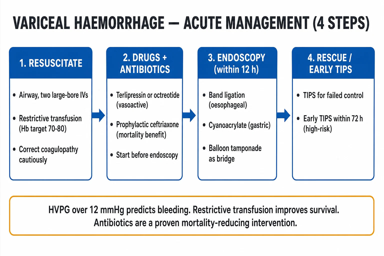
The first hour decides survival. The resuscitation bundle must run concurrently with the diagnostic plan, not after it.[1][2]
1. Airway. Intubate early if there is ongoing massive haematemesis, active grade 3 to 4 encephalopathy, agitation precluding safe endoscopy, or hypoxaemia from aspiration. A protected airway is also a prerequisite for safe, unhurried endoscopic therapy. [1]
2. Circulation — the restrictive transfusion strategy. This is one of the two interventions proven to reduce mortality. The Villanueva trial (NEJM 2013) randomised patients with acute upper GI bleeding to a restrictive strategy (transfuse to Hb 70 g/L, target 70 to 80) versus a liberal strategy (Hb 90, target 90 to 100). The restrictive strategy improved survival, lowered rebleeding, and reduced complications — because over-transfusion raises central venous and portal venous pressure, dislodging clots and worsening bleeding, and because stored blood has dysfunctional platelets and clotting factors.[2]
Exceptions — transfuse more generously in: [1]
- Massive active exsanguinating bleeding — resuscitate to maintain perfusion, not to a number.
- Coronary artery disease / unstable angina — these subgroups were excluded from the trial; target higher (around 80 to 90).
- Shock at presentation — restore perfusion first, then ease to the restrictive target. [1]
3. Access and fluid. Two large-bore peripheral cannulae (14G/16G). Resuscitate with balanced crystalloid (Hartmann or PlasmaLyte); avoid excessive saline in cirrhosis. Apply the massive transfusion protocol (1:1:1 red cells : FFP : platelets) for ongoing major haemorrhage. Insert a urinary catheter to titrate resuscitation to a urine output of at least 0.5 mL/kg/h. [1]
4. Coagulopathy correction — judicious, not automatic. The cirrhotic "coagulopathy" is a rebalanced haemostasis (procoagulant and anticoagulant factors fall together); an isolated raised INR does not by itself predict bleeding. Give: [1]
- Vitamin K 10 mg IV slow (corrects vitamin-K-dependent factor deficiency from cholestasis or antibiotics).
- Fresh frozen plasma and platelets only if there is active bleeding with INR over 1.5 or platelets under 50 — over-correction risks volume overload and worsened portal pressure.
- Tranexamic acid — routine use is not recommended (the HALT-IT trial showed no mortality benefit and possible harm).
- Prothrombin complex concentrate may be considered in life-threatening bleeding with anticoagulant-related coagulopathy. [1]
5. Vasoactive drug — start before endoscopy. The aim is splanchnic vasoconstriction, reducing portal inflow and pressure, slowing bleeding and buying time for endoscopy. Give before endoscopy and continue for 2 to 5 days.[1]
- Terlipressin — 2 mg IV bolus, then 1 mg every 4 hours for 48 hours, then 1 mg every 4 hours (reduced to 0.5 mg if tolerated) up to 5 days. The agent of choice in the UK and India. Caution: hyponatraemia, ischaemia (coronary, mesenteric, peripheral), and avoid in pregnancy unless essential.
- Octreotide — 50 microgram IV bolus, then 50 microgram per hour infusion for 3 to 5 days. Common in North America. Less potent than terlipressin but better tolerated; side effects hyperglycaemia and abdominal cramps.
- Somatostatin — 250 microgram bolus then 250 microgram per hour. An alternative where available. [1]
6. Prophylactic antibiotic — start before endoscopy, give for up to 7 days. This is the second mortality-reducing intervention. Infection both precipitates and complicates variceal bleeding, and prophylactic antibiotics reduce infection, rebleeding and death.[3]
- Ceftriaxone 1 g IV once daily for 7 days — the preferred agent (superior to oral norfloxacin/ciprofloxacin in trials, particularly in high-resistance regions).
- Oral norfloxacin 400 mg twice daily or ciprofloxacin — alternatives in low-resistance settings or as step-down. [1]
Management — Definitive & Stepwise
Step 1 — Combination therapy (drugs plus endoscopy) — first-line definitive treatment. Combining a vasoactive drug with endoscopic band ligation is superior to either alone in controlling acute bleeding and reducing 5-day mortality. Both should be in place by 12 hours.[1]
Step 2 — Endoscopic therapy, within 12 hours. [1]
- Oesophageal varices — endoscopic band ligation (EBL). A rubber band is suction-applied onto each varix, ligating and strangulating it. First-line therapy. Achieves haemostasis in 80 to 90 percent. Repeat sessions every 2 to 4 weeks until variceal obliteration; surveillance thereafter.
- Sclerotherapy (injection of a sclerosant such as ethanolamine oleate or sodium tetradecyl sulphate) — largely superseded by EBL (more complications: ulceration, strictures, sepsis); reserved for bleeding that cannot be banded.
- Gastric varices — cyanoacrylate (glue) injection. A tissue adhesive (N-butyl-2-cyanoacrylate, often mixed with lipiodol) is injected into the varix, polymerising and occluding it. First-line for gastric (especially fundal) varices, where band ligation is less effective and more prone to rebleed. Risks: glue embolisation (pulmonary, cerebral, splenic).
- Self-expanding metal stents (SEMS) — covered oesophageal metal stents placed endoscopically as a bridge for uncontrolled oesophageal bleeding, especially if balloon tamponade is unavailable or contraindicated. [1]
Step 3 — Balloon tamponade as a temporary bridge. A Sengstaken-Blakemore tube (gastric and oesophageal balloons) or a Linton / Minnesota tube (gastric balloon only) is inserted and the gastric balloon inflated against the gastro-oesophageal junction, tamponading the bleeding varices. Used only when endoscopic control fails, as a bridge of no more than 24 hours to definitive therapy (TIPS). Risks: aspiration, oesophageal ulceration and rupture, airway obstruction — the patient must be intubated and in an ICU. [1]
Step 4 — Rescue and pre-emptive TIPS. The transjugular intrahepatic portosystemic shunt (TIPS) creates a channel between the hepatic vein and the intrahepatic portal vein via a stent, decompressing the portal system. It is the rescue therapy for uncontrolled or recurrent variceal bleeding. [1]
- Rescue TIPS — for failure to control bleeding despite drugs plus endoscopy. Mortality is high but TIPS can be life-saving.
- Pre-emptive (early) TIPS — within 72 hours of presentation (ideally within 24 hours), in high-risk patients defined by Baveno:[1][5]
- Child-Pugh C, score 7 to 13, OR
- Child-Pugh B with active bleeding at endoscopy.
- Pre-emptive TIPS reduces both rebleeding and mortality in these groups (Monescillo 2004; Garcia-Pagan 2010). Do not apply to Child-Pugh over 13 or to patients with uncontrolled sepsis, severe cardiopulmonary disease, or extensive portal vein thrombosis (relative contraindications).
- Complications of TIPS — hepatic encephalopathy (in up to one-third; the shunted blood bypasses hepatic detoxification), heart failure (increased venous return), haemolysis, and shunt dysfunction (stenosis/thrombosis).
Step 5 — Prevention. Once the acute episode is controlled, every patient moves into secondary prevention (below). The goal throughout is to lower portal pressure. [1]
Prevention ladder — primary and secondary
Primary prevention (the patient who has varices but has never bled): [1]
- Indication — medium or large varices, or small varices with red wale signs or Child-Pugh B/C.
- Option A — non-selective beta-blocker (NSBB):
- Propranolol 20 to 40 mg orally twice daily (titrate to heart rate 55 to 60).
- Nadolol 40 to 80 mg orally daily.
- Carvedilol 6.25 to 12.5 mg orally daily — now preferred in many Baveno VII-aligned pathways because its alpha-1 blockade lowers portal pressure more than propranolol (Tripathi 2008).[6]
- Target haemodynamic response: HVPG fall to below 12 mmHg or by at least 20 percent.
- Option B — endoscopic band ligation — for those intolerant of NSBB or with contraindications (asthma, heart block, hypotension). Serial sessions until obliteration, then surveillance.
- Choice — NSBB and EBL are broadly equivalent for first-bleed prevention; combine the two only in selected high-risk cases (additive side-effect burden).
Secondary prevention (the patient who has survived a variceal bleed): [1]
- Without secondary prevention, rebleed risk is 60 to 70 percent within 1 year.
- Combination therapy — NSBB plus serial band ligation gives the lowest rebleed rate and is first-line.[8]
- TIPS — if rebleeding despite combination therapy, or intolerance of NSBB.
- Liver transplantation — the definitive treatment for decompensated cirrhosis; refer early (MELD-driven).
Specific Subtypes & Scenarios
- Isolated gastric varices (IGV1, fundal) — bleed less often than oesophageal but more torrentially when they do. First-line endoscopic therapy is cyanoacrylate injection; band ligation is less effective. TIPS if glue fails or recurrent. BRTO (balloon-occluded retrograde transvenous obliteration) is an alternative where the gastrorenal shunt allows access, particularly useful when TIPS is contraindicated.
- Portal hypertensive gastropathy — a diffuse mosaic/snakeskin mucosal vascular ectasia of the stomach, NOT discrete varices. Presents with chronic occult bleeding and anaemia rather than massive haematemesis. Treated with NSBB (propranolol/carvedilol), not endoscopy; TIPS for refractory cases.
- Gastric antral vascular ectasia (GAVE, "watermelon stomach") — linear vascular streaks radiating from the pylorus; associated with cirrhosis and autoimmune disease. Treated with argon plasma coagulation (endoscopic thermal therapy), not NSBB.
- Ectopic varices — duodenal, stomal (post-colectomy), rectal, periumbilical. Rare; often in non-cirrhotic portal hypertension (e.g. portal vein thrombosis, post-surgical). Managed with TIPS, BRTO, or local therapy depending on site and anatomy.
- Non-cirrhotic portal hypertension — portal vein thrombosis (acute: anticoagulation; chronic with varices: endoscopic therapy, consider shunt), schistosomiasis (preserved liver function; manage varices as in cirrhosis), extrahepatic portal vein obstruction in children (the leading cause of paediatric variceal bleeding; endoscopic therapy first, surgery if refractory).
- The high-risk acute patient — Child-Pugh C, MELD over 18, active bleeding at endoscopy, hepatocellular carcinoma, infection at presentation, or age over 60. These patients warrant pre-emptive TIPS and/or ICU-level care; their mortality approaches 30 to 40 percent per bleed. [1]
Complications & Pitfalls
Complications of the bleed itself: [1]
- Death — 15 to 25 percent 6-week mortality; the outcome metric of every trial.
- Early rebleeding (within 5 days) — risk highest in the first 48 to 72 hours; predicts death. Drives the rescue-TIPS decision.
- Hepatic encephalopathy — precipitated by the ammonia load from blood digested in the gut. Treat with lactulose (titrate to 2 to 3 soft stools per day) and rifaximin 550 mg twice daily.
- Infection — SBP, pneumonia, bacteraemia, UTI. Infection both precedes and follows bleeding; prophylactic antibiotics are mandatory.
- Acute kidney injury and hepatorenal syndrome (HRS-AKI) — precipitated by hypovolaemia, infection, and nephrotoxins. Volume-resuscitate; albumin 20 percent at 1 g/kg on day 1; terlipressin plus albumin for HRS.
- Aspiration pneumonia — from an unprotected airway during massive haematemesis or sedated endoscopy; intubate at-risk patients. [1]
Complications of therapy: [1]
- Balloon tamponade — oesophageal rupture, aspiration, airway obstruction, pressure necrosis. Never leave inflated beyond 24 hours.
- TIPS — hepatic encephalopathy (most consequential), heart failure, haemolysis, shunt stenosis/thrombosis.
- Endoscopic band ligation — post-banding ulcer bleeding, oesophageal stricture, transient dysphagia, chest pain.
- Cyanoacrylate — glue embolisation (pulmonary, cerebral, splenic) — rare but serious.
- Terlipressin — hyponatraemia (common), ischaemia (coronary, mesenteric, digital), peripheral vasoconstriction. [1]
Classic pitfalls (the ones examiners reward avoiding): [1]
- Over-transfusing to a normal Hb — raises portal pressure and worsens rebleeding (the Villanueva lesson).[2]
- Forgetting prophylactic antibiotics — the most common omission and the easiest mortality win.[3]
- Delaying endoscopy to "normalise the INR" — the INR in cirrhosis is a rebalanced haemostasis, not a bleeding predictor; endoscopy is therapeutic and must not wait.
- Leaving a balloon tamponade in too long — beyond 24 hours the risk of oesophageal necrosis and rupture is unacceptable.
- Not offering pre-emptive TIPS to a Child-Pugh C patient with active bleeding — a clear mortality-reducing intervention missed.[5]
- Not addressing prevention after the acute episode — secondary prevention prevents the next (often fatal) bleed.
Prognosis & Disposition
Prognostic drivers in variceal haemorrhage
Mortality tracks four things: liver reserve (Child-Pugh, MELD), control of bleeding, infection, and the underlying aetiology. The 6-week mortality of 15 to 25 percent has fallen over three decades as combination therapy, antibiotics, restrictive transfusion, and pre-emptive TIPS have been adopted, but it remains substantial in decompensated cirrhosis. [1]
Predictors of poor outcome:[1][5]
- Child-Pugh C (especially 10 to 13) and MELD over 18.
- Active bleeding at endoscopy.
- HVPG over 20 mmHg (a steep rise in failure and death).
- Infection at presentation (SBP, bacteraemia).
- Hepatocellular carcinoma and portal vein thrombosis.
- Renal failure at admission (HRS-AKI doubles mortality).
- Age over 60 and significant comorbidity. [1]
Disposition: [1]
- ICU or high-dependency unit for all acute variceal bleeds, at least for the first 24 to 48 hours.
- Endoscopy within 12 hours — not as an emergency to be done in parallel with resuscitation, but once the airway and circulation are secured.
- Pre-emptive TIPS within 72 hours for high-risk patients (above).
- After stabilisation — begin secondary prevention (NSBB plus serial EBL) and arrange surveillance endoscopy every 3 to 6 months.
- Transplant referral for decompensated cirrhosis (MELD-driven) or for those with refractory ascites, recurrent encephalopathy, or recurrent variceal bleeding despite optimal therapy. [1]
Special Populations
- Pregnancy — variceal bleed risk is highest in the second trimester, when the gravid uterus compresses the IVC and increases splanchnic venous pressure. Pregnant patients with known varices should be screened and treated before conception where possible. In acute bleed: endoscopic band ligation is first-line (safe in pregnancy); use terlipressin with caution (uterine vasoconstriction, risk of ischaemia; octreotide preferred by some); balloon tamponade and TIPS are last resorts, TIPS only after the second trimester. Vaginal delivery is generally advised with a covered second stage; caesarean for obstetric indications.
- Children — the leading cause of variceal bleeding in children is extrahepatic portal vein obstruction (cavernous transformation), with preserved liver synthetic function. Manage with endoscopic band ligation or sclerotherapy; surgical portosystemic shunt (meso-Rex bypass) for refractory cases, which also restores portal flow to the liver.
- Elderly — higher per-bleed mortality, atypical presentation (confusion, falls, painless melaena), more comorbidity, more anticoagulant and antiplatelet use. Lower threshold to intubate for airway protection. Stop NSAIDs.
- Anticoagulated / antiplatelet patient — hold the agent; reversal only if life-threatening bleeding (prothrombin complex concentrate for warfarin, specific reversal agents for DOACs where available). Resume anticoagulation as soon as haemostasis is secure — the thrombotic risk off-treatment is substantial.
- Renal impairment — dose-adjust terlipressin (and beware hyponatraemia, which is common and sometimes severe); monitor for hepatorenal syndrome; albumin-based resuscitation.
- The patient with HCC — portal vein tumour thrombus worsens portal hypertension and complicates TIPS; treat the HCC alongside the varices. [1]
Evidence, Guidelines & Regional Differences
Landmark trials and consensus: [1]
- Baveno VII (2022) — the current international consensus on portal hypertension. Introduced the pre-emptive TIPS criteria, endorsed carvedilol as the preferred NSBB in many, and refined the non-invasive CSPH thresholds (liver stiffness over 25 kPa).[1]
- Villanueva 2013 (NEJM) — restrictive (Hb 70) versus liberal (Hb 90) transfusion in acute UGIB: restrictive improved survival and reduced rebleeding.[2]
- Bernard 1999 meta-analysis and subsequent trials — prophylactic antibiotics reduce bacterial infection, rebleeding and mortality in cirrhotic UGIB.[3]
- Monescillo 2004 (Gut) — early TIPS in high-risk patients reduced treatment failure and mortality.[4]
- Garcia-Pagan 2010 (NEJM) — confirmed pre-emptive TIPS (within 72 hours) in Child-Pugh C 10 to 13 or B with active bleeding reduces 1-year mortality.[5]
- Tripathi 2008 (Hepatology) — carvedilol versus band ligation for first-bleed prevention; carvedilol non-inferior and better tolerated, with greater portal-pressure reduction.[6]
- Sarin 1992 — the classification of gastric varices (GOV1/2, IGV1/2) still in use.[7]
- AASLD 2007 (Garcia-Tsao, updated) — comprehensive practice guideline on prevention and management of varices.[8]
- HALT-IT trial (2020) — tranexamic acid showed no mortality benefit and possible harm in acute GI bleeding; not recommended routinely.
Regional deltas: [1]
[1] [1] [1]Controversies: [1]
- Tranexamic acid — no longer recommended after HALT-IT.
- NSBB choice — carvedilol versus propranolol: carvedilol preferred where portal pressure reduction is the goal, but caution in hypotensive or ascitic patients.
- Self-expanding metal stents versus balloon tamponade — SEMS appear safer and at least as effective as a bridge to definitive therapy; availability varies.
- TIPS in the elderly — higher encephalopathy risk; balance individualised.
- When to start oral feeding — early (after endoscopy) appears safe and is increasingly recommended. [1]
Exam Pearls
Acute variceal bleed — the four-step bundle
RATE
Airway, two large-bore IVs, restrictive transfusion (Hb 70 to 80)
Ceftriaxone 1 g IV OD plus terlipressin 2 mg IV then 1 mg q4h — both BEFORE endoscopy
Band ligation (oesophageal) or cyanoacrylate (gastric) within 12 h; rescue or pre-emptive TIPS
Within 72 h for Child-Pugh C 7 to 13 or Child-Pugh B with active bleeding at endoscopy
The three HVPG thresholds
VIP
Form when HVPG reaches 10 to 12 mmHg
HVPG over 10 mmHg — CSPH
HVPG over 12 mmHg — varices bleed; reduce to below 12 or by at least 20 percent
- Three numbers to memorise: 5, 12, 20. HVPG 5 = portal hypertension; over 12 = bleeding threshold; reduce by 20 percent (or below 12) = haemodynamic response to NSBB.
- Two interventions that reduce mortality: prophylactic ceftriaxone and restrictive transfusion (Hb 70 to 80). Both are easy marks in an MCQ.[2][3]
- Terlipressin is the vasoactive of choice in the UK/India; octreotide in the US. Both started before endoscopy, continued 2 to 5 days.
- Band ligation for oesophageal; cyanoacrylate for gastric (fundal); balloon tamponade only as a 24-hour bridge.
- Pre-emptive TIPS within 72 h for Child-Pugh C 7 to 13 or Child-Pugh B with active bleeding — a clear mortality benefit (Garcia-Pagan 2010).[5]
- Primary prevention: NSBB (now carvedilol preferred) OR band ligation for medium/large varices. Secondary: NSBB plus serial band ligation.
- Carvedilol lowers portal pressure more than propranolol (alpha-1 blockade) and is the modern NSBB of choice.[6]
- Sarin classification of gastric varices: GOV1 (lesser curve, behaves like oesophageal) versus GOV2/IGV1 (fundal, more severe bleeds).
- A normal early Hb does not exclude major bleeding — equilibration takes hours; trend and assess by perfusion and lactate.
- Tranexamic acid is NOT recommended after the HALT-IT trial.
- Endoscopy within 12 hours — do not delay to normalise the INR. The cirrhotic INR reflects rebalanced haemostasis, not bleeding risk.
Exam application bank (NEET-PG / INICET)
One-line answer
Variceal haemorrhage is massive upper gastrointestinal bleeding from ruptured oesophageal or gastric varices — dilated submucosal portosystemic collaterals that form when portal venous pressure rises. It is defined by a hepatic venous pressure gradient (HVPG) over 12 mmHg, and almost always occurs in cirrhosis. Each bleed carries 15 to 25 percent 6-week mortality. Acute management follows a four-step bundle: resuscitation with a restrictive transfusion strategy (Hb target 70 to 80), vasoactive drug (terlipressin or octreotide) plus prophylactic ceftriaxone (both proven to reduce mortality), urgent endoscopy within 12 hours for band ligation (or cyanoacrylate for gastric varices), and rescue or pre-emptive TIPS for failure or high-risk patients. Prevention is primary (non-selective beta-blocker or endoscopic band ligation for medium or large varices) and secondary (beta-blocker plus seria
Worked stems (answer without another resource)
Stem 1 — Classic presentation. Map symptoms to mechanism; name the first investigation and first treatment step with dose/route if drug therapy is standard. [1]
Stem 2 — Unstable / complicated. List red flags that force immediate resuscitation, theatre, ICU, antidote, or reperfusion — and what you do in the first 15 minutes. [1]
Stem 3 — Atypical group. Elderly, pregnancy, child, or immunocompromised: how presentation and thresholds change. [1]
Stem 4 — Differential trap. Name the three closest mimics and one discriminator for each. [1]
Stem 5 — Disposition. Who goes home with safety-netting, who is admitted, who needs HDU/ICU/theatre, and what follow-up is mandatory. [1]
Rapid viva checklist
- Definition + classification
- Pathophysiology chain
- Bedside signs / criteria
- Score with exact components (if any)
- Emergency bundle
- Definitive therapy with doses
- Complications of disease and of treatment
- Special populations
- Guideline/trial name if classic
- Three exam traps
Coverage self-check
If you cannot answer any stem above from this page alone, re-read the matching section — the page is intended to be self-sufficient for final-prof and NEET-PG/INICET questions on Variceal Haemorrhage.
References
- [1]de Franchis R, Bosch J, Garcia-Tsao G, et al. Baveno VII - Renewing consensus in portal hypertension J Hepatol, 2022.PMID 35120736
- [2]Villanueva C, Colomo A, Bosch A, et al. Transfusion strategies for acute upper gastrointestinal bleeding N Engl J Med, 2013.PMID 23281973
- [3]Bernard B, Grange JD, Khac EN, Amiot X, Opolon P, Poynard T. Antibiotic prophylaxis for the prevention of bacterial infections in cirrhotic patients with gastrointestinal bleeding: a meta-analysis Hepatology, 1999.PMID 10347104
- [4]Monescillo A, Martinez-Lagares F, Ruiz-del-Arbol L, et al. Influence of portal hypertension and its early decompression by TIPS placement on the outcome of variceal bleeding Hepatology, 2004.PMID 15382120
- [5]Garcia-Pagan JC, Caca K, Bureau C, et al. Early use of TIPS in patients with cirrhosis and variceal bleeding N Engl J Med, 2010.PMID 20573925
- [6]Tripathi D, Ferguson JW, Kochar N, et al. Randomized controlled trial of carvedilol versus variceal band ligation for the prevention of the first variceal bleed Hepatology, 2009.PMID 19610055
- [7]Sarin SK, Lahoti D, Saxena SP, Murthy NS, Makwana UK. Prevalence, classification and natural history of gastric varices: a long-term follow-up study in 568 portal hypertension patients Hepatology, 1992.PMID 1446890
- [8]Garcia-Tsao G, Sanyal AJ, Grace ND, Carey W. Prevention and management of gastroesophageal varices and variceal hemorrhage in cirrhosis Hepatology, 2007.PMID 17879356