General Surgery · General Surgery
Abdominal Aortic Aneurysm
Also known as AAA · Aortic aneurysm · Triple A · Ruptured AAA · Non-ruptured AAA
Abdominal aortic aneurysm (AAA) is a permanent, localised dilation of the abdominal aorta to 3 cm or more (or 1.5 times the expected normal diameter), with over 90% infrarenal. Most are asymptomatic until rupture. Risk: male sex, age over 65, smoking (the dominant modifiable factor), family history, hypertension. Screening: one-off ultrasound for men at 65 (NHS AAA Screening Programme); USPSTF for men 65 to 75 who ever smoked. Surveillance thresholds: under 3 cm normal; 3.0 to 4.4 cm yearly; 4.5 to 5.4 cm every 3 months; over 5.5 cm refer for elective repair (open or EVAR). Rupture triad: severe abdominal/back/flank pain plus hypotension plus a pulsatile abdominal mass (complete in only half). Ruptured AAA mortality 80 to 90% overall; 40 to 50% perioperative. Elective mortality 2 to 5% open, 1.5 to 2% EVAR. Laplace law: Wall Tension = Pressure x Radius / Wall Thickness explains the vicious cycle of growth. EVAR is less invasive but needs lifelong surveillance for endoleak.
On this page & tools
Your progress
Saved locally on this device.
Exam tags
Red flags
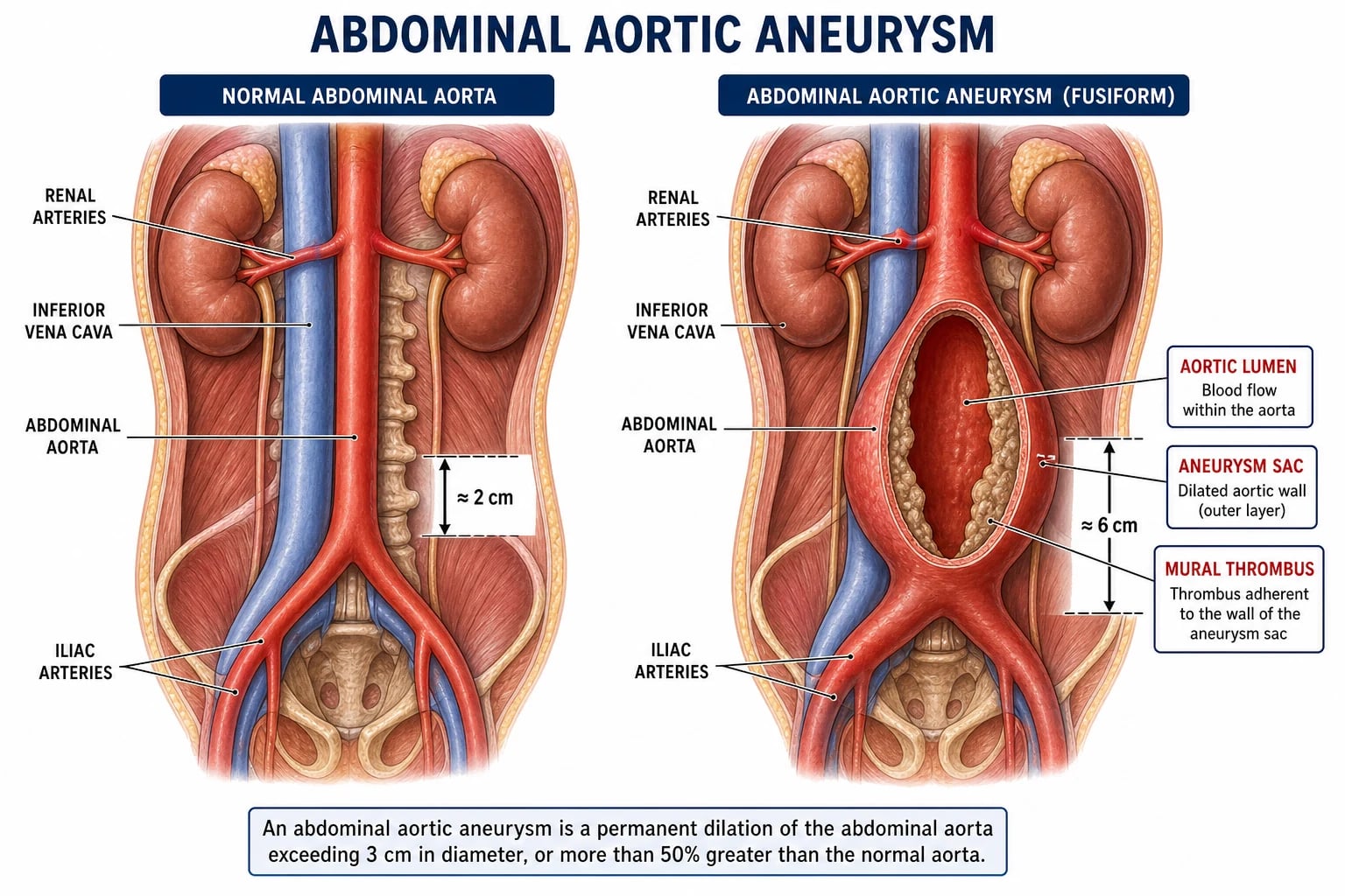
Overview & Definition
An abdominal aortic aneurysm (AAA) is a permanent, localised dilation of the abdominal aorta in which the maximum anteroposterior diameter reaches 3 cm or more — that is, 1.5 times the expected normal suprarenal or infrarenal diameter for age and sex. The normal adult infrarenal aorta measures roughly 1.7 to 2.2 cm in men and 1.5 to 1.9 cm in women, so the 3 cm threshold represents a substantial, pathological enlargement rather than a borderline change. AAA is a true aneurysm: all three layers of the arterial wall (intima, media, adventitia) are involved, distinguishing it from a pseudoaneurysm in which only adventitia and periadventitial tissue contain the flow.[2]
The clinical importance of AAA lies entirely in the risk of rupture, a catastrophic event that kills the majority of sufferers before they reach surgical care. The entire strategy of management — community screening, ultrasound surveillance, risk-factor modification, and elective repair — exists to detect and treat the aneurysm before it ruptures. The natural history is one of progressive, painless enlargement over years, followed in a minority by the dramatic, often fatal presentation of rupture. Because most AAAs are silent, the condition is a paradigm of a disease conquered by screening: the Multicentre Aneurysm Screening Study (MASS) demonstrated that a single ultrasound in older men halves aneurysm-related mortality and is highly cost-effective.[1][5]
An important conceptual point for the viva: AAA is not simply atherosclerosis. Although atherosclerosis coexists in nearly every case and the two share risk factors, AAA is fundamentally a degenerative disease of the aortic media driven by proteolytic destruction of elastin and collagen, chronic inflammation, and smooth-muscle cell apoptosis. This explains why some patients with severe atherosclerosis never develop an aneurysm, and why the search for medical therapy has focused on inhibiting matrix metalloproteinases and inflammation rather than merely treating atherosclerosis.[2]
Classification
By size — the classification that drives every management decision: [1]
| AP diameter | Category | Rupture risk per year | NHS surveillance / action |
|---|---|---|---|
| Under 3 cm | Normal aorta (no AAA) | negligible | discharge from screening |
| 3.0 to 4.4 cm | Small AAA | under 1% | rescan at 12 months |
| 4.5 to 5.4 cm | Medium AAA | 1 to 5% | rescan at 3 months |
| 5.5 cm or more | Large AAA | 5 to 25%+ | refer to vascular surgery for repair |
| Symptomatic or rapidly expanding (over 0.5 cm in 6 months) | Any size | high | urgent repair regardless of absolute size |
The 5.5 cm threshold is the single most examined number in this topic. It is the point at which rupture risk exceeds operative mortality in an average-risk patient, established by the UK Small Aneurysm Trial and confirmed by its 12-year follow-up.[8]
By anatomical extent:
- Infrarenal (commonest, around 95%) — the aneurysm begins below the renal arteries; the standard anatomy for both open and endovascular repair.
- Juxtarenal — begins immediately at the level of the renal arteries; the neck is absent or extremely short, complicating EVAR and often requiring suprarenal clamping at open repair.
- Suprarenal and pararenal — extend above the renal arteries; demand complex branched/fenestrated endografts or open repair with renal revascularisation.[1]
By morphology:
- Fusiform (commonest) — circumferential, spindle-shaped dilation of the whole circumference.
- Saccular — localised outpouching of one wall; more often seen in mycotic, post-traumatic, or penetrating atherosclerotic ulcer aneurysms. [1]
By clinical status (the classification that drives urgency):
- Asymptomatic / intact — detected on screening or incidental imaging; managed by surveillance or elective repair.
- Symptomatic non-ruptured — new pain attributable to the aneurysm; an urgent surgical problem even if small, because pain often heralds expansion or leak.
- Ruptured — a surgical emergency with mortality of 80 to 90%. [1]
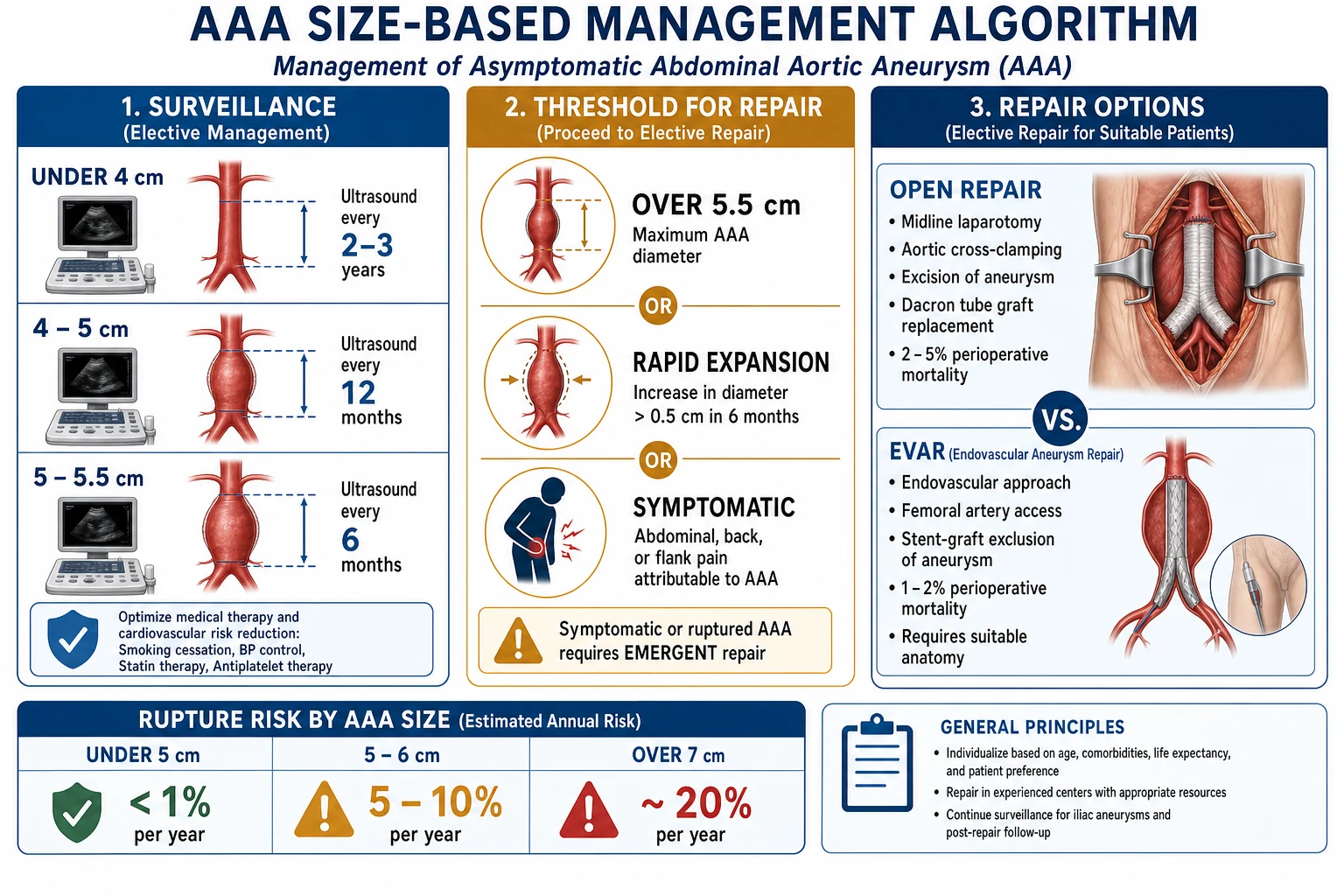
AAA — the numbers that matter
Epidemiology & Risk Factors
AAA is overwhelmingly a disease of older men of Northern European descent. Large community ultrasound surveys place the prevalence at 4 to 8% in men over 65; in women the figure is roughly ten-fold lower at 0.5 to 1.5%, giving a male-to-female ratio of approximately 6 to 1. The mean age at diagnosis is around 70 years, and AAA is rare before 55. Prevalence is highest in Scandinavian and North American white populations and substantially lower in African, South Asian, and East Asian populations, a difference only partly explained by reporting and screening intensity.[2]
Risk factors, in descending order of importance for the examination: [1]
- Smoking — the single most important modifiable risk factor. Ever-smoking confers a 3- to 5-fold increase in risk, accounts for the majority of attributable deaths, and predicts faster growth and higher rupture rates. Duration matters more than current status.
- Age — prevalence rises sharply after 55; the screening programmes target the 65 to 75 band because that is where yield and benefit are greatest.
- Male sex — 6 times more common in men; this is why screening is sex-selective.
- Family history — a first-degree relative with AAA roughly doubles to quadruples risk and produces AAAs a decade earlier; some authorities advise screening male relatives from age 60, or 10 years younger than the affected relative's age at diagnosis.[1]
- Hypertension — modest independent contribution, particularly to rupture risk.
- Caucasian ethnicity — white men carry the highest prevalence.
- Atherosclerosis and its correlates — coronary disease, peripheral arterial disease, hyperlipidaemia, and a high ABI in the contralateral limb are all associated.
- Chronic obstructive pulmonary disease (COPD) — an independent risk factor, likely reflecting shared proteolytic destruction of elastin in lung and aorta.[2]
A useful viva framing: AAA shares every risk factor with occlusive atherosclerosis except diabetes — diabetics, paradoxically, have a lower incidence of AAA, possibly because diabetic arterial stiffening and medial sclerosis limit aneurysmal dilation.[4]
Pathophysiology
AAA is best understood as a multifactorial degeneration of the aortic wall in which proteolysis, inflammation, biomechanical stress, and genetic susceptibility converge on a final common pathway of progressive weakening and dilation.[2]
1. Matrix degradation. The structural integrity of the aortic wall depends on elastin (which bears load at physiological pressures) and collagen (which takes over as the wall stretches). In AAA, matrix metalloproteinases (MMPs) — particularly MMP-2 and MMP-9 — and cysteine proteases are over-expressed by infiltrating macrophages, smooth muscle cells, and fibroblasts. These enzymes fragment elastin and cleave collagen faster than they can be resynthesised. The loss of elastin is the critical early event: elastin content in an aneurysmal wall may fall to less than 20% of normal. With elastin gone, the load shifts onto collagen, which remodels, fatigues, and ultimately fails.[2]
2. Chronic inflammation. The aneurysm wall is infiltrated by macrophages, T-lymphocytes, and B-cells, which produce cytokines (IL-1beta, IL-6, TNF-alpha, MCP-1) that recruit more inflammatory cells and up-regulate MMP production. This inflammatory infiltrate, together with the production of reactive oxygen species and the activation of angiotensin II signalling, sustains a self-perpetuating cycle of proteolysis. In a minority of cases the inflammation is so dense that the aneurysm is termed an inflammatory AAA (see Subtypes). [1]
3. Smooth muscle cell apoptosis. Loss of medial smooth muscle cells, the cells that synthesise replacement elastin and collagen, further erodes the wall's capacity to repair itself. [1]
4. Biomechanics — Laplace's law. The physics of aneurysm growth and rupture is governed by Laplace's law, which states that the wall tension of a cylinder is proportional to its radius and the transmural pressure, and inversely proportional to wall thickness: [1]
Wall Tension (T) = Pressure (P) x Radius (r) / Wall Thickness [1]
This single equation explains the entire natural history. As the aneurysm dilates, the radius increases and the wall thins (because the same amount of tissue lines a larger circumference). Both effects raise wall tension, which in turn drives further dilation — a positive feedback loop (vicious cycle) that culminates in rupture. It is also why hypertension accelerates growth (higher P) and why rupture risk rises non-linearly with diameter: doubling the radius roughly doubles the wall stress.[3]
5. Genetics and signalling. Familial clustering (15 to 20% of AAA patients have an affected first-degree relative) implicates genetic susceptibility. Associated variants cluster around extracellular-matrix genes and inflammatory pathways. The inverse relationship with diabetes hints that insulin signalling pathways may be protective, an observation now being explored therapeutically.[4]
6. The mural thrombus paradox. Most large AAAs contain a laminated mural (intraluminal) thrombus. Far from being protective, the thrombus is biologically active: it harbours neutrophils and proteases, prevents oxygen diffusion to the inner wall, and may weaken the underlying media. Chunks can embolise distally, producing the blue toe syndrome, renal infarcts, or livedo reticularis. The thrombus thus contributes to both growth and distal embolic complications.[2]
[1]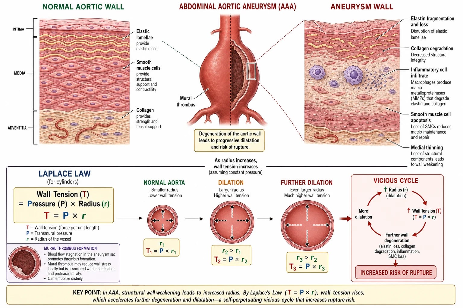
Clinical Presentation
The clinical challenge of AAA is that the first symptom is often death. Most aneurysms are detected either by screening or incidentally, and the management of rupture depends on recognising a presentation that mimics far commoner conditions. [1]
Asymptomatic (the majority). AAAs are silent. They are found on:
- screening ultrasound (the NHS AAA Screening Programme offers a single scan to men in their 65th year);
- incidental imaging — ultrasound, CT, or MRI performed for another reason (prostate, gallstones, abdominal pain, trauma);
- clinical examination — a pulsatile mass felt incidentally. [1]
A note on examination findings: the sensitivity of palpation for an AAA rises with diameter but is only moderate (around 40% overall, over 75% for AAAs over 5 cm), and falls markedly in obese patients. A palpable mass is therefore useful when present but does not exclude AAA when absent.[1]
The pulsatile abdominal mass. A true AAA is expansile — it expands outwards in all directions, best appreciated by placing the hands on either side of the mass and feeling it push the fingers apart with each beat. This distinguishes it from transmitted pulsation from a mass overlying a normal aorta (e.g. a gastric tumour), which simply moves up and down. The mass typically lies in the epigastrium or periumbilical region and is best felt with the patient relaxed and the knees flexed to soften the abdominal wall. An expansile mass over 4 cm wide in an older man should prompt ultrasound the same day. [1]
Symptomatic non-ruptured (expanding or tender) AAA. New symptoms in a known aneurysm are a danger sign:
- Back or abdominal pain — typically deep, constant, and non-colicky, from pressure on the lumbar vertebrae or stretching of the adventitia.
- Flank pain — from retroperitoneal irritation.
- A tender aneurysm on examination is symptomatic until proven otherwise. [1]
Symptomatic non-ruptured AAA is treated as urgent — pain often heralds imminent rupture, and these patients should be referred for repair within days rather than weeks.[3]
Ruptured AAA — the emergency. The classic triad of ruptured AAA is:
- Sudden, severe abdominal, back, or flank pain — often radiating to the back, groin, or testes; classically tearing, but may mimic renal colic.
- Hypotension / shock — tachycardia, cool peripheries, confusion, oliguria.
- A pulsatile abdominal mass. [1]
This triad is complete in only about half of cases, and rupture is frequently misdiagnosed as renal colic, pancreatitis, mesenteric ischaemia, or lumbar back pain. The rupture typically occurs posterolaterally into the retroperitoneum, producing a contained retroperitoneal haematoma that may tamponade the bleed and allow the patient to reach hospital. Free intraperitoneal rupture causes rapid exsanguination and is usually fatal before arrival. A patient may transiently stabilise (the "lucid interval") as the retroperitoneal tamponade holds, only to collapse again — this is a trap for the unwary, who may be falsely reassured.[1]
Atypical presentations (deliberately examined):
- Aortocaval fistula — the aneurysm erodes into the inferior vena cava, producing a massive arteriovenous shunt: high-output cardiac failure, loud abdominal bruit, leg swelling, and renal failure.
- Primary aortoenteric fistula — the aneurysm erodes into the duoderon (D3/D4), classically presenting with a small "herald" upper GI bleed followed hours later by catastrophic haematemesis. Any patient with an AAA and unexplained GI bleeding has a fistula until proven otherwise.
- Embolisation — distal emboli from the mural thrombus cause blue toes, livedo, or renal infarcts, occasionally the presenting feature.
- Spinal cord ischaemia — rare presentation with paraplegia from erosion into segmental supply.
- Inflammatory AAA — chronic dull back/abdominal pain with raised inflammatory markers, ureteric obstruction, and a dense fibrotic rind on CT. [1]
Asymptomatic AAA
incidental / screening
- **Painless**, found on ultrasound or CT
- Management by **surveillance or elective repair**
- **Most common** presentation
- Goal of screening programmes
Symptomatic (intact)
expanding / tender
- **New back/abdominal pain** or tenderness
- **Pain often heralds rupture**
- **Urgent repair** within days
- Treated like imminent rupture
Ruptured AAA
surgical emergency
- **Triad**: pain + hypotension + pulsatile mass (50%)
- **Mortality 80 to 90%**
- **Straight to theatre** if unstable
- Permissive hypotension, MTP
Differential Diagnosis
The ruptured AAA is one of medicine's great mimics. A confident diagnosis of any of the following, in an older male smoker, should be made only after the AAA has been excluded. [1]
| Differential | Distinguishing feature |
|---|---|
| Renal colic | The classic misdiagnosis. Colicky pain, visible haematuria, no hypotension, normal pulsations. Any older smoker with apparent renal colic needs an ultrasound to exclude AAA. |
| Acute pancreatitis | Epigastric pain radiating to back, raised serum lipase/amylase, gallstones or alcohol history. Hypotension possible but no pulsatile mass. |
| Perforated peptic ulcer | Sudden epigastric pain becoming generalized, board-like rigidity, free intraperitoneal gas on erect CXR. |
| Acute mesenteric ischaemia | Pain out of proportion to examination, atrial fibrillation, metabolic acidosis, raised lactate. |
| Myocardial infarction | Inferior MI may present with epigastric pain and hypotension; ECG is the discriminator. |
| Mycoplastic / mechanical lumbar back pain | No haemodynamic compromise, reproduced by movement, normal pulse. |
| Aortic dissection | Tearing chest-to-back pain, pulse deficits, widened mediastinum on CXR (may coexist with AAA). |
| Diverticulitis / appendicitis | Localised peritonism, fever, raised white count; no pulsatile mass. |
| Aortoenteric fistula (in known AAA or graft) | Herald GI bleed followed by massive haematemesis — exclude with endoscopy and CT angiography. |
The cardinal pitfall is misdiagnosing rupture as renal colic. The history (older male smoker, hypotension disproportionate to the degree of pain, a palpable mass) should trigger immediate ultrasound. A normal CT-KUB that does not include the aorta, or that is read only for stones, has killed patients by missing the AAA.[1]
Clinical & Bedside Assessment
Focused abdominal examination:[3]
- Inspect the abdomen for visible pulsation in the epigastrium.
- Palpate for an expansile (not merely transmitted) mass in the epigastrium/periumbilical region; assess its width.
- Auscultate for an abdominal bruit (present in a minority; an aortocaval fistula produces a loud continuous machinery murmur).
- Weigh the patient; remember that obesity reduces sensitivity of palpation to well under 50%.
General vascular assessment:
- Examine the distal pulses — femoral, popliteal, posterior tibial, dorsalis pedis. AAA coexists with peripheral arterial disease, and an associated popliteal aneurysm is found in up to 40% of men with AAA (and bilateral in half of those). Finding a popliteal aneurysm should prompt aortic imaging, and vice versa.
- Blood pressure in both arms (to exclude subclavian stenosis and to document baseline), and Ankle-Brachial Index to detect coexisting occlusive disease. [1]
Assessment of the suspected rupture — ABCDE:
- Airway and Breathing: high-flow oxygen, 15 L via non-rebreather mask.
- Circulation: two large-bore (14 to 16 G) cannulae; send bloods (group and save, crossmatch 6 to 10 units of packed red cells plus fresh frozen plasma and platelets), full blood count, coagulation, U&E, lactate, troponin; activate the massive transfusion protocol.
- Disability: Glasgow Coma Scale — confusion reflects hypoperfusion.
- Exposure: full examination, identify the pulsatile mass, exclude other sources of shock.
- Continuous ECG, SpO2, and non-invasive blood pressure (an arterial line can wait). [1]
Critical bedside decision: a haemodynamically unstable patient with a suspected ruptured AAA goes straight to theatre — do not delay for imaging. A haemodynamically stable patient with suspected rupture should have an urgent CT angiogram to confirm rupture and assess suitability for endovascular repair.[1]
Investigations
The investigations chosen depend on whether the question being asked is is there an AAA?, how fast is it growing?, or how do we repair it? [1]
Ultrasound (the screening and surveillance tool). Abdominal ultrasound is the first-line investigation for detection and follow-up. It is cheap, non-invasive, repeatable, and accurate to within 2 to 3 mm of the maximum anteroposterior diameter (measured outer-to-outer wall). It does not reliably show the proximal neck, iliac involvement, or accessory renal arteries, and therefore cannot be used to plan repair. It is the modality of the screening programmes and of all surveillance intervals.[1]
CT angiography (the planning tool). Thin-slice CT angiography with arterial and venous phase is the gold standard for repair planning. It defines:
- exact maximum diameter and saccular vs fusiform morphology;
- the proximal neck — length, diameter, angulation, thrombus, and calcification (the determinants of EVAR suitability);
- the distal landing zone and common iliac arteries — for the distal seal and device access;
- branch vessels — renal, accessory renal, inferior mesenteric, and internal iliac arteries;
- access vessels — femoral and iliac diameters, calcification, and tortuosity, which determine whether a stent-graft can be delivered;
- contrast extravasation or retroperitoneal haematoma — confirming rupture in the stable patient. [1]
A CTA is mandatory before elective EVAR and strongly preferred before open repair for complex anatomy.[3]
Investigations in suspected rupture.
- Unstable patient: none beyond a bedside ultrasound if needed to confirm — go straight to theatre. Do not image the unstable patient.
- Stable patient: urgent CT angiogram confirms rupture, defines anatomy, and selects for emergency EVAR (rEVAR) versus open repair. [1]
Routine pre-operative bloods and cardiac work-up (elective):
- FBC, U&E, coagulation, group and save/crossmatch, LFTs, glucose, HbA1c, lipids.
- ECG — ischaemic heart disease is the commonest comorbidity and the leading cause of perioperative death.
- Echocardiogram and cardiopulmonary exercise (CPX) testing where available, to risk-stratify.
- Lung function in smokers/COPD.
- ** serum creatinine and eGFR** — baseline renal function predicts contrast nephropathy and post-op AKI.[1]
Ultrasound
screening & surveillance
- **Detection and growth monitoring**
- Accurate to within 2 to 3 mm
- **Cheap, repeatable, no contrast**
- **Cannot plan repair** (no neck/access detail)
CT angiography
operative planning
- **Gold standard for repair planning**
- Shows neck, access, branch vessels
- **Confirms rupture** in the stable patient
- Radiation and contrast load
Management — Resuscitation
Ruptured AAA is a surgical emergency in which every minute matters. The objectives are to get the patient to operative control of the aorta as fast as possible while preserving the retroperitoneal tamponade.[1]
Immediate management of suspected ruptured AAA (unstable patient)
**Two large-bore IV cannulae (14 to 16 G)**; bloods including group and save, crossmatch 6 to 10 units, coagulation, lactate.
**Activate the massive transfusion protocol** — packed red cells, fresh frozen plasma, and platelets in a 1:1:1 ratio; use group O-negative emergency blood if needed.
**Permissive hypotension** — maintain SBP 70 to 80 mmHg and the patient conscious until cross-clamping. Do NOT resuscitate to normotension: raising the pressure dislodges the retroperitoneal tamponade and re-starts fatal bleeding.
**Analgesia**: small aliquots of IV morphine or fentanyl (titrated, careful not to drop BP further).
**Alert theatre, anaesthetist, and vascular surgeon** simultaneously — do not wait to be asked.
**Straight to theatre** if unstable — no CT. Induction only after the patient is draped and the surgeon scrubbed, because induction abolishes sympathetic tone and the tamponade may fail.
**Supracoeliac clamping** for proximal control, then definitive repair.
Exam application bank (NEET-PG / INICET)
One-line answer
Abdominal aortic aneurysm (AAA) is a permanent, localised dilation of the abdominal aorta to 3 cm or more (or 1.5 times the expected normal diameter), with over 90% infrarenal. Most are asymptomatic until rupture. Risk: male sex, age over 65, smoking (the dominant modifiable factor), family history, hypertension. Screening: one-off ultrasound for men at 65 (NHS AAA Screening Programme); USPSTF for men 65 to 75 who ever smoked. Surveillance thresholds: under 3 cm normal; 3.0 to 4.4 cm yearly; 4.5 to 5.4 cm every 3 months; over 5.5 cm refer for elective repair (open or EVAR). Rupture triad: severe abdominal/back/flank pain plus hypotension plus a pulsatile abdominal mass (complete in only half). Ruptured AAA mortality 80 to 90% overall; 40 to 50% perioperative. Elective mortality 2 to 5% open, 1.5 to 2% EVAR. Laplace law: Wall Tension = Pressure x Radius / Wall Thickness explains the vicio
Worked stems (answer without another resource)
Stem 1 — Classic presentation. Map symptoms to mechanism; name the first investigation and first treatment step with dose/route if drug therapy is standard. [1]
Stem 2 — Unstable / complicated. List red flags that force immediate resuscitation, theatre, ICU, antidote, or reperfusion — and what you do in the first 15 minutes. [1]
Stem 3 — Atypical group. Elderly, pregnancy, child, or immunocompromised: how presentation and thresholds change. [1]
Stem 4 — Differential trap. Name the three closest mimics and one discriminator for each. [1]
Stem 5 — Disposition. Who goes home with safety-netting, who is admitted, who needs HDU/ICU/theatre, and what follow-up is mandatory. [1]
Rapid viva checklist
- Definition + classification
- Pathophysiology chain
- Bedside signs / criteria
- Score with exact components (if any)
- Emergency bundle
- Definitive therapy with doses
- Complications of disease and of treatment
- Special populations
- Guideline/trial name if classic
- Three exam traps
Coverage self-check
If you cannot answer any stem above from this page alone, re-read the matching section — the page is intended to be self-sufficient for final-prof and NEET-PG/INICET questions on Abdominal Aortic Aneurysm.
Management — Definitive & Stepwise
The elective management of AAA divides into conservative (best medical therapy with surveillance) and surgical (open or endovascular repair), decided by the size of the aneurysm, its rate of growth, the presence of symptoms, and the patient's fitness for surgery.[3]
Conservative management — best medical therapy for all patients
Every patient with an AAA, regardless of size, should receive best medical therapy aimed at slowing growth and reducing overall cardiovascular risk: [1]
- Smoking cessation — the single most effective intervention; slows growth and reduces rupture risk. Refer to stop-smoking services; offer nicotine replacement, varenicline, or bupropion.
- Blood pressure control — target clinic BP under 140/90 mmHg (under 130/80 if high risk); beta-blockers may be particularly useful pre-operatively by lowering both pressure and the rate of pressure rise (dP/dt), the component of wall stress that most stresses the aneurysm.
- Lipid lowering — a statin for all; besides lowering cardiovascular events, statins appear to slow AAA growth modestly.
- Antiplatelet therapy — low-dose aspirin (75 mg once daily) for secondary cardiovascular prevention; not proven to affect growth or rupture.
- Lifestyle — regular exercise, weight reduction, and management of diabetes and COPD. [1]
No drug has yet been shown to prevent AAA growth or rupture in a randomised trial. Doxycycline (MMP inhibition), roxithromycin (anti-chlamydial), and angiotensin-receptor blockade have all been studied and none is recommended routinely. Surveillance and risk-factor control remain the cornerstone.[4]
Surveillance
Asymptomatic AAAs under 5.5 cm are managed by ultrasound surveillance at intervals scaled to rupture risk: [1]
| AAA diameter | NHS rescan interval | Rationale |
|---|---|---|
| Under 3 cm | discharge | no AAA |
| 3.0 to 4.4 cm | every 12 months | growth slow, rupture under 1%/yr |
| 4.5 to 5.4 cm | every 3 months | growth accelerates, rupture 1 to 5%/yr |
| over 5.5 cm | refer for repair | rupture exceeds operative risk |
Escalation triggers that demand earlier repair at any size: a rapid growth rate (over 0.5 cm in 6 months, or over 0.7 cm per year for a 3 to 4 cm AAA), new symptoms attributable to the aneurysm, or a patient who becomes symptomatic. The surveillance pathway is safe for small AAAs because the UK Small Aneurysm Trial showed no survival advantage of early surgery below 5.5 cm.[8]
Elective surgical repair — indications
Repair is offered when:
- the AAA is 5.5 cm or more in an average-risk man;
- the AAA is 5.0 cm or more in a woman (whose aorta is smaller and ruptures at lower diameters), or in a rapidly expanding or symptomatic aneurysm at any size;
- the patient is fit enough to benefit (life expectancy over 2 years; acceptable operative risk).[1]
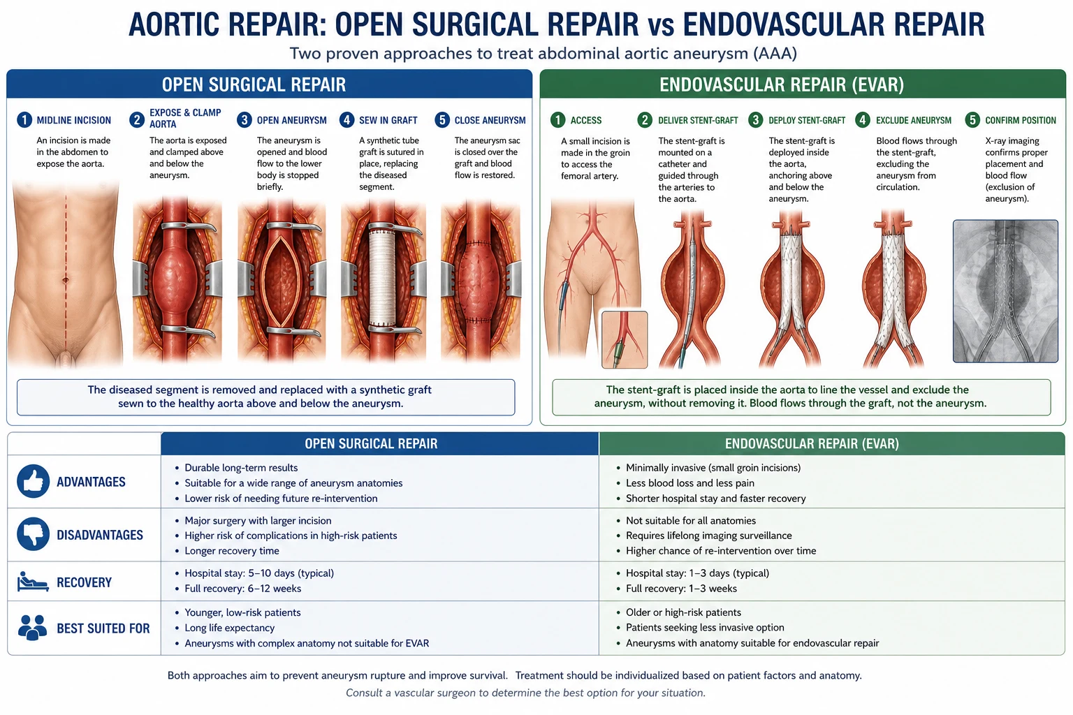
The two options are open surgical repair and endovascular aneurysm repair (EVAR). The choice is dictated by anatomy, fitness, and patient preference, decided at the multi-disciplinary vascular meeting. [1]
Open surgical repair — the traditional gold standard
Open repair is performed under general anaesthesia via a midline laparotomy (or, selectively, a left retroperitoneal approach for juxtarenal/suprarenal aneurysms). The steps are:
- Mobilisation of the small bowel and duodenum to expose the aorta.
- Proximal control by clamping the infrarenal aorta (or suprarenal/supracoeliac for juxtarenal aneurysms).
- Distal control of the common iliac arteries, and IV heparin (e.g. 100 units/kg) before clamping.
- Longitudinal aortotomy, evacuation of mural thrombus, and ligation of lumbar and inferior mesenteric arteries.
- Insertion of a Dacron (or PTFE) tube or bifurcated graft anastomosed end-to-end proximally and distally.
- Closure of the aneurysm sac over the graft. [1]
Open repair is durable: once healed, the graft rarely fails and no lifelong surveillance is needed. It remains the standard for complex anatomy (juxtarenal, suprarenal, mycotic, infected-field, or EVAR-ineligible) and for young, fit patients. Its elective 30-day mortality is 4 to 5%, and long-term graft-related complications are rare. The trade-off is a major laparotomy, prolonged recovery (6 to 12 weeks), and significant cardiopulmonary stress.[1]
Endovascular aneurysm repair (EVAR)
EVAR deploys a stent-graft (a Dacron graft supported by a metal skeleton) via the common femoral arteries (percutaneous or open cut-down), under fluoroscopic guidance, to exclude the aneurysm from circulation. Blood then flows through the graft, depressurising the sac which shrinks over time. EVAR is far less invasive: no laparotomy, no aortic cross-clamp, shorter ICU and hospital stay, and faster recovery.[3]
Anatomical requirements for EVAR (the EVAR eligibility criteria):
- a proximal aneurysm neck of adequate length (over 10 to 15 mm), diameter (under 32 mm), angulation (under 60 degrees), and free of heavy thrombus/calcification — for the graft's proximal seal;
- access vessels (common femoral and iliac arteries) of adequate calibre (over 7 mm) and acceptable tortuosity/calcification — for sheath delivery;
- a distal landing zone in the common iliac arteries. [1]
Only about 50 to 60% of infrarenal AAAs are anatomically suitable for standard EVAR; the rest require open repair or complex fenestrated/branched endografts.[4]
Outcomes. The EVAR-1 and DREAM trials together established that EVAR reduces 30-day mortality (1.5 to 2% vs 4 to 5% for open repair) but that this early advantage does not translate into long-term survival benefit — late endograft complications (endoleak, re-intervention, and rare graft rupture) erode the difference. EVAR-2, in patients unfit for open repair, showed no overall survival benefit because the patients were so sick that AAA did not determine their outcome.[6][7] EVAR is therefore preferred for older, less fit patients with suitable anatomy, while open repair remains reasonable for young, fit patients who want a durable one-off repair and can tolerate the operation.
Open repair
traditional gold standard
- **Durable** — no lifelong surveillance needed
- **Any anatomy** — incl. juxtarenal, suprarenal, mycotic
- 30-day mortality **4 to 5%** (elective)
- **MI is the commonest** cause of perioperative death
- Major laparotomy; recovery 6 to 12 weeks
EVAR
endovascular
- **Less invasive** — shorter recovery, no laparotomy
- 30-day mortality **1.5 to 2%** (elective)
- **Requires suitable anatomy**: neck, angulation, access
- **Lifelong CT/US surveillance** for endoleak
- Late re-intervention in up to 30%
Endoleak — the defining complication of EVAR
An endoleak is persistent blood flow into the aneurysm sac after EVAR, keeping it pressurised and at risk of rupture. They occur in 10 to 20% of EVARs and are graded by source: [1]
| Type | Cause | Risk | Management |
|---|---|---|---|
| I | Inadequate seal at proximal (Ia) or distal (Ib) graft end | High — sac pressurised, rupture risk | Urgent repair (cuff, balloon expandable stent) |
| II | Branch-vessel backflow (lumbar, IMA, accessory renal) — commonest | Low — usually self-limiting | Observe; intervene if sac grows |
| III | Graft component separation (IIIa) or fabric tear (IIIb) | High — graft failure | Urgent repair (bridging stent, relining) |
| IV | Graft-wall porosity within 30 days | Low — self-limiting | Observe (resolves) |
| V | Endotension — sac enlargement without identifiable leak | Intermediate | Investigate; often CTA; may need relining |
Type I and III endoleaks threaten rupture and need urgent re-intervention; type II (the commonest) is usually benign and managed by surveillance, with intervention only if the sac enlarges.[3]
Endoleak classification — I-II-III-IV-V
12345
graft-end seal failure (proximal or distal) — URGENT repair
branch backflow (lumbar, IMA) — commonest; OBSERVE
component separation or fabric tear — URGENT repair
graft porosity (within 30 days) — self-limiting
endotension — sac grows, no leak found; investigate
Ruptured AAA — emergency repair
The rupture may be managed by emergency open repair or, where anatomy and a rEVAR service allow, emergency EVAR (rEVAR). The protocol:
- Permissive hypotension (SBP 70 to 80 mmHg) until cross-clamping — see Resuscitation.
- Open repair: rapid laparotomy, supracoeliac clamp for immediate proximal control, then definitive repair once proximal control is secured; the infrarenal clamp is applied when feasible.
- rEVAR: offered in centres with a 24/7 service and an anatomically suitable aneurysm; the IMPROVE trial suggested a mortality benefit in women and a faster recovery.
- Massive transfusion with correction of coagulopathy, acidosis, and hypothermia (the trauma lethal triad).
- Post-operative ITU care for organ support; mortality remains 40 to 50% of those who reach theatre.[1]
Lifelong follow-up after EVAR
Every EVAR patient needs lifelong imaging surveillance — typically a CT angiogram at one month, then CT/US alternating annually — to detect endoleak, graft migration, limb occlusion, and sac enlargement. Open-repair patients need no such surveillance once recovered. This surveillance burden, and the re-interventions it generates, is the price of the lower perioperative mortality of EVAR.[4]
Specific Subtypes & Scenarios
-
Inflammatory AAA. A dense, fibrotic inflammatory rind invests the aortic wall and often encases surrounding structures, particularly the ureters (causing hydronephrosis), the duodenum, and the IVC. Patients have chronic back/abdominal pain and raised CRP/ESR, and the CT shows a characteristically enhancing retroperitoneal mass. Operative risk is higher, and steroids or immunosuppression may calm the inflammation before repair. Most are now treated by EVAR if feasible.[2]
-
Mycotic (infected) AAA. A rare aneurysm caused by bacterial infection of the aortic wall (Staphylococcus, Salmonella, Streptococcus). Presents with fever, sepsis, and back/abdominal pain, often saccular and atypical in location. Management combines long-term targeted antibiotics with excision and extra-anatomic bypass or in situ grafting with antibiotic-impregnated material; EVAR has a role as a bridge or in the very unfit.[3]
-
Aortoenteric fistula. Primary fistula arises from erosion of an AAA into the duodenum (D3); secondary (far commoner) follows previous aortic grafting. The hallmark is a herald upper GI bleed — a small, self-limiting haematemesis that is followed hours or days later by catastrophic exsanguination. Any GI bleed in a patient with an AAA or graft needs urgent endoscopy and CT angiography. Treatment is emergency surgery.[1]
-
Juxtarenal and suprarenal AAA. Absent or short necks at the renal level; standard EVAR fails. Options are open repair with suprarenal clamping and renal revascularisation, or fenestrated/branched EVAR in experienced centres, which preserves renal and visceral branches while excluding the aneurysm. [1]
-
Symptomatic non-ruptured AAA. Pain or tenderness in a known aneurysm is treated as imminent rupture — refer for urgent repair regardless of absolute size, ideally within days. [1]
-
Popliteal aneurysm. Found in up to 40% of men with AAA; bilateral in half. Detect by palpating the popliteal pulses — a widened, aneurysmal popliteal pulse. Asymptomatic popliteal aneurysms over 20 mm are considered for repair to prevent limb-threatening thrombosis or embolism.[3]
Complications & Pitfalls
Complications of the disease itself:
- Rupture — overall mortality 80 to 90%; the defining event.
- Distal embolisation — blue toe syndrome, renal infarcts, livedo, from the mural thrombus.
- Aortocaval fistula — high-output cardiac failure, leg swelling.
- Aortoenteric fistula — herald then massive GI bleed.
- Ureteric obstruction — in inflammatory AAA. [1]
Complications of open repair:
- Myocardial infarction — the leading cause of perioperative death; meticulous cardiac assessment is mandatory.
- Acute kidney injury — from cross-clamping, contrast, hypoperfusion.
- Colonic ischaemia — from sacrifice of the inferior mesenteric artery; may progress to ischaemic colitis (left colon) with bloody diarrhoea and peritonism.
- Spinal cord ischaemia — paraplegia, especially after suprarenal clamping.
- Respiratory failure — common in COPD after laparotomy.
- Bleeding, infection (graft), and incisional hernia. [1]
Complications of EVAR:
- Endoleak (types I-V) — see above; the defining complication.
- Graft migration, limb thrombosis, kinking — causing acute limb ischaemia.
- Access vessel injury — iliac rupture, femoral bleeding.
- Contrast-induced nephropathy.
- Post-implantation syndrome — transient fever, raised CRP, and back pain in the first week, self-limiting.
- Graft infection — rare but devastating; may present years later as an aortoenteric fistula.[3]
Classic pitfalls:
- Misdiagnosing rupture as renal colic — always ultrasound the older smoker.
- CT-KUB read for stones that misses the AAA.
- Over-resuscitating the ruptured patient to normotension and dislodging the retroperitoneal tamponade.
- Delaying theatre for an unnecessary CT in the unstable patient.
- Forgetting lifelong surveillance after EVAR — the sac can re-pressurise and rupture years later.
- Repairing a small AAA without heeding the patient's fitness — operative mortality may exceed rupture risk in the very frail.
- Missing a popliteal aneurysm on the distal pulse exam. [1]
Prognosis & Disposition
Rupture risk by size (the core examination numbers): [1]
| AAA diameter | Approximate annual rupture risk |
|---|---|
| under 5 cm | under 1% |
| 5 to 5.9 cm | 3 to 5% |
| 6 to 6.9 cm | around 10% |
| 7 cm or more | over 20 to 25% |
These are population averages; women rupture at smaller diameters, symptomatic AAAs at any size, and rapidly expanding AAAs more readily.[2]
Elective repair. Mortality is 4 to 5% open and 1.5 to 2% EVAR at 30 days; five-year survival is around 70%, limited not by the graft but by coexisting cardiovascular disease (which kills most AAA patients eventually). [1]
Ruptured AAA. Overall mortality is 80 to 90%: about half die before reaching hospital, and of those who reach theatre alive, 40 to 50% die. Among survivors, five-year survival is around 60%. [1]
Surveillance (under 5.5 cm). Rupture risk is under 1% per year. The UK Small Aneurysm Trial and its 12-year follow-up confirmed that surveillance is safe in this range — the operative mortality of early surgery exceeded the rupture risk it prevented.[8]
Disposition. Detected AAAs are referred to a vascular surgeon for counselling and follow-up; surveillance is shared between primary care and the screening service. After repair, the patient is followed by the vascular team (life-long for EVAR) and aggressively managed for cardiovascular risk, since the dominant threat to long-term survival is myocardial infarction, not the aneurysm itself.[1]
Special Populations
-
Women. AAA is six times less common in women, but ruptures at smaller diameters (their aortas are smaller). The ESVS 2024 guidelines suggest considering repair at 5.0 cm rather than 5.5 cm in women. Screening is not routinely offered to women because of low prevalence, but women with a family history or smoking history warrant individual assessment.[4]
-
Elderly and frail. EVAR is preferred where anatomy allows, because the lower perioperative mortality matters most when physiological reserve is limited. In the very frail or those with life expectancy under two years, surveillance alone may be the kinder choice. [1]
-
Family history. First-degree relatives of AAA patients have 2- to 4-fold increased risk and develop AAA a decade earlier. Many guidelines advise a one-off screening ultrasound of male first-degree relatives from age 60 (or 10 years younger than the affected relative's age at diagnosis).[1]
-
Patients on anticoagulation. Anticoagulation does not increase rupture risk and should be continued for its underlying indication during surveillance; it is managed peri-operatively in the usual way. [1]
-
Renal impairment. Contrast for CT and the clamp of open repair threaten the kidney. Pre-hydration, nephrotoxin avoidance, and preferential EVAR (no clamp) are considered where feasible. [1]
-
Low- and middle-income settings (e.g. India). No national screening programme exists; AAA is under-diagnosed and many patients present with rupture. Open repair predominates because EVAR infrastructure and lifelong CT follow-up are concentrated in a few centres. The threshold for repair is often dictated by presentation rather than by screening.[2]
Evidence, Guidelines & Regional Differences
The evidence base for AAA is built on a handful of landmark randomised trials that define every threshold in modern practice. [1]
UK Small Aneurysm Trial (UKSAT)
Lancet 1998; 12-yr follow-up BJS 2007 (PMID 17514693)
RCT and long-term follow-up of patients with AAA 4.0 to 5.5 cm: early open surgery vs ultrasound surveillance.
Key finding
Early surgery offered NO survival advantage over surveillance for AAA under 5.5 cm; at 12 years the early operative mortality was balanced by long-term benefit, and surveillance remained safe below 5.5 cm.
Practice change
Established the 5.5 cm elective repair threshold and the safety of surveillance below it.
MASS - Multicentre Aneurysm Screening Study
Lancet 2002 (PMID 12443589)
Population-based RCT: one-off ultrasound screening of men aged 65 to 74 vs no screening.
Key finding
Screening reduced AAA-related mortality by 42% at 4 years and was highly cost-effective.
Practice change
Underpins the NHS AAA Screening Programme and screening recommendations worldwide.
EVAR-1 - Endovascular vs open repair in fit patients
Lancet 2004 (PMID 15351191)
RCT: patients fit for open repair with AAA over 5.5 cm, EVAR vs open repair.
Key finding
EVAR reduced 30-day mortality (1.7% vs 4.7%) but had no overall long-term survival advantage, offset by late graft-related deaths and re-interventions.
Practice change
Established EVAR's lower perioperative mortality and the need for lifelong surveillance for endoleak.
DREAM - Dutch Randomised Endovascular Aneurysm Management
N Engl J Med 2004 (PMID 15483279)
RCT: conventional open vs endovascular repair of AAA over 5 cm.
Key finding
Confirmed EVAR's lower operative mortality (1.2% vs 4.6% at 30 days), with equivalence in cumulative survival by 2 years and more secondary interventions in the EVAR group.
Practice change
Corroborated EVAR-1; together they define the EVAR vs open repair decision.
Guideline deltas
ESVS 2024 Guidelines (PMID 38307694). Repair threshold 5.5 cm in men, 5.0 cm in women; emphasise shared decision-making, patient-reported outcomes, and the role of fenestrated/branched EVAR for complex anatomy. Recommend lifelong imaging after EVAR and global cardiovascular risk reduction for all.[4]
Australia and New Zealand. Population screening varies by state; many advocate a one-off ultrasound at 65 for ever-smoking men, mirroring UK practice. Repair thresholds follow ESVS/ACC guidance; rEVAR is offered in major vascular centres.
Controversies and evolving areas
- Lifetime cost of EVAR surveillance — re-interventions and CT radiation accumulate; some advocate ultrasound-only follow-up after a clean early CT.
- Fenestrated and branched EVAR — extending endovascular treatment to juxtarenal and thoracoabdominal aneurysms, traditionally open-repair territory.
- Women and screening — the risk-benefit balance of screening women remains debated, given lower prevalence but earlier rupture.
- Medical therapy — no drug yet prevents growth; doxycycline and other MMP inhibitors have disappointed in trials.
- rEVAR vs open — the IMPROVE trial showed no clear overall mortality benefit, but possible benefit in women and faster discharge.[4]
Exam Pearls
- Over 3 cm = AAA. Over 5.5 cm = repair (5.0 cm in women). Screen men at 65 with one ultrasound.[1]
- Rupture triad: abdominal/back/flank pain plus hypotension plus pulsatile mass — complete in only half. Mortality 80 to 90%.[3]
- Laplace law: Wall Tension = Pressure x Radius / Wall Thickness — the vicious cycle of growth and the reason rupture risk rises non-linearly with size.
- Unstable rupture = straight to theatre, no CT. Permissive hypotension SBP 70 to 80 mmHg until cross-clamping. Do not over-resuscitate (you will dislodge the retroperitoneal tamponade).[1]
- Surveillance intervals: 3 to 4.4 cm yearly, 4.5 to 5.4 cm 3-monthly, over 5.5 cm refer for repair.
- EVAR eligibility: infrarenal neck over 15 mm and under 60 degrees angulation; access vessels over 7 mm; only about half of AAAs qualify.
- Endoleaks: Type I (graft-end, urgent repair), Type II (branch backflow, commonest, observe), Type III (component failure, urgent repair), Type IV (porosity), Type V (endotension). MI is the commonest cause of perioperative death after repair.
- EVAR-1 and DREAM: lower perioperative mortality, no long-term survival advantage, lifelong surveillance for endoleak.[6][7]
- Misdiagnosis pitfall: never attribute severe back/flank pain in an older male smoker to renal colic without an ultrasound.
- Screen first-degree relatives from age 60; women considered for repair at 5.0 cm.
- Best medical therapy for all: stop smoking, statin, antiplatelet, BP control — but no drug prevents growth.[4]
References
- [1]Isselbacher EM, Preventza O, Hamilton Black J 3rd, et al. 2022 ACC/AHA Guideline for the Diagnosis and Management of Aortic Disease: A Report of the American Heart Association/American College of Cardiology Joint Committee on Clinical Practice Guidelines Circulation, 2022.PMID 36322642
- [2]Kent KC Clinical practice. Abdominal aortic aneurysms N Engl J Med, 2014.PMID 25427112
- [3]Chaikof EL, Dalman RL, Eskandari MK, et al. The Society for Vascular Surgery practice guidelines on the care of patients with an abdominal aortic aneurysm J Vasc Surg, 2018.PMID 29268916
- [4]Wanhainen A, Verzini F, Van Herzeele I, et al. Editor's Choice -- European Society for Vascular Surgery (ESVS) 2024 Clinical Practice Guidelines on the Management of Abdominal Aorto-Iliac Artery Aneurysms Eur J Vasc Endovasc Surg, 2024.PMID 38307694
- [5]The Multicentre Aneurysm Screening Study Group The Multicentre Aneurysm Screening Study (MASS) into the effect of abdominal aortic aneurysm screening on mortality in men: a randomised controlled trial Lancet, 2002.PMID 12443589
- [6]Greenhalgh RM, Brown LC, Kwong GP, Powell JT, Thompson SG; EVAR trial participants Comparison of endovascular aneurysm repair with open repair in patients with abdominal aortic aneurysm (EVAR trial 1), 30-day operative mortality results: randomised controlled trial Lancet, 2004.PMID 15351191
- [7]Prinssen M, Verhoeven EL, Buth J, et al. A randomized trial comparing conventional and endovascular repair of abdominal aortic aneurysms N Engl J Med, 2004.PMID 15483279
- [8]United Kingdom Small Aneurysm Trial Participants Final 12-year follow-up of surgery versus surveillance in the UK Small Aneurysm Trial Br J Surg, 2007.PMID 17514693