General Surgery · General Surgery
Acute Appendicitis
Also known as Appendicitis · Grumbling appendix · Appendicular mass · Appendicular abscess
Acute appendicitis is the most common acute surgical abdomen. Classic presentation: periumbilical pain migrating to the right iliac fossa over 12 to 24 hours, with anorexia, nausea, and low-grade fever. Pathophysiology: luminal obstruction (fecalith, lymphoid hyperplasia) → bacterial overgrowth → increased intraluminal pressure → venous obstruction → ischemia → gangrene → perforation (appendicular artery is an end artery). Alvarado score (MANTRELS) guides bedside diagnosis. CT is gold standard in adults; ultrasound first-line in children/pregnancy. Laparoscopic appendectomy is definitive treatment. Appendicular mass: conservative management → interval appendectomy 6 to 8 weeks. Perforated peritonitis: emergency laparotomy.
On this page & tools
Your progress
Saved locally on this device.
Exam tags
Red flags
Overview & Definition
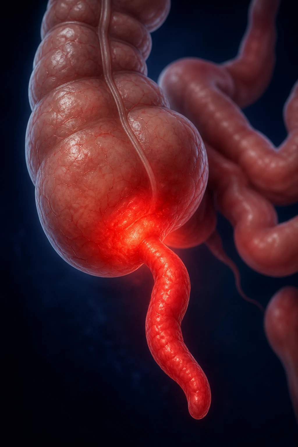
Acute appendicitis is acute suppurative inflammation of the vermiform appendix, almost always initiated by luminal obstruction that culminates, if untreated, in ischaemia, gangrene and perforation. It is the most common acute surgical condition of the abdomen, with a lifetime risk of roughly 7 to 8% and a slight male predominance. Although any age can be affected, the peak incidence is between 10 and 30 years; the disease is uncommon under 5 years and rare in infancy, yet it remains the most common general surgical emergency across the entire paediatric and young-adult population.[1]
The appendix is a blind-ending, worm-shaped tube (vermis, Latin: worm), 6 to 10 cm long, arising from the posteromedial wall of the caecum approximately 2 cm below the ileocaecal valve. Its base is remarkably constant in position and is marked on the surface where the three teniae coli of the caecum converge — a landmark used to localise the appendix at open surgery. The tip, in contrast, is freely mobile and may lie in any of five recognised positions: retrocaecal (about 65%, behind the caecum and abutting psoas), pelvic (about 20%, descending into the pelvis and abutting bladder, rectum or adnexa), subcaecal, pre-ileal, or post-ileal. This anatomic variability is the single most important reason appendicitis has such a protean clinical picture.[1]
The blood supply is through the appendicular artery, a branch of the ileocolic artery (occasionally the posterior caecal artery) that runs in the mesoappendix. It is functionally an end artery with no significant collateral circulation. As intraluminal pressure rises in obstructive appendicitis, the thin-walled capacitance veins draining the appendix wall are compromised first; oedema and congestion follow, and eventually the artery itself is throttled. The result is rapid transmural ischaemia, gangrene and rupture — typically within 24 to 36 hours of symptom onset. This single anatomical fact is why delay is dangerous and why the disease is so time-critical.[1]
Classification
Uncomplicated
simple acute
- Inflamed but **intact** appendix; **no** gangrene, perforation, or abscess
- Catarrhal (early mucosal inflammation) → suppurative (fibrinopurulent serosa)
- Treatment: **laparoscopic appendectomy** (gold standard)
- Excellent prognosis; mortality under 0.1%
Complicated
gangrene / perforation / abscess / mass
- **Gangrenous**: full-thickness wall necrosis, risk of imminent rupture
- **Perforated**: hole in wall — localized abscess or generalized peritonitis
- **Appendicular mass (phlegmon)**: omentum and bowel wall off inflamed appendix
- **Appendicular abscess**: walled-off pus collection; needs drainage
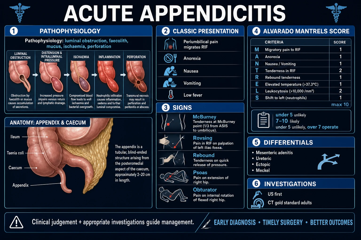
Clinically, the term uncomplicated is reserved for cases without gangrene, perforation, abscess or phlegmon; complicated describes any of those four states. The distinction matters because it changes the operative plan, antibiotic duration and prognosis. A further practical subgroup is the appendicular mass (a palpable, inflammatory conglomerate discovered on presentation, usually after 3 to 5 days of symptoms) and the appendicular abscess (a walled-off collection of pus) — both are managed initially without immediate appendectomy.[1]
Epidemiology & Risk Factors
Appendicitis at a glance
The lifetime risk of appendicitis is approximately 7 to 8% — roughly one in thirteen people will develop it in their lifetime — making it the most common cause of the acute surgical abdomen worldwide. The peak incidence is in the second and third decades, with a second, smaller peak described in elderly patients. There is a modest male predominance (1.2 to 1.4:1), and White populations are affected more often than other ethnic groups in cohort studies. A familial tendency is recognised, suggesting a genetic contribution to appendiceal morphology or immune reactivity.[1]
Recognised risk factors include a low-fibre Western diet (associated with harder stool and faecolith formation, though the link is debated), recurrent viral or bacterial enteric infections (which drive lymphoid hyperplasia, especially in children), and HIV or other immunodeficiency states (which raise the rate of atypical and complicated presentations). The incidence is rising rapidly in newly industrialised countries adopting a Western diet — historically Japan and Korea, now across South Asia — paralleling the lifestyle transition. A slight seasonal increase in summer has been reported in temperate climates.[1]
India / South Asia: Appendicitis is one of the leading causes of acute abdomen presenting to emergency departments. Parasitic obstruction (Ascaris lumbricoides, Enterobius vermicularis) is a recognised cause in endemic areas and may present as a worm protruding through an appendiceal perforation. Access to cross-sectional imaging is often limited in rural settings — ultrasound and the Alvarado score are therefore the diagnostic workhorses. Ileocaecal tuberculosis is a critical differential that mimics appendicitis, particularly with weight loss, evening rise of fever and night sweats; the index of suspicion must be high in endemic regions. Open appendectomy remains common in smaller centres, and the negative appendectomy rate is consequently higher.
Pathophysiology
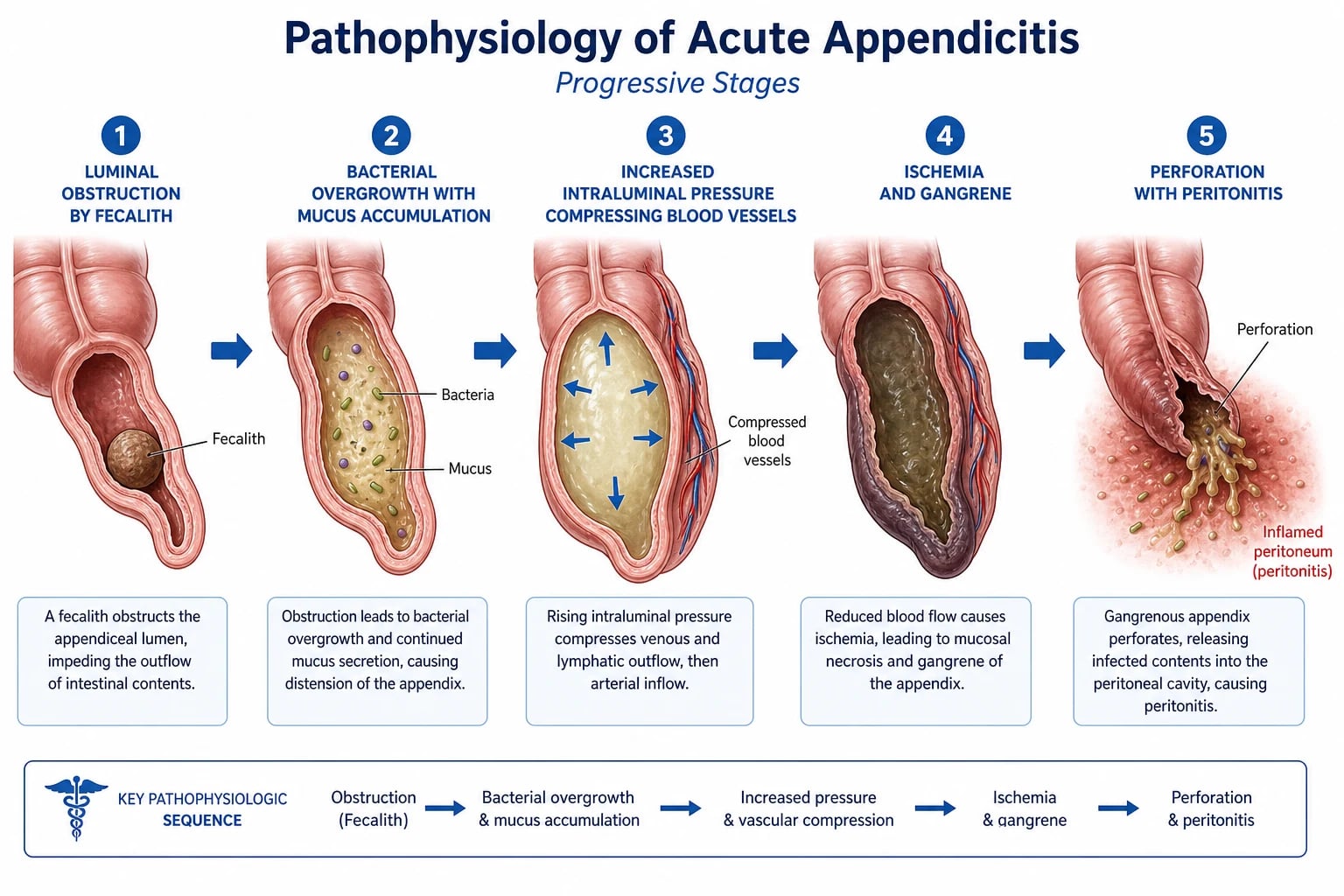
Appendicitis is the textbook example of a progressive obstructive-inflammatory cascade in a hollow viscus with an end-arterial supply. The sequence is best understood in five stages:[1]
1. Luminal obstruction. The trigger in most adults is a fecalith (also called an appendicolith or coprolith) — hardened, layered faecal material that lodges in the narrow appendiceal lumen, accounting for about 60% of adult cases. In children and adolescents, the dominant cause is lymphoid hyperplasia of the submucosal lymphoid follicles, often following a viral upper respiratory or enteric infection; the swollen follicles occlude the lumen. Less common causes include parasites (Ascaris, Enterobius), foreign bodies (seeds, gallstones), and tumours — classically a carcinoid tumour of the appendix tip, or a caecal carcinoma in the elderly that obstructs the appendiceal orifice and produces a "secondary" appendicitis.[1]
2. Mucus accumulation and bacterial overgrowth. Behind the obstruction, goblet cells continue to secrete mucus, the lumen distends, and intraluminal pressure rises. The stagnant mucus becomes an ideal culture medium. Resident colonic organisms multiply rapidly: Escherichia coli, Bacteroides fragilis, Peptostreptococcus, Pseudomonas and a host of other enteric aerobes and anaerobes. The mixed aerobic-anaerobic flora is what drives the polymicrobial peritonitis and abscess formation seen after perforation. Viridans and other streptococci may also be cultured.[1]
3. Increased intraluminal pressure → vascular compromise. As pressure climbs, the thin-walled capacitance veins in the appendix wall are compressed first. Venous obstruction causes congestion, oedema and serosal exudation — the gross appearance of early "catarrhal" appendicitis. Bacterial invasion of the wall follows; the mucosa ulcerates and a fibrinopurulent exudate coats the serosa (suppurative appendicitis). Eventually the rising pressure exceeds the appendicular artery pressure. Because this vessel is an end artery without collaterals, perfusion fails abruptly.[1]
4. Ischaemia and gangrene. Loss of arterial inflow produces transmural necrosis — gangrenous appendicitis. The wall becomes greenish-black, friable and studded with fibrin; the serosa may already have micro-perforations. Pain often transiently eases at this point as the visceral afferents die (the dangerous "lull before the storm" that can mislead clinician and patient alike). [1]
5. Perforation and peritonitis. The gangrenous wall gives way, spilling faeces, pus and mucus into the peritoneal cavity. If the omentum and adjacent small bowel have already walled off the area, the result is a localised abscess or an appendicular mass (phlegmon). If the inflammatory process has not been contained, the patient develops generalised faecal peritonitis — a surgical emergency with high morbidity. [1]
Histologically the appendix is rich in lymphoid tissue (submucosal follicles, particularly prominent in children and adolescents, which atrophy after the third decade — one reason appendicitis becomes less common with age). Some authors propose an immune-protective role for the appendix ("safe house" hypothesis for gut flora), but no clinical practice relies on this. Microscopically, early disease shows mucosal ulceration and neutrophil infiltration of the muscularis; advanced disease shows transmural necrosis with fibrinopurulent serositis. A histopathology specimen is examined routinely after every appendectomy not only to confirm the diagnosis but to exclude an incidental carcinoid tumour, mucinous neoplasm or adenocarcinoma, the management of which differs substantially. [1]
The migrating pain — a clinical pearl explained by embryology. The classic shift from periumbilical to right iliac fossa pain is one of the most elegant demonstrations of referred pain in medicine. The appendix, like all midgut structures, is innervated by visceral afferents entering the spinal cord at T10. Early obstruction distends the lumen and stimulates these visceral fibres, producing a dull, poorly localised pain in the T10 dermatome around the umbilicus. Once inflammation reaches the parietal peritoneum of the anterior abdominal wall — a somatically innervated structure — pain becomes sharp and exquisitely localised to the right iliac fossa (T12-L1). The temporal lag of roughly 12 to 24 hours between the two phases mirrors the time taken for transmural inflammation to reach the parietal peritoneum.[1]
Clinical Presentation
[1]The textbook patient is a young adult who describes visceral periumbilical pain that wakes them or worsens over hours, followed by nausea, anorexia and a low-grade fever, with the pain eventually settling in the right iliac fossa. Pain is typically worsened by movement and coughing — the patient prefers to lie still, often with the right hip flexed. A low-grade fever (around 38°C) is the rule; a fever over 39°C suggests perforation or an alternative diagnosis (pyelonephritis, pancreatitis). The pulse rate is mildly raised. The sequence matters more than any single feature: migratory pain + anorexia + RIF localisation is the diagnostic triad.[1]
Atypical presentations follow the tip position and are deliberately tested by examiners:[1]
- Retrocaecal (about 65%): pain may be in the right flank, loin or back rather than the RIF; abdominal signs may be muted because the inflamed appendix is cushioned by the caecum. A psoas sign (pain on extending the right hip with the patient on the left side) is typical — the inflamed appendix lies on the psoas muscle. May mimic pyelonephritis or cholecystitis.
- Pelvic (about 20%): pain is suprapubic; the patient may have urinary frequency, dysuria, tenesmus or diarrhoea because the appendix abuts the bladder or rectum. There may be no abdominal tenderness, but digital rectal or vaginal examination reveals marked right-sided tenderness. An obturator sign (pain on internal rotation of the flexed right hip) suggests a pelvic appendix lying against obturator internus.
- Subcaecal: pain localises near the inguinal ligament and may mimic groin pathology or ureteric colic.
- Pre-ileal / post-ileal: rare; closer to the umbilicus, may cause generalised central pain. [1]
Atypical presentations by patient group — each is examined as a deliberate "trap": [1]
Why the atypical case is atypical
POSTURED
gravid uterus elevates the caecum; pain may be RUQ by the 3rd trimester
elderly: vague pain, delayed presentation, 70% perforation rate at first assessment
under 5: non-verbal, rapid progression, 50% perforated at presentation
diabetics: blunted inflammatory response, muted signs
pelvic appendix causes urinary symptoms; mislabelled as UTI
right flank pain, abdominal signs muted, psoas sign positive
HIV, chemotherapy, steroids: minimal signs, high perforation
mass or abscess at first presentation after 3 to 5 days
Differential Diagnosis
The right iliac fossa contains a wide variety of structures, and the differential is correspondingly broad. The can't-miss mimics are ruptured ectopic pregnancy (always exclude with a β-hCG in any woman of reproductive age) and testicular torsion in males (referred pain to the RIF — examine the scrotum in every male with abdominal pain).[1]
| System | Condition | Key distinguishing features |
|---|---|---|
| GI | Mesenteric adenitis | Children; recent viral URTI; pain less localised, no peritonism; self-limiting |
| GI | Terminal ileitis (Crohn disease) | Chronic history; diarrhoea; weight loss; perianal disease; mass |
| GI | Meckel diverticulitis | Difficult to distinguish clinically; often intra-operative diagnosis |
| GI | Gastroenteritis | Vomiting precedes pain; watery diarrhoea; diffuse cramping; no peritonism |
| GI | Intussusception | Children under 2; redcurrant-jelly stool; sausage-shaped mass |
| GI | Right-sided diverticulitis | Older patients; more common in East Asian populations |
| GI | Perforated duodenal ulcer | Fluid tracks down the right paracolic gutter → RIF pain; erect CXR shows free air |
| GI | Acute pancreatitis | Epigastric pain radiating to back; raised amylase/lipase |
| Gynaecological | Ruptured ectopic pregnancy | Women of reproductive age; amenorrhoea; β-hCG positive; shock; shoulder-tip pain |
| Gynaecological | Ovarian cyst torsion or rupture | Sudden onset; cyclical pain history; PV examination; urgent US |
| Gynaecological | Pelvic inflammatory disease / salpingitis | Bilateral lower abdominal pain; vaginal discharge; cervical motion tenderness; high fever |
| Gynaecological | Mittelschmerz | Mid-cycle; self-limiting; no fever or peritonism |
| Urological | Right ureteric colic | Pain radiates to groin; haematuria; no fever or anorexia; colicky (not constant) |
| Urological | Pyelonephritis | High fever, rigors; marked urinary symptoms; CVA tenderness |
| Urological | Testicular torsion | Referred pain to RIF; examine the scrotum; surgical emergency |
| Surgical | Acute cholecystitis | RUQ pain; Murphy sign |
| Respiratory | Right basal pneumonia | Referred pain via T8-T11; cough, sputum, fever; CXR diagnostic |
| Medical | Diabetic ketoacidosis | Abdominal pain with acidosis; check glucose and ketones |
| Medical | Porphyria, sickle-cell crisis, Henoch-Schönlein purpura | Characteristic systemic features |
Clinical & Bedside Assessment
The clinical examination is built around the named peritoneal signs and the Alvarado (MANTRELS) score. Although imaging has displaced clinical diagnosis in many settings, the bedside examination still drives the decision to image or operate, and a number of signs predict appendix position and peritoneal involvement.[1][1]
| Sign | Technique | Positive finding | Meaning |
|---|---|---|---|
| McBurney point tenderness | Press at the point one-third of the way from the ASIS to the umbilicus | Maximum tenderness at the appendix base | The single most reliable clinical sign |
| Rovsing sign | Deep palpation in the left iliac fossa | Pain is felt in the right iliac fossa | Peritoneal irritation — gas/fluid shifts the inflammatory fluid |
| Psoas sign | Patient on the left side; passively extend the right hip (or active extension against resistance) | Pain in the right lower abdomen or flank | Retrocaecal appendix irritating the psoas muscle |
| Obturator sign | Flex the right hip and knee to 90°, then internally rotate the hip | Pain in the hypogastrium / RIF | Pelvic appendix irritating obturator internus |
| Blumberg sign (rebound tenderness) | Press deeply, then release quickly | Sharp pain on release | Peritoneal inflammation (peritonism); implies advanced disease |
| Aaron sign | Pressure on McBurney point | Referred pain or distress in the epigastrium or precordium | Confirms deep parietal irritation |
| Bastedo sign | Insufflation of the colon via the rectum | Pain at the site of caecal distension | Localised caecal or appendiceal inflammation |
| Dunphy sign | Cough | Pain in the RIF | Peritoneal irritation |
| Murphy triad | Sequential observation | Pain → vomiting → fever (in that order) | Classic appendicitis sequence |
Tenderness on digital rectal or vaginal examination is elicited on the right side in pelvic appendicitis and should never be omitted in an equivocal case. Generalised guarding, rigidity, board-like abdomen and absent bowel sounds indicate established generalised peritonitis — a surgical emergency. [1]
Alvarado score (MANTRELS) — bedside scoring
The Alvarado score (MANTRELS) is a 10-point bedside tool derived by Alfredo Alvarado in 1986 from 305 patients hospitalised with suspected appendicitis. It uses three symptoms, three signs and two laboratory findings.[1]
| Component | Points |
|---|---|
| Migration of pain | 1 |
| Anorexia | 1 |
| Nausea / vomiting | 1 |
| Tenderness in right iliac fossa | 2 |
| Rebound tenderness | 1 |
| Elevated temperature (≥37.3°C) | 1 |
| Leukocytosis (WBC over 10 ×10⁹/L) | 2 |
| Shift to left (neutrophilia) | 1 |
| Total | 10 |
Interpretation:[1]
- 1 to 4: appendicitis unlikely — consider alternative diagnosis, discharge with safety-net advice.
- 5 to 6: possible — observe, repeat assessment, image (CT in adults, US in children).
- 7 to 8: probable — proceed to appendectomy (or image to confirm in low-resource settings).
- 9 to 10: very probable — appendectomy.
Reported sensitivity is around 70% and specificity around 80% at a cut-off of 7; in children the score performs less well and should always be paired with imaging. The AIR (Appendicitis Inflammatory Response) score and the AAS (Adult Appendicitis Score) are newer, more granular alternatives that include CRP and polymorph proportion, but Alvarado remains the most widely taught.[1]
Alvarado = MANTRELS — eight letters, ten points
MANTRELS
pain moves from centre to RIF (1 pt)
loss of appetite (1 pt)
after pain onset (1 pt)
maximum at McBurney point (2 pts)
Blumberg sign (1 pt)
≥37.3°C (1 pt)
WBC over 10 ×10⁹/L (2 pts)
neutrophilia on differential (1 pt)
Self-test — Alvarado at the bedside (click to reveal)
A 22-year-old man has 24 hours of central abdominal pain that has moved to the RIF, with anorexia, one episode of vomiting, temperature 37.8°C, RIF tenderness and rebound, WBC 13.5 ×10⁹/L with neutrophilia. Alvarado score: M(1) + A(1) + N(1) + T(2) + R(1) + E(1) + L(2) + S(1) = 10/10 — very probable appendicitis. Proceed to imaging (CT in adults) and surgical review.
A scoring-system comparison is helpful at the viva: the Alvarado score uses only history, exam and a basic blood test, which is why it survives in resource-limited settings; the AIR score (Andersson) adds CRP and a polymorph proportion and performs better in teenagers; the AAS (Adult Appendicitin Score) is the most granular but requires a calculator. All three converge on the same truth — clinical judgement and imaging together outperform any single score. [1]
Investigations
Investigation is directed by pre-test probability (Alvarado), age, and reproductive status. The goal is to confirm the diagnosis, exclude dangerous mimics, and detect complications. No single blood test is diagnostic; imaging has supplanted "watchful waiting" in many adults.[1][1]
Bedside and laboratory:
- FBC: leukocytosis (WBC over 10 ×10⁹/L) with neutrophilia is present in 80% of cases. The CRP is typically elevated (over 10 mg/L) and rises with disease duration — a normal WBC and CRP at 24 hours of symptoms makes appendicitis unlikely. Combined WBC and CRP have a sensitivity of about 95 to 97%.[1]
- CRP: rises in proportion to gangrene and perforation; a CRP over 50 mg/L increases the likelihood of complicated disease.
- Urinalysis: excludes UTI. Mild pyuria may occur with a pelvic appendix abutting the bladder and does NOT exclude appendicitis — a common pitfall. Haematuria suggests ureteric colic.
- β-hCG: ALWAYS in women of reproductive age to exclude ectopic pregnancy, even if imaging is planned.
- U&E, LFTs, amylase/lipase, lactate: baseline; exclude alternative diagnoses and sepsis (raised lactate suggests perforation or mesenteric ischaemia).
Imaging:
- Ultrasound (graded compression): first-line in children, pregnant women and slim young adults. A non-compressible appendix over 7 mm in outer diameter, with wall thickening, peri-appendiceal fluid, or an appendicolith, is diagnostic. Sensitivity 75 to 90%, specificity over 90%; operator- and body-habitus-dependent. The chief value is to exclude gynaecological pathology in young women. A normal ultrasound does NOT exclude appendicitis.
- CT abdomen and pelvis (with IV contrast): gold standard in adults and in patients with equivocal ultrasound. Sensitivity around 98%, specificity 96%. Findings include an enlarged appendix over 6 mm, peri-appendiceal fat stranding, appendicolith, free fluid or free gas (perforation), and abscess. CT reduces the negative appendectomy rate to under 5% and is cost-effective in adults.
- MRI: in pregnant women if ultrasound is inconclusive — no ionising radiation; sensitivity and specificity over 95%.
- Plain abdominal X-ray: not routine; may show an appendicolith, a sentinel loop, or free air (perforation). Erect chest X-ray may show free air under the diaphragm from a perforated viscus.
- Diagnostic laparoscopy: both diagnostic and therapeutic; useful in women with persistent diagnostic uncertainty. [1]
Management — Resuscitation
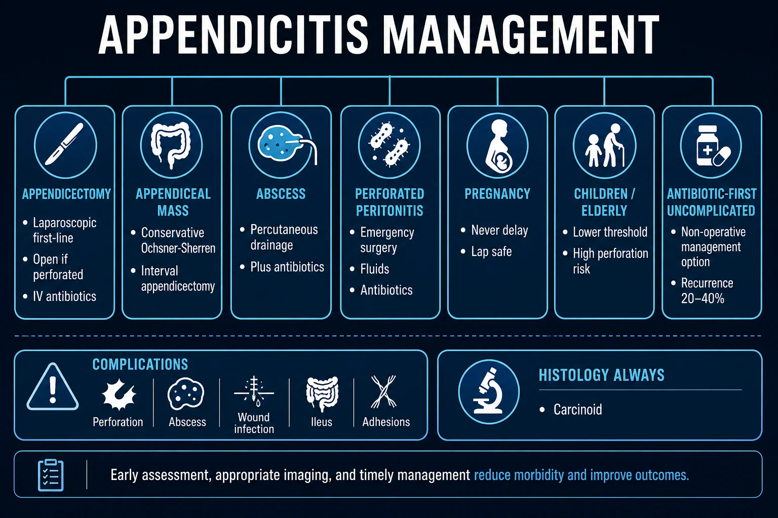
The resuscitation bundle is identical for any acute surgical abdomen and should run in parallel with the diagnostic work-up:[1]
Pre-operative bundle
Nil by mouth
Restrict oral intake to prepare for theatre and limit aspiration risk.
IV access and fluids
Two large-bore cannulae; crystalloid (Hartmann's or 0.9% saline) to correct dehydration.
Analgesia
Opioid (morphine 0.1 mg/kg IV); evidence confirms analgesia does NOT mask peritoneal signs.
Antiemetics
Ondansetron 4 mg IV; reduces nausea and aspiration risk.
IV antibiotics
Cefuroxime 1.5 g IV + metronidazole 500 mg IV, or co-amoxiclav 1.2 g IV; within 60 min of incision.
Consent
Discuss bleeding, infection, conversion to open, stump appendicitis, possible additional procedures.
Group and save
Cross-match if perforation or anaemia suspected.
Sepsis bundle if perforated and shocked: oxygen, two wide-bore cannulae, 1 L Hartmann's stat then titrated, urinary catheter, arterial blood gas and lactate, broad-spectrum antibiotics (piperacillin-tazobactam 4.5 g IV), and urgent surgical review.[1]
Management — Definitive & Stepwise
1. Acute uncomplicated appendicitis
Laparoscopic appendectomy is the gold standard for uncomplicated appendicitis and the default approach wherever the equipment and skills are available.[4]
Technique (laparoscopic):
- Three ports: umbilical camera port (10 mm), a 5 mm port in the left iliac fossa, and a 5 mm suprapubic port. Some centres use a single-incision (SILS) approach.
- Patient position: supine, head-down (Trendelenburg), tilted left-side down to expose the caecum.
- Identify the appendix by tracing the three teniae coli on the caecum down to their confluence at the appendix base.
- Mesoappendix: the appendicular artery (in the mesoappendix) is controlled with diathermy, clips, or a sealing device (LigaSure).
- Appendix base: secured with endoloops (two pre-cuts on the stump side plus one on the specimen side) or a linear stapler, leaving a short, well-perfused stump — a long stump risks stump appendicitis later.
- Specimen removed in a retrieval bag to prevent port-site contamination.
- Lavage of the RIF and pelvis if any spillage. [1]
Open appendectomy (Lanz or gridiron incision at McBurney point): a muscle-splitting approach through a transverse skin-crease (Lanz — better cosmesis) or oblique (gridiron) incision. The caecum is delivered, the mesoappendix ligated, the base crushed and ligated with absorbable suture, and the stump cauterised or invaginated with a purse-string. Indications: laparoscopy unavailable, a large appendicular mass, severe sepsis with haemodynamic instability, or surgeon inexperience.[1]
Advantages of laparoscopy (per the 2018 Cochrane review): less postoperative pain, fewer wound infections, shorter hospital stay, faster return to normal activity, and a diagnostic benefit (the entire abdomen can be inspected, particularly valuable in young women with possible gynaecological pathology). Trade-offs are longer operative time, higher equipment cost, and a small absolute increase in intra-abdominal abscess in earlier trials.[4]
2. Appendicular mass (phlegmon)
If the patient presents late (usually 3 to 5 days after onset) with a palpable, tender mass in the right iliac fossa and is systemically well (no peritonitis, no sepsis):[1]
- Conservative management (Ochsner-Sherren regimen): NBM, IV fluids, IV antibiotics (cefuroxime + metronidazole), analgesia, and strict serial observations — temperature, pulse rate, abdominal examination every 4 to 6 hours.
- If the mass resolves over 48 to 72 hours: continue antibiotics, then discharge.
- Interval appendectomy at 6 to 8 weeks after inflammation settles — historically routine, now selectively applied (recurrence rate is low).
- Conversion criteria (failure of conservative regimen): rising fever, rising tachycardia, increasing pain, enlarging mass, or signs of peritonitis → emergency surgery. [1]
3. Appendicular abscess
- Percutaneous drainage under ultrasound or CT guidance + IV antibiotics is the treatment of choice; surgery in the acute phase is technically difficult and risks bowel injury.[1]
- After resolution: interval appendectomy at 6 to 8 weeks (or conservative follow-up per patient preference).
- If drainage is impossible or the patient is unstable: open drainage ± appendectomy.
4. Perforated appendix with generalised peritonitis
- Emergency laparotomy (midline incision for full access), appendectomy, peritoneal lavage with warm saline, and broad-spectrum IV antibiotics (piperacillin-tazobactam 4.5 g or ceftriaxone + metronidazole).[1]
- Consider a pelvic drain if significant contamination.
- May require HDU/ITU post-operatively; mortality rises to 1 to 5%.
5. Antibiotic-first approach (APPAC and CODA trials)
Two landmark trials have established antibiotics as a legitimate first-line option for selected, uncomplicated appendicitis:[2][3]
APPAC (2015, JAMA) — Salminen et al.
JAMA 2015;313(23):2340-2348
Multicentre open-label non-inferiority RCT, Finland. 530 adults (273 appendectomy, 257 antibiotics) with CT-confirmed uncomplicated appendicitis. Antibiotic arm: ertapenem 1 g IV daily x 3 days then oral levofloxacin 500 mg + metronidazole 500 mg for 7 days.
Key finding
Antibiotics did NOT meet non-inferiority (27.3% recurrence at 1 yr vs 0.4% surgical success), BUT ~73% of antibiotic patients avoided surgery at 1 yr and the overall complication rate was lower in the antibiotic group.
Practice change
Antibiotics are a reasonable option for selected uncomplicated cases; ~1 in 3 recurs. 5-year follow-up showed 39% recurrence and a sustained lower complication rate.
CODA (2020/2021, NEJM) — Davidson/Flum et al.
N Engl J Med 2021;385:2395-2397
Pragmatic non-inferiority RCT, USA. 1552 adults (776 antibiotics, 776 appendectomy) with imaging-confirmed appendicitis (including appendicolith). Antibiotic arm: 10-day course of ertapenem then oral (metronidazole + ciprofloxacin or amoxicillin-clavulanate).
Key finding
At 90 days, 71% of antibiotic group avoided surgery; at 1 year, 29% had appendectomy; at 4 years, ~35%. Appendicolith subgroup had higher recurrence and complication rates.
Practice change
Shared decision-making: surgery remains definitive; antibiotics are acceptable for selected uncomplicated cases. The presence of an appendicolith predicts failure of antibiotic therapy.
Antibiotic regimens in trials: APPAC used ertapenem followed by levofloxacin + metronidazole; CODA used a 10-day course of either IV ertapenem followed by oral (metronidazole + ciprofloxacin or amoxicillin-clavulanate). Real-world practice often uses a shorter course of co-amoxiclav or ceftriaxone + metronidazole.[2][3]
Caveats: antibiotic-first applies only to uncomplicated appendicitis (no gangrene, perforation, abscess or phlegmon); complicated disease, immunosuppression, pregnancy (in some guidelines), and patient preference for definitive treatment all favour surgery. Patients must be informed of the roughly 30 to 40% recurrence at 1 to 5 years. [1]
Specific Subtypes & Scenarios
- Acute catarrhal appendicitis: earliest stage; mucosal inflammation only; may resolve spontaneously.
- Acute suppurative appendicitis: fibrinopurulent serosa; appendix red, swollen, tender.
- Acute gangrenous appendicitis: full-thickness wall necrosis; imminent perforation.
- Perforated appendicitis: free pus; localised abscess or generalised peritonitis.
- Appendicular mass / abscess: late presentation; managed non-operatively in the first instance.
- Stump appendicitis: inflammation of the residual appendiceal stump after incomplete appendectomy; rare but recognised, often presenting months or years later. Treatment: completion appendectomy.[1]
- Chronic / recurrent appendicitis: repeated episodes of RIF pain resolving without surgery; a legitimate diagnosis in selected patients after exclusion of other pathology. Treatment: elective appendectomy.
- Appendiceal tumours: carcinoid (neuroendocrine) tumour is the most common (often incidental finding at appendectomy); simple appendectomy is curative if under 1 cm and at the tip, but right hemicolectomy is indicated for tumours over 2 cm, at the base, with mesoappendiceal invasion, or with high-grade histology. Mucinous neoplasms and adenocarcinoma are rarer but may present with pseudomyxoma peritonei.
- Para-appendicular pathology: Crohn disease of the terminal ileum, caecal carcinoma, ileocaecal tuberculosis, or amoeboma may produce "secondary" appendicitis or be discovered at appendectomy — always inspect the caecum and terminal ileum.[1]
Complications & Pitfalls
Of the disease:
- Perforation (20 to 30% at presentation, higher at the extremes of age) → localised abscess, generalised peritonitis, or septic shock.[1]
- Appendicular mass / abscess — typically after 3 to 5 days of untreated symptoms.
- Pylephlebitis (portal pyaemia): rare but lethal — septic thrombophlebitis of the portal vein, classically with Bacteroides, leading to multiple liver abscesses. Suspect in any post-appendicitis patient with rigors, jaundice and a tender liver; treat with prolonged IV antibiotics (e.g. piperacillin-tazobactam) and drainage of liver abscesses.[1]
- Sepsis and septic shock — established perforation with peritonitis.
- Adhesions and intestinal obstruction — late complication; the most common cause of small-bowel obstruction in patients with prior appendectomy.
- Enterocutaneous fistula — after drainage of an appendix abscess or inadvertent enterotomy.
Of surgery:
- Wound infection (5 to 10% open, 1 to 3% laparoscopic) — reduced by prophylactic antibiotics and a laparoscopic approach.[4]
- Intra-abdominal / pelvic abscess — the principal serious complication after perforation; risk may be marginally higher after laparoscopic than open appendectomy for perforated disease.
- Post-operative ileus — usually self-limiting.
- Bleeding from the appendicular artery stump or mesoappendix — early, requires re-operation.
- Stump appendicitis — long residual stump; rare.
- Incisional hernia — open approach.
- Visceral injury — caecum, ileum, bladder, or iliac vessels at port insertion.
- Negative appendectomy — appendix removed but not inflamed; rate approximately 10 to 15%, lower with routine CT (under 5%) and higher in women because of gynaecological mimics.[1]
Prognosis & Disposition
- Uncomplicated appendicitis: mortality under 0.1%. Laparoscopic appendectomy: typically discharged within 24 hours, return to normal activity in 1 to 2 weeks.[4]
- Perforated / peritonitis: mortality 1 to 5%, rising in the elderly and diabetic; longer hospital stay (5 to 7 days), IV antibiotics, possible HDU.
- Elderly (over 65): higher morbidity and mortality — delayed presentation, atypical symptoms, ~70% perforation rate at first assessment. Consider underlying caecal carcinoma.[1]
- Pregnancy: risk of premature labour and fetal loss increases markedly with perforation. Early surgery is recommended — delay increases perforation risk and fetal morbidity. Maternal mortality is unchanged from non-pregnant baseline; fetal loss rises to 20 to 35% with perforation.
- Negative appendectomy rate: ideally under 5% with routine CT; higher when imaging is restricted.
Special Populations
- Children: the disease progresses rapidly — roughly half are perforated at presentation, because the omentum is underdeveloped and cannot wall off the inflammation. Atypical presentations are common (irritability, limp, refusal to walk). Ultrasound is first-line. Operate promptly once the diagnosis is made; dehydration develops quickly — aggressive fluid resuscitation. Laparoscopic appendectomy is safe in children of all ages at centres with paediatric expertise.[1]
- Elderly (over 65): vague symptoms, delayed presentation, 70% perforation rate, and a higher incidence of underlying neoplasia (caecal carcinoma, carcinoid). Low threshold for CT. Mortality is roughly 5%. Comorbidities drive the higher risk.
- Pregnancy: appendicitis is the most common non-obstetric surgical emergency in pregnancy (about 1 in 1500 pregnancies). The gravid uterus progressively elevates the caecum — by the third trimester the appendix may sit in the right upper quadrant, so pain is often atypically localised. Ultrasound is the first-line imaging modality (no ionising radiation); MRI if ultrasound is inconclusive; CT only if MRI is unavailable and the diagnosis remains uncertain. Laparoscopic appendectomy is safe in all trimesters with left-lateral tilt to avoid aortocaval compression. Delay is the single biggest predictor of fetal loss — operate early.[1]
- Diabetic patients: a blunted inflammatory response may mask peritoneal signs and fever; lower the threshold for imaging and surgery.
- Immunocompromised (HIV, chemotherapy, transplant, chronic steroids): atypical presentations, higher perforation rate, higher risk of unusual organisms (CMV, Kaposi, lymphoma). Low threshold for imaging and early surgery.
- Anticoagulated patients (warfarin, DOACs): peri-operative bridging depends on the indication and bleeding risk; warfarin is reversed with vitamin K and/or prothrombin complex concentrate if surgery is urgent, while DOACs may be paused (and idarucizumab or andexanet used for dabigatran or rivaroxaban/apixaban respectively in emergencies). Coordinate early with haematology. The risk of intra-operative bleeding from a stapled or tied appendiceal stump is modest, but pelvic and port-site bleeds dominate the morbidity.
- Inflammatory bowel disease: appendicitis can coexist with Crohn disease of the terminal ileum; intra-operative findings may change the operative plan (limited appendectomy, ileocaecectomy, or conservative closure of an inflamed but non-obstructed bowel). Biopsy any abnormal caecum.
Evidence, Guidelines & Regional Differences
Alvarado score (1986, Ann Emerg Med): Derived from 305 patients hospitalised with abdominal pain. Eight weighted components (MANTRELS) — Tenderness and Leukocytosis carry 2 points each. Widely validated; sensitivity approximately 70%, specificity approximately 80% at a cut-off of 7. Remains the most widely taught bedside score.[1]
APPAC trial (2015, JAMA): 530 adults with CT-confirmed uncomplicated appendicitis randomised to open appendectomy or antibiotics (ertapenem then oral levofloxacin + metronidazole). Antibiotics failed non-inferiority but approximately 73% of antibiotic patients avoided appendectomy at 1 year. Five-year follow-up showed a 39% recurrence and a lower overall complication rate in the antibiotic group.[3]
CODA trial (2020/2021, NEJM): 1,552 patients randomised to antibiotics vs appendectomy (including appendicolith). At 90 days, 71% of antibiotic patients avoided surgery; recurrence 29% at 1 year and approximately 35% at 4 years. Appendicolith was the strongest predictor of failure. The CODA Collaborative recommends shared decision-making.[2]
Jaschinski/Sauerland Cochrane review (2018): 67 RCTs comparing laparoscopic vs open appendectomy. Laparoscopy reduced wound infection, pain, hospital stay and time to return to activity; operative time was longer; intra-abdominal abscess was slightly more common after laparoscopy for complicated disease.[4]
WSES 2020 Jerusalem guidelines and EAES 2021 promote a risk-stratified, individualised approach — imaging in adults, laparoscopy as first-line, antibiotic-first for selected uncomplicated cases, and conservative management of abscesses. [1]
ANZ / UK / US: Routine CT in adults with equivocal ultrasound; laparoscopic appendectomy as standard; antibiotic-first offered as a patient-centred option under shared decision-making; structured scoring (Alvarado, AIR, AAS) integrated into pathways.
India / resource-limited: Clinical diagnosis + ultrasound + Alvarado remain the principal diagnostic tools. Open appendectomy is still common in smaller hospitals. Ileocaecal tuberculosis is a critical mimic to exclude. Parasitic obstruction is well described. Negative appendectomy rate is consequently higher.
Exam Pearls
- MANTRELS = Alvarado score. M(1) A(1) N(1) T(2) R(1) E(1) L(2) S(1) = 10.[1]
- Migration of pain from umbilicus to RIF is the single most discriminating clinical feature.[1]
- McBurney point: junction of the outer one-third with the inner two-thirds of the line from ASIS to umbilicus.[1]
- Rovsing sign: LIF palpation causes RIF pain — peritoneal irritation.
- Psoas sign → retrocaecal. Obturator sign → pelvic.[1]
- Appendicular artery is an end artery (branch of ileocolic) — explains rapid gangrene.
- Lanz incision (transverse skin crease) for open appendectomy — better cosmetic than gridiron.
- Interval appendectomy: 6 to 8 weeks after mass or abscess resolution.[1]
- Pregnancy: appendix displaced upward → may be RUQ pain; US first-line, MRI if needed; do NOT delay surgery.[1]
- ALWAYS check β-hCG in women of reproductive age.[1]
- Pain before vomiting = surgical; vomiting before pain = medical.[1]
- APPAC + CODA: antibiotics avoid surgery in ~70% at 1 yr but ~30% recurrence.[2][3]
- Cochrane 2018: laparoscopy reduces wound infection and shortens stay; small abscess signal in complicated disease.[4]
- Pylephlebitis: portal pyaemia with Bacteroides, multiple liver abscesses, rigors and jaundice.[1]
- Stump appendicitis after incomplete appendectomy — completion appendectomy is curative.
- Appendiceal carcinoid at the tip, under 1 cm: simple appendectomy suffices. Right hemicolectomy if over 2 cm, at the base, or with mesoappendiceal invasion.
Operative anatomy and surgical steps (exam detail)
The vermiform appendix arises from the posteromedial caecum about 2 cm below the ileocaecal valve, at the confluence of the three taeniae coli (a reliable finding landmark). Length is typically 6 to 10 cm. The mesoappendix carries the appendicular artery (branch of the ileocolic artery from the SMA) — an end artery, which is why luminal obstruction progresses rapidly to gangrene once venous and then arterial flow fail.[1]
Laparoscopic appendectomy (standard adult approach):
- General anaesthesia, supine, left-tilt optional, urinary catheter if prolonged.
- Three ports (typical): umbilical (camera), left lower quadrant and suprapubic (working) — variations exist.
- Survey for free fluid, perforation, Meckel diverticulum, gynaecological pathology, and other causes of pain.
- Identify the appendix at the taenia confluence; mobilise adhesions carefully.
- Divide the mesoappendix with energy device or clips; control the appendicular artery.
- Secure the base with endoloop, suture ligature, or stapler flush with caecum — avoid leaving a long stump (stump appendicitis risk) or injuring the caecal wall.
- Divide appendix; retrieve in a bag to reduce wound infection.
- Washout if free pus; drain only for established abscess cavities (not routine).
- Extract ports under vision; close fascia at larger ports. [1]
Open appendectomy: McBurney or Lanz incision; gridiron muscle-splitting approach; same identification and base ligation principles. Convert or start open for dense peritonitis, dense adhesions, or equipment failure.[4][1]
Antibiotic-first pathway (APPAC / CODA) — decision table
| Feature | Favours appendectomy | Favours antibiotic-first trial |
|---|---|---|
| Uncomplicated CT appearance | Either | Preferred if choosing non-op |
| Appendicolith | Preferred (higher failure) | Caution / often exclude |
| Sepsis, free perforation, abscess needing drainage | Required | Not suitable alone |
| Patient preference / access issues | Discuss | May accept ~30% 1-year recurrence |
| Pregnancy, child | Usually operate | Individualise; many units still operate |
Key numbers candidates must own: roughly 70% avoid surgery at 1 year on antibiotics in selected uncomplicated disease, but about one in three recur and may need later appendectomy (CODA longer-term and APPAC lines of evidence).[2][3] Consent must name recurrence, not promise permanent cure.
Worked clinical stems (answer these without another book)
Stem A — Classic young man. 22 M, 20 h migratory pain, anorexia, RIF tenderness + rebound, T 37.9°C, WBC 13.8 with left shift.
Alvarado 10; CT or proceed per unit; NBM, IV fluids, cefuroxime 1.5 g + metronidazole 500 mg IV (or co-amoxiclav 1.2 g), laparoscopic appendectomy. [1]
Stem B — Pregnancy 20 weeks. RUQ/flank pain, nausea, raised WBC.
β-hCG already positive by definition of pregnancy context; appendix may be displaced superiorly; US first, MRI if needed; early surgical review; laparoscopy acceptable with obstetric input; do not delay for “observation only” if imaging supports appendicitis — perforation raises maternal–fetal risk. [1]
Stem C — Day-5 mass. 5 days of pain, palpable RIF mass, stable vitals, no peritonitis.
Appendicular phlegmon; Ochsner–Sherren (NBM/IV fluids/IV antibiotics/serial exam); interval appendectomy 6–8 weeks if resolves; if deteriorates → operate; if fluctuant abscess → drain percutaneously when feasible. [1]
Stem D — Elderly vague pain. 78 M, mild RIF discomfort, afebrile, WBC 11.
High perforation risk; low threshold for CT; consider neoplasm if mass/appicolith patterns atypical; early surgery when indicated. [1]
Stem E — Woman of reproductive age. RIF pain, no migration, spotting.
β-hCG first; ectopic, torsion, PID on differential; US pelvis + RIF; never go to theatre without pregnancy status. [1]
OSCE / short-case performance script
- Wash hands, introduce, consent to examine.
- Inspect for scars, distension, movement with respiration.
- Palpate lightly then deeply — start away from RIF; localise McBurney; test rebound carefully.
- Demonstrate Rovsing, psoas, obturator with explanation of position.
- State you would do PR/PV when indicated, urinalysis, β-hCG, FBC/CRP, and arrange imaging.
- Communicate working diagnosis, need for surgery/antibiotics, risks (infection, conversion, stump issues), and safety-net if discharge after negative work-up. [1]
Extended viva bank (model outlines)
- Why is the appendicular artery dangerous when obstructed? End artery → no collateral → rapid gangrene.
- Define negative appendectomy rate and how CT changed it (~under 5% with CT pathways).
- Histology of carcinoid tip under 1 cm → appendectomy alone; when right hemicolectomy (size over 2 cm, base, mesoappendiceal invasion, positive nodes).
- Stump appendicitis — incomplete base resection; completion appendectomy.
- Pylephlebitis — septic thrombophlebitis of portal vein; Bacteroides classic; liver abscesses; prolonged antibiotics ± source control.
- Laparoscopy vs open evidence — less wound infection, shorter stay; discuss abscess signal in complicated disease from meta-analyses.[4]
- Children — lymphoid hyperplasia common trigger; US first; high perforation if delayed.
- Antibiotic choice timing — within 60 minutes of incision for prophylaxis; therapeutic continuum if perforation.
Common exam traps (fail patterns)
- Treating mild pyuria as “UTI only” and missing pelvic appendicitis.
- Sending a woman to CT without β-hCG.
- Operating on a walled-off mass on day 5 without a trial of conservation when stable.
- Claiming antibiotics never fail after APPAC/CODA.
- Forgetting conversion indications and consent.
- Missing free air / generalised peritonitis as “still laparoscopic only” dogma. [1]
Self-check coverage map
| Examiner dimension | Covered? |
|---|---|
| Definition/classification | Yes — simple/complicated/mass/abscess |
| Epidemiology | Yes — age peak, pregnancy |
| Pathophysiology | Yes — obstruction → end-artery death |
| Presentation + atypical | Yes — elderly, child, pregnancy |
| Differentials with discriminators | Yes — gynae, urinary, ileitis, Meckel |
| Bedside signs + Alvarado exact | Yes — MANTRELS 10-point table |
| Imaging thresholds | Yes — US/CT/MRI roles |
| Resuscitation + antibiotics + doses | Yes |
| Operative steps | Yes — lap/open |
| Non-operative pathway numbers | Yes — ~70% / ~30% |
| Complications | Yes — including pylephlebitis, stump |
| Special populations | Yes |
| Evidence (APPAC, CODA, Cochrane) | Yes |
| Exam pearls | Yes |
If any cell feels weak in your revision, re-read that section — the page is built to be the only resource you need for NEET-PG/INICET appendicitis stems. [1]
References
- [1]Alvarado A. A practical score for the early diagnosis of acute appendicitis Ann Emerg Med, 1986.PMID 3963537
- [2]CODA Collaborative, Davidson GH, Flum DR, et al. Antibiotics versus Appendectomy for Acute Appendicitis - Longer-Term Outcomes N Engl J Med, 2021.PMID 34694761
- [3]Salminen P, Paajanen H, Rautio T, et al. Antibiotic Therapy vs Appendectomy for Treatment of Uncomplicated Acute Appendicitis: The APPAC Randomized Clinical Trial JAMA, 2015.PMID 26080338
- [4]Jaschinski T, Mosch CG, Eikermann M, et al. Laparoscopic versus open surgery for suspected appendicitis Cochrane Database Syst Rev, 2018.PMID 30484855