General Surgery · General Surgery
Burns
Also known as Thermal injury · Burn injury · Scald · Flame burn
Burns are tissue injuries caused by thermal, chemical, electrical, or radiation energy. Severity is determined by depth (superficial / superficial-partial / deep-partial / full-thickness / 4th degree), total body surface area (TBSA), and site (face, hands, feet, perineum, airway). Inhalational injury and burns over 25% TBSA (adults) or 10% (children) are life-threatening. Fluid resuscitation with the Parkland formula (4 mL Ringer-lactate x kg x %TBSA, first half in 8 h, second half over next 16 h) is critical for burns over 15% TBSA. Escharotomy for circumferential full-thickness burns compromising circulation or ventilation. Refer to a burns centre for over 10% TBSA, full-thickness burns, special sites, electrical or chemical injury, or inhalational injury. Complications: hypovolaemic (burn) shock, infection or sepsis, Curling's stress ulcer, compartment syndrome, contractures, hypertrophic scarring.
On this page & tools
Your progress
Saved locally on this device.
Exam tags
Red flags
Overview & Definition
A burn is an injury to the skin and deeper tissues caused by the transfer of thermal, chemical, electrical, or radiation energy into the body at a rate that overwhelms the tissue's capacity to dissipate it. Heat above approximately 44 degrees Celsius denatures proteins; higher temperatures and longer contact produce deeper, irreversible necrosis. The clinical severity of any burn is defined by three variables: depth (how many tissue layers are destroyed), total body surface area or TBSA (the percentage of body surface involved by partial-thickness and deeper burns), and site (burns to the face, hands, feet, perineum, eyes, ears, or major joints carry disproportionate functional and cosmetic importance).[1]
Although a burn is fundamentally a local wound, a major burn (over 20 to 25% TBSA in adults) is a systemic disease. The physiological consequences are profound: massive fluid shifts from capillary leak cause hypovolaemic "burn shock" in the first 24 to 48 hours; a sustained hypermetabolic response roughly doubles or triples the basal metabolic rate and drives catabolism for weeks; immunosuppression predisposes to invasive sepsis, the leading cause of late death; and inhalational injury from heat, smoke, and toxic gases (carbon monoxide, cyanide) can kill before any skin sign is obvious. The goal of management is therefore to secure the airway, restore circulating volume, prevent infection, control the hypermetabolic state with nutrition, promote wound healing, and preserve function and appearance through surgery and rehabilitation.[1]
Burns are among the few surgical emergencies where a correctly executed first hour — cooling, airway protection, and the start of formula-based resuscitation — measurably changes survival. Examiners test the structured, reproducible decisions: depth assessment, TBSA estimation, the Parkland formula, escharotomy indications, and the recognition of inhalational injury. This topic builds each of those decisions into a single defensible algorithm. [1]
Classification
By depth (the primary descriptor)
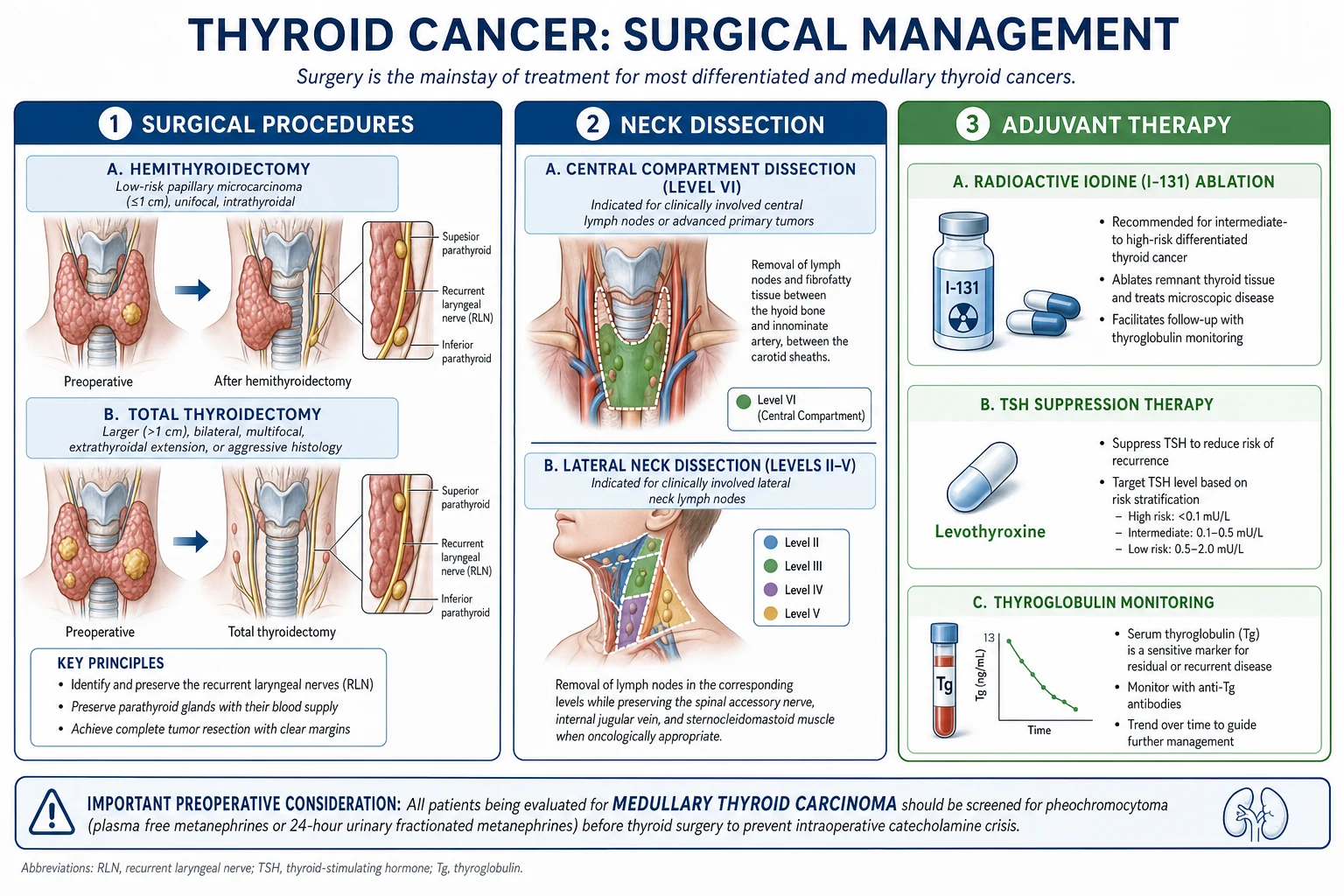
Burn depth determines healing potential, the need for surgery, and the analgesia requirement. Depth is a clinical judgement based on appearance, sensation, and blanching, and it can evolve over the first 48 to 72 hours as the zone of stasis declares itself.[1]
| Depth | Layers affected | Appearance | Sensation | Healing | Typical cause |
|---|---|---|---|---|---|
| Superficial (1st degree) | Epidermis only | Red, dry, no blisters, blanches | Painful | 3 to 5 days, desquamation, no scar | Sunburn, brief flash |
| Superficial-partial (2nd degree, superficial) | Epidermis + superficial dermis | Red, moist, blistering, brisk blanching | Very painful | 10 to 14 days, no scar (or mild pigment change) | Brief scald |
| Deep-partial (2nd degree, deep) | Epidermis + deep dermis | Pale, waxy, mottled, sluggish or absent blanching | Decreased sensation | 3 to 8 weeks with scar, or surgery (graft) | Prolonged scald, flame, contact |
| Full-thickness (3rd degree) | Entire skin (epidermis + dermis) | White, grey, or charred, dry, leathery eschar, no blanching, painless | Requires surgery (skin graft) | Flame, prolonged contact, deep chemical | |
| Full-thickness 4th degree | Skin + subcutaneous fat, fascia, muscle, tendon, or bone | Charred, thrombosed vessels visible, painless | Surgery, often flap or amputation | High-voltage electrical, prolonged flame, molten metal |
The single best clinical discriminator is sensation and blanching. A burn that is painful, blanching, and blistering is partial-thickness (viable dermis survives underneath). A burn that is dry, leathery, non-blanching, and painless is full-thickness (the entire dermis including its nerve endings and microcirculation is destroyed).[1]
Partial-thickness
dermis survives, will heal
- **Painful** (nerve endings intact)
- **Brisk blanching** on pressure (blood flow present)
- **Moist, blistering** surface
- Heals in 10 days to 8 weeks depending on depth; deep-partial may need grafting
Full-thickness
entire dermis destroyed
- **Painless** at the burn site (nerves destroyed)
- **No blanching** (no dermal blood flow)
- **Dry, leathery eschar**, white or charred
- Cannot heal spontaneously; needs surgical excision and skin graft
By TBSA — Wallace Rule of 9s (adult)
TBSA is estimated for partial-thickness and deeper burns only; superficial (epidermal, 1st-degree) burns are excluded from the calculation. The Wallace rule of 9s (1951) gives a rapid bedside estimate in adults:[1]
- Head and neck: 9%
- Each upper limb (arm): 9%
- Each lower limb (leg): 18% (anterior thigh and leg 9% each)
- Anterior trunk: 18%
- Posterior trunk: 18%
- Perineum and genitalia: 1%
- Total: 100% [1]
By TBSA — Lund and Browder chart (children)
Children have proportionally larger heads and smaller legs than adults, so the rule of 9s systematically under-estimates head burns and over-estimates leg burns. The Lund and Browder chart corrects each body region for age: in an infant the head is approximately 18% (vs 9% in the adult) while each leg is about 14% (vs 18% in the adult), and the proportions converge toward the adult values with growth. The Lund and Browder chart is the standard for accurate paediatric TBSA estimation.[3]
Palmar method
For small, scattered, or irregular burns, the patient's own palm including the fingers represents approximately 1% of TBSA. The area of the burn is compared with the patient's palm to give a quick estimate. This is especially useful in outpatient assessment and for chemical or contact burns.[1]
By severity and referral tier
Major burn — refer to a burns centre: over 10% TBSA partial-thickness (adults) or over 5% (children); any full-thickness burn over 2% TBSA; burns to special sites (face, hands, feet, perineum, eyes, ears, major joints); inhalational injury; electrical, chemical, or lightning injury; circumferential burns; burns with associated trauma or significant comorbidity; extremes of age.[1]
Moderate burn — admit to a general surgical ward: 10 to 20% TBSA partial-thickness in adults without inhalational injury or special-site involvement. [1]
Minor burn — manage as outpatient: under 10% TBSA partial-thickness, no special site, no inhalational injury, reliable follow-up. [1]
Epidemiology & Risk Factors
Burns are a major global health problem, disproportionately affecting low- and middle-income countries. The World Health Organization estimates that burns cause over 180,000 deaths annually worldwide, the vast majority in low- and middle-income countries, and that non-fatal burns are a leading cause of disability-adjusted life-years lost in young adults and children. Mortality is concentrated where open fires, unsafe cookstoves, overcrowded housing, and limited access to clean water and burns care coincide.[1]
Risk factors cluster in recognisable groups: [1]
- Occupational — firefighters, electricians, welders, chemical-plant workers, and kitchen staff exposed to hot oil, grease, and steam.
- Domestic (LMIC) — open-flame cooking on chulha stoves, kerosene lamps and stoves, loose synthetic clothing (the nylon sari tragedy), and floor-level cooking around toddlers.
- Children — scalds from pulled kettles, hot beverages, and bath water are the commonest mechanism; lack of supervision and thinner skin produce deeper burns at lower temperatures.
- Elderly — reduced mobility slows escape, thinner skin deepens injury, and comorbidity worsens outcome.
- Alcohol and substance intoxication — impair escape and reduce pain awareness; a common factor in house-fire deaths.
- Epilepsy and syncope — convulsive or collapsed patients fall into fire or hot water and sustain deep contact burns.
- Cognitive or physical disability — inability to withdraw from a heat source.
- Non-accidental injury (NAI) — immersion scalds with clear "stocking-glove" waterline, skip-area sparing of flexion creases, delayed presentation, and inconsistent history demand safeguarding assessment, especially in children.[3]
In high-income settings, smoke alarms, sprinkler systems, smoke-free building codes, regulated water-heater temperatures (set to 49 to 50 degrees Celsius at the tap), and child-resistant cigarette lighters have driven a steep fall in both incidence and severity. Prevention remains the single most cost-effective burns intervention.[1]
Pathophysiology
Local response — Jackson's three zones
The application of heat (or chemical/electrical energy) creates a stereotyped concentric pattern of injury described by Jackson in 1947:[1]
- Zone of coagulation (centre) — the area of maximum heat transfer. Proteins are denatured, blood vessels thrombose, and cells undergo irreversible necrosis. This tissue cannot recover; it becomes eschar and must separate or be excised.
- Zone of stasis (surrounding the coagulation zone) — viable but ischaemic tissue. Vasoconstriction, microthrombi, and mediator-driven capillary leak reduce perfusion. This zone is salvageable: adequate resuscitation, cooling, and avoidance of hypothermia and infection can restore flow; under-resuscitation, hypothermia, or infection converts it into a further zone of coagulation, deepening the burn. This is the physiological justification for early cooling and prompt fluid resuscitation.
- Zone of hyperaemia (outermost) — vasodilation, increased blood flow, and an inflammatory response. Tissue recovers fully within about 7 days. [1]
Local mediator cascade
Within seconds of injury, damaged cells release histamine, serotonin, bradykinin, and prostaglandins, which increase capillary permeability and produce the pain and erythema of the burn. Thromboxane A2 from platelets causes intense vasoconstriction and microthrombosis in the zone of stasis; leukotrienes and platelet-activating factor amplify the inflammatory injury. Over hours, neutrophils release free radicals and proteases that extend tissue damage. The net local effect is coagulation necrosis, increased capillary permeability, progressive oedema, eschar formation, and — when circumferential — compartment syndrome as the inelastic eschar and rising tissue pressure occlude distal flow.[1]
Systemic response (burns over 20 to 25% TBSA)
A sufficiently large burn triggers a systemic inflammatory response syndrome (SIRS) driven by catecholamines, cortisol, vasopressin, and a storm of cytokines (tumour necrosis factor-alpha, interleukin-1, interleukin-6, interleukin-10). The systemic consequences are:[1][2]
- Capillary leak and burn shock (first 24 to 48 hours). The inflammatory mediators increase capillary permeability throughout the body, not just at the burn. Fluid, electrolytes, and protein shift from the intravascular to the interstitial compartment, producing hypovolaemia, haemoconcentration, hypoproteinaemia, and gross oedema. This is the basis of burn shock and the rationale for formula-based crystalloid resuscitation in the first 24 hours.
- Hypermetabolic response (from day 2 to 3, lasting weeks). Core temperature rises by 1 to 2 degrees, metabolic rate doubles or triples (up to 2 times basal metabolic rate sustained for weeks), heart rate and cardiac output rise, and the patient becomes hyperglycaemic and profoundly catabolic, losing muscle mass rapidly. Driven by catecholamines, cortisol, and inflammatory cytokines, this response demands aggressive nutritional support.
- Immunosuppression. Depressed T-cell function, reduced immunoglobulin, and an open, protein-rich wound combine to allow bacterial colonisation and invasive infection. Sepsis is the leading cause of death after the first 48 hours.
- Inhalational injury. Heat and toxic gases (carbon monoxide, cyanide from burning plastics and furnishings) injure the upper airway directly; smoke particles and chemicals deposit in the lower airway, causing bronchospasm, mucosal sloughing, mucociliary paralysis, and acute respiratory distress syndrome (ARDS) over 24 to 72 hours.[1]
Key burn numbers to recall
Clinical Presentation
Focused history
The history establishes mechanism, time, and the likelihood of inhalational or associated injury — the variables that drive every subsequent decision.[1]
- Mechanism — flame, scald, contact, chemical, electrical, or radiation. Mechanism predicts depth (flame and contact are deeper; brief scalds are partial) and specific complications (electrical = arrhythmia and rhabdomyolysis; chemical = ongoing injury until neutralised or diluted).
- Time of injury — the Parkland clock starts at the time of burn, not the time of arrival. A patient presenting 4 hours after a burn has only 4 hours of the first 8-hour window remaining.
- Enclosed space? — a fire in a closed room implies inhalational injury from carbon monoxide, smoke, and cyanide even when the skin burn is small.
- Duration of contact and temperature — longer and hotter means deeper.
- First aid already given — cooling within 3 hours reduces depth; home remedies (butter, toothpaste, ice) worsen injury.
- Tetanus status, comorbidities, medications, allergies, and (for women) pregnancy. [1]
Inhalational injury — recognise it early (RED FLAG)
Inhalational injury roughly doubles the mortality for a given TBSA and is the commonest cause of death in the first 24 hours. Look for:[1]
- Facial burns, singed nasal and facial hairs, singed eyebrows.
- Carbonaceous (sooty) sputum and soot in the mouth or oropharynx.
- Hoarse voice, change in voice, or stridor — upper-airway oedema is imminent.
- Dyspnoea, wheeze, hypoxia, increased work of breathing.
- Confusion, headache, nausea, cherry-red skin — carbon monoxide poisoning (check carboxyhaemoglobin).
- History of entrapment in an enclosed-space fire, even without external signs. [1]
Airway oedema progresses rapidly over hours; the safe window to intubate electively closes. If you suspect inhalational injury, secure the airway early with a smaller-than-expected endotracheal tube before oedema makes intubation anatomically impossible. [1]
Circumferential burns — recognise it early (RED FLAG)
A full-thickness burn encircling a limb or the chest acts as an inelastic eschar. As oedema develops beneath it, tissue pressure rises until venous then arterial flow is occluded (limb) or ventilation is mechanically restricted (chest). Examine for: [1]
- Full-thickness burn encircling the limb or chest wall.
- Distal pulse, capillary refill, temperature, and sensation — compare both limbs.
- Reduced chest expansion and rising airway pressures in ventilated patients with circumferential chest burns. [1]
Atypical presentations
Examiners deliberately test atypical scenarios. Elderly patients have thinner skin and deeper burns at lower temperatures with blunted inflammatory signs. Diabetic and immunosuppressed patients may not mount fever or leucocytosis with sepsis. Pregnant women tolerate hypovolaemia poorly and resuscitation must protect both mother and fetus. Children with non-accidental burns present with a stocking-glove immersion pattern, sharply demarcated waterlines, sparing of flexion creases, and an inconsistent or delayed history.[3]
Differential Diagnosis
Several non-thermal conditions produce blistering, erythema, or skin loss that can be confused with a burn. The history and distribution usually separate them.[1]
| Condition | Key distinguishing feature |
|---|---|
| Toxic epidermal necrolysis (TEN) / Stevens-Johnson syndrome (SJS) | Drug reaction (anticonvulsants, sulphonamides, NSAIDs); widespread epidermal sloughing with prominent mucosal involvement, positive Nikolsky sign; not caused by heat; distribution does not match a heat source |
| Staphylococcal scalded skin syndrome (SSSS) | Caused by Staphylococcal exfoliative toxin; superficial epidermal peeling, usually in young children; positive Nikolsky; responds to anti-staphylococcal antibiotics; mucosa spared |
| Severe contact or allergic dermatitis | Erythema and blistering in the pattern of allergen or irritant exposure (e.g., plant, chemical, nickel); pruritic rather than painful; relevant exposure history |
| Photodermatitis / phytophotodermatitis | Sun-exposed or plant-juice-plus-sun-exposed areas; linear streaked pattern; itchy; history of photosensitising drug or plant (lime, celery, bergamot) |
| Friction burn / abrasion | Mechanism of road traffic accident or fall; abraded skin with foreign material; trauma history |
| Cold injury (frostbite) | Cold exposure; numb, white, hard tissue that rewarms with erythema and blistering; not a heat mechanism |
Clinical & Bedside Assessment
Assessment follows ABCDE with two burns-specific priorities: protect the airway in any suspected inhalational injury, and estimate depth and TBSA accurately.[1]
- A — Airway with cervical spine control. Look for the inhalational-injury signs above. If any are present, plan early elective intubation before oedema closes the airway. Use a smaller endotracheal tube than predicted because the oedematous airway is narrowed. Consider an awake fibreoptic technique if the airway is already compromised.
- B — Breathing. Assess oxygenation, respiratory rate, and work of breathing. Circumferential chest burns restrict ventilation and need escharotomy. Note that standard pulse oximetry overestimates arterial oxygen saturation in carbon monoxide poisoning because CO-haemoglobin is read as oxyhaemoglobin; confirm with an arterial blood gas and co-oximetry measuring carboxyhaemoglobin.
- C — Circulation. Two large-bore intravenous cannulae, ideally through unburned skin. Begin formula-based fluid resuscitation for major burns. Monitor heart rate, blood pressure, capillary refill, and — once catheterised — hourly urine output as the primary resuscitation endpoint.
- D — Disability. Glasgow Coma Scale and pupils; carbon monoxide and cyanide poisoning cause confusion, agitation, and coma.
- E — Exposure with environmental control. Remove all clothing and jewellery (rings, watches, and bangles must come off before swelling begins — they can otherwise constrict and ischaemia a digit). Keep the patient warm: burns patients lose thermoregulation through damaged skin and hypothermia deepens the zone of stasis. [1]
Burn-specific assessment then quantifies the injury: [1]
- Depth — assess appearance, sensation (pin-prick), and blanching in a warm, well-lit room. Depth matures over 48 to 72 hours; reassess at 48 hours.
- TBSA — Wallace rule of 9s in adults; Lund and Browder in children; palmar method for small scattered burns. Count partial-thickness and deeper only.
- Photograph the burn for documentation, monitoring, and medico-legal record.
- Special sites — examine the eyes (corneal burn with fluorescein), ears (cartilage involvement), and perineum, and check distal pulses in any circumferential limb burn. [1]
Investigations
Investigations are guided by burn severity, mechanism, and the suspicion of inhalational or electrical injury.[1]
- Bloods — full blood count, urea and electrolytes (monitor potassium, which rises as damaged cells lyse and falls with fluid dilution), glucose, albumin, and group and save or crossmatch before surgery.
- Carboxyhaemoglobin — if any enclosed-space fire; levels over 10% are significant, over 25% severe, and over 50% usually fatal. The half-life of carbon monoxide on room air is 4 to 5 hours; on 100% oxygen it falls to 40 to 80 minutes.
- Creatine kinase (CK) — in electrical burns or deep muscle injury; very high levels indicate rhabdomyolysis and the risk of pigment nephropathy.
- Arterial or venous blood gas with lactate — assesses perfusion, acid-base status, and base deficit as a resuscitation target.
- ECG — in all electrical injuries and any patient with chest pain or a cardiac history; arrhythmias (ventricular fibrillation, atrial fibrillation) may be the presenting feature.
- Urinalysis — myoglobin produces red-brown urine and a positive blood stick with no red cells; it indicates rhabdomyolysis requiring aggressive fluid and urinary alkalinisation.
- Chest X-ray — often normal initially in inhalational injury; ARDS and pulmonary oedema develop over 24 to 72 hours.
- Bronchoscopy — the diagnostic standard for inhalational injury, showing soot, erythema, mucosal necrosis, and carbonaceous material in the airway.
- Wound swabs — baseline and surveillance cultures to guide later antibiotic choices. [1]
Management — Resuscitation
Resuscitation is a sequence of time-critical bundles. The order never changes: scene first aid, airway, breathing, circulation, then adjuncts.[1][2]
Step 1 — First aid at the scene
- Cool the burn with cool (not ice-cold) running water for 20 minutes, most effective within 3 hours of injury. Cooling lowers the temperature in the zone of stasis, halts ongoing denaturation, and reduces pain and depth.[1]
- Remove clothing and all jewellery before oedema develops — a ring left on a swelling finger can ischaemiae the digit.
- Cover with cling film (does not stick, allows inspection) or a clean non-adherent dressing. Keep the patient warm to avoid hypothermia.
- Do NOT apply ice, butter, toothpaste, henna, or any home remedy; do not burst blisters at the scene.
Step 2 — Airway and breathing
- If any inhalational-injury sign is present, perform early elective intubation with a small endotracheal tube before oedema makes it impossible.
- Give 100% oxygen via a non-rebreather mask to all patients with suspected carbon monoxide poisoning; the target is a carboxyhaemoglobin below 10%. 100% oxygen reduces the carbon monoxide half-life from 4 to 5 hours to 40 to 80 minutes. Consider hyperbaric oxygen for carboxyhaemoglobin over 25%, neurological signs, pregnancy, or cardiac ischaemia. Suspected cyanide poisoning (soot, enclosed-space fire, raised lactate, low venous oxygen) is treated with hydroxocobalamin.[1]
- For circumferential full-thickness chest burns restricting ventilation, perform escharotomy at the bedside.
Step 3 — Circulation: fluid resuscitation (Parkland formula)
For partial-thickness and full-thickness burns over 15% TBSA in adults or 10% in children, start formula-based resuscitation.[2][4]
Fluid (mL in first 24 h) = 4 x weight (kg) x %TBSA of Ringer-lactate (Hartmann's solution). [1]
- Crystalloid only in the first 24 hours — the leaky capillaries exude colloid into the interstitium, so colloid is reserved for after 24 hours when capillary integrity recovers.
- First half in the first 8 hours from the TIME OF BURN (not arrival); second half over the next 16 hours.
- Children also need maintenance fluid containing dextrose in addition to the Parkland resuscitation, because their glycogen stores are limited.
- Titrate to urine output: target 0.5 mL/kg/h in adults, 1 mL/kg/h in children, and 1 to 1.5 mL/kg/h in electrical burns with myoglobinuria. If urine output is below target, increase the rate; if above, decrease.
- The formula is a starting estimate, not a protocol — resuscitation is titrated to clinical response (urine output, vital signs, lactate, base deficit). [1]
Parkland resuscitation — worked example
Calculate volume
Split into halves
Adjust for delay
Titrate
Step 4 — Escharotomy
Escharotomy releases the constricting full-thickness eschar in circumferential burns of a limb (threatening distal perfusion — compartment syndrome) or the chest (restricting ventilation). Incisions are made through the full thickness of the eschar down to subcutaneous fat along the lateral and medial aspects of the limb or across the chest wall in the anterior-axillary lines. Bleeding is minimal because the full-thickness burn has destroyed the dermal vessels. The procedure is performed at the bedside under sedation, and a fasciotomy (deeper release down to fascia) is added if deep-muscle compartment syndrome coexists, especially after electrical injury.[1]
Step 5 — Analgesia
Burns are severely painful (except the full-thickness burn itself, which is painless at its centre but very painful at the margin where nerve endings survive). Give intravenous morphine 0.1 mg/kg, titrated and repeated — avoid intramuscular opioids, whose absorption is unpredictable in shock. Ketamine (sub-dissociative dose) is valuable for dressing changes and debridement. Regular paracetamol and a non-steroidal anti-inflammatory (if not contraindicated) provide background analgesia.[1]
Step 6 — Tetanus prophylaxis
Burns are tetanus-prone wounds. Give tetanus toxoid booster (and immunoglobulin if the patient is unimmunised or the wound is contaminated) according to local guidelines.[1]
Step 7 — Nasogastric tube and urinary catheter
A nasogastric tube decompresses the stomach (paralytic ileus is common in major burns and causes aspiration risk) and enables early enteral feeding. A urinary catheter allows hourly urine-output measurement, the most important resuscitation endpoint. [1]
Management — Definitive & Stepwise
After resuscitation is established, definitive management addresses the wound, the metabolic state, and the prevention of infection and contracture.[1]
Wound care
- Cleaning and debridement. Wash the burn with saline or dilute chlorhexidine. Debride loose, necrotic skin and large, fluctuant blisters; small intact blisters may be left as a biological dressing. Remove tar and grease with medicinal liquid paraffin.
- Topical antimicrobials. The burn wound rapidly colonises with skin flora then hospital flora; topical agents suppress colonisation and invasive infection until the wound is closed or grafted.[1]
- Silver sulfadiazine (SSD) 1% cream — the standard, broad-spectrum (Gram-positive, Gram-negative, fungal including Candida); applied once or twice daily under dressings.
- Mafenide acetate — penetrates eschar better than SSD; the agent of choice for ears and nose (cartilage) because it reaches avascular cartilage, but it is painful on application and can cause metabolic acidosis (carbonic anhydrase inhibition).
- Nanocrystalline silver dressings (Acticoat) — sustained silver release, fewer dressing changes, useful for outpatient and paediatric burns.
- Mupirocin — for methicillin-resistant Staphylococcus aureus colonisation.
- Dressings. A non-adherent layer (paraffin gauze or Mepitel) next to the wound, an absorbent middle layer, and an outer conforming bandage. Hydrocolloid dressings suit superficial-partial burns; silver-impregnated dressings suit deeper colonised wounds.
- Biological and synthetic skin substitutes. For large-area burns and as a temporary cover while awaiting grafting: Biobrane (a nylon-silicone bilayer that adheres to a clean partial-thickness wound and reduces pain and fluid loss); Integra (a dermal regeneration template for full-thickness burns, applied first and later over-grafted with thin autograft); allograft (cadaver skin) as temporary physiological cover; and cultured epithelial autografts for massive burns with minimal donor sites.
Surgical management
- Early excision and grafting (within 3 to 7 days). For full-thickness and deep-partial burns, tangential excision — shaving thin layers of eschar with a Watson or Goulian knife until viable, bleeding tissue is reached — followed by split-thickness skin graft (STSG). Early excision removes the necrotic infective focus, reduces the hypermetabolic and inflammatory burden, shortens hospital stay, and improves survival and scar outcome compared with conservative separation.[1]
- Skin graft types:
- Split-thickness skin graft (STSG) — epidermis plus a variable slice of dermis, harvested with a powered or hand dermatome, usually from the thigh. The standard graft for any full-thickness burn over about 2 cm that cannot be closed primarily; donor site re-epithelialises in 10 to 14 days.
- Full-thickness skin graft (FTSG) — entire skin; used for small defects on the face, hands, and joints where contracture prevention and cosmesis matter; donor site must be closed primarily.
- Meshed graft — an STSG expanded at ratios of 1:1.5 or 1:3 to cover a larger area; the interstices heal by secondary intention, leaving a recognised mesh pattern.
- Cultured epithelial autograft (CEA) — for massive burns (over 50 to 60% TBSA) where donor sites are insufficient; sheets of the patient's cultured keratinocytes are applied weeks after a biopsy.
Nutrition
The hypermetabolic response demands 1.5 to 2 times the basal metabolic rate in calories and high protein (1.5 to 2 g/kg/day). Early enteral feeding within 24 hours maintains gut mucosal integrity, reduces bacterial translocation and infection, and moderates the catabolic response; it should continue throughout the acute phase. A nasoduodenal tube is used if paralytic ileus prevents gastric feeding.[1]
Infection prevention
Burns patients are profoundly immunosuppressed and carry an open, protein-rich wound; sepsis is the leading cause of death after 48 hours. Prevention rests on strict aseptic technique and barrier nursing, surveillance wound and blood cultures, topical antimicrobials, meticulous line care, and early enteral nutrition to preserve the gut barrier. Systemic antibiotics are reserved for documented infection — routine prophylactic antibiotics do not prevent sepsis and select resistant organisms.[1]
Specific Subtypes & Scenarios
Inhalational injury
Inhalational injury has three components that may coexist:[1]
- Carbon monoxide poisoning. Carbon monoxide binds haemoglobin with over 200 times the affinity of oxygen, shifting the oxygen dissociation curve left and causing cellular hypoxia. Pulse oximetry is falsely reassuring. Treat with 100% oxygen; consider hyperbaric oxygen for carboxyhaemoglobin over 25%, neurological signs, pregnancy, or cardiac ischaemia.
- Upper-airway thermal injury. Direct heat oedema of the supraglottic tissues — the commonest cause of early airway obstruction. Manage with early intubation.
- Lower-airway chemical injury (smoke inhalation). Particulate and chemical irritants (hydrogen chloride, phosgene, ammonia, aldehydes) cause bronchospasm, mucosal sloughing, mucociliary paralysis, and ARDS over 24 to 72 hours. Manage with humidified oxygen, bronchodilators, pulmonary toilet, and lung-protective ventilation when ARDS develops.
- Cyanide poisoning. Burning plastics and furnishings release cyanide; suspect it in an enclosed-space fire with a high lactate and a low arteriovenous oxygen difference. Treat with hydroxocobalamin (preferred, forms cyanocobalamin) or a sodium thiosulphate kit. [1]
Electrical burns
Electrical injury is deeper than the skin suggests because current follows the path of least resistance through deep tissues (muscle, nerve, vessel), sparing the skin-conductor interface. Two patterns exist: low-voltage (domestic, deep contact burn at the entry point) and high-voltage (over 1000 V, extensive deep-muscle necrosis with relatively small skin wounds, plus an exit wound).[1]
Key management points: [1]
- Cardiac monitoring for at least 24 hours — ventricular fibrillation at the moment of contact is a common cause of immediate death; delayed arrhythmias (atrial fibrillation, ectopics) also occur. Obtain an ECG on arrival and monitor.
- Massive fluid resuscitation for rhabdomyolysis — target urine output 1 to 1.5 mL/kg/h, add urinary alkalinisation with sodium bicarbonate to keep the urine pH above 7, and consider mannitol for osmotic diuresis once the intravascular volume is restored.
- Fasciotomy, not escharotomy, for compartment syndrome — deep-muscle oedema beneath a relatively intact skin envelope requires release down to and including the fascia. Inspect muscle for viability; non-viable muscle is debrided and re-inspected at 24 to 48 hours because necrosis can progress ("second-look" surgery).
- Identify entry and exit wounds, examine for falls and associated trauma, and look for secondary injuries (cataract from high-voltage head contact is a late complication). [1]
Chemical burns
Chemical injury continues until the chemical is removed or inactivated, so the priority is immediate, prolonged, copious irrigation with water for at least 20 to 30 minutes.[1]
- Alkali burns (cement, drain cleaner, bleach) are worse than acid burns — alkalis saponify fat and denature protein, allowing deep penetration; they need longer irrigation.
- Acid burns coagulate surface protein, which limits penetration somewhat.
- Hydrofluoric acid is uniquely dangerous: the fluoride ion penetrates deeply, binds calcium and magnesium, and causes severe pain, deep tissue necrosis, hypocalcaemia, hypomagnesaemia, and lethal ventricular arrhythmias. Treat with calcium gluconate gel (massaged into the burn) and, for severe burns, intra-arterial or intravenous calcium gluconate. Do not neutralise any chemical — the neutralisation reaction generates heat and worsens the burn.
- Phosphorus burns (military, industrial) keep burning in air; irrigate and keep the wound wet, and remove visible particles; copper sulphate solution helps identify phosphorus particles.
- Tar and bitumen burns — cool with water, then remove with medicinal liquid paraffin or a lipid solvent; do not rip off adherent tar. [1]
Special sites
Burns to the face, hands, feet, perineum, eyes, ears, and major joints warrant burns-unit or burns-centre referral regardless of TBSA, because of functional, cosmetic, and specialised-treatment implications:[1]
- Face and eyes — corneal burns (examine with fluorescein), eyelid oedema risking exposure keratopathy, and inhalational-injury risk.
- Ears and nose — cartilage is avascular and prone to suppurative chondritis; mafenide acetate penetrates eschar to reach cartilage.
- Hands — preserve function with early specialist input, splinting in the position of safety, and grafting of deep burns to prevent contracture.
- Perineum — difficile hygiene, risk of urethral injury, and need for specialist nursing and a Foley catheter.
- Joints — early grafting and physiotherapy to prevent contracture. [1]
Complications & Pitfalls
Complications fall into early, intermediate, and late phases that mirror the burn's pathophysiology.[1]
Timeline of burn complications
Curling's ulcer is a stress ulcer of the duodenum (occasionally stomach) classically described after severe burns, presenting with upper-gastrointestinal bleeding in the first week. It is prevented by early enteral feeding and proton-pump inhibitor or H2-blocker prophylaxis in major burns.[1]
Heterotopic ossification — ectopic bone formation, usually near the elbow, in deep burns — restricts joint movement and may need surgical excision once mature. [1]
Marjolin's ulcer — a squamous cell carcinoma arising in a chronic, non-healing burn scar or unstable scar years to decades after the original injury. Any non-healing ulcer in an old burn scar must be biopsied. [1]
Classic pitfalls: [1]
- Underestimating or overestimating TBSA — counting superficial epidermal burns (which are excluded); failing to use the Lund and Browder chart in children.
- Starting the Parkland clock at arrival rather than at the time of burn, under-resuscitating.
- Not intubating early in inhalational injury — waiting until oedema makes intubation impossible, converting an elective airway into a surgical one.
- Using colloid in the first 24 hours — it leaks into tissue through the leaky capillaries; crystalloid only for the first 24 hours.
- Not checking distal pulses in circumferential burns and missing compartment syndrome.
- Applying ice or home remedies — ice causes frostbite and worsens tissue damage; butter and toothpaste retain heat and infect the wound.
- Over-resuscitation ("fluid creep") — delivering far more than the formula because the rate is not titrated down as urine output rises; it causes pulmonary oedema and abdominal compartment syndrome. [1]
Prognosis & Disposition
Mortality is predicted by age, TBSA, and inhalational injury. The classic Baux rule states that mortality roughly equals age plus %TBSA (a Baux score of 100 is almost universally fatal in older formulations). The revised Baux score adds 17 points for inhalational injury (revised Baux = age + %TBSA + 17 if inhalational injury present) and predicts mortality more accurately. Comorbidity (diabetes, cardiac disease, chronic lung disease) and extremes of age worsen outcome for any given TBSA.[1]
Survival has improved dramatically over recent decades thanks to protocolised resuscitation, early excision and grafting, modern infection control, intensive nutritional support, and lung-protective ventilation. Modern burns centres achieve survival even for burns over 80% TBSA in young, otherwise healthy adults — once uniformly fatal.[1]
Disposition matches severity to the level of care: [1]
- Outpatient — minor burns (under 10% TBSA partial-thickness, no special site, no inhalational injury, reliable patient). Daily or specified-interval dressing changes, oral analgesia, tetanus prophylaxis, and review at 48 hours to reassess depth.
- Inpatient general surgical or ward — moderate burns (about 10 to 20% TBSA in adults) without inhalational injury or special-site involvement.
- Burns centre or ICU — major burns (over 20 to 25% TBSA), inhalational injury, electrical or chemical burns, special-site burns, circumferential burns, comorbidity, and extremes of age. The American Burn Association and the UK National Burn Care Review both define explicit referral thresholds (see below). [1]
Special Populations
- Children. Scalds predominate; thinner skin deepens injury at lower temperatures, and the proportionally larger head raises fluid requirements. Use the Lund and Browder chart for TBSA. Always consider non-accidental injury — stocking-glove or immersion distribution with a clear waterline, sparing of flexion creases (the child flexes away from hot water), delayed presentation, and an inconsistent history — and make a safeguarding referral if suspected. Children need maintenance dextrose in addition to Parkland resuscitation.[3]
- Elderly. Thinner skin deepens burns, comorbidity and frailty worsen outcome, and the threshold for burns-centre referral is lower. The Baux score rises steeply with age.
- Pregnancy. Burns in pregnancy carry higher maternal and fetal mortality; aggressive resuscitation protects the fetus, and a lower threshold for burns-centre transfer applies. Avoid hyperbaric oxygen where logistically risky.
- Diabetic and immunosuppressed patients. Higher infection risk, slower healing, and blunted septic signs; a lower threshold for surgery and a longer course of wound care.
- Anticoagulated patients. Higher risk of bleeding from excision and grafting; balance anticoagulation reversal against thrombotic risk, in consultation with haematology.
Evidence, Guidelines & Regional Differences
Landmarks and formulas
The Parkland formula (Baxter, 1968) — 4 mL x kg x %TBSA Ringer-lactate — is the most widely taught and used resuscitation formula, validated over decades. Its principal criticism is fluid creep: when the formula is applied without titration to urine output, patients receive far more fluid than intended, causing pulmonary oedema, abdominal compartment syndrome, and prolonged ventilation. The rule is therefore: calculate the formula, then titrate to urine output and clinical endpoints.[2][4]
The Modified Brooke formula — 2 mL x kg x %TBSA — uses less fluid and is advocated by some military and burns units to limit fluid creep. Both are starting points; clinical titration is essential.[2]
The Rule of Nines (Wallace, 1951) gives a rapid, memorable adult TBSA estimate; the Lund and Browder chart (1944) corrects for the child's larger head and smaller legs and is more accurate across ages.[1]
[1]The International Society for Burn Injuries (ISBI) publishes global practice guidelines and supports burns-care development in low- and middle-income countries. The WHO emphasises prevention — safe cookstoves, smoke alarms, regulated water-heater temperatures, and fire-safe building codes — as the most cost-effective intervention, given that the vast majority of burn deaths occur in low-resource settings.
Prevention
Prevention is the most cost-effective branch of burns medicine. Effective measures fall into environmental, regulatory, and educational layers:[1]
- Environmental — smoke alarms and residential sprinklers, fire-safe building codes and escape routes, safe cookstoves replacing open chulha flames, and regulated water-heater temperatures (tap water set to about 49 to 50 degrees Celsius to prevent scalds).
- Regulatory — child-resistant cigarette lighters, fire-safe cigarette standards, flame-retardant furniture and children's sleepwear standards, and industrial chemical-handling regulations.
- Educational — public first-aid teaching (cool a burn for 20 minutes, remove clothing and jewellery, cover with cling film, do not apply home remedies), kitchen and firework safety (Diwali), and supervision of children around hot liquids.
- Workplace — enforcement of electrical and chemical safety standards, personal protective equipment, and rapid-access emergency showers and eye-wash stations for chemical exposure. [1]
Burn depth — the clinical discriminator
PBDP
partial thickness (nerve endings intact)
partial thickness (dermal blood flow present)
full thickness (eschar, no moisture)
full thickness (nerves destroyed)
Exam Pearls
- Rule of 9s (adult): head 9, each arm 9, each leg 18, anterior trunk 18, posterior trunk 18, perineum 1. Children: use Lund and Browder (infant head is 18). Only count partial-thickness and deeper.[1]
- Parkland: 4 mL x kg x %TBSA Ringer-lactate. Half in 8 h, half in next 16 h. FROM TIME OF BURN. Titrate to urine output (0.5 mL/kg/h adult, 1 mL/kg/h child).[2]
- Crystalloid only for the first 24 h (capillary leak means colloid leaks too). Colloid may be added after 24 h when capillary integrity recovers.[2]
- Depth: painful + blistering + blanching = partial thickness. Painless + dry + leathery eschar = full thickness.[1]
- Inhalational injury: facial burns, singed nasal hairs, carbonaceous sputum, hoarseness. EARLY INTUBATION before oedema closes the airway. Carbon monoxide: 100% oxygen.[1]
- Escharotomy for circumferential full-thickness burns (limb = compartment syndrome; chest = ventilatory restriction). Fasciotomy, not escharotomy, for electrical burns.[1]
- Chemical: copious irrigation for at least 20 to 30 minutes; do NOT neutralise. Hydrofluoric acid: calcium gluconate (hypocalcaemia and lethal arrhythmia risk). Alkali worse than acid.[1]
- Electrical: deeper than it looks. ECG monitoring 24 h for arrhythmia. Myoglobinuria = rhabdomyolysis — target urine 1 to 1.5 mL/kg/h, alkalinise urine, consider mannitol. Fasciotomy for compartment syndrome.[1]
- Topical antimicrobials: silver sulfadiazine 1% (standard); mafenide acetate for ears and nose (penetrates cartilage).[1]
- Early excision and split-thickness skin grafting for full-thickness and deep-partial burns. STSG is the standard graft; FTSG for face and hands.[1]
- Sepsis is the leading cause of death after 48 h. Curling's ulcer = duodenal stress ulcer; prevent with PPI and early enteral feeding. Marjolin's ulcer = SCC in an old burn scar. Heterotopic ossification near the elbow.[1]
- Baux score = age + %TBSA (mortality roughly equals the number). Revised Baux adds 17 for inhalational injury.[1]
- Cool with running water 20 min within 3 h. Do NOT apply ice, butter, toothpaste. Remove rings and jewellery before swelling.[1]
Resuscitation maths and procedures (exam detail)
TBSA estimation
- Wallace rule of nines (adult): head 9, each arm 9, anterior trunk 18, posterior trunk 18, each leg 18, perineum 1.
- Palmar surface of patient's hand ≈ 1% TBSA (including fingers) for small patches.
- Lund–Browder preferred in children (head proportion larger).
- Count partial + full thickness only for Parkland fluid; exclude simple erythema. [1]
Parkland formula (must reproduce)
Total crystalloid in first 24 h from time of burn = 4 mL × body weight (kg) × %TBSA
- Give half in first 8 h from injury (not from arrival).
- Give remaining half over next 16 h.
- Fluid of choice traditionally Ringer's lactate / Hartmann's.
- Titrate to urine output: adults 0.5 mL/kg/h (about 30–50 mL/h); electrical / myoglobinuria often 1–1.5 mL/kg/h.
- Children often start with 3 mL × kg × %TBSA variants plus maintenance — state age-specific unit protocol.[1]
Worked example: 70 kg adult, 40% TBSA
4 × 70 × 40 = 11,200 mL in 24 h → 5,600 mL in first 8 h from burn, then 5,600 mL over 16 h. If presentation is delayed 2 h after burn, the first-half volume must still be completed within the remaining 6 h of that 8-hour window. [1]
Escharotomy indications (circumferential deep burns):
- Chest: impaired ventilation.
- Limb: absent distal pulses / Doppler, numbness, compartment threat.
- Lines along mid-axial lines; avoid major nerves; aftercare. [1]
Inhalational injury
- Suspect: closed-space fire, soot in airway, facial burns, singed nasal hair, carbonaceous sputum, hoarseness, stridor.
- Early senior airway assessment; carboxyhaemoglobin level; 100% oxygen until CO cleared.
- Cyanide consideration in smoke (industrial/plastic); hydroxocobalamin where protocols exist. [1]
Wound / infection / nutrition high-yield
- Cool running water 20 minutes (avoid ice, avoid hypothermia).
- Dressings: paraffin gauze, silver sulfadiazine (avoid in pregnancy/sulfa allergy; not on face preferentially), or modern silver dressings per unit.
- Early excision and grafting for deep burns reduces sepsis.
- Sepsis is leading cause of death after 48 h.
- Hypermetabolism: early enteral nutrition; stress ulcer prophylaxis (Curling ulcer) with PPI.
- Suxamethonium: avoid from about 24 h post-burn to 1 year (upregulation of ACh receptors → hyperkalaemia). [1]
Worked clinical stems (answer these without another book)
Stem A — Parkland. 80 kg, 25% partial/full.
4×80×25 = 8,000 mL / 24 h → 4,000 mL in first 8 h from burn; UO target 0.5 mL/kg/h; transfer criteria check. [1]
Stem B — Airway. Closed room fire, facial burns, hoarse, soot.
Assume inhalational injury; 100% O2; early intubation before swelling; COHb; burn centre. [1]
Stem C — Circumferential arm. Cool pale hand, Doppler weak.
Limb escharotomy; reassess pulses; pain control; elevate. [1]
Stem D — Electrical. High-voltage contact, small entry wound, dark urine.
Deep tissue injury > skin; ECG monitor; fluids to high UO; CK/myoglobin; compartment watch; surgical review. [1]
Stem E — Chemical alkali.
Prolonged irrigation; do not neutralise with acid; remove contaminated clothing; ophthalmology if eye. [1]
Stem F — Child 10 kg, 20%.
Lund–Browder; paediatric formula + maintenance; glucose monitoring; NAI consideration if history inconsistent; safeguarding. [1]
OSCE / short-case performance script
- Safety (stop burning process), first aid cooling.
- ABCDE with early airway decision.
- Estimate TBSA with rule of nines; draw on chart.
- Calculate Parkland aloud; set UO target.
- Analgesia (IV titrated opioid), tetanus, dressings.
- Referral criteria to burn centre (ABA/national): partial >10% (extremes of age), any full-thickness significant, face/hands/perineum/major joints, electrical/chemical, inhalational, special needs. [1]
Extended viva bank (model outlines)
- Rule of nines vs Lund–Browder.
- Parkland worked numbers.
- Why not count erythema.
- Escharotomy vs fasciotomy.
- CO vs cyanide features.
- Suxamethonium hyperkalaemia window.
- Curling ulcer.
- Marjolin ulcer definition.
- Fluid creep and abdominal compartment syndrome.
- Psychological sequelae and rehab. [1]
Common exam traps (fail patterns)
- Starting Parkland from arrival time not burn time.
- Including erythema in %TBSA.
- Ice on burns; overcooling children.
- Delayed airway until complete obstruction.
- Using suxamethonium day 3 post major burn.
- Underestimating electrical deep injury. [1]
Self-check coverage map
| Examiner dimension | Covered? |
|---|---|
| Depth classification | Yes |
| Epidemiology | Yes |
| Pathophysiology (zones of Jackson) | Yes |
| Presentation + inhalational | Yes |
| Differentials | Yes |
| TBSA methods | Yes |
| Parkland + UO targets | Yes |
| Escharotomy / surgery | Yes |
| Special burns (electrical/chemical) | Yes |
| Infection / nutrition | Yes |
| Special populations | Yes |
| Referral criteria | Yes |
| Evidence / regional | Yes |
| Exam pearls | Yes |
References
- [1]Jeschke MG, van Baar ME, Choudhry MA, et al. Burn injury Nat Rev Dis Primers, 2020.PMID 32054846
- [2]Guilabert P, Usua G, Martin N, et al. Fluid resuscitation management in patients with burns: update Br J Anaesth, 2016.PMID 27543523
- [3]Jeschke MG, Herndon DN Burns in children: standard and new treatments Lancet, 2014.PMID 24034453
- [4]Alvarado R, Chung KK, Cancio LC, et al. Burn resuscitation Burns, 2009.PMID 18539396