General Surgery · General Surgery
Carotid Artery Disease
Also known as Carotid stenosis · Carotid atherosclerosis · Carotid bifurcation disease
Carotid artery disease is atherosclerotic narrowing of the extracranial carotid artery (commonest: bifurcation/internal carotid). Presents as TIA (focal deficit under 24 h), amaurosis fugax (transient monocular blindness from retinal embolus), or completed stroke. Or asymptomatic (incidental bruit). NASCET method measures stenosis: (normal ICA diameter − minimal lumen) / normal ICA diameter × 100. Symptomatic stenosis 70–99%: CEA within 2 weeks (NASCET, ARR 17%, NNT 6). Symptomatic 50–69%: CEA for selected (modest benefit). Asymptomatic over 60–70%: CEA if perioperative risk under 3% (ACAS/ACST). All patients receive best medical therapy (antiplatelet + high-intensity statin + BP control + smoking cessation + diabetes control). CEA: open plaque removal + patch closure. CAS: endovascular stent for high-risk surgical patients (CREST).
On this page & tools
Your progress
Saved locally on this device.
Exam tags
Red flags
Overview & Definition
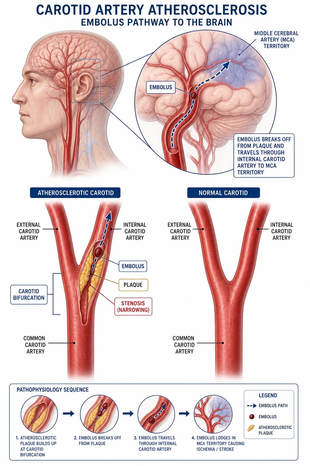
Carotid artery disease is atherosclerotic narrowing of the extracranial carotid arteries, almost always centred on the carotid bifurcation — the origin of the internal carotid artery (ICA) and the carotid bulb. It is one of the three great causes of ischaemic stroke, alongside small-vessel lipohyalinosis (lacunar) disease and cardioembolism, and is the single largest surgically preventable cause of stroke.[1]
The disease runs on a spectrum from asymptomatic (an incidental bruit, or stenosis found on screening or after a contralateral event) to symptomatic — defined as a focal neurological deficit of ischaemic origin in the territory of the affected carotid within the preceding six months. Symptomatic disease presents as transient ischaemic attack (TIA), amaurosis fugax, or a completed stroke. The central clinical question is not whether to image (always image), but whether to intervene — by carotid endarterectomy (CEA), carotid artery stenting (CAS), or best medical therapy alone — a decision driven by the degree of stenosis, symptom status, and the patient's perioperative risk and life expectancy.[1][8]
Mechanistically, the danger of a carotid plaque is embolic first, haemodynamic second. Most strokes attributable to carotid disease arise when an unstable plaque ruptures or ulcerates, releasing platelet-fibrin or cholesterol debris into the cerebral circulation; only in critical ("string-sign") stenosis does pure flow limitation become the dominant problem. This embolic truth is why the magnitude of benefit from surgery depends so sharply on how recently the patient became symptomatic: the unstable, embologenic plaque is at its most dangerous in the first hours to weeks after a TIA.[2]
Classification
Carotid disease is classified along two axes that together determine management — the degree of stenosis (measured by the NASCET method) and the symptom status (symptomatic within 6 months versus asymptomatic).[1]
The NASCET method
Stenosis is measured on angiography (conventional, CTA, or MRA) by the NASCET (North American Symptomatic Carotid Endarterectomy Trial) formula:[4]
Percent stenosis = (D-normal − D-minimal) / D-normal × 100 — where D-normal is the diameter of the distal, disease-free internal carotid artery (where the walls are parallel, beyond the bulb) and D-minimal is the narrowest residual lumen at the stenosis.[4]
The older European Carotid Surgery Trial (ECST) method used the estimated original diameter at the bulb as the denominator, which inflates the percentage for the same anatomy; the two methods agree closely only at severe stenosis. After Rothwell's pooled re-analysis reconciling NASCET and ECST data, the NASCET method is the international standard and all thresholds below are NASCET values.[5]
Severity bands and management (NASCET)
The severity band sets the surgical decision for both symptomatic and asymptomatic disease.[1]
| Stenosis band (NASCET) | Symptomatic (within 6 months) | Asymptomatic |
|---|---|---|
| Mild 0–49% | Best medical therapy only — surgery no benefit | Best medical therapy + risk-factor control |
| Moderate 50–69% | CEA for selected patients, soon (modest benefit, NNT ≈ 6 at 5 yr) | Medical therapy; CEA rarely justified |
| Severe 70–99% | CEA within 2 weeks — strong benefit (ARR 17%, NNT 6) | CEA if perioperative risk under 3% and life expectancy over 5 yr |
| Occluded 100% | Medical therapy — CEA not useful (no lumen to repair) | Medical therapy |
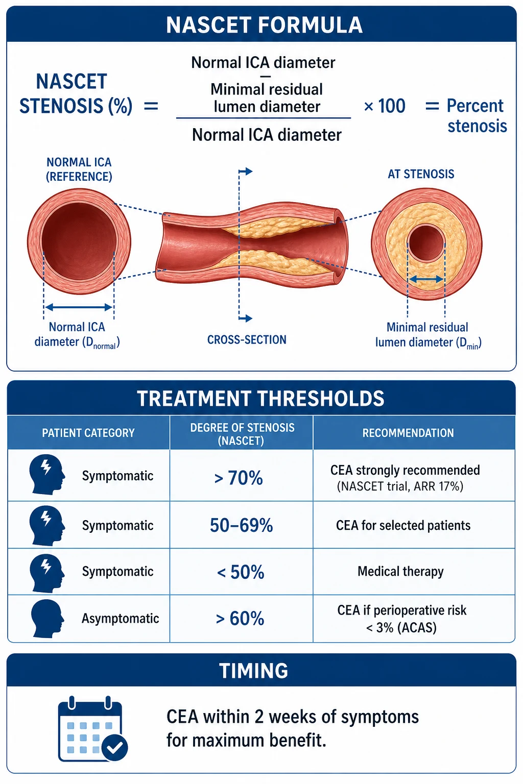
NASCET stenosis severity and the surgical decision
Severe 70–99%
Strong benefit, ARR 17%, NNT 6
The numbers that drive management
Epidemiology & Risk Factors
Carotid bifurcation atherosclerosis accounts for approximately 15–20% of all ischaemic strokes — roughly one in six — making it the largest individually preventable contributor after hypertension. Asymptomatic carotid stenosis of over 50% (NASCET) is found in about 4–5% of adults over 65, rising to 10% in those over 80 and in patients with established atherosclerosis elsewhere (coronary, peripheral arterial, aortic aneurysmal disease).[1][8]
The risk factors are those of systemic atherosclerosis, because carotid disease is a local manifestation of a systemic process — wherever you find one plaque, look for others.[1]
- Smoking — the dominant modifiable risk factor; roughly doubles stroke risk and accelerates plaque progression.
- Hypertension — the single largest population-attributable risk for stroke worldwide.
- Diabetes mellitus — a two- to four-fold increase in stroke risk.
- Hyperlipidaemia — elevated LDL drives plaque growth; statins reduce stroke risk.
- Age — prevalence rises sharply after 65.
- Male sex — higher prevalence, though women fare worse perioperatively.
- Atrial fibrillation — a key differential to exclude (cardioembolic source), not a cause of carotid stenosis.
- Family history of premature atherosclerosis or stroke.
- Known atherosclerotic disease elsewhere — coronary, PAD, AAA (systemic disease).
- Obesity, physical inactivity, obstructive sleep apnoea — contributory and modifiable. [1]
Epidemiology — who has it
Pathophysiology
Why the bifurcation?
The carotid bifurcation is a region of turbulent, oscillatory shear stress: blood dividing into the external and internal carotid branches creates low and oscillatory wall shear stress at the outer wall of the carotid bulb. Low shear stress promotes endothelial dysfunction, permits LDL infiltration and retention in the intima, and reduces the clearance of adhesion molecules — the seedbed for an atherosclerotic plaque. This is why the plaque sits at the bulb and the proximal ICA, and why the external carotid and the distal ICA are usually spared.[1]
A plaque matures from a fatty streak to a fibrous cap atheroma with a lipid-rich necrotic core. The danger is not the plaque's size alone but its stability. A thin fibrous cap over a large lipid core, with intra-plaque haemorrhage and inflammatory cell (macrophage) activity, is a vulnerable plaque — prone to rupture.[1]
Two mechanisms of cerebral injury
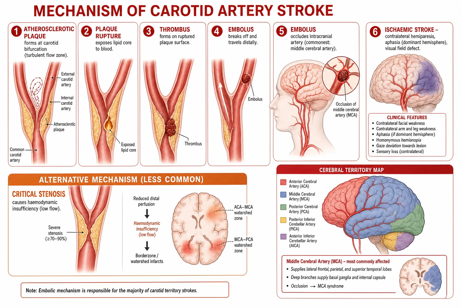
The two mechanisms by which a carotid plaque harms the brain explain both the typical anterior-circulation syndrome and the urgency of recently symptomatic disease.[1]
-
Artery-to-artery embolism (the commonest mechanism). The fibrous cap ruptures, exposing the thrombogenic lipid core to blood. Platelets adhere and aggregate, forming a thrombus on the plaque surface; fragments break off and embolise distally into the cerebral circulation. A platelet-fibrin embolus lodges transiently in a cortical or deep artery, producing a TIA; a larger or more persistent embolus causes a completed infarct. The commonest destination is the middle cerebral artery (MCA) territory, hence the motor and language signature of carotid-territory stroke.[1]
-
Haemodynamic insufficiency (less common). In critical ("string-sign", over 95%) stenosis or acute occlusion, flow distal to the stenosis falls below the brain's autoregulatory reserve, especially if the circle of Willis is incomplete or contralateral disease is present. A drop in perfusion pressure — hypotension, arrhythmia — then produces a watershed infarct between arterial territories.[1]
Why the MCA territory — and amaurosis fugax
Because the ICA supplies the anterior circulation (eye via ophthalmic artery, and the MCA/anterior cerebral artery via the circle of Willis), emboli from the bifurcation produce anterior-circulation symptoms: contralateral hemiparesis and hemisensory loss (face and arm more than leg, the MCA pattern), aphasia if the dominant (usually left) hemisphere is affected, homonymous hemianopia, and neglect in the non-dominant hemisphere.[1]
Amaurosis fugax is the retinal signature of the same process: a cholesterol crystal embolus (a shiny, refractile Hollenhorst plaque) from the carotid bifurcation lodges transiently in a branch of the central retinal artery. The patient describes a "curtain descending" or a "shade" over one eye, lasting seconds to minutes, with full recovery. It is, for practical purposes, pathognomonic of ipsilateral carotid disease and must be treated as a carotid-territory TIA.[1]
Clinical Presentation
Symptomatic carotid stenosis
Symptomatic disease is defined as a focal neurological deficit of ischaemic origin in the territory of the affected carotid within the preceding 6 months. Three presentations are recognised.[1]
- Transient ischaemic attack (TIA): sudden-onset focal neurological deficit lasting under 24 hours (typically minutes), with complete resolution, in the anterior (carotid) circulation — contralateral hemiparesis and hemisensory loss (face and arm more than leg), aphasia (dominant hemisphere) or neglect (non-dominant hemisphere), and monocular visual loss (amaurosis fugax). Use the ABCD squared score to stratify early stroke risk after a TIA.
- Amaurosis fugax: transient monocular blindness — already described; the retinal embolic equivalent of a hemispheric TIA.
- Completed stroke: a persistent focal deficit from established cerebral infarction in the carotid/MCA territory. [1]
Asymptomatic carotid stenosis
The patient has no referable symptoms. It is detected either as a carotid bruit on auscultation or as an incidental finding on carotid ultrasound performed for another reason (e.g. pre-coronary bypass, contralateral symptomatic disease, or screening). A bruit is a systolic, high-pitched murmur best heard at the angle of the jaw over the carotid bifurcation. Crucially, the presence and loudness of a bruit correlates poorly with stenosis severity: a very tight stenosis may produce no bruit because flow is so reduced ("silent occlusion"), and a moderate stenosis may be loud. A bruit is a reason to image, not a measure of severity.[1]
Anterior (carotid) circulation
internal carotid to ophthalmic, MCA, ACA
- **Contralateral hemiparesis / hemisensory loss** (face + arm greater than leg, the MCA pattern)
- **Aphasia** (dominant/left hemisphere) or **neglect** (non-dominant/right)
- **Monocular visual loss** — amaurosis fugax (retinal embolus)
- **Homonymous hemianopia** (optic radiation / occipital)
Posterior (vertebrobasilar) circulation
vertebral to basilar to PCA
- **Bilateral or crossing** motor/sensory deficit
- **Diplopia, vertigo, ataxia** (brainstem / cerebellum)
- **Dysarthria, dysphagia** (bulbar)
- **Loss of consciousness** / drop attacks
- **Visual field loss** (homonymous, from occipital cortex)
Atypical presentations
Examiners test the corners. Silent infarction on imaging in an asymptomatic patient with stenosis is now an indication for more aggressive workup. Crescendo TIA (two or more TIAs in 24 hours) and stroke-in-evolution denote a highly active, embologenic plaque and are urgent — they shorten the window for CEA. Elderly and diabetic patients may report vague symptoms (limb heaviness, word-finding difficulty) that are misattributed to age — a careful history and imaging are essential.[2]
Differential Diagnosis
When a patient presents with a focal neurological deficit or a bruit, the differential is wide. The task is to distinguish carotid disease from its mimics and from the other causes of stroke.[1]
| Condition | Key distinguishing feature |
|---|---|
| Cardioembolic stroke (AF, mural thrombus, valvular) | Multiple territory infarcts on imaging; AF on ECG; echocardiogram shows source; no carotid stenosis |
| Lacunar (small-vessel) stroke | Pure motor / sensory / ataxic-hemiparesis syndrome; small deep infarct on CT/MRI; hypertension, diabetes; no cortical signs, no carotid stenosis |
| Vertebrobasilar (posterior circulation) TIA | Diplopia, vertigo, ataxia, bilateral symptoms, loss of consciousness — not carotid territory |
| Intracerebral haemorrhage | Headache, vomiting, hypertension, rapid coma; non-contrast CT shows haemorrhage (dense area) — must be excluded before any antithrombotic or surgery |
| Subdural / extradural haematoma | Head injury, fluctuating consciousness; CT shows extra-axial collection |
| Brain tumour | Progressive (not sudden) deficit; headache, seizures; CT/MRI mass with oedema |
| Hypoglycaemia | Rapid onset, confusion, sweating; blood glucose low; reverses with glucose — always check glucose in any focal deficit |
| Seizure (post-ictal Todd's paresis) | Preceded by seizure activity; deficit resolves over hours; EEG abnormal |
| Carotid dissection | Younger patient, neck pain, Horner's syndrome; trauma or connective-tissue disease; flame-shaped ICA on imaging |
| Giant cell (temporal) arteritis | Older patient, headache, jaw claudication, raised ESR/CRP; can cause retinal ischaemia — a differential of amaurosis fugax |
| Functional / non-organic | Inconsistent examination, normal imaging |
Clinical & Bedside Assessment
A focused assessment answers three questions: Is this a stroke/TIA? Is it carotid-territory? Is there a surgical lesion?[1]
Neurological examination begins with the Glasgow Coma Scale (GCS) to grade consciousness, and the NIH Stroke Scale (NIHSS) — the standardised stroke severity score (0 = normal, 42 = deepest coma), which also stratifies thrombolysis decisions and must be documented on every suspected stroke. A focused cortical and motor exam then maps the deficit to anterior versus posterior circulation: carotid-territory stroke favours face/arm weakness, aphasia, and visual field loss. Fundoscopy looks for a Hollenhorst plaque (a refractile cholesterol crystal at a retinal artery bifurcation), the retinal stigmata of carotid embolism.[1]
Cardiovascular examination centres on auscultating for a carotid bruit at the angle of the jaw along the carotid course — systolic, high-pitched, present in roughly half of significant stenoses and whose absence does not exclude stenosis — followed by pulse, blood pressure (both arms), and heart sounds: AF (irregularly irregular), murmurs (valvular source), pulse deficit (dissection), unequal BP (subclavian/aortic disease). Because carotid disease is systemic, also examine the peripheral arteries — palpate for PAD and abdominal aortic aneurysm, and listen for renal bruits. Finally, a risk-factor and functional assessment documents smoking pack-years, diabetic control (HbA1c), lipid profile, blood pressure, and exercise tolerance, which together determine medical optimisation and operative fitness.[1][8]
Investigations
The goals of investigation are to confirm the diagnosis of stroke/TIA, exclude haemorrhage and mimics, quantify the carotid stenosis, and search for alternative embolic sources.[1]
Carotid imaging — the vascular ladder
- Carotid duplex ultrasound — the first-line modality. Non-invasive, cheap, bedside, and widely available. It measures peak systolic velocity (PSV) and end-diastolic velocity (EDV) at the stenosis and in the distal ICA; velocity criteria correlate with stenosis severity (e.g. PSV over 230 cm/s and ICA/CCA PSV ratio over 4 suggest over 70% stenosis). It also assesses plaque morphology — echolucency, ulceration, and intra-plaque haemorrhage are markers of an embologenic, vulnerable plaque. It is operator-dependent and less reliable distally and at very high (near-occlusion) grades.
- CT angiography (CTA) — the usual confirmatory test after an abnormal duplex. Fast, widely available, gives anatomical detail and allows formal NASCET measurement on a 3D dataset; also visualises the aortic arch and intracranial circulation. Requires iodinated contrast (renal caution).
- MR angiography (MRA) — contrast-enhanced MRA is highly accurate and avoids radiation; useful when CTA is contraindicated and for assessing the intracranial circulation and plaque composition. Time-of-flight MRA can overestimate stenosis due to turbulence.
- Conventional (catheter) digital subtraction angiography (DSA) — the gold standard for stenosis measurement, but invasive and carries an approximately 1% risk of procedure-related stroke. Reserved for discordant non-invasive imaging, suspicion of unusual lesions (dissection, fibromuscular dysplasia, vasculitis), or as part of CAS. [1]
The accepted standard is two concordant non-invasive modalities (duplex + CTA or MRA) before surgery; if they disagree, a third (or DSA) is obtained.[8]
Brain imaging
Brain imaging confirms ischaemia and excludes haemorrhage and mimics before any antithrombotic or surgical decision.[1]
- Non-contrast CT brain — the first imaging test in suspected stroke. Its primary job is to exclude haemorrhage (a dense area). Early ischaemia may be subtle: loss of the insular ribbon, obscuration of the lentiform nucleus, and the hyperdense MCA sign (thrombus in the M1). It determines thrombolysis eligibility.
- MRI brain (diffusion-weighted imaging, DWI) — the most sensitive test for early (within hours) ischaemia; identifies small and lacunar infarcts missed by CT. Used when the diagnosis is uncertain or for precise mapping. [1]
Cardiac and general workup
Because cardioembolism is the chief alternative source, every patient with a carotid-territory event is screened for a cardiac cause.[1]
- ECG — atrial fibrillation (the chief alternative embolic source); consider prolonged monitoring (Holter) if AF is paroxysmal.
- Echocardiogram (transthoracic, with or without transoesophageal) — mural thrombus, valvular vegetations, patent foramen ovale, low ejection fraction.
- Bloods — FBC, U&E, lipid profile, HbA1c, clotting, ESR/CRP (giant cell arteritis in the older amaurosis patient), glucose (hypoglycaemia mimic).
- Chest X-ray — heart size, and a baseline for anaesthetic assessment. [1]
Imaging at a glance — what and why
Management — Resuscitation
The acute presentation of carotid disease is the stroke or TIA itself. Resuscitation is generic stroke care, because carotid surgery (if any) is elective and deferred even in symptomatic patients — it is never the immediate resuscitative intervention.[1]
- ABCDE approach; secure the airway if GCS is depressed or bulbar failure is present.
- Oxygen only if hypoxic (SpO₂ under 94%) — routine high-flow oxygen is not beneficial and may worsen outcome.
- Blood glucose — treat hypoglycaemia (a stroke mimic) promptly; avoid hyperglycaemia (worsens outcome).
- Blood pressure — do not lower aggressively in the acute phase; permissive hypertension preserves penumbral perfusion. Lower only if over 220/120 mmHg, or if thrombolysis is planned (target under 185/110 mmHg).
- Thrombolysis — if an acute ischaemic stroke meets criteria (within 4.5 hours of onset, no haemorrhage on CT, no exclusions), intravenous alteplase 0.9 mg/kg (max 90 mg, 10% bolus then infusion over 60 min) is given before any carotid workup. Thrombectomy is considered for large-vessel anterior-circulation occlusion.
- Admit to a stroke unit; keep NPO until swallow assessed; give VTE prophylaxis; arrange continuous cardiac monitoring for AF.
Once the patient is stabilised and the diagnosis of a carotid-territory event is confirmed, the definitive carotid question is addressed: how severe is the stenosis, and is the patient a candidate for CEA or CAS?[1]
Management — Definitive & Stepwise
Management has three tiers, applied to every patient: (1) best medical therapy, (2) the surgical decision — CEA, CAS, or neither, and (3) risk-factor modification for life.[1][8]
Best medical therapy (BMT) — for ALL patients, regardless of surgery
Best medical therapy is the foundation for every patient, whether or not they proceed to surgery, and has markedly lowered the stroke risk of asymptomatic carotid stenosis in the modern era.[1]
Best medical therapy — every patient, every time
Carotid endarterectomy (CEA) — indications and timing
CEA is the evidence-based standard for revascularisation. Its benefit depends critically on two variables: the degree of stenosis and the time from the last symptom.[4][5]
CEA decision in symptomatic carotid stenosis
Confirm stenosis by two concordant non-invasive imaging modalities (duplex + CTA/MRA).
Apply NASCET severity band: 70–99% = strong benefit; 50–69% = selected; under 50% = no benefit.
Assess operative fitness and life expectancy (over 5 years) and confirm unit perioperative stroke/death rate under 6% for symptomatic disease.
Schedule CEA within 2 weeks of the index symptom for maximal benefit (NNT 5 if under 2 wk vs 125 if over 12 wk).
Start or continue best medical therapy throughout; antiplatelet and statin on the day of surgery.
Timing is the single most important modifiable factor. The benefit of CEA after a TIA or minor stroke is greatest in the first 2 weeks and declines steeply thereafter — the unstable plaque stabilises with time, and so the strokes surgery prevents have already happened. "Time is brain." The number needed to treat (NNT) to prevent one ipsilateral stroke is approximately 5 if CEA is performed within 2 weeks, rising to 125 if delayed beyond 12 weeks.[2]
NASCET (1991, 1998) — symptomatic severe stenosis
RCT: CEA + medical vs medical alone, symptomatic stenosis
Key finding
For 70–99% stenosis, CEA reduced 2-year ipsilateral stroke from 26% (medical) to 9% (CEA) — ARR 17%, NNT 6. Benefit durable. For 50–69% a modest benefit (5-year ARR 6%); under 50% no benefit.
ECST + NASCET pooled analysis (Rothwell, 2003)
Pooled individual-patient data from NASCET, ECST, VA-309
Key finding
Re-expressed all stenosis in NASCET terms. Benefit confirmed: greatest for 70–99% (NNT 3 to prevent one stroke at 2 yr in the first days after symptoms), modest for 50–69%, absent under 50%. Established NASCET as the international standard.
ECST (1991) — severe symptomatic stenosis
RCT: CEA vs medical, symptomatic carotid stenosis (MRC European Carotid Surgery Trial)
Key finding
Interim results confirmed CEA benefit for severe (70–99%) stenosis and no benefit for mild (under 30%) stenosis. Together with NASCET this defined the surgical thresholds; the ECST method of measuring stenosis was later reconciled with NASCET by Rothwell's pooled analysis.
CEA technique
CEA is the open surgical removal of the atheromatous plaque from the carotid bifurcation and proximal ICA.[8]
- Anaesthesia — general or local/regional (regional allows awake neurological monitoring: the patient performs tasks during clamping, detecting ischaemia in real time).
- Position and incision — supine, head turned away; a longitudinal incision along the anterior border of sternocleidomastoid from the angle of the jaw downward.
- Exposure — divide the facial vein; expose and control the common, external, and internal carotid arteries, slinging each. Identify and protect the hypoglossal (XII), vagus (X), and marginal mandibular branch of the facial (VII) nerves.
- Heparinisation — intravenous heparin before clamping.
- Clamping and cerebral protection — clamp the common, external, then internal carotid. If intra-operative monitoring shows ischaemia (or stump pressure is low, under 40–50 mmHg, or under regional anaesthesia the patient loses function), deploy a temporary intraluminal shunt to maintain cerebral perfusion while the artery is opened.
- Endarterectomy — longitudinal arteriotomy from CCA across the bulb into the ICA; dissect the plaque in the subintimal plane, removing it cleanly with a feathered endpoint in the distal ICA to avoid a flap.
- Closure — close the arteriotomy, ideally with a patch angioplasty (vein or prosthetic) to widen the lumen and reduce restenosis. Flush thoroughly to remove debris before final closure.
- Haemostasis and closure in layers over a drain; observe for haematoma. [1]
Carotid artery stenting (CAS)
CAS is the endovascular alternative: femoral access, a catheter guided into the carotid under fluoroscopy, an embolic protection device (filter) deployed distal to the stenosis, pre-dilatation balloon angioplasty, then self-expanding stent deployment across the lesion. Post-dilatation refines the result; the filter is retrieved.[7]
CAS is preferred when open surgery is high-risk: previous neck surgery or radiotherapy, recurrent stenosis after CEA, a "hostile neck", high lesions (above C2, surgically inaccessible), or contralateral laryngeal nerve palsy. The trade-off is a higher peri-procedural stroke risk than CEA, partly offset by lower MI and cranial-nerve-injury rates.[1]
CREST (Brott, 2010) — CEA vs CAS
RCT: CAS vs CEA in symptomatic and asymptomatic stenosis
Key finding
Composite endpoint (stroke + MI + death) similar. **Peri-procedural stroke higher with CAS (4.1% vs 2.3%)**; **peri-procedural MI higher with CEA (2.3% vs 1.1%)**; **cranial nerve injury only with CEA**. Age effect: CEA better in patients over 70; CAS acceptable in younger patients. Long-term ipsilateral stroke risk similar after the peri-procedural period.
CEA (endarterectomy)
open surgery
- **Lower peri-procedural stroke** risk (2.3%)
- **Higher MI** and **cranial nerve injury** risk
- Better in **over-70s** (CREST)
- **Durable** — no routine surveillance needed
- Gold standard for most symptomatic patients
CAS (stenting)
endovascular
- **Higher peri-procedural stroke** risk (4.1%)
- **Lower MI** and **no cranial nerve injury**
- Better for **high-risk surgery** patients
- Indicated for: previous neck surgery/radiotherapy, recurrent stenosis, hostile neck, high lesion
- Age effect: acceptable in younger, less so in over-70s
Asymptomatic carotid stenosis — the surgical decision
For asymptomatic stenosis, surgery is far more selective. The original ACAS (1995) and the ACST (2004) showed that CEA reduced the 5-year stroke risk from about 11–12% (medical) to roughly 5–6% (CEA) — an absolute risk reduction of about 5–6% (NNT 17) — but only if the perioperative stroke/death rate was under 3%. With modern intensive medical therapy narrowing the medical-arm risk, the net benefit of operating on asymptomatic patients has shrunk and is individualised: consider CEA for asymptomatic stenosis over 60–70% in a fit patient with a life expectancy over 5 years and a surgeon/unit with audited perioperative risk under 3%.[6]
ACAS (1995) & ACST (2004) — asymptomatic stenosis
RCT: CEA + medical vs medical alone, asymptomatic stenosis over 60%
Key finding
CEA halved the 5-year stroke risk (from approx 11% to 5–6%; ARR 5–6%, NNT 17) — but only if perioperative stroke/death rate was under 3%. Benefit modest and much smaller than for symptomatic disease; modern BMT has narrowed it further.
Specific Subtypes & Scenarios
Each subtype changes the surgical decision and must be recognised on imaging and history.[1]
- Bilateral carotid stenosis: CEA on the symptomatic side first, within 2 weeks if symptomatic. The contralateral CEA is staged 4–6 weeks later — simultaneous bilateral CEA risks bilateral cranial nerve injury, bilateral hyperperfusion, and haemodynamic instability.
- Carotid near-occlusion / "string sign": a thread-like residual lumen with collapse of the distal ICA ("slim" distal ICA). The peri-procedural stroke risk is higher, and the benefit of CEA less certain than for discrete 70–99% stenosis; managed in a specialist unit, often with careful DSA definition.
- Complete carotid occlusion: no lumen to repair — CEA is not useful. Medical therapy; chronic occlusion carries an annual stroke risk of 2–5% (higher if symptomatic). Extracranial-to-intracranial bypass is investigational.
- Carotid dissection: younger patient, neck pain, Horner's syndrome (ptosis, miosis, anhidrosis), trauma or connective-tissue disease. Managed medically (antiplatelet or anticoagulant); stenting reserved for high-risk or evolving deficit.
- Post-CEA restenosis: myointimal hyperplasia within 2 years (smooth, non-atherosclerotic). CAS is often preferred over re-do CEA (hostile scarred neck).
- Carotid body tumour (paraganglioma): a differential of a neck mass at the bifurcation; separate entity, not atherosclerotic, but anatomically relevant.
- Crescendo TIA / stroke-in-evolution: denotes a highly active embologenic plaque — treat as urgent; CEA within days if stenosis is severe and the deficit is not devastating. [1]
Complications & Pitfalls
Complications of CEA
The perioperative stroke/death rate is the central audit metric — if the unit rate exceeds the ceiling, the surgery causes more strokes than it prevents.[1]
CEA — the audit threshold for perioperative stroke/death
The recognised complications of CEA, in order of clinical importance, are:[1]
- Perioperative stroke or death — the principal risk, caused by intraoperative embolisation (manipulation of the plaque), hypoperfusion during clamping (mitigated by shunting), or postoperative thrombosis/embolism. The unit's audited rate must be under 6% for symptomatic and under 3% for asymptomatic patients; above these, the benefit of surgery is lost.
- Myocardial infarction — the commonest cause of perioperative death; these patients have coronary disease too. Continue statin and beta-blocker, optimise cardiac status.
- Cranial nerve injury — hypoglossal (XII) (tongue deviation to the operated side, dysarthria), vagus/recurrent laryngeal (X) (hoarseness, vocal cord palsy), and the marginal mandibular branch of VII (mouth droop). Usually transient; bilateral injury (e.g. staged surgery) can cause airway compromise.
- Wound haematoma — can expand rapidly and compromise the airway (a true surgical emergency — open the wound at the bedside to decompress before returning to theatre).
- Cerebral hyperperfusion syndrome — occurs days after revascularisation: severe headache, seizures, focal deficit, hypertension, and risk of intracerebral haemorrhage from restored flow exceeding autoregulation. Manage with tight BP control and seizure treatment.
- Restenosis — myointimal hyperplasia (early, under 2 yr) or recurrent atherosclerosis (late). Reduced by patch closure. Symptomatic restenosis is often treated by CAS.
- Bleeding, infection, hypertensive crisis (carotid sinus manipulation) — perioperative. [1]
Complications of CAS
CAS carries its own distinct risk profile centred on embolic and access complications.[7]
- Peri-procedural stroke — higher than CEA (embolic debris during wire/stent manipulation, despite embolic protection devices).
- Bradycardia / hypotension — carotid sinus stimulation during balloon inflation; atropine standby.
- Contrast nephropathy, access-site complications (groin haematoma), and in-stent restenosis/thrombosis. [1]
Classic pitfalls
The classic errors that lose benefit or cause harm are avoidable with disciplined timing and workup.[1]
- Delaying CEA beyond 2 weeks — benefit declines sharply with time; the strokes surgery was meant to prevent have already happened.
- Operating on an asymptomatic patient without confirming low perioperative risk — if the unit rate exceeds 3%, surgery causes more strokes than it prevents.
- Treating a bruit as a measure of severity — a bruit mandates imaging, not a percentage.
- Missing cardioembolism — AF is a common alternative source; always check the ECG and image the heart.
- Lowering blood pressure aggressively in acute stroke — permissive hypertension protects the penumbra.
- Forgetting BMT in surgical patients — even after CEA, antiplatelet, statin, BP and smoking control are lifelong. [1]
Prognosis & Disposition
Without treatment
The natural history depends on symptom status and stenosis severity.[1]
- Symptomatic 70–99% stenosis: approximately 13% 2-year risk of ipsilateral stroke (untreated); the risk is highest in the first days to weeks after the index event.
- Symptomatic 50–69%: lower but still significant annual stroke risk.
- Asymptomatic over 70%: roughly 3–5% annual stroke risk untreated, though modern BMT has reduced this.
- Asymptomatic under 60%: under 1% per year. [1]
After CEA
CEA converts the high early risk of a recently symptomatic plaque into a low long-term risk.[4][6]
- Symptomatic severe stenosis: stroke risk falls to approximately 3–9% at 2 years (from 13% untreated) — the magnitude of the NASCET benefit. Long-term annual ipsilateral stroke risk under 1–2%.
- Asymptomatic: annual stroke risk reduced to approximately 1–2% from 3–5%. [1]
After CAS
Long-term ipsilateral stroke prevention is similar to CEA (CREST), once the higher peri-procedural period is survived.[7]
Disposition: symptomatic patients are admitted under a stroke unit with vascular surgery referral; CEA (if indicated) is arranged within the 2-week target. Asymptomatic patients are managed in an outpatient vascular / stroke-prevention clinic with risk-factor optimisation and surveillance duplex imaging. After CEA or CAS, lifelong BMT, risk-factor control, and (for some) periodic surveillance imaging of the contralateral side continue.[8]
Special Populations
The surgical choice and perioperative risk shift with the patient, so revascularisation is individualised rather than protocolised.[1]
- Elderly (over 70): higher peri-procedural stroke risk with CAS; CEA preferred (CREST age effect). Overall operative risk is higher due to comorbidities, so selection is careful — but age alone is not a contraindication.
- Women: smaller carotid arteries and possibly higher perioperative risk; the benefit of CEA in asymptomatic women is debated and generally more selective.
- Diabetics: higher stroke risk and worse outcomes; aggressive risk-factor control. Renal function must be checked before CTA contrast.
- Renal impairment: iodinated contrast for CTA is a risk — prefer duplex-first, and consider MRA or pre-hydration/contrast-sparing protocols.
- Anticoagulated patients (AF, valves): warfarin/DOAC management peri-CEA — bridge or withhold as appropriate; AF remains the alternative embolic source to exclude.
- Previous neck surgery or radiotherapy: "hostile neck" — CAS preferred over re-do open surgery.
- Young patients (under 50): suspect non-atherosclerotic causes — carotid dissection, fibromuscular dysplasia, vasculitis, hypercoagulable state — and investigate accordingly; CAS is more acceptable in younger patients needing revascularisation. [1]
Evidence, Guidelines & Regional Differences
The landmark trials — and what they changed
The modern management of carotid disease rests on five randomised trials and their pooled analysis; each changed practice.[5]
NASCET (1991/1998)
symptomatic
- CEA + medical vs medical, symptomatic stenosis
- 70–99%: 2-yr ipsilateral stroke 26% then 9% (ARR 17%, NNT 6)
- 50–69%: modest benefit; under 50%: no benefit
- Established the NASCET measurement standard
ECST (1991) + pooled (2003)
symptomatic
- European trial confirming benefit of CEA for severe stenosis
- Rothwell pooled analysis reconciled NASCET + ECST in NASCET terms
- Benefit greatest 70–99%, modest 50–69%, none under 50%
- NASCET became the international standard denominator
ACAS (1995) / ACST (2004)
asymptomatic
- CEA halved 5-yr stroke (11–12% to 5–6%; ARR 5–6%, NNT 17)
- Only if perioperative stroke/death rate under 3%
- Modest benefit; modern BMT has narrowed it
- Justifies selective CEA in fit asymptomatic over 60–70%
CREST (2010)
CEA vs CAS
- CAS vs CEA, symptomatic + asymptomatic
- Composite outcome similar; CAS more stroke, CEA more MI
- CEA better in over-70s; CAS acceptable younger
- CAS for high-risk-surgery anatomical/clinical groups
Regional guideline differences
India / LMIC context: atherosclerosis and stroke prevalence are high, but access to CEA is limited to major centres and CAS availability is even more restricted. Best medical therapy (antiplatelet, statin, BP control, diabetes management) is the most widely applicable and impactful intervention and must be optimised for every patient. Smoking cessation remains the single most powerful modifiable risk factor. Where surgery is available, the same evidence-based thresholds and the 2-week symptomatic target apply.[1]
Controversies
Two live debates shape contemporary practice.[1]
- Asymptomatic carotid stenosis and modern BMT: with contemporary intensive medical therapy lowering the medical-arm stroke risk to 1–2% per year, the net benefit of prophylactic CEA has narrowed; some centres no longer operate routinely on asymptomatic stenosis, reserving surgery for high-risk plaque features (echolucent, progressing, silent infarction on imaging). Ongoing trials (e.g. ACST-2, ECST-2) inform this debate.
- CEA versus CAS: the peri-procedural trade-off (stroke with CAS vs MI/nerve injury with CEA) is individualised by age and anatomy; there is no single best procedure for all.
- Timing after thrombolysis: CEA is generally deferred a few days after thrombolysis to avoid haemorrhagic transformation, but within the 2-week window. [1]
CEA timing — time is brain
FAST
CEA within 2 weeks of symptom onset
absolute risk reduction for symptomatic 70–99%
number needed to treat if within 2 weeks (vs 125 if over 12 weeks)
maximum benefit from earliest intervention
Exam Pearls
The high-yield facts examiners reward, each stated precisely.[1]
- NASCET: symptomatic 70–99% = CEA within 2 weeks (ARR 17%, NNT 6). 50–69% = selected. Under 50% = medical therapy only.[4]
- NASCET method: (normal distal ICA − minimal lumen) / normal distal ICA × 100. All thresholds are NASCET values.[5]
- Timing dominates: NNT 5 if CEA within 2 weeks vs 125 if over 12 weeks. "Time is brain."[2]
- Amaurosis fugax = transient monocular blindness ("curtain descending") = retinal cholesterol embolus (Hollenhorst plaque) from the carotid bifurcation = ipsilateral carotid stenosis until proven otherwise.[1]
- Asymptomatic over 60–70%: CEA only if perioperative risk under 3% and life expectancy over 5 yr (ACAS/ACST, ARR 5–6%, NNT 17).[6]
- Mechanism: embolic first (plaque rupture then MCA territory), haemodynamic second (critical stenosis then watershed).[1]
- Anterior (carotid): hemiparesis (face + arm), aphasia, monocular visual loss. Posterior (vertebrobasilar): diplopia, vertigo, bilateral, ataxia.[1]
- CEA complications: perioperative stroke (under 6% symptomatic / under 3% asymptomatic), MI (commonest cause of death), cranial nerve XII injury, wound haematoma (airway emergency), hyperperfusion syndrome.[1]
- CREST: CAS higher stroke, CEA higher MI and cranial nerve injury. CEA better in over-70s; CAS for high-risk-surgery patients (hostile neck, restenosis, high lesion).[7]
- Bruit correlates poorly with severity — a reason to image, not a measure of degree.[1]
- Every patient gets best medical therapy: aspirin 75 mg (or clopidogrel 75 mg), atorvastatin 80 mg, BP under 130/80, smoking cessation — even after surgery.[1]
Exam application bank (NEET-PG / INICET)
One-line answer
Carotid artery disease is atherosclerotic narrowing of the extracranial carotid artery (commonest: bifurcation/internal carotid). Presents as TIA (focal deficit under 24 h), amaurosis fugax (transient monocular blindness from retinal embolus), or completed stroke. Or asymptomatic (incidental bruit). NASCET method measures stenosis: (normal ICA diameter − minimal lumen) / normal ICA diameter × 100. Symptomatic stenosis 70–99%: CEA within 2 weeks (NASCET, ARR 17%, NNT 6). Symptomatic 50–69%: CEA for selected (modest benefit). Asymptomatic over 60–70%: CEA if perioperative risk under 3% (ACAS/ACST). All patients receive best medical therapy (antiplatelet + high-intensity statin + BP control + smoking cessation + diabetes control). CEA: open plaque removal + patch closure. CAS: endovascular stent for high-risk surgical patients (CREST).
Worked stems (answer without another resource)
Stem 1 — Classic presentation. Map symptoms to mechanism; name the first investigation and first treatment step with dose/route if drug therapy is standard. [1]
Stem 2 — Unstable / complicated. List red flags that force immediate resuscitation, theatre, ICU, antidote, or reperfusion — and what you do in the first 15 minutes. [1]
Stem 3 — Atypical group. Elderly, pregnancy, child, or immunocompromised: how presentation and thresholds change. [1]
Stem 4 — Differential trap. Name the three closest mimics and one discriminator for each. [1]
Stem 5 — Disposition. Who goes home with safety-netting, who is admitted, who needs HDU/ICU/theatre, and what follow-up is mandatory. [1]
Rapid viva checklist
- Definition + classification
- Pathophysiology chain
- Bedside signs / criteria
- Score with exact components (if any)
- Emergency bundle
- Definitive therapy with doses
- Complications of disease and of treatment
- Special populations
- Guideline/trial name if classic
- Three exam traps
Coverage self-check
If you cannot answer any stem above from this page alone, re-read the matching section — the page is intended to be self-sufficient for final-prof and NEET-PG/INICET questions on Carotid Artery Disease.
References
- [1]Bonati LH, Jansen O, de Borst GJ, Brown MM. Management of atherosclerotic extracranial carotid artery stenosis Lancet Neurol, 2022.PMID 35182512
- [2]Naylor AR. Time is brain! Surgeon, 2007.PMID 17313125
- [3]European Carotid Surgery Trialists' Collaborative Group. MRC European Carotid Surgery Trial: interim results for symptomatic patients with severe (70-99%) or with mild (0-29%) carotid stenosis. European Carotid Surgery Trialists' Collaborative Group Lancet, 1991.PMID 1674060
- [4]Barnett HJ, Taylor DW, Eliasziw M, et al. (NASCET). Benefit of carotid endarterectomy in patients with symptomatic moderate or severe stenosis. North American Symptomatic Carotid Endarterectomy Trial Collaborators N Engl J Med, 1998.PMID 9811916
- [5]Rothwell PM, Eliasziw M, Gutnikov SA, et al. Analysis of pooled data from the randomised controlled trials of endarterectomy for symptomatic carotid stenosis Lancet, 2003.PMID 12531577
- [6]Halliday A, Mansfield A, Marro J, et al. (ACST Collaborative Group). Prevention of disabling and fatal strokes by successful carotid endarterectomy in patients without recent neurological symptoms: randomised controlled trial Lancet, 2004.PMID 15135594
- [7]Brott TG, Hobson RW II, Howard G, et al. (CREST). Stenting versus endarterectomy for treatment of carotid-artery stenosis N Engl J Med, 2010.PMID 20505173
- [8]Twine CP, Venermo M, de Borst GJ, et al. (ESVS Carotid Guidelines Committee). Editor's Choice - European Society for Vascular Surgery (ESVS) 2023 Clinical Practice Guidelines on the Management of Atherosclerotic Carotid and Vertebral Artery Disease Eur J Vasc Endovasc Surg, 2023.PMID 35598721