General Surgery · General Surgery
Colorectal Carcinoma
Also known as Colorectal cancer · CRC · Colon cancer · Rectal cancer · Bowel cancer
Colorectal carcinoma (CRC) arises from the colonic or rectal epithelium (adenocarcinoma in 95%). Third most common cancer worldwide; risk factors include age over 50, family history, Lynch syndrome (HNPCC), FAP, IBD (ulcerative colitis), red/processed meat, smoking, obesity. Presents with altered bowel habit, rectal bleeding, weight loss, abdominal pain; right-sided tumours cause anaemia/weight loss (insidious), left-sided/rectal cause obstruction, tenesmus, fresh bleeding. Diagnosis by colonoscopy + biopsy; staging by CT chest/abdomen/pelvis + MRI rectum (for rectal) and CEA. TNM staging drives treatment: surgery is curative for stages I to III (TME for rectal); adjuvant chemotherapy (FOLFOX/CAPOX) for stage III; neoadjuvant chemoradiotherapy for locally advanced rectal cancer. Screening from age 45 to 50 (colonoscopy, FIT) saves lives.
On this page & tools
Your progress
Saved locally on this device.
Practise this topic
Exam tags
Red flags
Overview & Definition
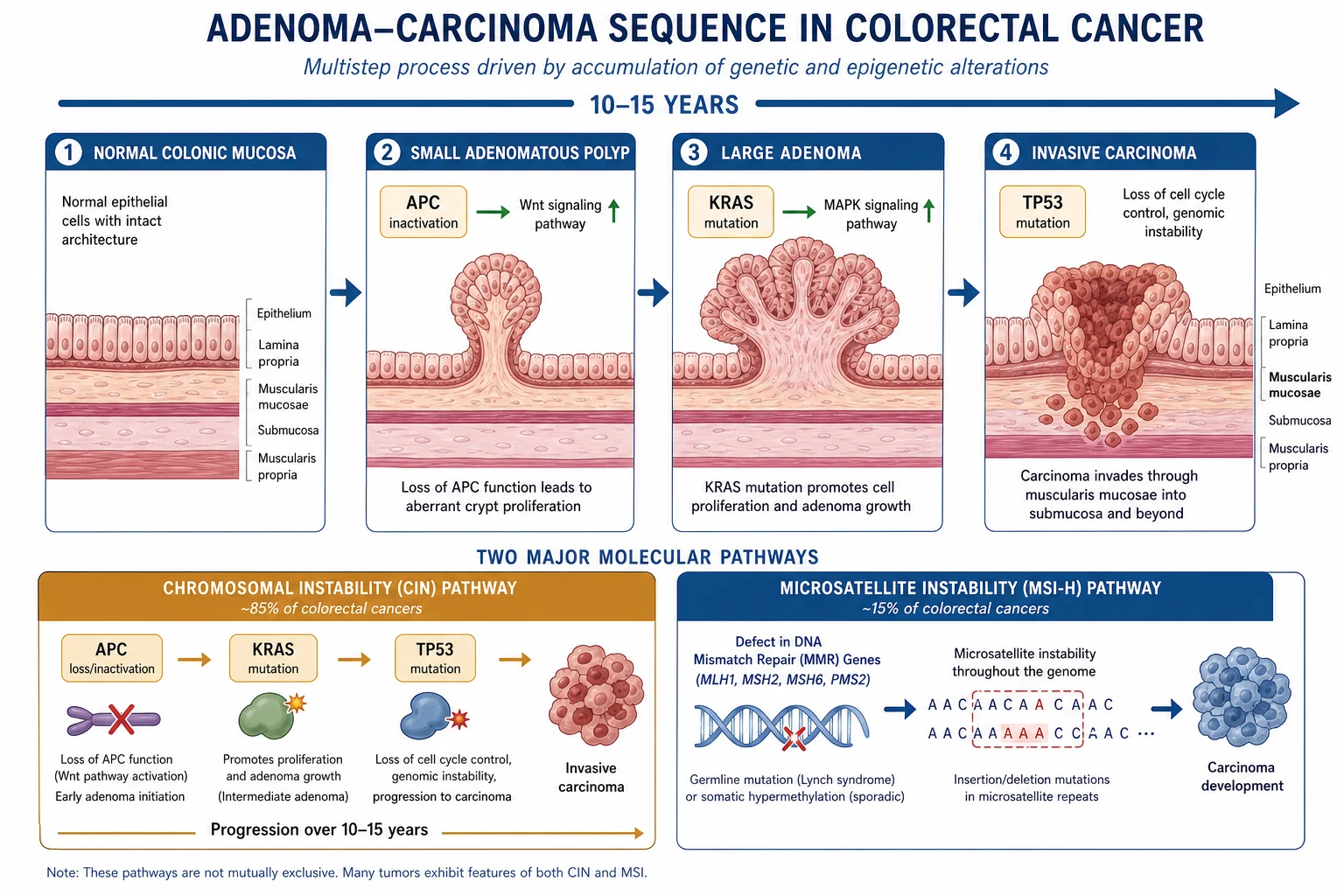
Colorectal carcinoma (CRC) is a malignant epithelial tumour of the colon or rectum, with adenocarcinoma accounting for over 95% of cases. It develops through the adenoma-carcinoma sequence — a stepwise accumulation of genetic mutations (APC, KRAS, TP53) over 10 to 15 years that transforms normal mucosa into an adenomatous polyp and then invasive carcinoma.[1]
CRC is the third most commonly diagnosed cancer and the second leading cause of cancer death worldwide. The clinically critical distinctions are: right colon vs left colon vs rectum (different presentation, surgical approach, and chemosensitivity), sporadic vs hereditary (Lynch syndrome, FAP), and curative (stages I to III) vs palliative (stage IV) intent.[1]
Classification
By anatomical site (the split that changes presentation and management):[1]
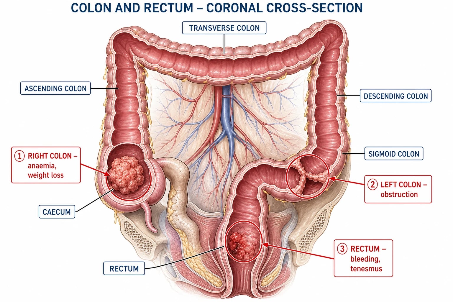
- Right colon (caecum, ascending, proximal transverse) — insidious: iron-deficiency anaemia, weight loss, vague right iliac fossa pain. Tumours are often fungating/cauliflower.
- Left colon (distal transverse, descending, sigmoid) — obstructive symptoms: altered bowel habit (constipation, overflow diarrhoea), colicky pain, abdominal distension. Tumours tend to be annular/constricting ("napkin-ring").
- Rectum (distal 15 cm) — fresh rectal bleeding, tenesmus, change in stool calibre, feeling of incomplete evacuation. Digital rectal examination (DRE) may palpate the tumour. [1]
By molecular pathway: [1]
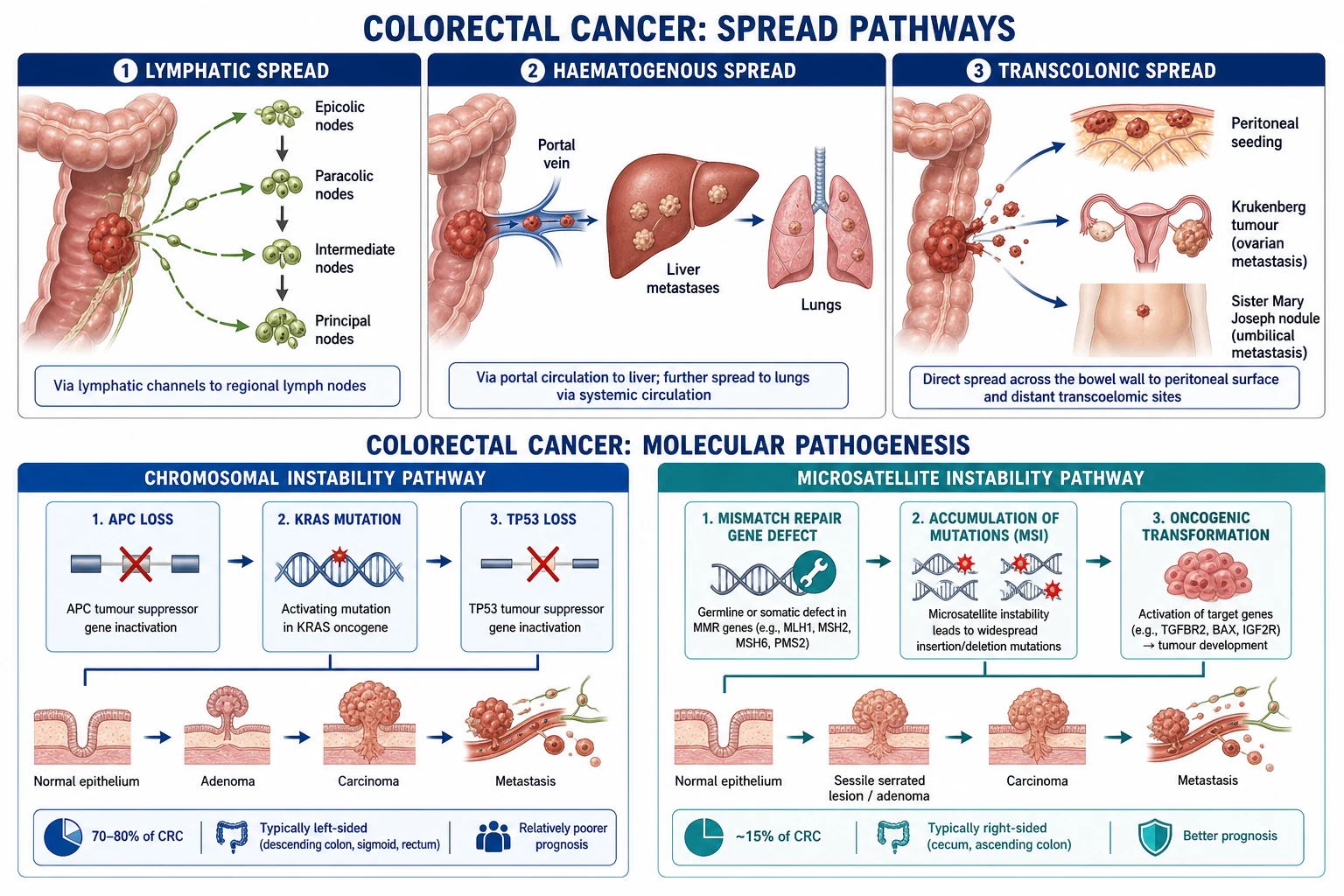
- Chromosomal instability (CIN) — APC mutation then KRAS then TP53 (the classic adenoma-carcinoma sequence); accounts for ~70 to 80%.
- Microsatellite instability (MSI-H) — defective DNA mismatch repair (Lynch syndrome/HNPCC or sporadic MLH1 hypermethylation); ~15%; better prognosis, resistance to 5-FU monotherapy, predicts response to immunotherapy.
- CpG island methylator phenotype (CIMP) — epigenetic silencing, often with BRAF mutation; associated with serrated pathway. [1]
By TNM stage (AJCC 8th edition):[1]
| Stage | TNM (simplified) | 5-year survival |
|---|---|---|
| I | T1 to T2, N0, M0 | ~90% |
| II | T3 to T4, N0, M0 | 60 to 80% |
| III | Any T, N1 to N2, M0 | 40 to 65% |
| IV | Any T, Any N, M1 | ~15% |
TNM 8th Edition — Detailed Staging Reference
The AJCC 8th edition (2017) staging system is the current standard and is built on the TNM components plus prognostic stage groups.[1]
T (Tumour) categories — depth of primary tumour invasion: [1]
- Tis (carcinoma in situ) — intramucosal carcinoma (limited to the lamina propria or muscularis mucosae; has not breached the basement membrane). Managed by polypectomy alone if completely removed with clear margins.
- T1 — tumour invades the submucosa (through muscularis mucosae into submucosal layer). Polyp cancer staging uses the Haggitt level for pedunculated polyps (1 = head, 2 = neck, 3 = stalk, 4 = invasion of submucosa of bowel wall below stalk) and the Kikuchi sm level for sessile polyps (sm1 = upper third, sm2 = middle third, sm3 = lower third of submucosa). T1 sm1/2 with no poor histology may be managed by endoscopic mucosal resection (EMR) or transanal local excision (TEMS/TAMIS); T1 sm3, poor differentiation, or lymphovascular invasion warrants formal oncological resection.
- T2 — tumour invades the muscularis propria (the deep muscle layer of the bowel wall).
- T3 — tumour invades through the muscularis propria into the pericolic/perirectal tissues (subserosa). T3 is subdivided in the 8th edition by depth of invasion beyond muscularis propria: T3a under 1 mm, T3b 1 to 5 mm, T3c 5 to 15 mm, T3d over 15 mm — deeper invasion carries worse prognosis.
- T4 — tumour invades the visceral peritoneum (T4a) or directly invades adjacent organs or structures (T4b). T4 disease requires en-bloc multivisceral resection to achieve an R0 (negative-margin) resection. [1]
N (Node) categories — regional lymph node involvement: [1]
- N0 — no regional lymph node metastasis. A minimum of 12 nodes must be examined for accurate staging.
- N1 — 1 to 3 regional nodes involved: N1a = 1 node, N1b = 2 to 3 nodes, N1c = no nodes positive but tumour deposits in the subserosa/mesentery/non-peritonised pericolic tissues (without residual nodal architecture).
- N2 — 4 or more regional nodes involved: N2a = 4 to 6 nodes, N2b = 7 or more nodes.
- Note: nodal positivity is the strongest prognostic factor in colon cancer and the principal indication for adjuvant chemotherapy. [1]
M (Metastasis) categories: [1]
- M0 — no distant metastasis.
- M1a — metastasis confined to one organ (liver or lung or non-regional nodes).
- M1b — metastasis to more than one organ (still no peritoneal involvement).
- M1c — metastasis to the peritoneal surface, with or without other organ involvement. Peritoneal disease carries the worst prognosis in the M1 category. [1]
Prognostic stage groups (AJCC 8th edition): [1]
Stage I = T1 to T2, N0, M0 — 5-year survival ~90%. Stage IIA = T3, N0, M0. Stage IIB = T4a, N0, M0. Stage IIC = T4b, N0, M0 (note: although deeper T stage, the 5-year survival for stage IIC is paradoxically lower than for stage IIIA in some series due to the T4 peritoneal involvement). Stage IIIA = T1 to T2, N1 (or T1, N2a). Stage IIIB = T3 to T4a, N1 (or T2 to T3, N2a). Stage IIIC = any T4b, N2. Stage IVA = any T, any N, M1a. Stage IVB = any T, any N, M1b. Stage IVC = any T, any N, M1c.[1]
Critical staging rules: [1]
- Circumferential resection margin (CRM) in rectal cancer — the closest distance from the tumour to the mesorectal fascia on MRI; CRM under 1 mm defines an involved (or threatened) margin and mandates neoadjuvant long-course chemoradiotherapy before TME.
- Extramural vascular invasion (EMVI) — tumour within vessels beyond the muscularis propria on MRI; an independent poor prognostic factor in rectal cancer.
- Tumour regression grade (TRG) after neoadjuvant therapy — assesses response: TRG 0 (complete response) to TRG 3 (poor response); complete clinical responders may be considered for the "watch and wait" strategy (no surgery, intensive surveillance).
- Lymphovascular and perineural invasion — both worsen prognosis and upstage risk of recurrence; routinely reported in the pathology specimen.
Epidemiology & Risk Factors
CRC is the third most common cancer (after breast and lung) and the second most common cause of cancer death globally. Lifetime risk is approximately 4 to 5% in the general population, rising sharply with the identified risk factors below.[1]
Non-modifiable risk factors: [1]
- Age — over 90% of cases are in people aged 50 or older; incidence rises steeply after 50.
- Family history — one first-degree relative doubles the risk; the risk is higher if diagnosed before 60 or if multiple relatives are affected.
- Hereditary syndromes — Lynch syndrome (HNPCC): autosomal dominant, mismatch repair gene mutation (MLH1, MSH2, MSH6, PMS2), accounts for ~3% of CRC, presents young (under 50), right-sided, synchronous/metachronous tumours. Familial adenomatous polyposis (FAP): APC gene mutation, hundreds to thousands of colonic polyps from adolescence, 100% lifetime CRC risk without colectomy.
- Inflammatory bowel disease — long-standing ulcerative colitis (and Crohn disease to a lesser extent) increases CRC risk, especially with pancolitis and duration over 8 to 10 years.
- Personal history of CRC, adenomatous polyps, or ovarian/endometrial cancer. [1]
Modifiable risk factors: red and processed meat, low fibre intake, smoking, alcohol, obesity, physical inactivity.[1]
Pathophysiology
The adenoma-carcinoma sequence is the central paradigm: normal colonic epithelium acquires mutations in a predictable order over 10 to 15 years:[1]
- APC gene loss (5q) — initiates adenoma formation (Wnt pathway dysregulation, beta-catenin accumulation).
- KRAS mutation (12p) — drives adenoma growth from small to large.
- TP53 loss (17p) — converts large adenoma to invasive carcinoma. [1]
This 10 to 15-year window is the basis for screening and polypectomy — removing an adenoma before it becomes malignant prevents CRC.[1]
MSI-H pathway (Lynch syndrome and 15% sporadic): defective mismatch repair leads to accumulation of microsatellite length errors, rapid mutation accumulation, and right-sided, mucinous, poorly differentiated tumours with tumour-infiltrating lymphocytes — paradoxically better stage-for-stage prognosis.[1]
Spread: direct extension through the bowel wall (T stage), lymphatic to regional nodes (N stage — epicolic, paracolic, intermediate, principal), haematogenous to liver (via portal vein) then lungs, and transcoelomic drop metastases (peritoneum, ovary — Krukenberg). [1]
CRC survival by stage — the numbers
Clinical Presentation
Right colon: insidious and late — iron-deficiency anaemia (microcytic, hypochromic), fatigue, weight loss, vague right iliac fossa mass or pain. Obstruction is rare (liquid stool, wide lumen). Bleeding is occult (mixed in stool, not visible).[1]
Left colon: earlier symptoms due to narrower lumen and more solid stool — altered bowel habit (constipation alternating with diarrhoea from partial obstruction), colicky lower abdominal pain, visible blood or mucus in stool, abdominal distension. Obstruction is common (annular "napkin-ring" tumour).[1]
Rectum: painless fresh rectal bleeding (on the surface of stool, not mixed), tenesmus (feeling of incomplete evacuation), change in stool calibre (pencil-thin), rectal mass on DRE (hard, irregular, non-tender, bleeding on contact).[1]
Atypical presentations:
- Acute large bowel obstruction — distension, no flatus/stool, colicky pain, empty rectum on DRE; emergency presentation (commonest surgical emergency from CRC).
- Perforation — sudden severe pain, peritonism, free gas on erect CXR; typically at the tumour site or caecum (diastatic perforation behind an obstructing left-sided lesion).
- Iron-deficiency anaemia in a male or postmenopausal woman — investigate for right-sided CRC.
- Young patient (under 45) — consider Lynch syndrome; index of suspicion must be high despite young age. [1]
Right colon
caecum, ascending
- **Insidious, late** presentation
- **Iron-deficiency anaemia**, weight loss
- **Fungating/cauliflower** tumour
- Obstruction **rare** (liquid stool, wide lumen)
Left colon
descending, sigmoid
- **Altered bowel habit**, obstruction
- **Fresh bleeding** mixed in stool
- **Annular constricting** (napkin-ring) tumour
- Colicky lower abdominal pain
Rectum
distal 15 cm
- **Fresh rectal bleeding**, tenesmus
- **Change in stool calibre** (pencil-thin)
- **Palpable mass** on DRE
- Contact bleeding
Differential Diagnosis
| Condition | Key distinguishing feature |
|---|---|
| Haemorrhoids | Bright red blood on toilet paper / dripping into toilet; no weight loss or anaemia; proctoscopy confirms |
| Diverticular disease | Acute LLQ pain, fever, localised peritonism; CT shows pericolic fat stranding / abscess; no mass |
| Inflammatory bowel disease (UC) | Chronic bloody diarrhoea, tenesmus, extra-intestinal features; colonic biopsies show mucosal inflammation, dysplasia surveillance |
| Irritable bowel syndrome | Chronic alternating bowel habit but no weight loss, no bleeding, no anaemia, normal colonoscopy; diagnosis of exclusion |
| Colonic polyps | Asymptomatic or incidental bleeding; diagnosed and removed at colonoscopy; histology is adenoma vs hyperplastic |
| Ischaemic colitis | Acute pain + bloody diarrhoea in elderly/vasculopathic patient; "thumbprinting" on imaging; self-limiting in most |
| Infective colitis | Acute onset, fever, positive stool culture (Campylobacter, Salmonella, Shigella, E. coli); resolves in days |
Clinical & Bedside Assessment
Digital rectal examination (DRE) — mandatory in any patient with rectal bleeding or altered bowel habit. Assesses: position, fixity, surface of a rectal mass; sphincter tone (important for surgical planning); prostate/bladder involvement; blood or mucus on the glove. A low rectal tumour is palpable as a hard, irregular, non-tender, fixed or mobile mass, often with contact bleeding.[1]
Rigid/flexible proctosigmoidoscopy — visualises the tumour, allows biopsy, and determines the distance from the anal verge (critical for surgical planning — determines whether APR or anterior resection is feasible).[1]
Abdominal examination — palpable mass (right iliac fossa for caecal tumour), hepatomegaly (liver metastases), ascites (peritoneal disease), distension (obstruction). [1]
General examination — pallor (anaemia), cachexia, lymphadenopathy (Virchow's node — left supraclavicular), Sister Mary Joseph nodule (umbilical metastasis). [1]
Investigations
Diagnostic (confirming the tumour): [1]
- Colonoscopy with biopsy — the gold standard. Visualises the entire colon, allows biopsy for histology, identifies synchronous lesions (3 to 5%), and enables polypectomy of incidental adenomas. If incomplete (poor prep, obstruction), CT colonography is the alternative.[1]
Staging (assessing extent): [1]
- CT chest, abdomen, pelvis (CT CAP) — the standard staging scan. Assesses local invasion, regional nodes, liver/lung metastases. Contrast-enhanced (portal venous phase for liver).
- MRI rectum — essential for rectal cancer. Assesses depth of invasion through the rectal wall (T stage), mesorectal fascia involvement (circumferential resection margin), and extramural vascular invasion (EMVI) — all critical for deciding neoadjuvant therapy and surgical approach.[1]
- Serum CEA (carcinoembryonic antigen) — a tumour marker; not diagnostic (elevated in smokers, IBD, other cancers), but useful for monitoring response to treatment and detecting recurrence post-resection. Check pre-operatively as a baseline.
Pre-operative assessment: [1]
- Full blood count — anaemia (especially right-sided tumours).
- LFTs — may be abnormal with liver metastases (alkaline phosphatase rise is typical).
- U&Es, coagulation, group and save — surgical preparation. [1]
Molecular testing (guides therapy): [1]
- Mismatch repair (MMR) / MSI status — tested on the tumour (immunohistochemistry for MLH1, MSH2, MSH6, PMS2, or PCR for MSI). MSI-H/dMMR suggests Lynch syndrome (refer for genetic counselling), predicts better prognosis, and indicates response to immune checkpoint inhibitors (pembrolizumab) in metastatic disease.
- RAS (KRAS/NRAS) and BRAF mutation testing — in metastatic CRC, determines eligibility for anti-EGFR therapy (cetuximab, panitumumab) — only RAS wild-type tumours respond. [1]
Management — Resuscitation
In the emergency presentation (obstruction or perforation) treat as an acute abdomen / sepsis pathway first, then decide the operation: [1]
- ABC / sepsis six mindset. High-flow oxygen if hypoxic; two large-bore cannulae; crystalloid bolus for hypovolaemia/sepsis (e.g. 500 mL balanced crystalloid, reassess); urinary catheter for output; large-bore NG tube for obstruction (decompress stomach, reduce aspiration risk); early lactate, FBC, U&E, ABG/VBG, group-and-save/crossmatch.
- Antibiotics (peritonitis, perforation, or septic obstruction): start within the first hour after cultures if septic — e.g. piperacillin-tazobactam 4.5 g IV every 8 hours, or co-amoxiclav 1.2 g IV TDS + metronidazole 500 mg IV TDS; escalate for hospital-acquired/ESBL risk per local policy. Analgesia with titrated IV morphine; antiemetics.
- Blood products and optimisation. Crossmatch 2 to 4 units if bleeding/perforation/planned major resection. Correct severe hypokalaemia/acidosis as able pre-op; involve critical care early for shock.
- Imaging when stable enough. Erect CXR may show free gas; CT CAP with IV contrast (if renal function allows) defines transition point, perforation, metastases, and guides whether SEMS bridge is even a consideration.
- Urgent surgical decision.
- Right-sided obstruction in a fit patient: often resection + primary anastomosis (ileocolic).
- Left-sided obstruction, unstable or contaminated: Hartmann's procedure (resection + end colostomy + rectal stump) — prioritises survival over immediate restoration of continuity.
- Selected stable malignant left obstruction without peritonism: SEMS as bridge to elective oncological resection in experienced centres (perforation/migration risks; not for low rectal lesions or suspected perforation).
- Perforation with peritonitis: source control — resection of perforated segment, washout, usually stoma; damage-control laparotomy if in extremis.
- Thromboprophylaxis with LMWH when bleeding risk allows; ERAS principles resume after emergency recovery.[1]
Management — Definitive & Stepwise
Principle: surgery is the only curative treatment for localised CRC.[1]
Colon cancer surgery
- Right hemicolectomy — for caecal, ascending, and proximal transverse colon tumours. Resection with ileocolic, right colic, and middle colic vessel ligation + regional lymph nodes. Primary ileocolic anastomosis (can be laparoscopic).
- Left hemicolectomy — for descending and sigmoid colon tumours. Ligation of inferior mesenteric artery (IMA) branches + regional nodes; primary anastomosis.
- Extended right hemicolectomy — for mid-transverse colon tumours.
- Subtotal/total colectomy — for synchronous tumours or FAP. [1]
Oncological principles: proximal vascular ligation (high tie) to ensure adequate lymph node harvest (minimum 12 nodes for accurate staging); 5 cm proximal and distal margins; en-bloc resection of involved adjacent organs if adherent (T4).[1]
Rectal cancer surgery
- Total mesorectal excision (TME) — the gold standard. The entire mesorectum (containing lymphatics and nodes) is excised in an intact fascial envelope, ensuring a clear circumferential resection margin (CRM). Local recurrence rates dropped from 20 to 30% (pre-TME) to under 10% with TME.[5]
- Anterior resection (AR) — for upper and mid-rectal tumours (above ~5 to 6 cm from anal verge). Restores bowel continuity with a colorectal or coloanal anastomosis. A temporary defunctioning ileostomy protects the anastomosis.
- Abdominoperineal excision of rectum (APER) — for low rectal tumours (involving the sphincter or too low for a safe anastomosis). The rectum and anus are removed, leaving a permanent end colostomy. ELAPE (extralevator APER) reduces positive CRM rates.
- Transanal endoscopic microsurgery (TEMS/TAMIS) — for early T1 rectal cancer (well-differentiated, no lymphovascular invasion, under 3 cm) in unfit patients; local excision only.
Neoadjuvant therapy for rectal cancer
For stage II to III rectal cancer (T3 to T4, or N+), pre-operative therapy downstages the tumour and reduces local recurrence: [1]
- Short-course radiotherapy (25 Gy in 5 fractions over 1 week) followed by surgery within 1 week — simpler, less toxicity, preferred in many European centres.
- Long-course chemoradiotherapy (45 to 50.4 Gy with concurrent capecitabine or 5-FU over 5 to 6 weeks) — preferred for bulky tumours needing downstaging; surgery 6 to 8 weeks later. Better local control but more acute toxicity.[1]
Adjuvant chemotherapy
- Stage III (node-positive) colon cancer: FOLFOX (5-FU + leucovorin + oxaliplatin) or CAPOX (capecitabine + oxaliplatin) for 3 to 6 months. The MOSAIC trial established that adding oxaliplatin to 5-FU/leucovorin improved disease-free survival and overall survival in stage II to III colon cancer.[3][4]
- Stage II colon cancer: generally not routinely recommended; consider in high-risk features (T4, fewer than 12 nodes examined, perforation/obstruction, poorly differentiated, lymphovascular invasion). MSI-H stage II has excellent prognosis and may not benefit from 5-FU.
- Stage IV (metastatic): palliative systemic therapy — first-line FOLFOX or FOLFIRI +/- bevacizumab (anti-VEGF) or cetuximab/panitumumab (anti-EGFR, only if RAS wild-type). Pembrolizumab (anti-PD-1) for MSI-H/dMMR metastatic CRC. Palliative surgery/stenting for obstruction.[1]
Cytotoxic and Biologic Drug Doses (Adult, BSA-Indexed)
The following regimens and doses are the standard adult regimens for colorectal cancer; doses are calculated on body surface area (BSA in m²) and adjusted for toxicity, organ function, and performance status. Always cross-check renal function (creatinine clearance) for capecitabine and irinotecan, and UGT1A1 polymorphism testing for irinotecan where available. [1]
FOLFOX (standard for stage III colon adjuvant and first-line metastatic) — given every 2 weeks (one cycle = 14 days), for 12 cycles (6 months) adjuvantly:[3][4]
- Oxaliplatin 85 mg/m² IV over 2 hours on day 1 of each 2-week cycle. Cumulative dose-related sensory neuropathy is the dose-limiting toxicity — numbness/dysaesthesia of hands, feet, and perioral region, exacerbated by cold (avoid cold drinks/food for 48 hours after infusion); typically dose-limiting above cumulative 800 to 1,000 mg/m².
- Leucovorin (folinic acid) 400 mg/m² IV over 2 hours on day 1, given to potentiate 5-FU by stabilising the thymidylate synthase–5-FU complex.
- 5-Fluorouracil (5-FU) 400 mg/m² IV bolus on day 1, followed by 2,400 mg/m² as a 46-hour continuous IV infusion (via portable pump), starting on day 1 and finishing on day 3. [1]
CAPOX (also called XELOX) — alternative to FOLFOX — given every 3 weeks for 8 cycles (6 months): Oxaliplatin 130 mg/m² IV over 2 hours on day 1 + Capecitabine 1,000 mg/m² orally twice daily on days 1 to 14 (14 doses total per cycle). The 3-week schedule reduces hospital visits but slightly more diarrhoea and hand-foot syndrome than FOLFOX. [1]
FOLFIRI (used for metastatic CRC and second-line) — given every 2 weeks: [1]
- Irinotecan 180 mg/m² IV over 90 minutes on day 1. Diarrhoea and neutropenia are the dose-limiting toxicities (UGT1A1 polymorphism predisposes to severe neutropenia). Premedication with atropine for acute cholinergic reaction (cramping, sweating, lacrimation) is sometimes given.
- Leucovorin 400 mg/m² IV over 2 hours on day 1.
- 5-Fluorouracil 400 mg/m² IV bolus, then 2,400 mg/m² 46-hour continuous infusion on days 1 to 3. [1]
Single-agent capecitabine (oral 5-FU prodrug) — used in stage III adjuvant when oxaliplatin is contraindicated (elderly, neuropathy): Capecitabine 1,250 mg/m² orally twice daily on days 1 to 14 of each 3-week cycle, for 6 to 8 cycles. Hand-foot syndrome (palmar-plantar erythrodysesthesia) and diarrhoea are the main toxicities; dose-reduce for grade 2 or worse. Severe renal impairment (CrCl under 30 mL/min) is a contraindication. [1]
Bevacizumab (anti-VEGF monoclonal antibody) — added to FOLFOX/FOLFIRI in metastatic CRC, irrespective of RAS status: Bevacizumab 5 mg/kg IV every 2 weeks (or 7.5 mg/kg every 3 weeks when paired with CAPOX). Continued until disease progression. Toxicities: hypertension, proteinuria, impaired wound healing, perforation (rare, ~1 to 2%), bleeding/thromboembolism. [1]
Cetuximab (anti-EGFR monoclonal antibody) — added to FOLFIRI or FOLFOX in metastatic CRC, but only in RAS/BRAF wild-type, left-sided tumours: Cetuximab 400 mg/m² IV loading dose, then 250 mg/m² IV weekly. Panitumumab (fully human) is an alternative: 6 mg/kg IV every 2 weeks. Skin rash (acneiform eruption) is the hallmark toxicity and is a positive predictor of response. KRAS/NRAS mutations predict no benefit — testing is mandatory before use. [1]
Pembrolizumab (anti-PD-1 immune checkpoint inhibitor) — first-line for unresectable/metastatic MSI-H or dMMR CRC: Pembrolizumab 200 mg IV every 3 weeks (or 400 mg every 6 weeks) until disease progression or up to 24 months. The KEYNOTE-177 trial established pembrolizumab as superior to chemotherapy in this subgroup. Contraindicated for patients with active autoimmune disease or on chronic immunosuppression. Immune-related adverse events: colitis, hepatitis, pneumonitis, endocrinopathies. [1]
Practical notes on dose modifications: 3 months of adjuvant CAPOX is non-inferior to 6 months of FOLFOX for stage III colon cancer (IDEA trial). For capecitabine, reduce to 750 to 1,000 mg/m² bid if toxicity persists. For oxaliplatin, reduce to 75 mg/m² or discontinue if grade 2 or higher neuropathy (cold-triggered dysaesthesia). [1]
Specific Subtypes & Scenarios
- Lynch syndrome (HNPCC) — autosomal dominant mismatch repair defect. Amsterdam II criteria: 3+ relatives with CRC (or Lynch-associated cancer), one being a first-degree relative of the other two, spanning 2+ generations, at least one diagnosed under 50. Surveillance colonoscopy from age 25 to 30, every 1 to 2 years. Prophylactic colectomy is considered.
- FAP — APC mutation. Colonoscopy from age 10 to 12, annually. Prophylactic colectomy by late teens to early 20s (100% cancer risk). Also surveil duodenum (side-viewing endoscopy) and abdominal wall for desmoid tumours.
- Obstructing CRC — emergency. Right-sided: resection + primary anastomosis. Left-sided: Hartmann's procedure (resection + end colostomy + rectal stump) in the unstable/unprepared patient; or on-table lavage + primary anastomosis in a stable, prepared patient. Self-expanding metal stent (SEMS) as a bridge to elective surgery in selected patients.
- Rectal cancer with threatened CRM on MRI — long-course chemoradiotherapy to downsize before TME. [1]
Hereditary Colorectal Cancer Syndromes — Detailed
Up to 5 to 10% of all CRC is hereditary. Recognising these syndromes changes screening age, modality, surgical extent, and family counselling. The two predominant syndromes are Lynch syndrome and Familial Adenomatous Polyposis (FAP); the recessively inherited MUTYH-Associated Polyposis (MAP) rounds out the differential. [1]
Lynch syndrome (Hereditary Non-Polyposis Colorectal Cancer, HNPCC) — the most common hereditary CRC syndrome, accounting for 2 to 4% of all CRC and 10 to 15% of right-sided CRC under age 50. Inheritance is autosomal dominant with high (70 to 80%) penetrance. The molecular defect is a germline mutation in a DNA mismatch repair (MMR) gene: MLH1 (~40%), MSH2 (~40%), MSH6 (~10%), or PMS2 (~5%); less often EPCAM deletion (causes MSH2 silencing). The defective MMR machinery leads to microsatellite instability (MSI-H) in tumour DNA, accelerating mutation accumulation during the adenoma-carcinoma sequence. Despite a much faster carcinogenesis (2 to 3 years per adenoma vs 10 years sporadic), the stage-for-stage prognosis is better than sporadic CRC because of prominent tumour-infiltrating lymphocytes and immune activation. Tumours are typically right-sided (proximal to splenic flexure), mucinous, poorly differentiated, and present under age 50. Clinical diagnostic criteria: Amsterdam II (3-2-1 rule) — 3 relatives with Lynch-associated cancer (CRC, endometrial, ovarian, gastric, small bowel, hepatobiliary, urinary tract, brain, sebaceous skin tumours), 2 successive generations affected, 1 first-degree relative of the other two, with at least 1 diagnosed under age 50; an alternative screening tool is the Bethesda guidelines (CRC under 50, synchronous/metachronous Lynch-associated tumours, MSI-H histology under 60, Lynch-associated cancer under 50 with CRC, CRC with first-degree relative with Lynch-related cancer under 50). Universal screening of all newly diagnosed CRC under age 70 (and selected cases over 70) for MSI/MMR status — by immunohistochemistry (IHC) for the four MMR proteins or by PCR for MSI — is now standard. Surveillance: colonoscopy every 1 to 2 years from age 20 to 25 (or 2 to 5 years before the youngest case in the family); annual endometrial sampling + transvaginal ultrasound for women from age 30 to 35; consider annual urine cytology and gastroscopy from age 30 to 35 in families with upper GI or urinary tract cancers. Surgical options for a CRC in a Lynch patient: (1) segmental resection (oncologically adequate) or (2) extended (subtotal/total) colectomy with ileorectal or ileosigmoid anastomosis — the latter is favoured because metachronous CRC risk is 16 to 25% at 10 years after segmental resection and up to 40 to 60% lifetime; lifetime colonoscopic surveillance is still required after segmental resection. First-degree relatives should be referred for germline genetic testing and cascade screening.[1]
Familial Adenomatous Polyposis (FAP) — accounts for ~1% of all CRC. Inheritance is autosomal dominant, caused by a germline mutation in the APC gene on chromosome 5q21 (a tumour-suppressor in the Wnt/β-catenin pathway). Classic FAP presents with hundreds to thousands of adenomatous polyps carpeting the colon from adolescence, with virtually 100% lifetime CRC risk by age 35 to 40 if untreated. Attenuated FAP (AFAP) is a milder phenotype with fewer polyps (typically 10 to 100), more right-sided distribution, and later cancer onset (average 55 years). Screening of at-risk relatives: annual flexible sigmoidoscopy or colonoscopy from age 10 to 12 (or late teens for AFAP), continuing lifelong. Polyp burden prevents endoscopic management and prophylactic colectomy is the definitive treatment — three surgical options: (1) total proctocolectomy with ileal pouch-anal anastomosis (IPAA) — preferred when rectal polyp burden is heavy; eliminates practically all CRC risk at the cost of a pelvic pouch and slightly higher operative morbidity. (2) Total abdominal colectomy with ileorectal anastomosis (IRA) — simpler, preserves continence; suitable when the rectum is polyp-free; residual rectal cancer risk of 10 to 15% over 30 years mandates lifelong yearly rectal surveillance. (3) Subtotal colectomy with ileosigmoid anastomosis — rarely used. Surgery is typically planned in the late teens or early 20s when polyp burden becomes unmanageable. Extra-colonic manifestations (Gardner syndrome variant) include duodenal/periampullary adenomas (5 to 10% lifetime risk of duodenal cancer — surveillance with side-viewing duodenoscopy every 1 to 3 years from age 25), desmoid tumours (abdominal wall and mesentery, particularly after surgery), CHPRE (congenital hypertrophy of retinal pigment epithelium), osteomas (skull, mandible), epidermoid cysts, supernumerary teeth, and thyroid cancer (papillary, ~2 to 3% risk).[1]
MUTYH-Associated Polyposis (MAP) — a recessively inherited polyposis syndrome due to biallelic MUTYH gene mutations (MUTYH is a base-excision repair glycosylase). Usually presents with 10 to 100 adenomas (overlap with AFAP) and a high lifetime CRC risk (~75 to 90% without surveillance). Importantly, MAP is autosomal recessive, so siblings have a 25% risk but obligate carriers (heterozygotes) generally do not — distinct counselling pattern from FAP. Surveillance: colonoscopy every 1 to 2 years from age 25 to 30. Management parallels FAP when polyp burden is high (prophylactic colectomy); total colectomy with IRA is preferred. [1]
Hamartomatous polyposis syndromes (rare, but recognised when CRC is diagnosed young with a polyp history): Peutz-Jeghers syndrome (STK11 mutation) — mucocutaneous pigmentation, hamartomatous GI polyps, >90% lifetime CRC risk by age 65; surveillance colonoscopies every 2 to 3 years from age 18, gastroscopy + capsule endoscopy for small-bowel polyps, breast and pancreas screening. Juvenile polyposis syndrome (SMAD4, BMPR1A) — multiple juvenile polyps, ~50% lifetime CRC risk; colonoscopic surveillance from age 15. Cowden syndrome (PTEN mutation) — mucocutaneous features, breast/thyroid/endometrial/CRC; colonoscopic surveillance from age 35. [1]
Serrated polyposis syndrome (formerly hyperplastic polyposis) — non-Mendelian but strongly familial; characterised by multiple serrated polyps in the proximal colon plus conventional adenomas; cumulative CRC risk is ~25 to 50%. Surveillance: colonoscopy every 1 to 3 years from diagnosis. [1]
[1]Complications & Pitfalls
Local: obstruction (most common surgical emergency from CRC), perforation (generalised peritonitis or localised abscess), fistula (colovesical — pneumaturia/faecaluria; colovaginal), bleeding (chronic anaemia or acute haemorrhage).[1]
Metastatic: liver (commonest site; resectable in ~20% — consider hepatic resection or ablation), lungs, peritoneum (pseudomyxoma peritonei from mucinous tumours), bone (late). [1]
Surgical complications: anastomotic leak (3 to 5%; presents days 5 to 7 with fever, pain, tachycardia; risk higher in low rectal anastomoses, malnutrition, steroids), stoma complications (parastomal hernia, retraction, necrosis), ureteric injury (especially in rectal surgery), sexual/urinary dysfunction (autonomic nerve damage during TME).[5]
Classic pitfalls:
- Not doing a DRE in a patient with "haemorrhoids" — a rectal tumour is missed.
- Attributing iron-deficiency anaemia to diet in an adult male — always exclude right-sided CRC with colonoscopy.
- Not screening first-degree relatives of CRC patients (colonoscopy 10 years before the index case's age, or age 40, whichever is earlier).
- Forgetting Lynch syndrome in a young patient with right-sided CRC.
- Inadequate lymph node harvest (under 12) — understaging, under-treatment. [1]
Prognosis & Disposition
Stage at diagnosis is the dominant prognostic factor. Stage I has a 5-year survival of approximately 90%; stage IV (metastatic) approximately 15%. Node-negative patients with clear margins are generally cured by surgery alone.[1]
Other prognostic factors: MSI-H/dMMR (better prognosis), BRAF mutation (worse prognosis in metastatic disease), perineural/lymphovascular invasion, positive CRM in rectal cancer (high local recurrence risk), elevated pre-operative CEA (worse prognosis), obstruction/perforation at presentation (worse prognosis). [1]
Follow-up after curative resection:
- CEA every 3 to 6 months for 3 years, then 6-monthly to 5 years. Rising CEA prompts imaging for recurrence.
- CT CAP annually for 3 to 5 years.
- Surveillance colonoscopy at 1 year post-resection, then 3-yearly (to detect metachronous tumours). [1]
Special Populations
- Lynch syndrome (HNPCC) — germline mismatch-repair defect (MLH1, MSH2, MSH6, PMS2, EPCAM). Tumours often right-sided, mucinous, MSI-H, with better stage-for-stage prognosis but high metachronous risk. Amsterdam II and revised Bethesda criteria select families/tumours for IHC/MSI testing; germline confirmation guides relatives. Colonoscopy from age 25 to 30 (or 10 years before the youngest CRC in the family), every 1 to 2 years. After CRC, strongly consider extended (subtotal) colectomy rather than segmental resection because of metachronous risk. Women: counsel endometrial and ovarian risk — discuss annual endometrial sampling/TVUS and risk-reducing surgery after childbearing per genetics team. Also screen stomach/urinary tract per local Lynch protocols. Metastatic MSI-H disease may respond to PD-1 blockade (pembrolizumab/nivolumab).[1]
- FAP and attenuated FAP — APC germline mutation; classic FAP develops hundreds to thousands of colorectal adenomas with near-100% lifetime CRC risk without intervention. Start flexible sigmoidoscopy/colonoscopy from age 10 to 12; offer prophylactic colectomy (IRA or restorative proctocolectomy with IPAA) typically by age 20 to 25, individualised by polyp burden. Post-colectomy: annual pouch/rectal surveillance. Screen duodenum (Spigelman stage) and consider thyroid (cribriform-morular PTC association) and desmoid risk. Genetic testing of first-degree relatives is mandatory once a pathogenic variant is known.[1]
- Ulcerative colitis / extensive Crohn's colitis — chronic inflammation drives dysplasia–carcinoma. Surveillance colonoscopy with chromoendoscopy + targeted biopsies (or random biopsies if chromo unavailable) generally from 8 years after symptom onset (earlier if PSC coexists — PSC-UC is high risk and often annual from diagnosis). Any confirmed dysplasia prompts colectomy discussion at MDT. After colectomy with IPAA, pouch surveillance continues if residual cuff/rectal tissue remains.
- Elderly/frail — physiological age outweighs chronological age. Prefer laparoscopic resection when feasible (faster recovery). Emergency laparotomy mortality is high — for obstructing left colon in comorbid patients, SEMS as bridge to elective resection in specialist centres may avoid Hartmann's if no perforation/peritonitis. Dose-adjust oxaliplatin carefully (neuropathy, falls risk); consider shorter 3-month CAPOX where IDEA evidence allows in lower-risk stage III.
- Pregnancy — CRC in pregnancy is rare; delay non-urgent CT; MRI without gadolinium preferred after first trimester for local staging when needed. Surgery in second trimester if unavoidable; chemotherapy (5-FU-based) only after detailed counselling, generally avoided in first trimester. Multidisciplinary maternal–fetal medicine involvement is essential.
- Immunosuppressed / transplant — higher CRC risk in some cohorts (long-standing IBD on immunosuppression; solid-organ transplant). Lower threshold for colonoscopy for anaemia or bleeding; coordinate timing of adjuvant chemo with transplant team.
- Young-onset CRC (<50 years) — rising incidence globally. Always take a three-generation pedigree, test tumour for MSI/MMR, and refer genetics even if Amsterdam criteria incomplete. Do not attribute rectal bleeding to haemorrhoids without endoscopic evaluation.
Evidence, Guidelines & Regional Differences
Screening (USPSTF 2021):[2]
- Age to start: 45 (lowered from 50 in 2021, reflecting rising incidence in young adults). Age to stop: 75 (individual decision 76 to 85; stop at 85).
- Modalities: colonoscopy every 10 years (gold standard), FIT (faecal immunochemical test) annually, guaiac FOBT annually, stool DNA-FIT every 1 to 3 years, flexible sigmoidoscopy every 5 years, or CT colonography every 5 years. [1]
Regional screening differences: [1]
- UK (NHS Bowel Cancer Screening): FIT every 2 years from age 50 to 74; colonoscopy if FIT positive.
- India — no national screening programme; opportunistic screening recommended from age 50 (or 40 with risk factors). FIT/colonoscopy in urban centres.
- Australia (National Bowel Cancer Screening): FOBT/FIT every 2 years from age 50 to 74. [1]
Screening modalities — comparative detail: [1]
- Colonoscopy — the gold-standard screening test and the only modality that is both diagnostic and therapeutic (polyps can be removed at the same procedure). Sensitivity for CRC is ~95%; specificity approaches 100% with adequate bowel prep. Bowel preparation with split-dose polyethylene glycol (PEG) the day before is preferred — minimum Boston Bowel Preparation Scale (BBPS) score 6 or above with all three colonic segments scored ≥2 is considered adequate; otherwise the study should be repeated within a year. Sedation (midazolam ± fentanyl, or propofol by anaesthetist) is standard. Complication rates: perforation (~1 in 1,400 diagnostic, 1 in 800 therapeutic), significant bleeding (~1 in 1,000, higher post-polypectomy), and sedation-related cardiorespiratory events (~1 in 200). Recommended interval after a normal colonoscopy: every 10 years, with earlier follow-up (typically 3 years) after polypectomy of advanced adenomas (≥1 cm, villous, high-grade dysplasia, or multiple tubular adenomas ≥3). Serrated polyps (particularly proximal sessile serrated lesions/SSPs) require shorter intervals (3 to 5 years) because of their rapid progression to CRC. Quality indicators: adenoma detection rate (ADR) ≥25% in men and ≥15% in women aged 50+, cecal intubation rate ≥95%, withdrawal time ≥6 minutes.[2]
- Faecal Immunochemical Test (FIT) — the cornerstone of population-based screening; replaces older guaiac FOBT because it is specific for human haemoglobin (no false-positives from dietary meat, vitamin C, or iron) and uses a single faecal sample collected at home. A positive FIT is defined as ≥10 µg Hb/g faeces (some programmes use ≥20 µg Hb/g for higher specificity). Sensitivity for CRC is ~75 to 80% at the lower threshold (lower for advanced adenomas, 20 to 30%); specificity ~94 to 96%. Any positive FIT mandates a colonoscopy — a FIT-positive result should NEVER be repeated as a re-test; ~50% of FIT-positive patients have neoplastic findings on colonoscopy, of which ~5 to 7% have CRC and ~30 to 40% have advanced adenomas. Recommended interval: annually. Major programmatic advantages: low cost, no bowel prep, no sedation, accessible to rural and elderly populations.[2]
- FIT-DNA (Cologuard, multi-target stool DNA) — combines FIT with nine DNA methylation/NGS biomarkers (KRAS mutations, BMP3, NDRG4, β-actin). Sensitivity ~92% for CRC, ~42% for advanced adenomas, vs FIT alone (74% CRC, 24% advanced adenoma); however, specificity is lower (~87% vs ~95%), generating more false positives and colonoscopies. Recommended by USPSTF as a 3-yearly option. Higher cost than FIT alone and not yet universally adopted; reimbursement is the limiting factor in many low-resource settings.
- CT colonography (CTC, "virtual colonoscopy") — a thin-slice CT with carbon-dioxide insufflation following full bowel prep. Sensitivity ~90% for CRC and large polyps; ~65 to 80% for 6 to 9 mm polyps. No sedation, requires full bowel prep and air insufflation. Does not permit polypectomy — a CTC-detected polyp of ≥6 mm requires a follow-up colonoscopy. Radiation dose ~5 to 8 mSv, comparable to 2 to 3 abdominal CT scans. Recommended interval: every 5 years (USPSTF). Particularly useful when colonoscopy is incomplete or contraindicated, in the elderly, and in patients on anticoagulation. Extracolonic findings (incidental adrenal, renal, aortic, lung nodules) are detected in up to 60 to 80% — sometimes helpful, sometimes trigger further testing.
- Flexible sigmoidoscopy — visualises the rectum and left colon only (splenic flexure and below); detects distal lesions but misses ~30 to 40% of CRC that are proximal to the splenic flexure. Recommended interval: every 5 years (USPSTF), or every 10 years combined with annual FIT in some UK and European programmes. Diagnostic flexibility: allows small polyp removal but proximal lesions require a follow-up colonoscopy.
- Capsule colonoscopy (PillCam COLON) — ingestible camera-based examination; sensitivity ~80 to 85% for CRC, but ~60 to 70% for polyps ≥6 mm. Still considered adjunctive; complete visualisation is less reliable than optical colonoscopy and polyps cannot be removed. Use when colonoscopy is contraindicated (severe frailty, anticoagulation concerns) and the patient accepts the caveats.
MOSAIC trial (landmark): established that adding oxaliplatin to 5-FU/leucovorin (FOLFOX4) in the adjuvant setting improved 3-year disease-free survival from 65.3% to 78.2% in stage II to III colon cancer; 6-year update confirmed overall survival benefit in stage III.[3][4]
TME (landmark surgical advance): Heald's total mesorectal excision reduced local recurrence from 20 to 30% to under 10% and is the global standard for rectal cancer surgery.[5]
APC-KRAS-TP53 — the mutation sequence
AKT
loss on 5q, initiates adenoma (Wnt pathway)
mutation on 12p, drives adenoma growth
loss on 17p, converts to invasive carcinoma
Exam Pearls
- Adenoma-carcinoma sequence: APC then KRAS then TP53 — takes 10 to 15 years — this is the window for screening and polypectomy.[1]
- Right colon = anaemia, weight loss, mass (insidious, late). Left colon = obstruction, altered habit, annular constricting tumour. Rectum = bleeding, tenesmus, DRE palpable mass.[1]
- Iron-deficiency anaemia in a male or postmenopausal woman = colonoscopy to exclude right-sided CRC.[1]
- Lynch syndrome (HNPCC): mismatch repair defect, right-sided, early onset (under 50), Amsterdam II criteria, colonoscopy from age 25 to 30.[1]
- FAP: APC mutation, hundreds of polyps, 100% cancer risk, prophylactic colectomy by age 20 to 25.[1]
- TME is the standard for rectal cancer — reduces local recurrence from 30% to under 10%. Anterior resection (upper/mid rectum) vs APER (low rectum, permanent colostomy).[5]
- Adjuvant FOLFOX/CAPOX for stage III colon cancer (MOSAIC trial). Neoadjuvant chemoradiotherapy for stage II to III rectal cancer.[3][4]
- Screening from age 45 (USPSTF 2021). Colonoscopy every 10 years or FIT annually.[2]
- Minimum 12 lymph nodes for accurate staging. High vascular tie (oncological resection).[1]
- CEA is for monitoring, NOT diagnosis. Rising CEA post-resection = investigate for recurrence.[1]
Exam application bank (NEET-PG / INICET)
One-line answer
Colorectal carcinoma (CRC) arises from the colonic or rectal epithelium (adenocarcinoma in >95%). Third most common cancer worldwide; risk factors include age over 50, family history, Lynch syndrome (HNPCC), FAP, IBD (ulcerative colitis), red/processed meat, smoking, obesity. Presents with altered bowel habit, rectal bleeding, weight loss, abdominal pain; right-sided tumours cause anaemia/weight loss (insidious), left-sided/rectal cause obstruction, tenesmus, fresh bleeding. Diagnosis by colonoscopy + biopsy; staging by CT chest/abdomen/pelvis + MRI rectum (for rectal) and CEA. TNM staging drives treatment: surgery is curative for stages I to III (TME for rectal); adjuvant chemotherapy (FOLFOX/CAPOX) for stage III; neoadjuvant chemoradiotherapy for locally advanced rectal cancer. Screening from age 45 to 50 (colonoscopy, FIT) saves lives.
Worked stems (answer without another resource)
Stem 1 — Classic presentation. Map symptoms to mechanism; name the first investigation and first treatment step with dose/route if drug therapy is standard. [1]
Stem 2 — Unstable / complicated. List red flags that force immediate resuscitation, theatre, ICU, antidote, or reperfusion — and what you do in the first 15 minutes. [1]
Stem 3 — Atypical group. Elderly, pregnancy, child, or immunocompromised: how presentation and thresholds change. [1]
Stem 4 — Differential trap. Name the three closest mimics and one discriminator for each. [1]
Stem 5 — Disposition. Who goes home with safety-netting, who is admitted, who needs HDU/ICU/theatre, and what follow-up is mandatory. [1]
Rapid viva checklist
- Definition + classification
- Pathophysiology chain
- Bedside signs / criteria
- Score with exact components (if any)
- Emergency bundle
- Definitive therapy with doses
- Complications of disease and of treatment
- Special populations
- Guideline/trial name if classic
- Three exam traps
Coverage self-check
If you cannot answer any stem above from this page alone, re-read the matching section — the page is intended to be self-sufficient for final-prof and NEET-PG/INICET questions on Colorectal Carcinoma.
References
- [1]Dekker E, Tanis PJ, Vleugels JLA, et al. Colorectal cancer Lancet, 2019.PMID 31631858
- [2]US Preventive Services Task Force, Davidson KW, Barry MJ, et al. Screening for Colorectal Cancer: US Preventive Services Task Force Recommendation Statement JAMA, 2021.PMID 34003218
- [3]Andre T, Boni C, Mounedji-Boudiaf L, et al. Oxaliplatin, fluorouracil, and leucovorin as adjuvant treatment for colon cancer N Engl J Med, 2004.PMID 15175436
- [4]Andre T, Boni C, Navarro M, et al. Improved overall survival with oxaliplatin, fluorouracil, and leucovorin as adjuvant treatment in stage II or III colon cancer in the MOSAIC trial J Clin Oncol, 2009.PMID 19451431
- [5]Votava J, Kachlik D, Hoch J Total mesorectal excision - 40 years of standard of rectal cancer surgery Acta Chir Belg, 2020.PMID 32200705