General Surgery · General Surgery
Common Fractures
Also known as Common Fractures
Common fractures covers the most frequently encountered fractures in clinical practice: Colles, Smith, scaphoid, neck of femur, intertrochanteric, tibial plateau, ankle, clavicular, humeral neck, and supracondylar fractures. Each has characteristic mechanisms, deformities, and management principles following AO/OTA classification and Garden classification for femoral neck fractures. Management follows the AO principles: anatomical reduction, stable fixation, preservation of blood supply, early mobilisation. Open fractures require emergency debridement (within 24h), IV antibiotics, tetanus prophylaxis, and stabilisation per Gustilo-Anderson classification.
On this page & tools
Your progress
Saved locally on this device.
Exam tags
Red flags
Overview
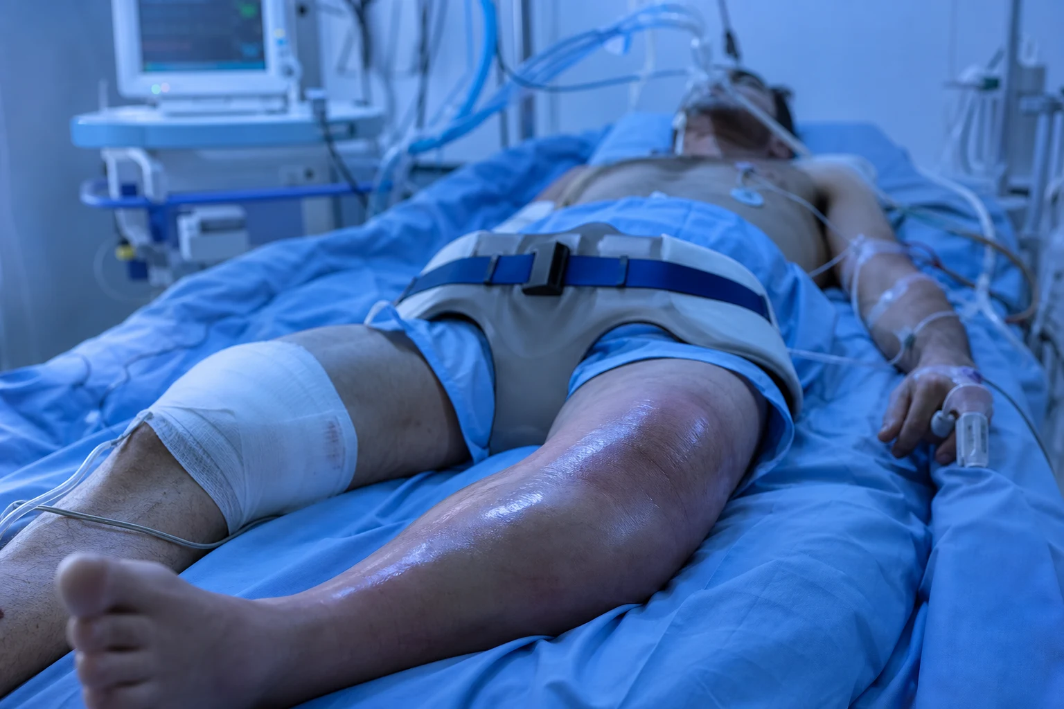
Fractures are disruptions in the continuity of bone. They range from undisplaced hairline cracks to comminuted, open injuries with significant soft tissue damage. Understanding common fracture patterns, their mechanisms, and standard management is fundamental to surgical practice. [1]
Gustilo-Anderson Classification (Open Fractures)
| Type | Description | Management | Infection Risk |
|---|---|---|---|
| I | Wound under 1 cm, minimal contamination | Debridement + primary closure + IV antibiotics 48h | Under 2% |
| II | Wound 1-10 cm, moderate soft tissue damage | Debridement + delayed closure + IV antibiotics 72h | 2-7% |
| IIIa | Wound over 10 cm, adequate soft tissue cover | Debridement + flap coverage + IV antibiotics 72h+ | 10-50% |
| IIIb | Requires flap coverage (extensive soft tissue loss) | Debridement + flap + IV antibiotics | 10-50% |
| IIIc | Vascular injury requiring repair | Debridiment + vascular repair + fasciotomies | 25-50% |
Upper Limb Fractures
Colles Fracture
- Mechanism: FOOSH (fall on outstretched hand) in elderly osteoporotic women
- Deformity: Dinner fork deformity — dorsal displacement, radial shortening
- X-ray: Extra-articular, transverse fracture of distal radius (within 2 cm of articular surface) with dorsal angulation
- Management: Closed reduction under haematoma block/Bier's block, below-elbow cast for 6 weeks; percutaneous pinning or volar locking plate if unstable or intra-articular
Smith's Fracture (Reverse Colles)
- Mechanism: Fall on dorsiflexed hand or direct blow to dorsum of wrist
- Deformity: Garden spade deformity — volar displacement
- Management: Closed reduction + cast; ORIF with volar plate if unstable
Scaphoid Fracture
- Mechanism: FOOSH in young adults
- Clinical: Anatomical snuffbox tenderness, scaphoid tubercle tenderness, pain on axial loading of thumb
- X-ray: May be NORMAL initially — repeat at 10-14 days if clinically suspected (bone resorption makes fracture visible) or MRI (gold standard for occult fracture)
- Management: Below-elbow cast with thumb spica for 6 weeks (waist fracture) to 3 months (proximal pole — avascular)
- Complication: Avascular necrosis (AVN) of proximal fragment (up to 100% for proximal pole fractures due to retrograde blood supply entering distally)
Supracondylar Fracture of Humerus (Children)
- Mechanism: FOOSH in children aged 5-8 years
- Classification: Gartland types I-IV (undisplaced, displaced with posterior cortex intact, fully displaced, multidirectionally unstable)
- X-ray: Posterior fat pad sign (always abnormal), anterior and posterior fat pads (sail sign)
- Management: Type I — above-elbow cast; Types II-IV — closed reduction and percutaneous K-wiring (CRIF)
- Critical complication: Volkmann's ischaemic contracture from brachial artery injury — check radial pulse; also median nerve injury
Clavicular Fracture
- Most common fracture in children
- Site: Middle third (80%), lateral third (15%), medial third (5%)
- Management: Broad arm sling/figure-of-8 bandage for 2-3 weeks (most heal non-operatively); ORIF for widely displaced, tented skin, or lateral third with coracoclavicular disruption
Lower Limb Fractures
Neck of Femur (NOF) Fracture
Tibial Plateau Fracture
- Mechanism: Axial loading (valgus/varus force) — bumper fracture, fall from height
- Classification: Schatzker I-VI
- Management: Undisplaced (under 2 mm step-off) — cast/knee brace; Displaced — ORIF with plate and screws; bone graft for depression
Ankle Fractures
- Weber classification (based on fibula fracture level relative to syndesmosis):
- Weber A: Below syndesmosis — stable, below-knee cast
- Weber B: At syndesmosis — may be unstable, ORIF if displaced
- Weber C: Above syndesmosis — unstable, ORIF + syndesmotic screw
Open Fracture Management
Key Exam Points
GUSTILO
SCAPHOID
WEBER
Fracture Healing and AO Principles
Fracture healing proceeds through three overlapping phases. The reactive phase (days 0 to 7) begins with haematoma formation at the fracture site, organisation into granulation tissue, and infiltration by inflammatory cells and macrophages. The reparative phase (weeks 1 to 12) generates a fibrocartilaginous soft callus, which progressively mineralises into hard (bony) callus by intramembranous and endochondral ossification. The remodelling phase (months to years) replaces woven bone with lamellar bone along lines of mechanical stress according to Wolff's law, restoring the original cortical architecture and medullary canal. [1]
Healing is classified as secondary (indirect) union — the natural callus-mediated pathway used by conservative treatment and bridging implants — versus primary (direct) healing, which occurs only with absolute stability (anatomical reduction and rigid compression, e.g. lag screws and compression plating) and proceeds by cutting cones of osteons crossing the fracture directly, without visible callus. The AO/OTA principles guide operative fixation: (1) anatomical reduction (articular surfaces absolute, diaphyseal shafts relative), (2) stable fixation matched to the biomechanical goal, (3) preservation of blood supply (biological plating, limited periosteal stripping), and (4) early active mobilisation and rehabilitation. Biological or bridge plating prioritises soft-tissue respect and callus formation over rigid anatomical restoration in comminuted shaft fractures. [1]
[1]Implants and Internal Fixation Devices
Implant selection follows fracture location, pattern, soft-tissue envelope, and patient factors. The dynamic hip screw (DHS) — a sliding lag screw in the femoral head engaged into a barrel-and-side-plate — allows controlled impaction across stable intertrochanteric and selected femoral neck fractures, converting axial load into compression at the fracture line. A 135 degree neck-shaft angle is standard; an additional anti-rotation screw may be added. The DHS is contraindicated in reverse-obliquity and subtrochanteric patterns, which tend to fail in varus. [1]
Intramedullary (IM) nails are load-sharing centromedullary devices ideal for long-bone diaphyseal fractures (femur, tibia, humerus) and unstable peritrochanteric fractures, where cephalomedullary nails (proximal femoral nail antirotation PFNA, gamma nail) carry a lag screw into the head and control rotation. Advantages over plating include a smaller incision, preserved periosteal blood supply, early weight-bearing, and biomechanical superiority as a lever arm close to the mechanical axis. Reamed nails accept a larger diameter implant and allow interlocking screws; unreamed nails reduce medullary trauma but risk nail failure. [1]
The volar locking plate (VLP) is the workhorse for unstable distal radius fractures (dorsally angulated Colles, volar Smith, intra-articular). Locking screws thread directly into the plate, creating a fixed-angle, angularly stable construct that buttresses the subchondral bone and permits early motion; the plate is applied to the strong volar distal radius (floor of the flexor carpi radialis sheath) to avoid flexor tendon irritation, with the distal edge proximal to the watershed line. Cannulated screws (6.5 to 7.3 mm partially threaded for the femoral neck, or 3.0 mm headless Herbert screws for the scaphoid) are inserted over a guide wire for femoral neck fixation (three parallel screws in an inverted triangle) and scaphoid fixation, providing compression with minimal soft-tissue dissection. Kirschner wires (K-wires) offer percutaneous, low-cost fixation (paediatric supracondylar humerus, unstable distal radius) but lack rigidity and may migrate. Locking compression plates (LCP) and limited-contact dynamic compression plates (LC-DCP) stabilise periarticular and comminuted shaft fractures. External fixators (spanning uniplanar/biplanar or ring Ilizarov) are used for severe open fractures, damage control in polytrauma, and temporisation until definitive soft-tissue cover. [1]
Management: Operative versus Conservative Indications
The choice between ORIF (open reduction and internal fixation) and conservative management balances fracture stability, articular involvement, neurovascular status, the soft-tissue envelope, and patient demands. Conservative treatment — closed reduction under haematoma block or sedation plus a cast or functional brace — suits stable, extra-articular, undisplaced fractures: undisplaced Colles, most midshaft clavicle fractures, Weber A ankle, and stable paediatric injuries. [1]
ORIF is indicated when the fracture is intra-articular with a step-off greater than 2 mm (tibial plateau, distal radius, pilon, Weber B/C ankle) to prevent post-traumatic osteoarthritis; when it is unstable or cannot be held reduced in plaster (comminuted Colles, displaced scaphoid, displaced neck of femur); when there is neurovascular compromise requiring exploration; when an open fracture needs debridement and stabilisation; for pathological fractures; after failed conservative treatment with secondary displacement; and in polytrauma requiring early definitive stabilisation (damage control orthopaedics) so the patient can be mobilised, nursed, and receive pulmonary toilet. [1]
Fracture-specific surgical defaults: displaced Garden III/IV femoral neck in the elderly is treated with hemiarthroplasty (cemented, bipolar) or, in the active and cognitively intact patient, total hip replacement, which lowers reoperation rates and improves function compared with internal fixation per NICE guidance[2]. Intertrochanteric fractures receive a DHS or, if unstable, a cephalomedullary nail. A displaced scaphoid is fixed with a percutaneous Herbert screw. Gartland II to IV paediatric supracondylar humerus fractures undergo closed reduction and percutaneous K-wiring. Displaced tibial plateau (Schatzker IV to VI) is managed with a locking plate plus bone graft or substitute for depressed articular segments. Weber B/C ankle injuries need ORIF of the fibula with a one-third tubular plate and a syndesmotic screw. A widely displaced or skin-tenting clavicle is plated or fixed with an intramedullary device.
Paediatric Fractures
Children's bones differ biomechanically and biologically: a thicker and more cellular periosteum, greater plasticity, open physes, and rapid remodelling capacity produce fracture patterns absent in adults. The greenstick fracture is an incomplete fracture in which the cortex fails in tension on one side while the opposite cortex bends but stays intact; the periosteal hinge preserves some alignment and reduction usually requires rotating the limb to a stable position or gently "completing" the break. [1]
The torus (buckle) fracture is a compression failure of the metaphyseal cortex — classic at the distal radius — producing a subtle cortical bulge without a frank cortical break; it is inherently stable and treated with a short period of splintage, increasingly a removable wrist splint for 3 to 4 weeks, with excellent outcome and minimal follow-up. Plastic (bowing) deformation is sustained microfailure along the concavity of a long bone (typically the ulna or fibula paired with a fracture of the other bone) with no discrete fracture line; it is corrected by gradual manual straightening under anaesthesia. Complete (displaced) paediatric fractures heal rapidly but demand accurate control of rotation and angulation, because remodelling preferentially corrects deformity in the plane of joint motion and is greatest near the physes in younger children. Critically, rotational deformity and angulation across the plane of motion do NOT reliably remodel and must be reduced anatomically at the index procedure. [1]
Torus (buckle)
- Compression injury — metaphyseal cortex buckles but is intact; inherently stable.
- Treatment: removable splint or short-arm cast for 3 to 4 weeks; near-zero displacement risk.
- Return to normal function by 6 weeks; no routine follow-up imaging required in many units.
Greenstick
- Tension-side cortex breaks; opposite cortex intact; periosteal hinge maintained.
- Reduce by completing or rotating to a stable position; above-elbow cast for 4 to 6 weeks.
- Re-angulation is common — review at 1 week with X-ray to detect loss of reduction.
Salter-Harris Physeal Classification
The Salter-Harris classification (1963) describes epiphyseal (physeal) injuries in children and predicts the risk of growth disturbance. Higher grades carry a greater risk of premature physeal closure, limb-length discrepancy, and angular deformity, because the resting cartilage cells of the growth plate are increasingly injured. [1]
| Type | Pattern | Prognosis |
|---|---|---|
| I | Separation through the physis (growth plate) | Excellent — usually reduces and heals without growth arrest |
| II | Through physis with a metaphyseal fragment (Thurston-Holland sign) | Good — the most common type; low risk of growth disturbance |
| III | Through physis and epiphysis into the joint (intra-articular) | Fair — needs anatomical articular reduction; moderate growth-arrest risk |
| IV | Through epiphysis, physis, and metaphysis (intra-articular) | Poor — highest growth-arrest risk; ORIF for anatomical reduction |
| V | Compression or crush injury of the physis | Poor — late, asymmetric growth arrest; often diagnosed in retrospect |
| VI | Partial physeal bridging / peripheral injury (Peterson) | Variable — bony bar formation and progressive angular deformity |
Type IV and V injuries demand long-term surveillance for asymmetric growth. A bony physeal bar can be resected, with fat or silicone interposition, to restore growth in children with more than two years of growth remaining; established deformity may need a corrective osteotomy or epiphysiodesis of the contralateral side to equalise length. [1]
Salter-Harris prognosis rises with the number
SALTER
Through the physis only — best prognosis.
Physis plus a metaphyseal fragment — most common, good outcome.
Through physis into the epiphysis — intra-articular, needs reduction.
Epiphysis, physis, and metaphysis — highest arrest risk, ORIF.
Compression of the plate — delayed growth arrest.
Complications of Fractures
Complications are grouped by timing. Immediate and early: haemorrhagic shock (pelvic and femoral fractures can each lose more than 1500 mL), fat embolism syndrome at 24 to 72 hours (the triad of hypoxaemia, confusion, and a petechial rash over chest, axillae, and conjunctivae), compartment syndrome (pain out of proportion, pain on passive stretch, a tense compartment, and late pulselessness), neurovascular injury, and acute carpal tunnel syndrome. The brachial artery is injured in roughly 10 to 15 percent of displaced paediatric supracondylar humerus fractures. [1]
Late: avascular necrosis (AVN) is the death of bone from interrupted blood supply — the scaphoid proximal pole (retrograde vascular supply entering distally, with up to 100 percent necrosis for proximal-pole fractures), the femoral head (around 40 percent of displaced intracapsular neck fractures, where reduction timing is critical), the lunate (Kienbock disease), and the talus. Volkmann's ischaemic contracture is the end-stage of an untreated forearm compartment syndrome or brachial artery injury: fibrosis and contracture of the flexor compartment produce a classic claw-like flexion deformity of the wrist and fingers — prevention by pulse monitoring and prompt fasciotomy is the only effective treatment. Complex regional pain syndrome type I (CRPS, formerly Sudeck's atrophy) follows injury, classically a distal radius fracture, presenting with disproportionate burning pain, allodynia, swelling, trophic skin and hair-change, stiffness, and patchy osteopenia; management is early mobilisation, desensitisation, graded physiotherapy, and analgesia, with most patients recovering substantial function within the first year[4].
Non-union (loss of healing potential) is classified as atrophic — poor biology and blood supply, needing bone graft with or without revision fixation — versus hypertrophic — adequate biology but inadequate stability, requiring more rigid fixation. Malunion is healing in a non-anatomical position producing deformity (a dorsally angulated Colles giving the dinner-fork deformity, or a shortened malrotated tibia) and may require corrective osteotomy. Delayed union sits between delayed healing and non-union. Other late problems include post-traumatic osteoarthritis after an articular step-off, osteomyelitis complicating open fractures or ORIF, and symptomatic or prominent implants requiring removal. [1]
Exam application bank (NEET-PG / INICET)
One-line answer
Common fractures covers the most frequently encountered fractures in clinical practice: Colles, Smith, scaphoid, neck of femur, intertrochanteric, tibial plateau, ankle, clavicular, humeral neck, and supracondylar fractures. Each has characteristic mechanisms, deformities, and management principles following AO/OTA classification and Garden classification for femoral neck fractures. Management follows the AO principles: anatomical reduction, stable fixation, preservation of blood supply, early mobilisation. Open fractures require emergency debridement (within 24h), IV antibiotics, tetanus prophylaxis, and stabilisation per Gustilo-Anderson classification.
Worked stems (answer without another resource)
Stem 1 — Classic presentation. Map symptoms to mechanism; name the first investigation and first treatment step with dose/route if drug therapy is standard. [1]
Stem 2 — Unstable / complicated. List red flags that force immediate resuscitation, theatre, ICU, antidote, or reperfusion — and what you do in the first 15 minutes. [1]
Stem 3 — Atypical group. Elderly, pregnancy, child, or immunocompromised: how presentation and thresholds change. [1]
Stem 4 — Differential trap. Name the three closest mimics and one discriminator for each. [1]
Stem 5 — Disposition. Who goes home with safety-netting, who is admitted, who needs HDU/ICU/theatre, and what follow-up is mandatory. [1]
Rapid viva checklist
- Definition + classification
- Pathophysiology chain
- Bedside signs / criteria
- Score with exact components (if any)
- Emergency bundle
- Definitive therapy with doses
- Complications of disease and of treatment
- Special populations
- Guideline/trial name if classic
- Three exam traps
Coverage self-check
If you cannot answer any stem above from this page alone, re-read the matching section — the page is intended to be self-sufficient for final-prof and NEET-PG/INICET questions on Common Fractures.
[1]Differential Diagnosis
A suspected fracture must be distinguished from conditions that mimic focal pain, deformity, or swelling after trauma, and from non-traumatic causes of acute limb pain. [1]
Traumatic mimics
- Severe ligament sprain (ankle inversion without fracture) — bony tenderness and Ottawa rules guide imaging.
- Joint dislocation (glenohumeral, patellar) — frequently coexists with fracture as a fracture-dislocation.
- Tendon or muscle rupture — Achilles, quadriceps, or biceps tendon rupture with a palpable gap and weak function.
- Soft-tissue contusion or haematoma — pain and swelling without a cortical break.
Non-traumatic and pathological
- Septic arthritis or osteomyelitis — fever, a hot swollen joint, and raised inflammatory markers.
- Pathological fracture through a tumour or bone cyst — simple bone cyst, giant cell tumour, metastasis, or myeloma.
- Stress or insufficiency fracture — repetitive load or osteoporotic bone with a normal early X-ray; confirm on MRI.
- Gout or pseudogout — acute monoarthritis that can mimic a periarticular fracture.
- Fatigue (stress) fracture — repetitive load (metatarsal, tibial shaft); normal early X-ray, diagnose on MRI.
- Pathological fracture — minimal-trauma fracture through abnormal bone; investigate with history, bloods, and imaging for malignancy or metabolic bone disease.
- Septic arthritis versus periarticular fracture — a hot, painful joint with fever can hide an occult fracture; aspirate if in doubt.
- Limping child (SUFE, Perthes, Osgood-Schlatter) — paediatric hip or knee pain may masquerade as trivial injury; always examine the hip in a child with knee pain. [1]
Drug Dosing: Antibiotics, Analgesia and Thromboprophylaxis
Standard drug therapy spans antimicrobial prophylaxis, analgesia, venous thromboembolism (VTE) prophylaxis, and tetanus cover. Open-fracture antibiotics are given within 1 hour of arrival and continued for 72 hours or until 24 hours after soft-tissue cover: Gustilo I and II — flucloxacillin 1000 mg IV six-hourly (cefuroxime 750 mg IV eight-hourly if penicillin-allergic without anaphylaxis); Gustilo III — add gentamicin 5 mg/kg IV once daily, dose-adjusted to renal function, and add metronidazole 500 mg IV eight-hourly for farmyard or soil contamination; give tetanus immunoglobulin 250 units IM to the unimmunised[1].
Perioperative prophylaxis for ORIF such as a hip hemiarthroplasty — cefuroxime 1.5 g IV at induction, repeated at 4 hours; vancomycin 15 mg/kg IV if there is MRSA risk or beta-lactam allergy. Analgesia: paracetamol 1000 mg six-hourly (maximum 4 g per day; 15 mg/kg per dose in children), stepped up to an oral opioid (oxycodone 5 mg four-hourly) or IV morphine 0.1 mg/kg for severe pain; an NSAID such as ibuprofen 400 mg eight-hourly for inflammatory pain, though some protocols advise avoiding NSAIDs in the first 48 hours of acute fracture healing because of a theoretical anti-proliferative effect on callus. Regional nerve blocks (fascia iliaca block for neck of femur, brachial plexus block for the upper limb) reduce opioid demand and aid early physiotherapy. [1]
VTE prophylaxis is essential because hip and lower-limb fractures carry a high risk of deep vein thrombosis and pulmonary embolism. NICE recommends low-molecular-weight heparin such as enoxaparin 40 mg subcutaneous once daily (20 mg if body weight is below 50 kg or eGFR is below 30), or fondaparinux 2.5 mg once daily, continuing for 28 to 35 days after hip-fracture surgery, with mechanical prophylaxis (anti-embolism stockings or intermittent pneumatic compression) added whenever possible. Stop LMWH 12 hours before neuraxial anaesthesia and for 8 hours afterwards. [1]
Follow-up and Rehabilitation
Follow-up tracks reduction maintenance, union, and complications. A typical protocol reviews the patient clinically and radiographically at 2 weeks (wound check, fracture position within cast), 6 weeks (union assessment, cast removal for stable fractures, and the start of mobilisation), 3 months, 6 months, and 12 months for high-risk injuries (neck of femur, scaphoid, tibial plateau). Children are reviewed at 1 week post-reduction to detect re-angulation, which is common in greenstick and supracondylar fractures. Red flags at follow-up include loss of reduction, persistent pain, loosening or breakage of an implant, and signs of infection or CRPS. [1]
Rehabilitation follows a stabilise, move, strengthen sequence: immobilisation in a correct reduction; early active motion of uninvolved joints and of the injured joint once it is stable, which prevents stiffness and CRPS; progressive weight-bearing (non-weight-bearing to partial to full as union advances on imaging); and then strengthening, proprioception, and functional or sport-specific training. Typical neck of femur milestones: mobilise bed-to-chair on day 0 to 2; partial weight-bearing at 6 weeks for an uncemented implant or weight-bearing as tolerated for a cemented stem; independent mobility by 3 months. Return to driving is usually cleared at 6 to 8 weeks for an upper-limb fracture, once motor control and a pain-free range of movement return. Multidisciplinary input (physiotherapist, occupational therapist, orthogeriatrics for fragility-fracture patients) plus a bone-health assessment — DEXA scan, calcium and vitamin D optimisation, and a bisphosphonate where osteoporosis is confirmed — completes secondary-fracture prevention. [1]
The AO fixation checklist
AORTA
Absolute for articular surfaces, relative for diaphyseal shafts.
Respect the soft tissues and blood supply — biological plating.
Absolute (compression) or relative (bridging) per the fracture.
Move uninvolved and stable joints early to prevent stiffness and CRPS.
Tailor implant, weight-bearing, and bone-health care to the patient.
Paediatric Fracture Management in Depth
Children's long bones fail in patterns no adult bone produces, and the open physis dictates both treatment and prognosis. Management decisions turn on three questions: Is the physis involved? Is the deformity in a plane that will remodel? Is the injury pattern consistent with the stated mechanism, or is non-accidental injury in play? [1]
Salter-Harris (with Rang type VI): pattern and management
The classic Salter-Harris types I to V are extended by Rang's type VI — a peripheral physeal injury (avulsion or periosteal/cartilaginous bridging at the margin of the plate) that produces a focal bony bar and progressive angular deformity as the uninjured remainder of the physis continues to grow. [1]
- Type I — physeal separation: a clean transphyseal split with an intact periosteal hinge. Usually undisplaced and stable. Management: closed reduction and a well-moulded cast for 3 to 4 weeks; percutaneous pinning only if unstable (e.g. proximal femoral capital epiphysis — slipped capital femoral epiphysis is a Salter I variant). Excellent prognosis; growth disturbance is rare.
- Type II — above the metaphysis: a physeal injury with a metaphyseal Thurston-Holland fragment on the compression side. The most common Salter-Harris injury (about 75 percent, classic at the distal radius). Management: closed reduction and cast; percutaneous K-wires only if reduction cannot be held. Good prognosis because the epiphyseal circulation and the resting cartilage cells on the tension side are preserved.
- Type III — into the epiphysis: an intra-articular fracture running through the physis and into the epiphysis, splitting the joint surface (classic at the distal tibia, Tillaux fragment). Management: anatomical reduction is mandatory to restore articular congruity — open reduction and internal fixation with small-fragment cannulated or headless screws if displacement exceeds 2 mm, avoiding crossing the physis where possible. Moderate risk of partial growth arrest.
- Type IV — through everything: the fracture line crosses epiphysis, physis, and metaphysis (classic at the lateral distal tibia, the juvenile Tillaux variant). Highest risk of premature physeal arrest because the healing callus can bridge the plate. Management: ORIF with anatomical joint-surface and physeal restoration, usually via small-fragment screws placed parallel to the physis in the epiphyseal and metaphyseal fragments; long-term growth surveillance is mandatory.
- Type V — crush/ram: an axial compression injury that appears normal on the initial X-ray but destroys the resting cartilage cells of the plate. Classic at the distal tibia and proximal radius. Management: non-operative — protected weight-bearing and cast — but counsel the family about delayed asymmetric growth arrest, which declares itself months later and may need epiphysiodesis of the contralateral limb or a corrective osteotomy.
- Type VI (Rang) — peripheral physeal bridge: a marginal periosteal or chondral injury that heals as a bony bar tethering one edge of the plate. Management: serial imaging for progressive angular deformity; definitive treatment is physeal bar resection with fat or silicone interposition if more than two years of growth remain, or contralateral epiphysiodesis to equalise length. [1]
Greenstick, torus and plastic deformation — distinctions
| Pattern | Mechanism | Stability | Management |
|---|---|---|---|
| Torus (buckle) | Compression of the metaphyseal cortex | Inherently stable | Removable wrist splint for 3 to 4 weeks; minimal follow-up |
| Greenstick | Tension-side cortex breaks, compression side bends | Stable but may re-angulate | Reduce by completing or rotating to a stable position; above-elbow cast 4 to 6 weeks; X-ray at 1 week |
| Plastic (bowing) | Sustained microfailure along the concavity of a long bone | Difficult to correct | Gradual manual straightening under anaesthesia; often paired with reduction of an ipsilateral complete fracture |
Remodelling potential by age
Remodelling is governed by four rules the examiner expects you to state: (1) it is greatest in the plane of joint motion — apex-volar angulation of a distal-radius fracture remodels well, whereas angulation perpendicular to that plane does not; (2) it is greater the closer the fracture is to a physis (metaphyseal injuries remodel more than diaphyseal); (3) it is greater in younger children (a child under 10 remodels dramatically; a teenager near skeletal maturity remodels little); and (4) rotational deformity and translation never reliably remodel and must be corrected at the index procedure. As a practical guide, up to about 20 degrees of angulation in the plane of motion in a distal-radius fracture in a child under 10 will remodel completely within 6 to 12 months; the same deformity in a 14-year-old will not. [1]
Non-accidental injury (NAI) patterns
Any paediatric fracture warrants an NAI screen when the pattern is inconsistent with the history, the child is non-mobile (under walking age), or there is delay in presentation. Red-flag fracture patterns include: classic metaphyseal lesions (corner or bucket-handle fractures) at the distal femur, proximal or distal tibia — a shearing injury to the immature metaphysis that is highly specific for NAI; posterior rib fractures near the costotransverse process from squeezing; scapular, spinous-process, and sternum fractures, which require high energy and are rarely accidental; femoral fractures in a non-ambulant infant; multiple fractures at different stages of healing; and skull fractures that are complex, depressed, or growing. Suspected NAI mandates a skeletal survey (not a single X-ray), referral to the paediatric safeguarding team, and documented clearly in the notes before any disclosure or confrontation. [1]
Compartment Syndrome
Compartment syndrome is a limb-threatening rise in interstitial pressure within a fascial osteofibrous compartment that drops perfusion below the threshold for tissue survival. It is most common after tibial shaft fractures, paediatric supracondylar humerus fractures, distal-radius and forearm fractures, and tight circumferential casts, and after reperfusion of an ischaemic limb. The window for irreversible damage is 6 to 8 hours. [1]
The six Ps are the traditional framework but are misleading if applied as a checklist, because the later signs indicate irreversible injury: Pain (out of proportion, and worse on passive stretch — the earliest and most reliable sign), Pressure (a tense, woody compartment on palpation), Paraesthesia (early ischaemia of fast-conducting sensory nerves), Paralysis (late motor nerve failure), Pallor, and Pulselessness. Critically, a pulse is usually still present in established compartment syndrome, because arterial systolic pressure far exceeds compartment pressure until very late; waiting for pulse loss is the classic catastrophic error. [1]
Diagnosis is clinical in a cooperative, awake patient. Pressure measurement (a hand-held Stryker intracompartmental pressure monitor) is reserved for the obtunded, sedated, or paediatric patient who cannot communicate pain, or when the clinical picture is equivocal. Two thresholds are accepted: an absolute compartment pressure over 30 mmHg, or a delta pressure (diastolic blood pressure minus compartment pressure) below 30 mmHg. The delta-pressure criterion is preferred because it adjusts for the patient's perfusion pressure and is more sensitive. [1]
Management is emergency fasciotomy: remove any constricting cast or dressing, prepare the limb, and decompress all compartments in the affected segment. For the leg, two-incision (anterolateral and posteromedial) fasciotomy decompresses the four compartments (anterior, lateral, superficial and deep posterior); for the forearm, the Henry (volar) extensile and dorsal incisions open the mobile wad and extensor compartments, with carpal tunnel release if median-nerve signs are present. Fasciotomy wounds are left open under vacuum-assisted closure and revised at 48 to 72 hours; delayed primary closure or skin grafting follows once swelling subsides. A delayed or incomplete fasciotomy leads to Volkmann's ischaemic contracture — the fibrotic, clawed flexion deformity described above. [1]
Fat Embolism Syndrome
Fat embolism syndrome (FES) follows long-bone and pelvic fractures, particularly closed femoral shaft fractures, typically within the 24 to 72 hour window after injury (a 72-hour window is the standard exam teaching). It arises from both mechanical embolisation of marrow fat into the pulmonary and cerebral circulation and a biochemical cascade in which free fatty acids injure the pulmonary endothelium. [1]
The classic triad is: (1) respiratory distress — hypoxaemia, dyspnoea, and diffuse alveolar infiltrates progressing to ARDS; (2) neurological signs — confusion, agitation, or depressed consciousness ranging to coma, often out of proportion to the hypoxaemia; and (3) a petechial rash distributed over the chest, axillae, conjunctivae, and palate, present in 50 to 60 percent of cases but virtually pathognomonic when seen. Tachycardia and pyrexia are common supporting features. Early proteinuria and a falling platelet count or haemoglobin support the diagnosis. [1]
Diagnosis is clinical (Gurd's criteria); there is no confirmatory test, though fat globules in the urine and bronchoalveolar lavage are supportive. Management is supportive — high-flow oxygen, lung-protective mechanical ventilation for respiratory failure, and haemodynamic support — because no specific therapy reverses the syndrome. Prevention is the best treatment: early splintage and stabilisation of long-bone fractures (within 24 hours, ideally with intramedullary nailing after resuscitation) reduces the incidence and severity of FES, the rationale for damage-control and early total care in polytrauma. Mortality is 5 to 15 percent. [1]
Complex Regional Pain Syndrome: Type I versus Type II
Complex regional pain syndrome (CRPS) is a disproportionate, regional pain disorder arising after a noxious event. The Budapest clinical criteria require continuing pain that is disproportionate to the inciting event, plus symptoms in at least three of four categories (sensory, vasomotor, sudomotor or oedema, and motor or trophic) and signs in at least two categories, with no alternative diagnosis. It most often follows a distal-radius fracture, with immobilisation in a cast and psychological stress as risk factors. [1]
The two types are distinguished by the presence of a named-nerve injury: CRPS type I (formerly reflex sympathetic dystrophy, Sudeck's atrophy) — no identifiable nerve injury, by far the more common after a fracture or sprain; and CRPS type II (formerly causalgia) — a confirmed nerve injury is present, for example after a median-nerve laceration or a gunshot wound to a major nerve trunk. The clinical presentation — burning allodynia, swelling, colour and temperature change, sweating changes, trophic skin and nail changes, hair-growth abnormalities, stiffness, and patchy osteopenia — is identical in both types; only the trigger differs. [1]
Management is multidisciplinary and early: graded desensitisation and active mobilisation physiotherapy is the cornerstone; analgesia (paracetamol, NSAIDs, neuropathic agents such as gabapentin or pregabalin, and short courses of oral corticosteroid within the first 3 months); vocational and psychological support; and, for refractory cases, sympathetic blocks or spinal cord stimulation. Most patients recover substantial function within 12 months, but a minority develop chronic, disabling pain and stiffness, making early recognition and prevention — by minimising cast time and encouraging early active motion — the priority[4].
References
- [1]Gustilo RB, Anderson JT. Prevention of infection in the treatment of one thousand and twenty-five open fractures of long bones: retrospective and prospective analyses J Bone Joint Surg Am, 1976.PMID 773941
- [2]Parker MJ, Handoll HHG. Replacement arthroplasty versus internal fixation for extracapsular hip fractures in adults Cochrane Database Syst Rev, 2006.PMID 16625528
- [3]Dias JJ, Dhukaram V, Abhinav A, Bhowal B. Clinical and radiological outcome of cast immobilisation versus surgical treatment of acute scaphoid fractures at a mean follow-up of 93 months J Bone Joint Surg Br, 2008.PMID 18591600
- [4]Bean DJ, Johnson MH, Heiss-Dunlop W, Kydd RR. Extent of recovery in the first 12 months of complex regional pain syndrome type-1: A prospective study Eur J Pain, 2016.PMID 26524108