General Surgery · General Surgery
Diverticular Disease
Also known as Diverticulosis · Diverticulitis · Complicated diverticular disease · Hinchey classification
Diverticular disease encompasses a spectrum from asymptomatic diverticulosis (70 to 80% of cases) through symptomatic uncomplicated diverticular disease (SUDD) to acute diverticulitis and its life-threatening complications. False (pseudodiverticula) = mucosa and submucosa herniate through the muscularis propria at sites of vasa recta penetration. The sigmoid colon is affected in 90% of Western populations (right-sided in 15 to 75% of Asians). Over 50% of people older than 60 are affected. Hinchey classification (stages I to IV) and the WSES 2015 severity grade guide surgical management of perforated diverticulitis. Management ranges from dietary modification to emergency Hartmann's procedure for faecal peritonitis.
On this page & tools
Your progress
Saved locally on this device.
Exam tags
Red flags
Overview & Definition
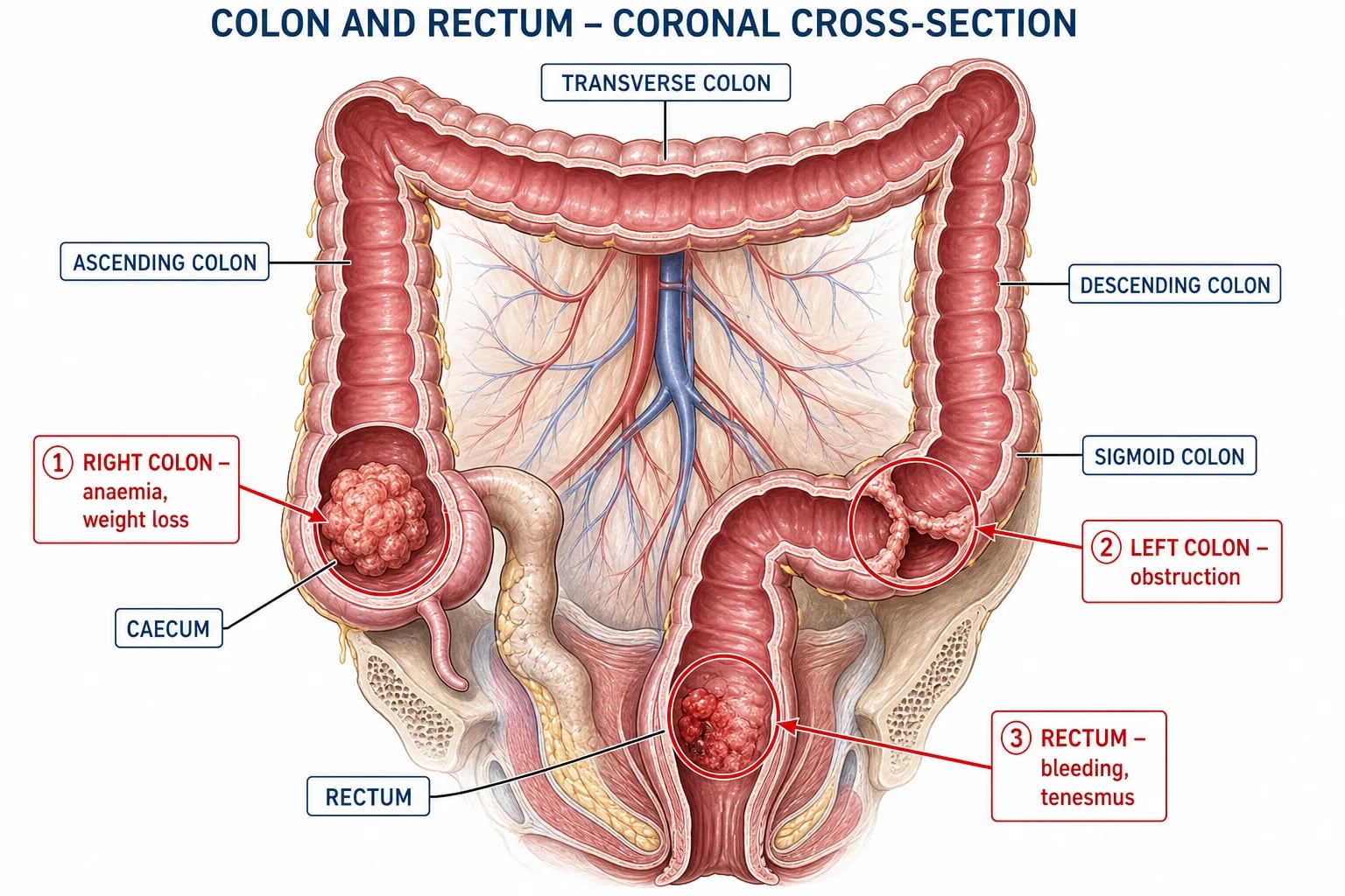
A diverticulum (plural: diverticula) is a localised outpouching of the wall of a hollow viscus. The distinction between a true and a false diverticulum is foundational and examiners test it relentlessly. A true diverticulum involves all layers of the bowel wall (mucosa, submucosa, muscularis propria, serosa) and is usually congenital — the paradigm is Meckel's diverticulum of the ileum. A false (pseudo) diverticulum involves only the mucosa and submucosa herniating through a defect in the muscularis propria; colonic diverticula in Western populations are almost always of this acquired pulsion type.[1]
It is essential to separate three terms that students conflate. Diverticulosis is the mere anatomical presence of diverticula, almost always asymptomatic. Diverticulitis is inflammation of one or more diverticula, usually from microperforation. Diverticular disease is the umbrella clinical term covering the whole spectrum, including SUDD and complicated disease. A patient with incidental diverticula found at colonoscopy has diverticulosis, not diverticular disease, and needs only reassurance.[1][2]
The clinical importance of diverticular disease lies in its sheer prevalence and the bimodal severity of its presentation. Most affected individuals never know they have it, but the minority who develop perforation face a mortality of up to 30%, and a small number bleed massively enough to need emergency angiography or colectomy. The disease is therefore best understood as a spectrum in which the challenge is to identify, at each end of that spectrum, the patient who needs urgent intervention rather than reassurance.[3]
Colonic Anatomy & the Mechanism of Diverticular Formation
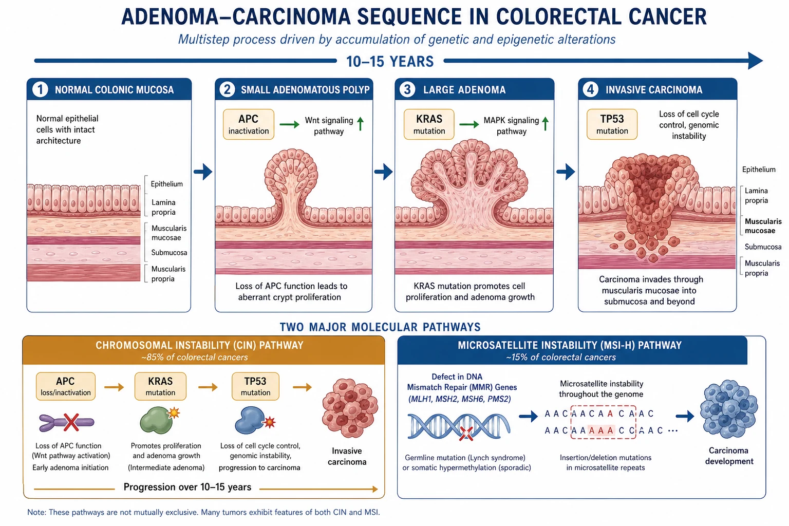
Understanding why diverticula form — and why they form in the sigmoid — requires a working knowledge of colonic wall anatomy. The colonic wall has four layers: mucosa, submucosa, muscularis propria (an inner circular and outer longitudinal layer), and serosa. The outer longitudinal muscle is not continuous but gathered into three flat bands called the taeniae coli. Between the taeniae, the circular muscle is exposed and, crucially, it is traversed by the vasa recta — the straight end-arteries that supply the colon wall. These penetrating vessels create points of inherent mechanical weakness, and it is precisely at these points that diverticula herniate.[1]
Anatomy of diverticular formation
The formation of a diverticulum is a pulsion event driven by raised intraluminal pressure. The dominant mechanistic model combines Laplace's law with the concept of segmentation. Laplace's law states that the pressure required to distend a tube is inversely proportional to its radius (T = P × r); consequently, the narrowest segment of colon sustains the greatest wall tension. The sigmoid colon is the narrowest colonic segment, which is why it bears the brunt of the disease. Layered on this is segmentation: when the colon contracts, it does so in discrete rings that compartmentalise the lumen into closed chambers. Each closed chamber behaves as a small-bore tube to which Laplace's law applies, generating pressures that can exceed 90 mmHg locally — far above the resting intraluminal pressure. Over years, these repeated pressure spikes extrude mucosa through the vasa-recta weak points.[1][3]
The five-step path from fibre-depleted stool to a diverticulum
Low dietary fibre reduces stool bulk and accelerates transit segmentation, increasing the number and force of colonic pressure waves (segmental contractions).
Each segmental contraction compartmentalises a short length of colon; by Laplace's law, the narrow sigmoid generates the highest wall tension in the organ.
Raised intraluminal pressure drives mucosa and submucosa outward through the inherent weak points where the vasa recta penetrate the circular muscle between the taeniae.
Once a pseudodiverticulum forms, an inspissated faecolith may lodge in its narrow neck, obstructing drainage and eroding the thin wall.
Erosion produces a microperforation: initially contained by pericolic fat and mesentery as a pericolic abscess, but potentially free into the peritoneal cavity as purulent or faecal peritonitis.
Three additional structural changes accompany and promote the disease. First, with age the colonic wall undergoes elastosis — increased deposition of elastin in the taeniae and circular muscle — which shortens and thickens the bowel (producing the characteristic palpable, cord-like sigmoid at laparotomy) and reduces wall compliance, so the same contractile force produces a higher pressure. Second, there is thickening of the circular and longitudinal muscle (myochosis) even in the absence of overt inflammation. Third, the diverticular wall, lacking a muscular layer, is structurally fragile: a faecolith impacted in its neck can erode through it, and a vasa recta draped over the fundus of the diverticulum can be eroded to produce the massive arterial-type diverticular bleed.[1]
This anatomy also explains the two anatomically distinct complications — inflammation versus bleeding. Diverticulitis arises from microperforation of the fundus; diverticular haemorrhage arises from erosion of the vasa recta at the neck/fundus into the diverticular lumen. The two are mechanistically separate: most patients who bleed have never had diverticulitis, and most patients with recurrent diverticulitis never bleed. Examiners deliberately test this distinction.[1][3]
Epidemiology & Risk Factors
Diverticular disease is a disease of affluent, low-fibre societies and of age. It was uncommon before the twentieth century and its rise tracks the industrial refinement of carbohydrate and the loss of dietary fibre in the West. Age is the strongest risk factor: the disease is rare before age 40 (under 5%), affects roughly 50% of people older than 60, and 65% of those older than 80. The prevalence is essentially equal in men and women, although women present slightly later.[1][2]
Headline epidemiology
Fibre is the central environmental factor. Low intake reduces stool bulk, increases segmentation, raises intraluminal pressure, and shortens the period over which the pulsion mechanism operates. The classical fibre hypothesis is supported by the near-absence of the disease in rural African and Asian populations on high-fibre diets and its appearance in those populations within a generation of migration to a Western diet. The protective effect of fibre is strongest for fruit, vegetable, and cereal fibre (particularly cellulose).[1]
Modifiable
lifestyle and drugs
- **Low dietary fibre** — the dominant modifiable risk factor
- **Obesity** — raises risk and severity, especially in the young
- **Physical inactivity** — reduces colonic motility and transit
- **Smoking** — increases risk of complicated (perforated) disease
- **NSAIDs and aspirin** — double the risk of diverticular bleeding and perforation
- **Opioids** — precipitate constipation and pseudo-obstruction
Non-modifiable
age, genetics, sex
- **Age** — strongest risk factor; wall elastosis and myochosis accumulate
- **Genetics** — twin studies show ~40 to 50% heritability; right-sided Asian pattern is familial
- **Connective tissue disease** — Ehlers-Danlos, Marfan — early, right-sided diverticula
- **Anatomy** — short sigmoid, narrow lumen concentrate pressure
- **Sex** — roughly equal; women present slightly later
Disputed / debunked
older teaching now revised
- **Nuts, seeds, popcorn** — NO increased risk; the historical avoidance advice is obsolete
- **Young age under 50** alone is NOT an indication for more aggressive surgery
- **Red meat** — modest association, less powerful than low fibre
- **Alcohol and caffeine** — no consistent causal link
The clinical course follows a predictable stepped progression that examiners expect you to articulate: of 100 patients with diverticulosis, 75 to 80 remain asymptomatic for life; 15 to 25 develop SUDD or a single episode of uncomplicated diverticulitis; of those who develop diverticulitis, roughly 20% develop a complication (abscess, fistula, perforation, stricture); and only 3 to 5% ever suffer a major diverticular bleed. Recurrence after a first episode of diverticulitis is around 20 to 30%, and recurrent episodes are increasingly likely to be complicated.[1][6]
Pathophysiology
The pathogenesis of diverticular disease is distinct from the pathogenesis of diverticular formation. The diverticula themselves arise from the pulsion mechanism above; the clinical disease arises when one or more diverticula become obstructed by a faecolith and undergo microperforation. The obstructed diverticulum distends, its thin wall is rendered ischaemic by the distension and by the chemical insult of retained stool, and the wall then gives way. The resulting perforation is initially microscopic and is contained by pericolic fat, mesocolon, and the omentum as a pericolic abscess or phlegmon — this is the substrate of uncomplicated to moderately complicated diverticulitis. If containment fails, the perforation extends to produce a distant abscess (Hinchey II), or it breaks free into the peritoneal cavity as purulent peritonitis (Hinchey III) or, when the faecolith and stool are extruded, faecal peritonitis (Hinchey IV).[1][3]
The inflammatory cascade: faecolith to peritonitis
A faecolith or food particle impacts in the narrow neck of a diverticulum, obstructing drainage and distending the fundus.
Distension, retained bacterial toxins, and chemical injury render the thin diverticular wall ischaemic; the vasa recta supply is compromised.
The wall undergoes microperforation; the leak is initially contained by pericolic fat and mesentery as a pericolic abscess/phlegmon (Hinchey I).
If containment is overwhelmed, pus tracks to a pelvic or distant intra-abdominal abscess (Hinchey II) or, with persistent leakage, produces generalised purulent peritonitis (Hinchey III).
Frank extrusion of faeces and a faecolith into the peritoneal cavity produces generalised faecal peritonitis (Hinchey IV) — the highest-mortality presentation.
Bacteriology. The colon harbours a dense mixed flora; the organisms cultured from a diverticular abscess or peritoneal washings reflect this. The dominant aerobes are Escherichia coli, Klebsiella, Proteus, and Enterococcus; the dominant anaerobes are Bacteroides fragilis, Clostridium, Peptostreptococcus, and Fusobacterium. Anaerobes outnumber aerobes by 1000 to 1, which is why empirical therapy must cover both Gram-negative aerobes and anaerobes, and why a single agent such as amoxicillin alone is inadequate. Antibiotic choice and duration are guided by severity and by local resistance; in regions with high rates of extended-spectrum beta-lactamase (ESBL) production, empirical piperacillin-tazobactam or a carbapenem may be required for severe disease.[4]
The microbiome and SUDD. Contemporary research implicates a low-grade chronic inflammation and an altered gut microbiota in the generation of SUDD (symptomatic uncomplicated diverticular disease) — the patient with recurrent left lower quadrant pain, bloating, and altered bowel habit but no acute inflammation on imaging. Biopsies show microscopic lymphocytosis and mucosal neutrophilia, and there is evidence of visceral hypersensitivity resembling irritable bowel syndrome. This is the rationale for the (still debated) use of mesalazine, rifaximin, and probiotics in SUDD, and it explains why SUDD and IBS are easily confused clinically.[1][6]
Classification
Three complementary classification systems are used: the clinical spectrum, the Hinchey classification (for perforated diverticulitis, 1978), and the WSES 2015 severity grade (Sartelli et al, 2016), which is CT-based and has largely superseded the older Ambrosetti grading in emergency practice.[4]
The clinical spectrum
| Stage | Description | Imaging | Clinical features |
|---|---|---|---|
| Diverticulosis | Asymptomatic diverticula | Outpouchings on colonoscopy/barium | Incidental; no inflammation |
| SUDD | Symptomatic uncomplicated diverticular disease | Diverticula, NO inflammation | Recurrent LLQ pain, bloating, altered bowel habit |
| Acute uncomplicated diverticulitis | Inflammation of diverticulum | Bowel wall thickening, fat stranding; NO abscess/perforation | LLQ pain, fever, leucocytosis |
| Complicated diverticulitis | Abscess, fistula, perforation, stricture, bleeding | Abscess, free gas, fistula | See Hinchey / specific complication |
The Hinchey classification (1978) — for perforated diverticulitis
The Hinchey classification is the bedrock of surgical communication in perforated diverticulitis. It grades the anatomical extent of peritoneal contamination and maps directly onto the surgical decision. The modified Hinchey classification (Wasvary / Kaiser) subdivides stages I and II to incorporate CT findings (pericolic air vs abscess, small vs large abscess) but the four-stage original remains the framework examiners expect.[5]
Hinchey I
pericolic abscess / phlegmon
- Pericolic abscess or paracolic inflammation **confined to the mesocolon**
- Manage with **IV antibiotics ± CT-guided percutaneous drainage**
- Most resolve; elective sigmoid resection only if recurrent
- Modified Ia = confined pericolic inflammation; Ib = confined pericolic abscess
Hinchey II
distant abscess
- **Distant abscess** — pelvic, intra-abdominal, or retroperitoneal
- **CT-guided percutaneous drainage + IV antibiotics**; drainage if ≥3 to 4 cm
- After recovery, interval sigmoid resection is usually advised
- Modified IIa = distant abscess; IIb = fistula formation
Hinchey III
generalised purulent peritonitis
- Free perforation with **pus but NO faecal contamination**
- **Requires surgery**: primary anastomosis ± defunctioning ileostomy, or Hartmann's, or laparoscopic lavage in selected cases
- Laparoscopic lavage (DILALA, LOLA, SCANDIV) is controversial; not routine
Hinchey IV
generalised faecal peritonitis
- Free perforation with **frank faecal contamination** — highest mortality (up to 30%)
- **Emergency surgery: Hartmann's procedure** — sigmoid resection, end colostomy, rectal stump
- Primary anastomosis generally avoided in unstable/contaminated patients
- Laparoscopic lavage is CONTRAINDICATED
The WSES 2015 severity grade — CT-based, decision-driving
The WSES guidelines (Sartelli 2016) introduce a CT-driven severity grade that directly links the imaging appearance to a management recommendation. It is now the dominant system in European emergency-surgical practice because it is objective and reproducible.[4]
WSES 2015 severity grades of acute left colonic diverticulitis
The WSES grade and the Hinchey stage are related but not identical: WSES grades 0 to 2 correspond broadly to the non-perforated or contained spectrum (uncomplicated to Hinchey I to II), while WSES grades 3 and 4 map onto Hinchey III and IV. The advantage of the WSES system is that it is decided at the CT scanner, before theatre, and it tells the surgeon whether percutaneous drainage (grades 1b to 2) or surgery (grades 3 to 4) is required.[4]
Clinical Presentation
The clinical presentation is dominated by left lower quadrant pain because the sigmoid is the most common site. The history and a deliberate abdominal examination will identify most cases before imaging.[2]
Acute uncomplicated diverticulitis
Complicated diverticular disease
The presentation pivots when a complication develops. The cardinal pattern to recognise is the loss of the localised picture — peritonitis becomes generalised, fever becomes septic, or a new symptom (pneumaturia, massive bleeding, obstruction) declares the complication.[1]
| Complication | Hallmark clinical feature | Implication |
|---|---|---|
| Pericolic abscess | Persistent fever, swinging temperature, LLQ mass, unwell despite antibiotics | Percutaneous drainage if ≥3 to 4 cm |
| Pelvic/distant abscess | Pelvic pain, urinary frequency, persistent sepsis | Drainage + interval resection |
| Generalised purulent peritonitis | Generalised abdominal tenderness, rigid abdomen, free gas | Emergency surgery |
| Faecal peritonitis (Hinchey IV) | Sudden severe pain, septic shock, rigid silent abdomen | Emergency Hartmann's; mortality up to 30% |
| Colovesical fistula | Pneumaturia, faecaluria, recurrent polymicrobial UTI (especially with enterococcus/anaerobes) | Elective sigmoid resection + primary anastomosis |
| Colovaginal fistula | Transvaginal passage of gas or faeces, recurrent vaginitis | Sigmoid resection + vaginal repair |
| Colocutaneous fistula | Faecal discharge through abdominal wall (often post-drainage or post-operative) | Conservative then elective repair |
| Stricture | Change in bowel habit, colicky pain, distension = large-bowel obstruction | Endoscopic dilation or resection; exclude cancer |
| Diverticular haemorrhage | Sudden, painless, massive haematochezia (maroon or bright red) | Resuscitate first, localise second (see bleeding algorithm) |
COMPLICATIONS — complicated diverticular disease
COMPLICATIONS
pneumaturia, faecaluria, recurrent UTI
subacute or acute large-bowel obstruction
contained pericolic abscess/phlegmon
swinging fever, LLQ mass
rare; by portal pyaemia
coloenteric, colocutaneous
transvaginal gas/faeces
painless massive haematochezia
rare extension in women
paralytic ileus from peritonitis
from stricture or sealed-off loop
ischaemic segment, rare
faecal peritonitis, multi-organ failure
Diverticular bleeding — the distinct clinical entity
Diverticular bleeding deserves emphasis because it is mechanistically separate from diverticulitis and is the single commonest cause of major lower gastrointestinal bleeding. The bleed arises when a vasa recta, draped over the fundus of a diverticulum, is eroded at the point where it penetrates the diverticular neck. The result is an arterial-type bleed: sudden, painless, and massive, producing maroon or bright red haematochezia (unlike the slow, occult bleeding of a caecal cancer). Around 75 to 80% of diverticular bleeds stop spontaneously, but 20 to 25% recur, and a minority require urgent endoscopic, radiological, or surgical haemostasis. Notably, right-sided diverticula bleed disproportionately relative to their frequency, because their vasa recta are exposed over a wider fundus.[1][3]
Differential Diagnosis
The differential of left lower quadrant pain with fever is broad, and the discriminator is the combination of persistent localised tenderness with systemic inflammation and a CT demonstrating bowel-wall thickening with fat stranding.[3]
Acute appendicitis
the key mimic in right-sided disease
- Migrating RLQ pain, anorexia, leucocytosis
- Distinguished by CT; in Asian right-sided diverticular disease the two are easily confused
- If uncertainty at laparoscopy — assess the caecum before committing
Colorectal cancer
must-not-miss
- Weight loss, change in bowel habit, occult bleeding, anaemia
- A diverticular stricture and a malignant stricture can be indistinguishable on imaging
- MANDATORY colonoscopy 6 to 8 weeks after any episode of diverticulitis
Inflammatory bowel disease
Crohn's, ulcerative colitis
- Chronic diarrhoea, blood/mucus, weight loss, perianal disease
- CT shows wall thickening but also mucosal enhancement, creeping fat (Crohn's)
- Distinguished by colonoscopy and biopsy
Ischaemic colitis
vascular mimic
- Sudden crampy pain followed by bloody diarrhoea
- CT: thumbprinting, pneumatosis in a vascular territory
- Risk factors: AF, vascular disease, hypotension
Ovarian pathology / PID
in women
- Tubal/ovarian abscess, torsion, ectopic pregnancy
- Bilateral adnexal tenderness, vaginal discharge
- Pelvic ultrasound and a pregnancy test resolve it
Perforated peptic ulcer
the surgical emergency
- Sudden epigastric then generalised pain, rigid abdomen
- Free gas under the right hemidiaphragm on erect CXR
- Differs from diverticulitis by upper-abdominal onset and CT distribution of gas/fluid
Gastroenteritis / infective colitis
self-limiting
- Watery diarrhoea, vomiting, low-grade fever
- No peritoneal signs; resolves within days
- Stool culture if persistent
The single most dangerous error is to attribute a diverticular-looking stricture or mass to diverticulitis without excluding cancer. Because the two can coexist and look identical on CT, every patient recovering from a first episode of diverticulitis must undergo colonoscopy 6 to 8 weeks after the acute inflammation has settled.[3]
Investigations
Investigation serves three purposes: to confirm diverticulitis, to grade severity (Hinchey / WSES), and to exclude mimics — principally colorectal cancer. The order of investigations follows the clinical question.[2][4]
| Investigation | Role |
|---|---|
| CT abdomen/pelvis with IV contrast | GOLD STANDARD for acute diverticulitis — confirms diagnosis, grades severity (Hinchey/WSES), identifies abscess/fistula/free gas; sensitivity >95% |
| FBC | Leucocytosis with left shift; check haemoglobin if bleeding; baseline for monitoring |
| CRP | Raised in acute inflammation; trends with severity and response to treatment |
| U&E, creatinine | Baseline renal function before contrast and antibiotics; septic AKI in perforation |
| LFTs, amylase/lipase | Exclude biliary/pancreatic pathology |
| Blood cultures | Before antibiotics if febrile/septic; guide tailoring |
| Urinalysis ± MSU | Exclude UTI; pneumaturia suggests colovesical fistula |
| Erect CXR | Free air under the right hemidiaphragm in perforation (Hinchey III-IV) — now largely supplanted by CT |
| Colonoscopy | 6 to 8 weeks AFTER the acute episode settles — mandatory to exclude malignancy after a first episode |
| CT cystogram / cystoscopy | For suspected colovesical fistula (contrast in bladder, air in bladder, fistulous tract) |
| Water-soluble contrast enema | Obstruction or stricture where colonoscopy cannot pass; barium CONTRAINDICATED acutely (perforation risk) |
CT findings and severity grading
The CT findings of acute diverticulitis are: bowel-wall thickening (>4 to 5 mm), pericolic fat stranding, diverticula, and — with increasing severity — pericolic fluid, abscess, free gas, or free fluid. The distribution of gas and fluid determines the WSES grade. A retrospective review of the SCANDIV trial data confirmed that preoperative CT accurately predicts the Hinchey/WSES grade in most patients, although it tends to under-stage faecal peritonitis, which is why a normal-looking CT does not over-rule a rigid septic abdomen.[4][11]
Management
Management is entirely severity-driven and follows the Hinchey/WSES grade. The principle is to reserve surgery for those who need it — abscess drainage for contained pus, emergency resection for faecal peritonitis, and elective resection for a carefully selected minority after recovery.[2][4]
Asymptomatic diverticulosis
The vast majority need only reassurance. Counsel the patient that 70 to 80% will never develop symptoms, advise a high-fibre diet (25 to 30 g per day) with whole grains, fruit, vegetables, and legumes, adequate fluid (2 L per day), regular exercise, and avoidance of unnecessary NSAIDs. The historical instruction to avoid nuts, seeds, and popcorn is obsolete — large cohort data show no increased risk and possibly a protective effect. There is no role for routine follow-up colonoscopy for diverticulosis found incidentally, other than participation in population screening.[1][6]
Acute uncomplicated diverticulitis (WSES 0 to 1a)
Most patients can be managed as outpatients. The bundle is oral antibiotics covering Gram-negatives and anaerobes, a brief period of clear fluids, and paracetamol-based analgesia, with a clear safety-net to return if pain or fever worsen.[2][6]
- Antibiotics: amoxicillin-clavulanate 625 mg PO TDS for 7 to 10 days (first line); alternatives are ciprofloxacin 500 mg PO BD + metronidazole 400 mg PO TDS, or co-trimoxazole 960 mg PO BD + metronidazole 400 mg PO TDS, or moxifloxacin 400 mg PO OD (monotherapy).
- Diet: clear fluids for 24 to 48 hours, advancing to a low-residue diet as symptoms settle.
- Analgesia: paracetamol (avoid NSAIDs — they increase perforation and bleeding risk; avoid opioids — they cause constipation and may precipitate pseudo-obstruction).
- Admit if the patient is systemically unwell, immunocompromised, unable to tolerate oral intake, elderly with comorbidity, or if there are high-risk features on CT (WSES 1b or above). [1]
Complicated diverticulitis — abscess (Hinchey I to II, WSES 1b to 2)
An abscess changes management. Admit, make the patient nil by mouth, give IV fluids and IV antibiotics, and decide on drainage.[4][5]
Abscess management by size (the 3-4 cm threshold)
Small pericolic abscess or phlegmon (under 3 to 4 cm) — IV antibiotics alone; most resolve. Hinchey I.
Larger pericolic or distant abscess (over 3 to 4 cm) — add CT-guided percutaneous drainage. Hinchey II. Drainage reduces the need for emergency surgery and converts a sick patient into a stable one for elective resection.
Abscess not amenable to percutaneous drainage (loculated, multiloculated, or inaccessible) — surgical drainage, often via a laparoscopic approach.
After recovery (4 to 6 weeks) — assess for elective sigmoid resection, especially if the abscess was large or recurrent, to prevent recurrence.
Perforation (Hinchey III to IV, WSES 3 to 4) — emergency surgery
Generalised peritonitis with free gas is a surgical emergency. Resuscitate aggressively (IV fluids, oxygen, IV antibiotics covering Gram-negatives and anaerobes, urinary catheter, central access if shocked), then operate. The operation chosen depends on the grade, the patient's physiology, and the surgeon's expertise.[4][5]
Hinchey III — purulent peritonitis
pus but no faeces
- Options: **primary anastomosis ± defunctioning loop ileostomy**, Hartmann's, or selected laparoscopic lavage
- DIVA trial: in fit patients, primary anastomosis gave a higher 12-month stoma-free rate than Hartmann's without excess morbidity
- Laparoscopic lavage is an option in selected stable patients with purulent (not faecal) peritonitis, by experienced surgeons
Hinchey IV — faecal peritonitis
frank faecal contamination
- **Hartmann's procedure is the default** — sigmoid resection, end colostomy, closed rectal stump
- Mortality up to 30%; often elderly, comorbid, shocked patients
- Primary anastomosis generally avoided in unstable/heavily contaminated patients
- Laparoscopic lavage is CONTRAINDICATED
Hartmann's Procedure — Detailed Surgical Technique
The Hartmann's procedure (resection with end colostomy and a closed rectal stump) is the operation examiners most often ask you to describe for perforated sigmoid diverticulitis. Master the steps in sequence.[5]
Indications. Hinchey IV (faecal peritonitis); Hinchey III in an unstable, comorbid, or heavily contaminated patient; failed conservative management with perforation; perforation in a frail patient unfit for the physiological insult of an anastomosis.[4]
Hartmann's procedure — operative steps
General anaesthesia, supine, urinary catheter; **midline laparotomy** (lower to upper midline for access).
Take cultures of peritoneal fluid/pus; perform copious **peritoneal lavage** with warm saline and quantify the contamination (pus vs faeces — confirms Hinchey grade).
Mobilise the sigmoid and descending colon by dividing the **lateral peritoneal reflection** up to and around the splenic flexure if more length is needed.
Identify, ligate, and divide the **inferior mesenteric artery** (sigmoid branches; preserve the marginal artery and, if possible, the superior rectal artery supply to the rectal stump).
Identify and divide the **inferior mesenteric vein** to gain colonic mobility.
Resect the diseased sigmoid and proximal rectum — **proximal transection** in healthy, non-inflamed descending colon; **distal transection at the sacral promontory** (top of the rectum), leaving a closed rectal stump in the pelvis.
Bring the proximal end out as an **end colostomy** through a trephine in the **left iliac fossa** (through the rectus muscle); mature to skin.
**Close the rectal stump** (stapled or hand-sewn); some surgeons mark it with a long non-absorbable suture or a pelvic drain to aid later identification.
Place a **pelvic drain**; re-lavage; close the abdomen in layers.
Hartmann's reversal (3 to 6 months later). Only 30 to 50% of patients ever undergo reversal — many are too old or comorbid, some decline, and some develop recurrence or other disease in the interval.[5]
- Pre-operative work-up: barium enema or flexible sigmoidoscopy to assess the rectal stump length and exclude pathology; assess fitness (cardiopulmonary); counsel that reversal is a major operation with an anastomotic leak rate of 5 to 10%.
- Technique: open or, increasingly, laparoscopic; the colo-rectal anastomosis is stapled (double-staple) or hand-sewn; a temporary defunctioning loop ileostomy is fashioned if there is concern about anastomotic integrity.
- Pitfalls: the rectal stump can be hard to find (dense pelvic adhesions); the left ureter is at risk during mobilisation; pelvic nerve damage (sexual/urinary dysfunction) is a recognised risk. [1]
In UK, US, and Australasian practice, the DIVA trial has shifted the default for fit Hinchey III patients toward primary anastomosis with a defunctioning loop ileostomy rather than Hartmann's, because the 12-month stoma-free survival is higher and the morbidity is comparable. Hartmann's remains the operation of choice for Hinchey IV and for unstable, comorbid, or grossly faecally contaminated patients, and for centres without expertise in primary anastomosis in the emergency setting. ASCRS, WSES, and NICE guidance all now individualise the choice rather than mandating Hartmann's for every perforation.[4][12]
Laparoscopic Lavage — DILALA, LOLA, and SCANDIV Trial Evidence
Laparoscopic peritoneal lavage (without resection) for Hinchey III purulent peritonitis was proposed as a less invasive alternative to resection. The premise is that the source has often sealed by the time of surgery, so washing out the pus and placing drains may suffice. Three landmark randomised trials have tested this, and their combined message has pulled practice back from routine lavage.[7][8][9][10]
DILALA (Angenente, 2016)
Sweden; n=83; Hinchey III
- Laparoscopic lavage vs open resection (Hartmann's or PA)
- Found lavage **feasible and safe**; shorter hospital stay; fewer stomas
- Small sample; single positive trial — could not exclude a real difference in serious adverse events
LADIES / LOLA (Vennix, 2015)
Netherlands; n=90; stopped early
- Laparoscopic lavage vs sigmoidectomy for purulent peritonitis
- LOLA arm **stopped early** for an excess of morbidity and mortality in the lavage group
- **Higher need for reintervention within 12 months** in the lavage arm
SCANDIV (Schultz, 2017; Azhar, 2021)
Norway/Sweden; n=199; long-term reported
- Laparoscopic lavage vs primary resection
- Lavage group had **more reoperations** and more sigmoid resections by 12 months
- Long-term (JAMA Surg 2021): no clear advantage to lavage; higher reintervention rate; stoma-free status not clearly better
The Cirocchi meta-analysis (Tech Coloproctol 2017) pooled these data and concluded that laparoscopic lavage is associated with a higher rate of reoperation and reintervention than resection, without a clear benefit in mortality or stoma-free survival.[10]
Primary Anastomosis versus Hartmann's — the DIVA Evidence
The traditional surgical dogma was that emergency perforated diverticulitis mandates a stoma (Hartmann's). The DIVA trial and contemporary reviews have challenged this for fit patients with Hinchey III disease, demonstrating that primary resection with anastomosis (with or without a defunctioning loop ileostomy) achieves a higher rate of stoma-free survival at 12 months, with comparable mortality and morbidity to Hartmann's. The trade-off is the risk of anastomotic leak (5 to 10%), which is mitigated by a defunctioning ileostomy. The decision is individualised: Hartmann's for the unstable, comorbid, or faecally contaminated patient; primary anastomosis ± ileostomy for the fit, stable patient with purulent peritonitis.[12]
Fistula Management
A fistula is a chronic complication arising when a peridiverticular abscess erodes into an adjacent organ. It almost always requires elective surgical resection, because the source (the diseased sigmoid) persists and conservative management rarely closes a diverticular fistula.[1][3]
Colovesical fistula is the commonest diverticular fistula. The triad is pneumaturia, faecaluria, and recurrent polymicrobial UTI (classically with mixed enterococci and anaerobes resistant to single-agent antibiotics). CT (air and contrast in the bladder, bladder wall thickening adjacent to the sigmoid) and CT cystogram (contrast passing into the colon) confirm the diagnosis; cystoscopy shows nonspecific inflammation around the fistula but rarely the tract itself. The Cuellar–Camarasa modification of the Hinchey staging is sometimes invoked here. Management is elective one-stage sigmoid resection with primary colorectal anastomosis; the bladder defect is usually small and heals with catheter drainage for 10 to 14 days, so a formal bladder repair is rarely needed. A stoma is usually unnecessary. The differential of pneumaturia is worth memorising: diverticulitis (commonest cause in the West), Crohn's disease, colorectal cancer, radiation, and post-surgical.[1]
Colovaginal fistula presents with transvaginal passage of gas or faeces and recurrent vaginitis, often in a woman who has had a hysterectomy (the vaginal vault is the weak point). Management is sigmoid resection with primary anastomosis and repair or omental interposition over the vaginal defect.[1]
Colocutaneous fistula typically follows percutaneous drainage of an abscess or a surgical anastomotic leak. Many close spontaneously once distal obstruction is excluded and nutrition is optimised; persistent fistulae require resection.[1]
Diverticular Bleeding Algorithm
Diverticular bleeding is an emergency of resuscitation and localisation, not of inflammation. The algorithm is sequential, and the order matters.[3]
Diverticular bleeding — the resuscitate-localise-treat algorithm
**Resuscitate first.** Two large-bore cannulae, IV crystalloid, crossmatch 4 to 6 units; correct coagulopathy (reverse warfarin/DOACs, replete vitamin K and clotting factors); transfuse to a haemoglobin target of 70 to 90 g/L. Stop NSAIDs and antiplatelets if safe.
**Stratify severity.** Major bleeding (haemodynamic compromise) vs minor (self-limiting). Around 75 to 80% of diverticular bleeds stop spontaneously.
**Localise the source.** Urgent **colonoscopy within 24 hours** (after bowel purge with polyethylene glycol) for ongoing bleeding; **CT angiography** for haemodynamically unstable patients to localise before angiography.
**Endoscopic haemostasis** — adrenaline injection, endoscopic clipping, band ligation, or argon-plasma coagulation of the bleeding diverticulum.
**Mesenteric angiography with superselective embolisation** — the vasa recta or marginal artery feeder; success rates 70 to 90%; small risk of colonic ischaemia.
**Surgery** — segmental resection (or subtotal colectomy if the source cannot be localised) for uncontrolled or recurrent bleeding, ideally after radiological localisation. Blind segmental resection is avoided.
Two principles dominate. First, diverticular bleed is arterial and massive but usually self-limiting — do not rush unstable patients to theatre before an attempt at localisation. Second, localise before you resect: a blind segmental colectomy for unlocalised bleeding has a high rebleed rate because diverticula are pan-colonic. Right-sided diverticula bleed disproportionately, so a presumed diverticular bleed localised to the right colon may warrant a right hemicolectomy.[1]
Recurrent Diverticulitis & Elective Resection Criteria
The question of when to offer elective sigmoid resection after diverticulitis has been one of the most revised areas in colorectal surgery, and examiners test the current (not the historical) view.[6]
Older teaching (now revised)
pre-2000s dogma
- Two episodes of diverticulitis → elective sigmoid resection
- Young age (under 50) → more aggressive surgery (fear of progression to perforation)
- These rules over-treated many patients and did not prevent most complications
Current guidance (AGA, ASCRS, WSES)
individualised
- **Individualised decision** — not a fixed number of episodes
- Surgery considered for: recurrent complicated episodes, persistent symptoms/fistula/stricture, immunocompromised, inability to exclude cancer, frequent severe recurrences impairing quality of life
- **Young age alone is NOT an indication** — a younger patient should be managed like any other
- Elective resection after a single complicated episode (abscess, perforation) is increasingly offered in fit patients
After recovery from an episode of complicated diverticulitis (especially perforation managed non-operatively or by drainage), an elective laparoscopic sigmoid resection with primary anastomosis is often advised, because recurrence and the risk of a future emergency are higher. The resection must remove the entire thickened sigmoid down to the proximal rectum; leaving diverticular bowel behind increases recurrence. The recurrence rate after a properly performed elective sigmoid resection is 5 to 10%.[5][6]
Detailed Antibiotic Protocols
Outpatient (uncomplicated) — 7 to 10 days oral
- Amoxicillin-clavulanate 625 mg PO TDS (covers Gram-negatives and anaerobes — first line).
- Alternative: ciprofloxacin 500 mg PO BD + metronidazole 400 mg PO TDS.
- Alternative: co-trimoxazole 960 mg PO BD + metronidazole 400 mg PO TDS.
- Alternative (monotherapy): moxifloxacin 400 mg PO OD.
- Adjunctive: clear fluids for 24 to 48 hours, then low-residue diet; paracetamol for analgesia (avoid NSAIDs, avoid opioids); safety-net advice. [1]
Inpatient (complicated, Hinchey I to IV) — IV, then step down
- Co-amoxiclav 1.2 g IV TDS + metronidazole 500 mg IV TDS.
- Alternative: cefuroxime 1.5 g IV TDS + metronidazole 500 mg IV TDS.
- Alternative (monotherapy, broad): piperacillin-tazobactam 4.5 g IV TDS to QDS.
- Alternative: ciprofloxacin 400 mg IV BD + metronidazole 500 mg IV TDS.
- Severe sepsis / faecal peritonitis / ESBL risk: meropenem 1 g IV TDS (de-escalate on culture).
- Duration: 7 to 14 days IV, step down to oral when the patient is afebrile, the WCC/CRP are falling, and pain is resolving; total course typically 10 to 14 days. [1]
The principle is empirical cover of Gram-negative aerobes and anaerobes, narrowed once cultures are available, with attention to local resistance (ESBL) and to the patient's allergies and renal function.[4]
Special Populations
- Young patients (under 50). Historically thought to follow a more aggressive course, justifying early elective surgery. Contemporary data do not support age alone as an indication; young patients are managed like any other, with surgery reserved for recurrent complicated disease or persistent symptoms.[6]
- The elderly. More likely to present atypically (confusion, minimal pain, near-normal WCC) and to bleed (diverticular haemorrhage peaks after 70). Higher operative mortality for emergency surgery; consider primary anastomosis cautiously, favour Hartmann's or damage-control in the very frail.
- The immunocompromised (transplant, chemotherapy, chronic steroids, HIV). Present later, with blunted signs; higher rates of perforation and sepsis; lower threshold to image and to operate; elective resection considered after a single complicated episode.[3]
- Pregnant women. Right-sided pain can be confusing; imaging with MRI or limited CT after first trimester if essential; surgical management by a colorectal and obstetric team, with a low threshold for early intervention to protect mother and fetus.
- Patients on anticoagulants/NSAIDs. Higher bleeding risk; reversible risk factors addressed during any bleeding episode; NSAIDs avoided long-term after diverticulitis.[1]
Complications of Disease and Surgery
Complications of the disease include abscess, perforation (purulent or faecal peritonitis), fistula (colovesical, colovaginal, colocutaneous, coloenteric), stricture with large-bowel obstruction, and diverticular haemorrhage; distant complications include portal pyaemia and liver abscess, and sepsis with multi-organ failure.[1]
Complications of surgery (Hartmann's, primary anastomosis, elective resection) include anastomotic leak (5 to 10% for colo-rectal anastomosis, less for elective), pelvic/abdominal collection, wound infection (higher after faecal peritonitis), incisional hernia, stoma complications (necrosis, retraction, parastomal hernia, skin excoriation), left ureteric injury during sigmoid mobilisation, pelvic nerve damage (urinary and sexual dysfunction), and the morbidity and mortality of a Hartmann's reversal that may never occur. The failure to reverse a Hartmann's (50 to 70% of patients) is itself a recognised long-term complication and is a key argument for primary anastomosis in fit patients.[5][12]
Prognosis & Follow-up
The prognosis is excellent at the benign end and grave at the severe end. Uncomplicated diverticulitis treated medically resolves in over 90% with no sequelae; recurrence is 20 to 30% over 5 to 10 years. Complicated diverticulitis carries higher recurrence and the risk of future emergency surgery. Faecal peritonitis (Hinchey IV) has a mortality of up to 30%, driven by sepsis and comorbidity in the elderly. Diverticular bleeding has an inpatient mortality of 2 to 5% in modern series, mostly from comorbidity rather than exsanguination.[1][3]
Follow-up after a first episode of diverticulitis is built around two principles: a colonoscopy at 6 to 8 weeks to exclude colorectal cancer, and lifestyle counselling (high-fibre diet, hydration, exercise, avoidance of unnecessary NSAIDs and smoking) to reduce recurrence. Patients who have had a complicated episode are reviewed in an outpatient clinic with a view to elective resection in selected cases. After a Hartmann's procedure, a decision about reversal is made at 3 to 6 months once the patient has recovered and has been fully investigated.[3][6]
India: the epidemiology is in transition. Right-sided disease (mimicking appendicitis) remains common in southern and rural populations, while left-sided disease is rising in urban, low-fibre, increasingly obese cohorts. Late presentation with abscess, perforation, or obstruction is commoner than in the West, and open Hartmann's remains the default emergency operation in many centres. Access to interventional radiology for percutaneous drainage and to mesenteric embolisation for bleeding is variable; in resource-limited settings, surgery may be the only definitive option. Tuberculosis of the colon is an important differential of a sigmoid/ileocaecal mass with stricture and must be excluded (quantiFERON, colonoscopic biopsy, AFB) before attributing the lesion to diverticular disease.[1]
Exam Pearls
When an examiner asks about diverticular disease, they are testing one of six discriminations: true vs false diverticulum, diverticulosis vs diverticulitis vs diverticular disease, the Hinchey stage and its operation, when NOT to scope (acute episode), the bleeding algorithm (resuscitate-localise-treat), and when to offer elective surgery (individualised, not a fixed count). Frame every answer around these axes.[2][4]
HINCHEY — the four stages and their operations
HINCHEY
pericolic abscess/phlegmon — IV antibiotics ± percutaneous drainage
distant abscess — CT-guided percutaneous drainage + antibiotics
generalised purulent peritonitis — surgery: primary anastomosis ± ileostomy, Hartmann's, or selected lavage
generalised faecal peritonitis — emergency Hartmann's; mortality up to 30%
High-yield MCQ topics. 1. False diverticulum = mucosa + submucosa only (vs Meckel's = true). 2. Hinchey IV = faecal peritonitis = Hartmann's. 3. Right-sided diverticular in Asians. 4. No colonoscopy/barium during an acute attack (perforation risk); wait 6 to 8 weeks. 5. Nuts/seeds/popcorn NOT contraindicated (debunked myth). 6. Colovesical fistula: pneumaturia, faecaluria, recurrent polymicrobial UTI. 7. Diverticular bleed: painless, massive, arterial-type; usually self-limiting. 8. Antibiotics: amoxicillin-clavulanate or cipro + metronidazole (cover anaerobes). 9. Laparoscopic lavage is NOT routine; DILALA positive but LOLA and SCANDIV negative; contraindicated in Hinchey IV. 10. Elective surgery is individualised — young age alone is not an indication. 11. Hartmann's = resection + end colostomy + closed rectal stump. 12. Diverticular bleed is localised by CTA/angiography and embolised superselectively before any surgery.[1][6]
Self-test: A 72-year-old presents with sudden painless massive maroon haematochezia and haemodynamic compromise. Known diverticulosis. What is the correct sequence of management?
Resuscitate first (IV access, fluids, crossmatch 4 to 6 units, reverse anticoagulants, transfuse to Hb 70 to 90 g/L). Then localise: haemodynamically unstable → CT angiography followed by mesenteric angiography with superselective embolisation of the bleeding vasa recta; stable → urgent colonoscopy within 24 h after bowel purge (adrenaline, clips, band ligation, APC). Around 75 to 80% stop spontaneously. Reserve surgery (segmental resection, ideally after localisation) for uncontrolled or recurrent bleeding. Do not rush to theatre before an attempt at localisation, because diverticula are pan-colonic and a blind resection rebleeds.[1][3]
Self-test: A 60-year-old with three days of LLQ pain and fever has CT showing a 5 cm pelvic abscess (Hinchey II). What is the management?
Admit, nil by mouth, IV fluids, and IV antibiotics covering Gram-negatives and anaerobes (e.g., co-amoxiclav 1.2 g IV TDS + metronidazole 500 mg IV TDS). Add CT-guided percutaneous drainage because the abscess is ≥3 to 4 cm. After resolution (4 to 6 weeks), perform colonoscopy to exclude malignancy and consider elective laparoscopic sigmoid resection with primary anastomosis, because complicated diverticulitis has a higher recurrence and future-emergency risk. This pathway converts an emergency into a planned, lower-risk operation.[4][5]
Exam application bank (NEET-PG / INICET)
One-line answer
Diverticular disease encompasses a spectrum from asymptomatic diverticulosis (70 to 80% of cases) through symptomatic uncomplicated diverticular disease (SUDD) to acute diverticulitis and its life-threatening complications. False (pseudodiverticula) = mucosa and submucosa herniate through the muscularis propria at sites of vasa recta penetration. The sigmoid colon is affected in 90% of Western populations (right-sided in 15 to 75% of Asians). Over 50% of people older than 60 are affected. Hinchey classification (stages I to IV) and the WSES 2015 severity grade guide surgical management of perforated diverticulitis. Management ranges from dietary modification to emergency Hartmann's procedure for faecal peritonitis.
Worked stems (answer without another resource)
Stem 1 — Classic presentation. Map symptoms to mechanism; name the first investigation and first treatment step with dose/route if drug therapy is standard. [1]
Stem 2 — Unstable / complicated. List red flags that force immediate resuscitation, theatre, ICU, antidote, or reperfusion — and what you do in the first 15 minutes. [1]
Stem 3 — Atypical group. Elderly, pregnancy, child, or immunocompromised: how presentation and thresholds change. [1]
Stem 4 — Differential trap. Name the three closest mimics and one discriminator for each. [1]
Stem 5 — Disposition. Who goes home with safety-netting, who is admitted, who needs HDU/ICU/theatre, and what follow-up is mandatory. [1]
Rapid viva checklist
- Definition + classification
- Pathophysiology chain
- Bedside signs / criteria
- Score with exact components (if any)
- Emergency bundle
- Definitive therapy with doses
- Complications of disease and of treatment
- Special populations
- Guideline/trial name if classic
- Three exam traps
Coverage self-check
If you cannot answer any stem above from this page alone, re-read the matching section — the page is intended to be self-sufficient for final-prof and NEET-PG/INICET questions on Diverticular Disease.
References
- [1]Tursi A, et al. Colonic diverticular disease Nat Rev Dis Primers, 2020.PMID 32218442
- [2]Wilkins T, et al. Diagnosis and management of acute diverticulitis Am Fam Physician, 2013.PMID 23668524
- [3]Swanson SM, et al. Acute Colonic Diverticulitis Ann Intern Med, 2018.PMID 29710265
- [4]Sartelli M, et al. WSES Guidelines for the management of acute left sided colonic diverticulitis in the emergency setting World J Emerg Surg, 2016.PMID 27478494
- [5]McDermott FD, et al. Minimally invasive and surgical management strategies tailored to the severity of acute diverticulitis Br J Surg, 2014.PMID 24258427
- [6]Peery AF, et al. AGA Clinical Practice Update on Medical Management of Colonic Diverticulitis: Expert Review Gastroenterology, 2021.PMID 33279517
- [7]Schultz JK, et al. One-year results of the SCANDIV randomized clinical trial of laparoscopic lavage versus primary resection for acute perforated diverticulitis Br J Surg, 2017.PMID 28631827
- [8]Vennix S, et al. Laparoscopic peritoneal lavage or sigmoidectomy for perforated diverticulitis with purulent peritonitis: a multicentre, parallel-group, randomised, open-label trial Lancet, 2015.PMID 26209030
- [9]Angenente E, et al. Laparoscopic Lavage Is Feasible and Safe for the Treatment of Perforated Diverticulitis With Purulent Peritonitis: The First Results From the Randomized Controlled Trial DILALA Ann Surg, 2016.PMID 25489672
- [10]Cirocchi R, et al. Laparoscopic lavage versus surgical resection for acute diverticulitis with generalised peritonitis: a systematic review and meta-analysis Tech Coloproctol, 2017.PMID 28197792
- [11]Azhar N, et al. Laparoscopic Lavage vs Primary Resection for Acute Perforated Diverticulitis: Long-term Outcomes From the Scandinavian Diverticulitis (SCANDIV) Randomized Clinical Trial JAMA Surg, 2021.PMID 33355658
- [12]Pellino G, et al. Laparoscopy and resection with primary anastomosis for perforated diverticulitis: challenging old dogmas Updates Surg, 2020.PMID 31993993