General Surgery · General Surgery
Ductal Carcinoma In Situ
Also known as Ductal Carcinoma In Situ
Ductal carcinoma in situ (DCIS) is a non-invasive breast neoplasm in which malignant epithelial cells proliferate within the ductal-lobular system but do not breach the basement membrane. It is a non-obligate precursor of invasive ductal carcinoma — untreated, about 30 to 50% progress over 10 to 20 years, but many lesions never become invasive. With screening mammography, DCIS now makes up about 20 to 25% of all new breast cancer diagnoses and is nearly always detected as microcalcifications. Treatment: breast-conserving surgery (wide local excision with at least 2 mm clear margins) plus whole-breast radiotherapy, or mastectomy for large or multicentric disease. Tamoxifen for ER-positive DCIS reduces ipsilateral and contralateral recurrence (NSABP B-24). The Van Nuys Prognostic Index (VNPI) guides treatment intensity. Pure DCIS cannot metastasise, so the axilla is not routinely staged.
On this page & tools
Your progress
Saved locally on this device.
Exam tags
Red flags
Overview & Definition
Ductal carcinoma in situ (DCIS) — also called intraductal carcinoma — is a clonal proliferation of malignant epithelial cells within the breast ductal-lobular system that, by definition, has not breached the basement membrane and therefore has no access to lymphatics or blood vessels. Because it lacks stromal invasion it cannot metastasise: there is, by definition, no lymph-node or distant spread. The clinical problem is not the DCIS lesion itself, which is curable in over 97% of patients, but the risk of progression to invasive carcinoma if left untreated.[5]
DCIS sits within the classical morphological spectrum of breast carcinogenesis — normal epithelium → usual ductal hyperplasia → atypical ductal hyperplasia (ADH) → ductal carcinoma in situ → invasive ductal carcinoma. The boundary between ADH and low-grade DCIS is partly quantitative (extent over 2 mm, or involvement of two complete separate spaces), which is why a small focus of suspicious cells on core biopsy may be "upstaged" to DCIS at excision. DCIS is thus best understood as the in situ phase of ductal carcinogenesis — the malignant clone has accumulated enough genetic hits to proliferate autonomously within the duct, but has not yet acquired the capacity to invade.[5]
The concept that makes DCIS intellectually distinctive — and a favourite of examiners — is that it is a non-obligate precursor. Unlike colonic adenomas, where the adenoma-carcinoma sequence is fairly predictable, DCIS behaves with profound biological heterogeneity. Some high-grade comedo DCIS lesions progress to invasive cancer within months to a few years; many low-grade lesions may never progress in a woman's natural lifespan. This uncertainty drives the modern debate about overdiagnosis and overtreatment of screen-detected DCIS, and motivates the contemporary movement toward de-escalation and active surveillance of low-risk disease in clinical trials.[5][6]
The clinical importance of DCIS at MBBS level rests on five exam-defining facts: it is non-invasive and cannot metastasise (so the axilla is not routinely staged); it is the commonest screen-detected lesion (microcalcifications); margin status is the strongest predictor of local recurrence; radiotherapy after breast-conserving surgery roughly halves recurrence; and endocrine therapy (tamoxifen for ER-positive DCIS) further reduces both ipsilateral and contralateral events. This topic is built so that any one of these can be answered from first principles.[1][2]
Epidemiology
DCIS by the numbers
Before population screening, DCIS was uncommon, accounting for only 1 to 5% of breast cancers, and usually presented as a palpable mass or nipple discharge. The introduction of organised mammographic screening transformed its epidemiology: DCIS now constitutes about 20 to 25% of all newly diagnosed breast cancers in screened populations, and the great majority (about 90%) are detected radiographically as microcalcifications before they become palpable. The mean age at diagnosis is the mid-50s, roughly a decade younger than invasive breast cancer, reflecting its position earlier in the carcinogenic cascade.[5]
The rise in DCIS incidence has provoked an important and continuing controversy about overdiagnosis — the detection by screening of indolent lesions that would never have become clinically apparent in a woman's lifetime. Modelling and observational data suggest that a substantial fraction of low-grade DCIS may be overdiagnosed, and that many of these lesions are nonetheless treated with surgery, radiotherapy and endocrine therapy of uncertain individual benefit. This tension — between the real risk of progression in some patients and the overtreatment of others — is the central unsolved problem of modern DCIS management and the rationale for de-escalation trials.[5][6]
Risk factors for DCIS largely overlap those for invasive breast cancer and cluster around cumulative lifetime oestrogen exposure: early menarche, late menopause, nulliparity, late age at first full-term pregnancy, postmenopausal obesity (peripheral aromatisation of androgens to oestrogen), combined hormone replacement therapy, and prior atypical ductal hyperplasia (which confers a roughly 4 to 5-fold risk). BRCA1/2 mutation carriers have an elevated risk of both DCIS and invasive cancer. A personal or family history of breast cancer and dense breast tissue further increase risk. About 75% of DCIS is ER-positive, mirroring the predominance of luminal biology in screen-detected disease.[5]
Pathophysiology & Natural History
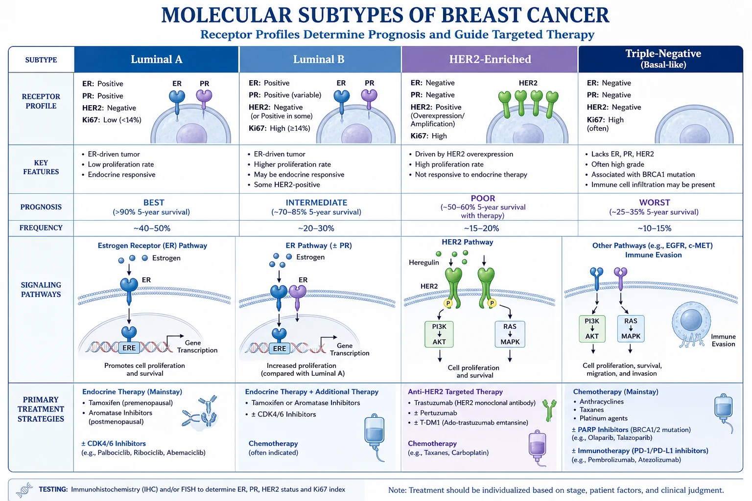
The non-obligate precursor concept
DCIS is the archetypal non-obligate (incomplete) precursor lesion. The natural-history data come largely from the biopsy-only era before screening — women in whom a breast lump was biopsied and reported as "intramammary carcinoma" (in situ) but, by error or choice, not treated. Long-term follow-up of these historical cohorts (notably the Nashville/Wellings series) showed that approximately 30 to 50% of women with untreated low- to intermediate-grade DCIS developed invasive breast cancer in the same breast over 10 to 20 years, almost always in the same quadrant as the original DCIS. Critically, the remaining 50 to 70% did not progress even after decades of follow-up — establishing that progression is probable but not inevitable, and that DCIS is heterogeneous.[5]
The risk of progression is strongly grade-dependent. High-grade comedo DCIS carries the highest and most rapid risk of invasion; low-grade cribriform or micropapillary DCIS carries a lower and more indolent risk. Because grade is therefore the single most important biological variable, every histopathology report on a DCIS specimen must state the nuclear grade, the presence or absence of comedo necrosis, and the architectural pattern.[5]
Molecular biology of progression
DCIS and the invasive carcinoma that may follow it share clonal driver lesions, confirming the lineage relationship. Low-grade DCIS is characterised by gain of 1q and 16p and loss of 16q, low proliferation, ER positivity, and a relatively stable genome; it tends to progress slowly, if at all. High-grade DCIS shows greater genomic instability, frequent HER2 amplification (50 to 70% of comedo-type), high Ki-67, p53 mutation, ER negativity in a subset, and activation of the PI3K/AKT/mTOR pathway — a molecular profile that overlaps heavily with high-grade invasive carcinoma. The decisive event in progression is acquisition of the capacity to degrade and breach the basement membrane, mediated by upregulation of matrix metalloproteinases, loss of cell adhesion molecules, and epithelial-to-mesenchymal transition. Once the basement membrane is breached, the cells become invasive ductal carcinoma and gain access to lymphatics and blood vessels — at which point metastasis becomes possible.[5]
An important corollary for the surgeon: because DCIS may coexist with or hide an occult invasive focus, 10 to 20% of excision specimens for what was DCIS on core biopsy reveal invasive carcinoma. This occult invasion then redefines the disease as invasive (with implications for axillary staging and systemic therapy), which is the basis for considering sentinel lymph node biopsy at the time of mastectomy for DCIS.[5]
India and South Asia. In India and other low- and middle-income settings, organised population-based mammographic screening is not widely implemented, so the DCIS-to-invasive ratio seen in Western screening programmes is not reproduced. Most breast pathology still presents symptomatically at a locally advanced invasive stage, and DCIS is a relatively smaller fraction of diagnoses. As access to mammography grows in urban centres, DCIS detection is rising. A practical consequence for exams and practice: a screen-detected, microcalcification-only DCIS in an Indian woman is still managed exactly as in international guidelines — wide local excision with clear margins plus whole-breast radiotherapy, or mastectomy — with endocrine therapy for ER-positive disease.
Classification
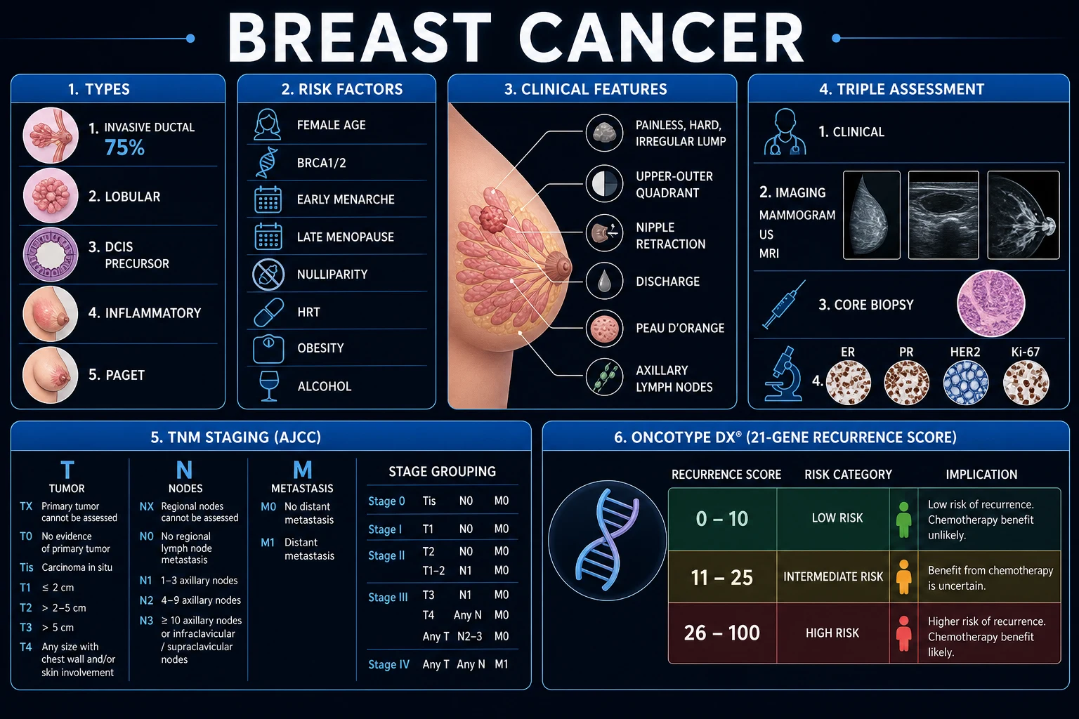
DCIS is classified along four independent axes, all of which must appear on the histopathology report because each influences prognosis and treatment: nuclear grade, architectural pattern, presence of comedo necrosis, and hormone receptor (ER/PR/HER2) status. The composite Van Nuys Prognostic Index integrates grade, size, margin and age into a single treatment-guiding score.[5]
Nuclear grade
Nuclear grade is the single most reproducible and prognostically important histological feature and is assessed by comparing the DCIS cell nuclei with normal ductal epithelial nuclei. [1]
| Grade | Nuclear features | Mitoses | Associated architecture | Prognosis |
|---|---|---|---|---|
| Low | Small, uniform, monomorphic nuclei (1.5 to 2x normal); fine evenly-distributed chromatin; inconspicuous nucleoli | Rare | Cribriform or micropapillary; no necrosis | Best; lowest progression risk |
| Intermediate | Nuclei 2 to 3x normal; slight pleomorphism; small nucleoli | Occasional | Mixed/solid; may have punctate necrosis | Intermediate |
| High (comedo) | Large pleomorphic nuclei (over 3x normal); coarse chromatin; prominent nucleoli | Frequent (over 1 per high-power field) | Solid with central comedo necrosis | Worst; highest risk of recurrence and invasion |
Comedo versus non-comedo necrosis
Comedo necrosis — cheese-like eosinophilic necrotic debris in the centre of a duct involved by high-grade DCIS — is a marker of aggressive biology. Comedo-type DCIS shows HER2 overexpression in 50 to 70%, a high Ki-67 proliferation index, frequent p53 mutation, and the highest rate of local recurrence and progression to invasion. It characteristically produces linear, branching (casting) microcalcifications on mammography that map out the necrotic ducts. Non-comedo patterns (cribriform, micropapillary, solid, papillary) are generally lower grade, more often ER-positive, and carry a better prognosis.[5]
Architectural patterns
| Pattern | Microscopic features | Usual grade | Prognosis |
|---|---|---|---|
| Cribriform | Sieve-like, "cookie-cutter" punched-out spaces within the duct | Low | Good |
| Micropapillary | Small finger-like projections into the lumen without fibrovascular cores; often extensive/multicentric | Low to intermediate | Good (but may be widespread) |
| Solid | Neoplastic cells fill the duct lumen completely | Variable | Variable |
| Papillary | True fronds with fibrovascular cores | Low to intermediate | Good |
| Comedo | High-grade cells with central necrosis | High | Worst |
Mixed patterns are common. Micropapillary DCIS deserves special mention because it tends to be extensive and multicentric and to involve more of the ductal tree than its apparent mammographic extent, which can push management toward mastectomy. Multiple architectural patterns may coexist within one lesion; the highest grade present determines the overall grade assignment. [1]
Van Nuys Prognostic Index (VNPI)
The Van Nuys Prognostic Index, developed by Silverstein and colleagues, is the classic composite tool that integrates the three strongest predictors of local recurrence into a single score that guides treatment intensity. It has been extended by the addition of patient age (the USC-VNPI).[5]
USC-VNPI — each factor scored 1 to 3, total 4 to 12
| Factor | 1 point | 2 points | 3 points |
|---|---|---|---|
| Tumour size | Under 1.5 cm | 1.5 to 4 cm | Over 4 cm |
| Margin width | Over 10 mm clear | 1 to 10 mm | Under 1 mm |
| Grade / pathology | Low grade, no necrosis | Low grade with necrosis, or intermediate | High grade with or without necrosis |
| Age (USC extension) | Over 60 | 40 to 60 | Under 40 |
Total score and management (classical Silverstein thresholds): [1]
- Score 4 to 6 (low risk): Excision alone may be sufficient — the local recurrence rate is low enough that the marginal benefit of radiotherapy is debated.
- Score 7 to 9 (intermediate): Excision plus whole-breast radiotherapy; consider endocrine therapy if ER-positive.
- Score 10 to 12 (high risk): Mastectomy recommended; radiotherapy cannot be given after mastectomy for DCIS and these high-risk lesions are not safely conservable. [1]
It is important to understand that the VNPI is a guide, not a rule. Modern practice increasingly relies on multidisciplinary team discussion and shared decision-making, and the SSO/ASTRO/ASCO consensus favours a 2 mm margin as "adequate" for breast-conserving therapy with whole-breast radiotherapy (see Management). The VNPI remains a high-yield exam concept because it crystallises the four determinants of local recurrence into one memorable tool.[5][6]
Molecular subtypes
Like invasive breast cancer, DCIS can be classified by receptor status, which guides endocrine therapy and reflects biology: [1]
| Subtype | ER | PR | HER2 | Ki-67 | Features |
|---|---|---|---|---|---|
| Luminal A | Positive | Positive | Negative | Low | Best prognosis; common in low-grade DCIS; tamoxifen-responsive |
| Luminal B | Positive | Variable | Variable | High | Intermediate; tamoxifen-responsive |
| HER2-enriched | Negative | Negative | Positive | High | High grade, comedo type; worse prognosis |
| Basal-like | Negative | Negative | Negative | High | Rare in pure DCIS; aggressive when present |
About 75% of DCIS is ER-positive, which is the rationale for offering tamoxifen (or anastrozole in postmenopausal women) as adjuvant endocrine prevention.[2][4]
Clinical Presentation
The defining feature of DCIS in the screening era is that most patients are asymptomatic — the diagnosis is made on imaging, not by the patient or the examining clinician.[5]
- Screen-detected microcalcifications (about 90%) — the dominant presentation. Clusters of pleomorphic, granular or branching calcifications in a ductal (segmental) distribution are found on routine screening mammography. The patient has no palpable abnormality.
- Palpable mass (10 to 15%) — in larger or higher-grade lesions, particularly comedo DCIS, a hard irregular area may be felt. A palpable DCIS raises the index of suspicion for an occult invasive component.
- Nipple discharge — rarely, spontaneous unilateral bloodstained or serosanguineous discharge from a single duct, when subareolar ducts are involved.
- Paget disease of the nipple — an eczematous, scaling, weeping erosion of the nipple-areolar complex caused by intraepidermal spread of malignant Paget cells from an underlying ductal carcinoma; underlying disease is DCIS in a proportion of cases.
- Incidental — found in a biopsy performed for another lesion, or in a mastectomy specimen for invasive cancer. [1]
Clinical breast examination remains essential: inspection (in four positions — arms by the side, on hips, raised, leaning forward) for asymmetry, skin or nipple change; and supine palpation of all four quadrants, the central areolar area and the axillary tail, plus regional nodes. In pure DCIS the examination is usually normal, but a palpable abnormality or nodal enlargement suggests coexisting invasive disease and mandates full triple assessment.[5]
Mammographic Features & Imaging
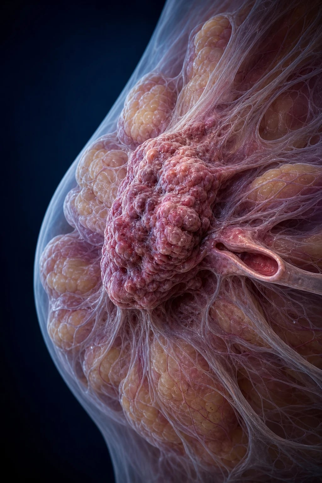
Mammography is the key imaging modality for DCIS because of its unique sensitivity for microcalcifications. The mammographic hallmark of DCIS is microcalcifications — and recognising their morphology and distribution is an exam favourite.[5]
Calcification morphology:
- Fine pleomorphic / granular calcifications — varying size and shape, "crushed-stone" appearance; typical of low- to intermediate-grade DCIS.
- Fine linear / fine linear-branching ("casting") calcifications — outline the necrotic ducts in a discontinuous linear or branching pattern; highly suspicious for high-grade comedo DCIS and carry the highest positive predictive value for malignancy. [1]
Calcification distribution (in increasing order of suspicion): diffuse → regional → grouped/clustered → linear → segmental. A segmental or linear (ductal) distribution of pleomorphic calcifications is the most characteristic and most suspicious pattern for DCIS, because it reflects filling of a ductal tree by the neoplastic process. Calcifications should be characterised as BI-RADS 4 (suspicious, biopsy) or BI-RADS 5 (highly suggestive of malignancy, biopsy).[5]
BI-RADS (Breast Imaging Reporting and Data System) assessment categories:
- BI-RADS 0 — incomplete, recall for additional imaging.
- BI-RADS 1/2 — negative / benign; routine screening.
- BI-RADS 3 — probably benign (under 2% malignancy risk); short-interval (6-month) follow-up.
- BI-RADS 4 — suspicious; biopsy. Subdivided 4a (low), 4b (moderate), 4c (high) suspicion.
- BI-RADS 5 — highly suggestive of malignancy (over 95%); biopsy.
- BI-RADS 6 — known biopsy-proven malignancy. [1]
Ultrasound is complementary rather than primary for DCIS, because pure in situ disease often produces no mass. It is used to assess any associated mass, to guide biopsy of a solid component, and to evaluate the axilla. Breast MRI is more sensitive than mammography for the extent of disease (multifocality, multicentricity, occult invasive focus, and chest-wall involvement) and is increasingly used pre-operatively in selected cases — particularly before mastectomy or when calcification extent is uncertain — but its high sensitivity and lower specificity can overestimate extent and lead to wider excisions, so it is used selectively.[5][6]
Investigations & Diagnostic Pathway
The diagnosis of DCIS is made by image-guided needle biopsy of the suspicious calcifications, confirmed histologically, and then mapped to plan surgery.[5]
| Investigation | Role in DCIS |
|---|---|
| Mammography | First-line and often the only positive test — microcalcifications in ductal/segmental distribution. Magnification views characterise morphology and extent. |
| Stereotactic core needle biopsy | Gold standard for sampling calcifications; a vacuum-assisted 9- to 11-gauge device under stereotactic x-ray guidance yields multiple cores with the calcifications (confirmed on specimen radiograph). 14-gauge ultrasound-guided core if a mass is present. |
| Specimen radiograph | Confirms that the excised tissue contains the targeted calcifications, ensuring adequate sampling/excision. |
| Excision (open) biopsy | Reserved for non-diagnostic cores, image-pathology discordance, or when calcifications cannot be sampled stereotactically (e.g. very posterior). |
| Histopathology | Confirms DCIS; must report nuclear grade, architectural pattern, comedo necrosis, ER/PR/HER2, size/extent, and margin status. |
| Breast MRI | Selected cases — to assess extent, multifocality, occult invasion, before mastectomy. |
| ER/PR/HER2 | On the core biopsy; determines eligibility for endocrine therapy. HER2 is prognostic in DCIS but anti-HER2 therapy is not given for pure DCIS. |
Differential Diagnosis
DCIS enters the differential at two points: the radiological differential of suspicious microcalcifications, and the histological differential of an intraductal epithelial proliferation.[5]
Benign causes of microcalcifications
often round, scattered, diffuse — but overlap exists
- **Sclerosing adenosis**: clustered, round or punctate calcifications; benign proliferative change
- **Fibrocystic change**: coarse, eggshell-like (cyst wall) or scattered round calcifications
- **Dermal / vascular calcifications**: parallel 'tram-track' vessel walls; skin calcifications are often lucent-centred
- **Secretory / plasma cell mastitis**: coarse, dense, rod-like calcifications in large ducts
- **Fat necrosis**: peripheral, ring-like or oil-cyst calcifications after trauma or surgery
Other in situ / precursor lesions
histological distinction at excision
- **Atypical ductal hyperplasia (ADH)**: shares cytology/architecture with low-grade DCIS but is smaller (under 2 mm or fewer than 2 spaces); ADH on core biopsy must be surgically excised (about 15 to 30% upstage to DCIS)
- **Lobular carcinoma in situ (LCIS)**: not cancer — a bilateral risk marker (6 to 10x); discohesive cells distending lobules; no calcifications typically; managed by surveillance and endocrine prevention, not surgery
- **Atypical lobular hyperplasia (ALH)**: a smaller LCIS-like lesion; also a risk marker
Malignant mimics
change management entirely
- **Invasive ductal carcinoma with extensive intraductal component (EIC)**: DCIS surrounding/within an invasive tumour; the invasive component mandates axillary staging and systemic therapy
- **Microinvasive carcinoma (T1mi)**: invasion up to 1 mm — technically staged and managed as minimal invasive disease
- **Paget disease of the nipple**: underlying DCIS or invasive carcinoma; biopsy the nipple
The histological distinction between ADH and low-grade DCIS is the single most common and most important boundary problem. The practical rule for the surgeon: ADH on a core biopsy mandates surgical excision, because a substantial minority harbour unsampled DCIS or invasion. The distinction between DCIS and LCIS is also exam-critical: DCIS is treated as a precursor lesion to be excised; LCIS is a risk marker managed by surveillance and endocrine prevention, not surgery.[5]
Management — Multidisciplinary Principles
DCIS is not a surgical emergency. After histological confirmation the case is presented at a multidisciplinary team (MDT) meeting (breast surgeon, radiation oncologist, medical oncologist, radiologist, pathologist, breast-care nurse), the extent of disease and receptor status are confirmed, and a tailored locoregional and endocrine plan is agreed with the patient. The twin goals are to prevent progression to invasive carcinoma and local recurrence while minimising overtreatment of lesions that may never have progressed.[5][6]
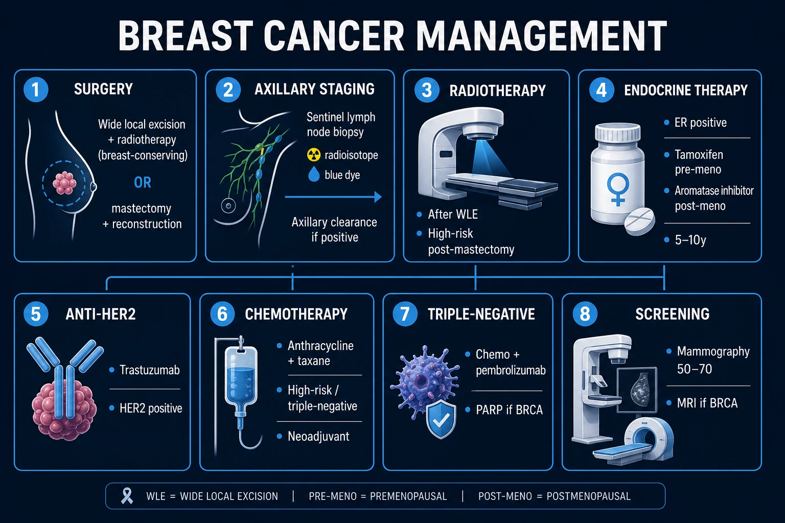
The treatment decision balances patient factors (age, comorbidity, life expectancy, breast size relative to DCIS extent, patient preference for conservation versus mastectomy, contraindications to radiotherapy such as pregnancy, prior chest irradiation or active connective-tissue disease, BRCA status) and tumour factors (size, nuclear grade, comedo necrosis, margin status, multifocality/multicentricity, ER status, extent of calcifications). Young age (under 40) and high nuclear grade are the strongest risk factors for local recurrence after breast-conserving therapy.[5]
Management — Surgery
Breast-conserving surgery (BCS): technique
Breast-conserving surgery — also called wide local excision (WLE), lumpectomy, or partial mastectomy — removes the DCIS with a rim of surrounding normal breast tissue while preserving the breast shape. Combined with whole-breast radiotherapy it constitutes breast-conserving therapy (BCT), the standard of care for most unifocal, conservable DCIS.[1]
Operative technique:
- The skin incision is placed to be cosmetically favourable (usually circumareolar, periareolar, or a curved incision following Langer's lines directly over the lesion) and is planned so that it could be incorporated within a future mastectomy flap if needed. Avoid radial incisions where possible.
- The DCIS is excised en bloc with a surrounding cuff of normal tissue, guided by the preoperative imaging and, if used, a localisation wire, radioactive seed (RSL), or magnetic/reflective localisation marker placed under ultrasound or stereotactic guidance.
- Specimen radiograph of the excised tissue is performed intra-operatively (when calcifications are targeted) to confirm that the microcalcifications have been removed and to orient the specimen for margin assessment.
- The specimen is inked (or strategically clipped) in the pathology laboratory so that the six margins (superior, inferior, medial, lateral, anterior, posterior) can be individually measured.
- The cavity is usually not closed by mobilising tissue flaps in a simple lumpectomy; careful glandular re-approximation preserves cosmesis. [1]
Margin requirements. The single strongest modifiable predictor of local recurrence after BCS for DCIS is margin status. The 2016 SSO/ASTRO/ASCO consensus guideline established that a margin of at least 2 mm is adequate for DCIS treated with whole-breast radiotherapy — wider margins do not meaningfully reduce recurrence, while positive or close margins (under 2 mm) significantly increase it. Positive margins mandate re-excision to achieve at least 2 mm; if clear margins cannot be obtained, mastectomy is recommended.[5][6]
Oncoplastic techniques
When the volume of tissue that must be excised to clear the DCIS threatens the cosmetic outcome, oncoplastic breast surgery combines oncological resection with plastic-surgical reconstruction of the defect in the same operation. Oncoplastic techniques allow wider, safer margins for larger DCIS while preserving or improving breast shape, and have expanded the proportion of women eligible for conservation. [1]
- Volume displacement (level I/II oncoplasty): local glandular and dermoglandular flaps are advanced to fill the defect — suitable for excisions up to about 20 to 50% of breast volume. Level I covers up to 20%, level II (therapeutic mammoplasty, batwing, round-block, J-mammoplasty, Grisotti for central/subareolar lesions) covers 20 to 50%.
- Volume replacement: distant or regional tissue (most commonly the latissimus dorsi miniflap or, less often, an intercostal artery perforator flap) is transferred to fill a partial defect, particularly in small breasts where displacement is impossible.
- Bilateral symmetrisation: contralateral reduction/mastopexy to match the operated breast when a large therapeutic mammoplasty has been performed. [1]
Oncoplastic resection is particularly valuable for extensive but unifocal/segmental DCIS that would otherwise require mastectomy, for central subareolar DCIS (where a Grisotti or round-block preserves the nipple), and in small breasts. It demands careful preoperative planning of imaging extent and incision design, and a two-team (surgical plus reconstructive) approach in many centres.[5][6]
Mastectomy
Mastectomy removes essentially all breast glandular tissue and is recommended when the DCIS is large (over about 4 cm), multicentric (in more than one quadrant), cannot be cleared with acceptable margins by BCS, when radiotherapy is contraindicated, or by patient choice. Because recurrence risk after a properly performed mastectomy for DCIS is under 2%, radiotherapy is NOT given after mastectomy for DCIS, and the mastectomy effectively cures the local disease.[5]
Types of mastectomy:
- Total (simple) mastectomy: removal of the entire breast parenchyma, nipple-areola complex and a small ellipse of overlying skin; the standard oncological operation.
- Skin-sparing mastectomy: removal of the breast parenchyma and nipple-areola complex through a periareolar or reduced incision, preserving the native skin envelope to optimise immediate reconstruction; oncologically equivalent to total mastectomy for DCIS.
- Nipple-sparing (subcutaneous) mastectomy: preservation of the entire skin envelope including the nipple-areola complex, with parenchyma removed through an inframammary, periareolar or lateral incision; reserved for selected, carefully counselled patients having immediate reconstruction, and generally avoided when the DCIS involves the subareolar ducts or is extensive.
- Risk-reducing (prophylactic) mastectomy: bilateral mastectomy in a BRCA1/2 carrier or woman at very high genetic risk; reduces breast cancer risk by about 90 to 95%. [1]
Immediate or delayed reconstruction (implant, tissue expander-to-implant, or autologous flap — DIEP, TRAM or latissimus dorsi) should be discussed with every woman facing mastectomy; it does not compromise oncological control in DCIS. Because 10 to 20% of mastectomy specimens harbour occult invasion, sentinel lymph node biopsy is performed at the time of mastectomy for DCIS.[5]
Management algorithm by VNPI
Combining the surgical options with the Van Nuys classification gives a memorable treatment algorithm:[5]
| USC-VNPI score | Recommended treatment |
|---|---|
| 4 to 6 (low risk) | Excision alone may suffice (radiotherapy benefit marginal; many centres still offer it) |
| 7 to 9 (intermediate) | Excision plus whole-breast radiotherapy; add endocrine therapy if ER-positive |
| 10 to 12 (high risk) | Mastectomy (with SLNB); radiotherapy not given after mastectomy |
Management — Radiotherapy
Whole-breast radiotherapy after breast-conserving surgery is the cornerstone of breast-conserving therapy and roughly halves the risk of ipsilateral local recurrence — both recurrent DCIS and subsequent invasive cancer. The benefit was established by the landmark NSABP B-17 and UK/ANZ DCIS randomised trials and confirmed by long-term follow-up.[1][3]
Whole-breast radiotherapy (after BCS):
- Dose and fractionation: the conventional regimen is 50 Gy in 25 fractions over 5 weeks to the whole breast. A hypofractionated regimen (40 Gy in 15 fractions over 3 weeks, or 42.5 Gy in 16 fractions) is now widely preferred and is at least as effective and cosmetically acceptable, with greater patient convenience.
- Tumour-bed boost: an additional 10 to 16 Gy (commonly 10 Gy in 4 fractions or 16 Gy in 8 fractions) is delivered to the excision cavity for high-grade DCIS and in younger women (under 45 to 50), who have the highest recurrence risk.
- Magnitude of benefit: radiotherapy reduces the 10-year local recurrence rate by approximately half (absolute risk reduction roughly 10 to 15% at 10 years), with the benefit sustained at long-term follow-up.
- Side effects: acute skin erythema and desquamation, fatigue and breast oedema; late effects of fibrosis, telangiectasia, a small risk of cardiac toxicity (more for left-sided treatment, minimised by deep-inspiration breath-hold and intensity-modulated techniques), pneumonitis, and a very small long-term excess of secondary angiosarcoma in the irradiated field. [1]
What the landmark radiotherapy trials showed
Accelerated partial breast irradiation (APBI) delivers radiotherapy to the tumour bed only (with a margin) rather than the whole breast, over 1 week or less, via interstitial or intracavitary brachytherapy (e.g. a balloon catheter device) or external-beam (3D-conformal or intensity-modulated) techniques. APBI is an option for a highly selected low-risk subgroup — older women (over 50 to 60) with small (under 2 to 2.5 cm), low- to intermediate-grade, ER-positive DCIS excised with widely clear margins. It is not standard for higher-risk DCIS, and several trials continue to define its safe niche. Radiotherapy is not given after mastectomy for DCIS.[5][6]
Management — Endocrine Therapy
For ER-positive DCIS (about 75% of cases), adjuvant endocrine therapy reduces both ipsilateral and contralateral breast events. It is offered in addition to surgery ± radiotherapy, and its benefit must be weighed against its side effects in the shared-decision context — particularly because DCIS itself is not life-threatening.[2][3][4]
Tamoxifen
Tamoxifen, a selective oestrogen receptor modulator (SERM), is the standard endocrine agent for DCIS and can be used in women of any menopausal status. [1]
- NSABP B-24 trial: 1,804 women with DCIS treated by lumpectomy + radiotherapy were randomised to tamoxifen 20mg daily versus placebo for 5 years. Tamoxifen reduced all breast-cancer events at 5 years (8.2% vs 13.4%); the benefit was concentrated in ER-positive DCIS, reducing both ipsilateral and contralateral breast cancer by roughly 30%.[2]
- UK/ANZ DCIS trial: a 2x2 factorial of radiotherapy and tamoxifen. Radiotherapy reduced both ipsilateral invasive and DCIS recurrence; tamoxifen reduced recurrent DCIS but did not significantly reduce ipsilateral invasive recurrence, and reduced contralateral events.[3]
- Dose: 20 mg oral once daily for 5 years.
Side effects of tamoxifen:
- Vasomotor: hot flushes (about 30%), night sweats — the commonest and most compliance-limiting.
- Thromboembolic: roughly 2 to 3-fold increased risk of deep vein thrombosis and pulmonary embolism — avoid in women with prior VTE.
- Endometrial: 2 to 3-fold increased risk of endometrial cancer, endometrial hyperplasia and polyps (oestrogen-agonist effect on the endometrium) — investigate any postmenopausal bleeding.
- Ocular: cataracts, rare retinopathy.
- Hepatic: fatty liver, mildly elevated transaminases.
- Gynaecological / menstrual: vaginal discharge, menstrual irregularity. [1]
Anastrozole and other aromatase inhibitors
For postmenopausal women, the aromatase inhibitor anastrozole is an alternative with a different side-effect profile. [1]
- IBIS-II DCIS trial: postmenopausal women with locally excised DCIS were randomised to anastrozole 1mg daily versus tamoxifen 20mg daily for 5 years. There was no significant difference in overall recurrence between the two (a non-significant trend favouring anastrozole), but the side-effect profiles differed: tamoxifen caused more gynaecological symptoms and venous thromboembolism, while anastrozole caused more musculoskeletal symptoms, fractures (osteoporosis) and vasomotor flushes. The conclusion: anastrozole is a reasonable alternative in postmenopausal women, with the choice guided by tolerability and side-effect risk.[4]
- Dose: anastrozole 1mg oral once daily for 5 years (postmenopausal only).
- Side effects of aromatase inhibitors: osteoporosis and fractures, arthralgia/myalgia, vasomotor symptoms, and (unlike tamoxifen) no increased thromboembolism or endometrial cancer.
Endocrine therapy in DCIS — by menopausal status
TAKE
any menopausal status, ER-positive DCIS — 20mg OD for 5 years. Beware VTE and endometrial cancer (NSABP B-24)
POSTmenopausal only — 1mg OD for 5 years. Equivalent efficacy, different toxicity (IBIS-II DCIS); more fractures/arthralgia
an aromatase inhibitor in a PREmenopausal woman does NOT suppress ovarian oestrogen (it can paradoxically rise) — never use without ovarian suppression
endocrine therapy is for ER-positive DCIS (~75%); it reduces both ipsilateral and contralateral events, but weigh benefit vs toxicity in a non-lethal disease
Management — Summary of the Standard Pathway
Low-risk DCIS
small (under 1-1.5 cm), low-grade, no necrosis, wide margins, older patient
- BCS with at least 2 mm margins ± whole-breast radiotherapy (VNPI 4-6 may allow excision alone, debated)
- Endocrine therapy (tamoxifen or anastrozole) if ER-positive — shared decision
- No routine axillary surgery (pure DCIS)
- Active surveillance is under trial (COMET, LORD, LORIS) for very low-risk, screen-detected disease
Intermediate-risk DCIS
1.5 to 4 cm, intermediate grade, or close margins
- BCS with at least 2 mm margins + whole-breast radiotherapy (VNPI 7-9)
- Boost to tumour bed for high-grade or young patients
- Endocrine therapy for 5 years if ER-positive
- Re-excise if margins under 2 mm
High-risk DCIS
over 4 cm, high-grade comedo, multicentric, or unachievable margins
- Mastectomy (VNPI 10-12); no post-mastectomy radiotherapy
- Sentinel lymph node biopsy at the time of mastectomy (occult invasion risk)
- Immediate reconstruction discussion
- Endocrine therapy for ER-positive disease even after mastectomy (reduces contralateral risk)
Surveillance After Treatment
The aims of follow-up are early detection of ipsilateral local recurrence (in the conserved breast or chest wall), contralateral new primary, and — if recurrence is invasive — timely salvage treatment. DCIS itself is highly curable, but recurrence, particularly invasive recurrence, is the event that threatens survival.[5][6]
Standard surveillance schedule (after BCS + RT, or mastectomy):
- Clinical breast and nodal examination every 6 to 12 months for the first 2 to 3 years, then annually. Most recurrences occur within the first 5 to 10 years.
- Annual mammography of the conserved (and contralateral) breast, beginning 6 to 12 months after completion of radiotherapy. After bilateral mastectomy, mammography is not routinely performed (chest-wall clinical examination suffices); mammography of a preserved contralateral breast continues.
- No routine blood tests, tumour markers, chest imaging, bone scans or CT in the asymptomatic patient — these do not improve survival in DCIS and are not recommended by ASCO/NCCN. Imaging is directed by symptoms.
- Endocrine-therapy adherence for the full 5 years, with baseline and periodic bone-density (DEXA) monitoring in women on an aromatase inhibitor, and prompt investigation of any vaginal bleeding on tamoxifen (endometrial cancer).
- Psychological support and breast-awareness counselling, addressing the anxiety of a "cancer" diagnosis for a disease that is rarely life-threatening, and supporting body-image recovery after surgery and radiotherapy. [1]
Most local recurrences after BCS + RT occur in or near the original tumour bed. About half of all recurrences are invasive ductal carcinoma (and half recurrent DCIS) — it is the invasive recurrences that carry mortality risk, which is why surveillance matters.[5]
Management of Recurrence
Local recurrence after treatment for DCIS is not failure of the original concept but a known risk that modern therapy minimises. The management is dictated by the type of recurrence (in situ versus invasive), its site, and the prior treatment.[5]
Ipsilateral recurrence after breast-conserving therapy:
- Recurrent DCIS: repeat wide local excision to clear margins. Because a second breast-conserving attempt within an irradiated breast is cosmetically and oncologically problematic, completion mastectomy is often the preferred salvage operation. Re-irradiation is rarely feasible.
- Invasive recurrence (about half of all recurrences): treat as a new primary invasive breast cancer — mastectomy (usually), sentinel lymph node biopsy for axillary staging (lymphatic mapping usually still possible), and full receptor staging with systemic therapy by subtype (endocrine if ER-positive; chemotherapy/anti-HER2 as indicated). [1]
Recurrence after mastectomy is uncommon (under 2%) and is managed by local excision of the chest-wall recurrence, with radiotherapy if not previously given, and systemic therapy by subtype. [1]
Contralateral new primary is treated as a new breast cancer in its own right, with its own triple assessment, staging and subtype-directed therapy; endocrine therapy for ER-positive DCIS reduces the contralateral event rate, which is one rationale for its use even after mastectomy. [1]
The single most important prognostic point: a recurrence that is invasive is what can kill the patient, whereas recurrent DCIS is again a local problem. This is the biological justification for aggressive prevention of recurrence in the first place (clear margins, radiotherapy, endocrine therapy).[5][6]
Complications & Pitfalls
Complications of surgery include seroma (the commonest, managed by aspiration), haematoma, wound infection, poor cosmetic outcome (especially after large or re-excision lumpectomies), and — after axillary surgery for occult invasion — lymphoedema (5 to 8% after sentinel node biopsy), shoulder stiffness, chronic pain and intercostobrachial nerve sensory loss. Immediate reconstruction carries its own set of complications (implant capsular contracture, flap loss, donor-site morbidity).[5]
Complications of radiotherapy include acute skin reactions and fatigue, and late fibrosis, breast shrinkage and asymmetry, cardiac toxicity (minimised with modern breath-hold and intensity-modulated techniques), pneumonitis, brachial plexopathy, and a small long-term excess of secondary angiosarcoma in the irradiated field (classically presenting years later as a violaceous skin plaque).[5]
Complications of endocrine therapy are as detailed above: tamoxifen-related VTE, endometrial cancer and vasomotor symptoms; aromatase-inhibitor-related osteoporosis, fractures and arthralgia. [1]
Classic pitfalls:
- Treating DCIS with the intensity of invasive cancer in a low-risk, elderly woman with indolent disease — overdiagnosis and overtreatment.
- Failing to achieve or re-excise a margin under 2 mm after BCS — the single strongest correctable recurrence risk.
- Omitting radiotherapy after breast-conserving surgery for higher-risk DCIS without a considered low-risk justification.
- Giving tamoxifen to a premenopausal woman with prior VTE, or an aromatase inhibitor to a premenopausal woman without ovarian suppression.
- Routine sentinel node biopsy for pure DCIS treated by BCS — not indicated; reserve for mastectomy or high suspicion of invasion.
- Confusing DCIS with LCIS — DCIS is treated as a precursor; LCIS is a risk marker managed by surveillance and prevention, not surgery.
- Mislabelling an ADH core biopsy as benign without surgical excision — ADH on core must be excised (about 15 to 30% upstage to DCIS or invasion).
- Forgetting contralateral and endocrine-prevention considerations even after mastectomy. [1]
Prognosis & Disposition
DCIS is highly curable. Breast cancer-specific survival is approximately 97 to 99% at 10 years, and overall mortality from DCIS itself is very low; the small excess mortality seen in DCIS cohorts is driven by the minority who develop invasive recurrence. After mastectomy, local recurrence is under 2%. After breast-conserving therapy (BCS + RT), local recurrence is roughly 10 to 15% at 10 years, with about half of these being invasive. Excision alone (selected low-risk cases) carries a higher recurrence rate of about 20 to 30% at 10 years, which is the rationale for adding radiotherapy in all but the lowest-risk cases.[1][5]
Risk factors for local recurrence after breast-conserving therapy:
- Positive or close margins (under 2 mm) — the strongest modifiable factor.
- Young age (under 40) — independent risk factor.
- High nuclear grade and comedo necrosis.
- Large size / extensive disease.
- Omission of radiotherapy and (to a lesser degree) omission of endocrine therapy for ER-positive disease.
- Positive family history / BRCA mutation. [1]
Disposition is to a multidisciplinary breast cancer follow-up programme with annual mammography and clinical examination, endocrine-therapy adherence and bone-density monitoring where relevant, and psychological and rehabilitation support. The contemporary shift is toward risk-adapted de-escalation — identifying the very-low-risk subgroup for whom excision alone or even active surveillance is appropriate, while continuing full therapy for higher-risk disease.[5][6]
Special Populations
Young women (under 40) have a higher recurrence rate after breast-conserving therapy, more frequently harbour occult invasion and BRCA mutations, and warrant careful extent-of-disease imaging (often MRI). Mastectomy is more frequently chosen. Genetic testing (BRCA1/2 and multigene panel) should be offered to young women with DCIS and a suggestive personal or family history. [1]
BRCA1/2 carriers with DCIS are managed with standard locoregional therapy but should be counselled about risk-reducing bilateral mastectomy and bilateral salpingo-oophorectomy, enhanced surveillance (annual breast MRI), and the value of endocrine prevention. Genetic testing is offered based on risk-prediction models and family history.[5]
Pregnancy-associated DCIS is rare. Mammography with abdominal shielding is safe; gadolinium MRI is avoided in the first trimester. Surgery is safe in any trimester; radiotherapy is deferred until after delivery; tamoxifen is absolutely contraindicated (teratogenic). Decisions are individualised within the MDT. [1]
Older and frail patients with low-grade, ER-positive DCIS may be excellent candidates for endocrine therapy with minimal surgery or, in some, active surveillance, weighing the competing risks of comorbidity and the often-indolent natural history. Under-treatment of highly curable disease in older women who could tolerate standard therapy is equally a pitfall.[5][6]
Evidence, Guidelines & the De-escalation Era
Modern DCIS management is built on the landmark randomised trials that established the roles of radiotherapy and endocrine therapy, and is now evolving toward risk-stratified de-escalation.[5]
- NSABP B-17 established that adding radiotherapy to lumpectomy roughly halves ipsilateral recurrence (DCIS and invasive) — the foundation of breast-conserving therapy for DCIS.[1]
- NSABP B-24 showed that tamoxifen after lumpectomy + radiotherapy reduces all breast-cancer events, with benefit concentrated in ER-positive DCIS.[2]
- UK/ANZ DCIS trial (2x2 factorial) confirmed that radiotherapy reduces both ipsilateral invasive and DCIS recurrence, while tamoxifen reduces DCIS (not invasive) recurrence and contralateral events.[3]
- IBIS-II DCIS trial showed anastrozole and tamoxifen have equivalent efficacy in postmenopausal DCIS with different toxicity profiles, supporting anastrozole as an alternative.[4]
- SSO/ASTRO/ASCO margin consensus (2016) established that 2 mm is an adequate margin for DCIS treated with whole-breast radiotherapy, ending the debate over wider margins and reducing re-excision rates.
- De-escalation trials — including COMET, LORD, and LORIS — are testing whether active surveillance (with or without endocrine therapy) is safe for low-risk, screen-detected DCIS, and may reshape the future of DCIS management for the lowest-risk subgroup.[6]
Exam Pearls
- DCIS is non-invasive and CANNOT metastasise — confined by the basement membrane. Therefore no routine axillary staging; exception is mastectomy for DCIS (SLNB done because of 10 to 20% occult invasion risk).[5]
- Detected as clustered microcalcifications on screening mammography (about 90%). Linear branching (casting) calcifications = high-grade comedo DCIS; granular calcifications = low/intermediate grade.[5]
- Margin of at least 2 mm required for BCS (SSO/ASTRO/ASCO consensus). Margin status is the strongest modifiable predictor of local recurrence. Positive/close margins → re-excision or mastectomy.[5]
- Radiotherapy after BCS halves local recurrence (NSABP B-17, UK/ANZ). Standard: 50 Gy/25# or hypofractionated 40 Gy/15#, boost 10 to 16 Gy for high-grade/young. No RT after mastectomy for DCIS.[1][3]
- Tamoxifen 20mg OD for 5 years for ER-positive DCIS (NSABP B-24) — any menopausal status. Side effects: VTE, endometrial cancer, hot flushes.[2]
- Anastrozole 1mg OD for 5 years is an alternative in postmenopausal women (IBIS-II DCIS) — equivalent efficacy, more fractures/arthralgia, less VTE/endometrial cancer.[4]
- VNPI scores Size, Margin, Grade (and Age in USC-VNPI), each 1 to 3: 4 to 6 excision ± RT; 7 to 9 excision + RT; 10 to 12 mastectomy.[5]
- DCIS ≠ LCIS. DCIS = precursor, treated by excision ± RT. LCIS = risk marker, managed by surveillance and endocrine prevention, not surgery.[5]
- ADH on core biopsy → surgical excision (15 to 30% upstage to DCIS or invasion).
- Breast cancer-specific survival over 97% at 10 years. About half of recurrences are invasive — these are what threaten survival.[5]
Exam application bank (NEET-PG / INICET)
One-line answer
Ductal carcinoma in situ (DCIS) is a non-invasive breast neoplasm in which malignant epithelial cells proliferate within the ductal-lobular system but do not breach the basement membrane. It is a non-obligate precursor of invasive ductal carcinoma — untreated, about 30 to 50% progress over 10 to 20 years, but many lesions never become invasive. With screening mammography, DCIS now makes up about 20 to 25% of all new breast cancer diagnoses and is nearly always detected as microcalcifications. Treatment: breast-conserving surgery (wide local excision with at least 2 mm clear margins) plus whole-breast radiotherapy, or mastectomy for large or multicentric disease. Tamoxifen for ER-positive DCIS reduces ipsilateral and contralateral recurrence (NSABP B-24). The Van Nuys Prognostic Index (VNPI) guides treatment intensity. Pure DCIS cannot metastasise, so the axilla is not routinely staged.
Worked stems (answer without another resource)
Stem 1 — Classic presentation. Map symptoms to mechanism; name the first investigation and first treatment step with dose/route if drug therapy is standard. [1]
Stem 2 — Unstable / complicated. List red flags that force immediate resuscitation, theatre, ICU, antidote, or reperfusion — and what you do in the first 15 minutes. [1]
Stem 3 — Atypical group. Elderly, pregnancy, child, or immunocompromised: how presentation and thresholds change. [1]
Stem 4 — Differential trap. Name the three closest mimics and one discriminator for each. [1]
Stem 5 — Disposition. Who goes home with safety-netting, who is admitted, who needs HDU/ICU/theatre, and what follow-up is mandatory. [1]
Rapid viva checklist
- Definition + classification
- Pathophysiology chain
- Bedside signs / criteria
- Score with exact components (if any)
- Emergency bundle
- Definitive therapy with doses
- Complications of disease and of treatment
- Special populations
- Guideline/trial name if classic
- Three exam traps
Coverage self-check
If you cannot answer any stem above from this page alone, re-read the matching section — the page is intended to be self-sufficient for final-prof and NEET-PG/INICET questions on Ductal Carcinoma In Situ.
[1]References
- [1]Fisher B, Dignam J, Wolmark N, et al. Lumpectomy and radiation therapy for the treatment of intraductal breast cancer: findings from National Surgical Adjuvant Breast and Bowel Project B-17 J Clin Oncol, 1998.PMID 9469327
- [2]Fisher B, Dignam J, Wolmark N, et al. Tamoxifen in treatment of intraductal breast cancer: National Surgical Adjuvant Breast and Bowel Project B-24 randomised controlled trial Lancet, 1999.PMID 10376613
- [3]UK Coordinating Committee on Cancer Research (UKCCCR) DCIS Working Party. Radiotherapy and tamoxifen in women with completely excised ductal carcinoma in situ of the breast in the UK, Australia, and New Zealand: randomised controlled trial Lancet, 2003.PMID 12867108
- [4]Forbes JF, Sestak I, Howell A, et al.; IBIS-II DCIS Investigators. Anastrozole versus tamoxifen for the prevention of locoregional and contralateral breast cancer in postmenopausal women with locally excised ductal carcinoma in situ (IBIS-II DCIS): a double-blind, randomised controlled trial Lancet, 2016.PMID 26686313
- [5]Nash AL, Hwang ES. The Landmark Series-Ductal Carcinoma in Situ: The Evolution of Treatment Ann Surg Oncol, 2023.PMID 37024766
- [6]Bucheit JT, Schacht D, Kulkarni SA. Update on Management of Ductal Carcinoma in Situ Clin Breast Cancer, 2024.PMID 38216382