General Surgery · General Surgery
Gallstone Disease
Also known as Cholelithiasis · Gallstones · Biliary colic · Acute cholecystitis · Choledocholithiasis
Gallstone disease encompasses the spectrum from asymptomatic stones (80%) through biliary colic (RUQ pain after fatty meal, 2 to 6 hours, resolves spontaneously) to acute cholecystitis (fever + Murphy sign + RUQ pain lasting over 6 hours) and its complications (choledocholithiasis, cholangitis, pancreatitis, gallstone ileus). Risk: the 5 Fs (Female, Fat, Forty, Fertile, Family). Ultrasound is first-line (95% sensitive for stones over 2 mm). Asymptomatic: observe. Biliary colic: elective laparoscopic cholecystectomy. Acute cholecystitis: early laparoscopic cholecystectomy within 72 hours. CBD stone: ERCP with sphincterotomy. Cholangitis: IV antibiotics + urgent ERCP. Courvoisier's law: palpable gallbladder + jaundice = NOT stones (malignant obstruction).
On this page & tools
Your progress
Saved locally on this device.
Exam tags
Red flags
Overview & Definition
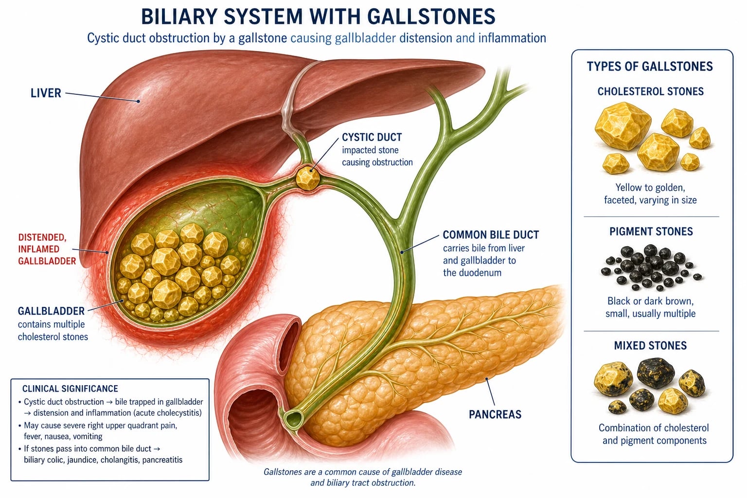
Gallstone disease (cholelithiasis) is one of the commonest surgical presentations in adult medicine and the single most frequent reason for elective abdominal surgery in the developed world. It encompasses the full clinical spectrum of solid formations within the gallbladder and biliary tree — from incidental asymptomatic stones discovered incidentally on imaging, through the recurrent postprandial pain of biliary colic, to acute cholecystitis and its life-threatening complications of choledocholithiasis, ascending cholangitis, gallstone pancreatitis, and gallstone ileus.[2]
The clinical importance of gallstones lies less in the stones themselves than in where they lodge and what they obstruct. A stone sitting quietly in the body of the gallbladder is harmless; the same stone impacted at the cystic duct causes cholecystitis, at the common bile duct (CBD) causes obstructive jaundice and cholangitis, and at the ampulla of Vater causes pancreatitis. The entire examinable clinical spectrum flows from this single anatomical principle.[1]
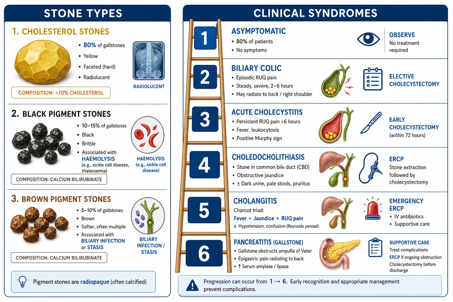
[1]A useful clinical framing is to think of gallstone disease as a progression through four tiers of severity, each with its own management: (1) asymptomatic stones (observe), (2) uncomplicated symptomatic stones — biliary colic (elective surgery), (3) acute cholecystitis (early surgery plus antibiotics), and (4) complicated disease — CBD stone, cholangitis, pancreatitis, ileus (targeted endoscopic, surgical, or radiological therapy). The exam rewards the candidate who can place any given patient on this ladder and act accordingly. [1]
Classification
Gallstones are classified by their chemical composition, which in turn reflects their underlying pathogenesis and guides the differential diagnosis when a stone type is identified at surgery or in a jaundiced work-up.[1]
Cholesterol stones (80%)
yellow, faceted, radiolucent
- Formed from **cholesterol supersaturation** of bile
- **Radiolucent** (not visible on plain X-ray)
- Risk: the **5 Fs** (Female, Fat, Forty, Fertile, Family)
- Light, faceted, yellow-green; often multiple
- Can be dissolved by ursodeoxycholic acid if small and floating
Pigment stones (20%)
black or brown
- **Black pigment**: haemolysis (sickle cell, hereditary spherocytosis), cirrhosis
- **Brown pigment**: biliary stasis and infection (Ascaris, Clonorchis, Asian populations)
- **Radiopaque** (contain calcium bilirubinate)
- Small, dark, irregular, multiple
- Not amenable to dissolution therapy
Mixed stones
cholesterol plus pigment
- Contain both cholesterol and calcium bilirubinate
- Most stones encountered clinically are mixed
- Often multiple and faceted
- Radiopacity varies with calcium content
- Treated as cholesterol stones for management purposes
A second, equally important classification is the clinical syndrome the stones produce — because this, not the stone composition, dictates management. The syndromes sit on a severity ladder, each step representing deeper obstruction or infection.[1]
Clinical syndromes — the severity ladder
Epidemiology & Risk Factors
Gallstones affect approximately 10 to 15% of the adult population in developed countries, with prevalence rising steadily with age and the global obesity epidemic. In Western populations, roughly one in five women and one in ten men will develop gallstones by middle age. Most remain asymptomatic — only 20 to 30% develop symptoms over their lifetime, and only 1 to 4% per year of asymptomatic carriers become symptomatic, which underpins the conservative approach to incidental stones.[2]
The prevalence varies sharply by ethnicity, geography, and diet. The highest rates in the world are seen in Native Americans (Pima Indians, up to 70% of women) and in Chilean Mapuche, reflecting strong genetic predisposition. In South Asia, gallstones are extremely common — particularly cholesterol stones in urban populations with rising obesity, and brown pigment stones in rural areas where Ascaris lumbricoides and Clonorchis sinensis infest the biliary tree.[1]
Risk factors — the 5 Fs
5 Fs
oestrogen increases cholesterol secretion into bile (2:1 female predominance)
obesity increases hepatic cholesterol secretion and bile saturation
risk rises with age (peak incidence 40 to 69)
pregnancy and parity increase risk (progesterone slows gallbladder motility)
genetic predisposition (ABO blood group, Native American ancestry, ABCG5/8 polymorphisms)
Beyond the mnemonic, the examinable risk factors divide neatly into those that increase cholesterol saturation of bile and those that impair gallbladder motility (causing stasis). Many risk factors act through both mechanisms.[2]
Increase cholesterol saturation
excess cholesterol relative to bile salts
- Obesity (BMI over 30) and metabolic syndrome
- Rapid weight loss (bariatric surgery, very-low-calorie diet)
- Oestrogen therapy: oral contraceptives, hormone replacement, pregnancy
- Fibrate therapy (gemfibrozil) — increases biliary cholesterol
- High-fat, low-fibre, refined-carbohydrate diet
Impair gallbladder motility
stasis allows nucleation
- Prolonged fasting / total parenteral nutrition (TPN)
- Somatostatin analogue therapy (octreotide)
- Diabetes mellitus (autonomic neuropathy)
- Pregnancy (progesterone)
- Spinal cord injury, vagotomy
Increase bilirubin load
pigment stones
- Chronic haemolysis: sickle cell disease, hereditary spherocytosis, thalassaemia
- Cirrhosis (impaired bilirubin conjugation)
- Biliary infection: Ascaris, Clonorchis, E. coli (beta-glucuronidase)
A handful of iatrogenic and circumstantial risk factors are heavily tested: ceftriaxone forms calcium-ceftriaxone sludge in bile (reversible on stopping), octreotide causes gallbladder stasis (used in acromegaly and variceal bleeding), and total parenteral nutrition deprives the gallbladder of cholecystokinin (CCK) stimulation, producing sludge in nearly all patients within weeks.[1]
Pathophysiology
The formation of a gallstone, like the formation of any crystal in a saturated solution, requires three sequential events. Understanding the mechanism explains why each risk factor behaves as it does and why dissolution therapy only works in a narrow subset.[1]
Step 1 — Cholesterol supersaturation of bile. Bile is a complex micellar solution in which hydrophobic cholesterol is kept dissolved by bile acids and phospholipids (lecithin). When the liver secretes bile with cholesterol in excess of the solubilising capacity of bile acids, the bile becomes "lithogenic" — supersaturated — and cholesterol begins to precipitate out of micelles into unilamellar vesicles. Oestrogen and obesity both push the liver towards this lithogenic bile by up-regulating HMG-CoA reductase (cholesterol synthesis) and down-regulating the bile acid pump. [1]
Step 2 — Nucleation. Supersaturation alone is not enough — most people with saturated bile never form stones. Nucleation is the rate-limiting step, driven by mucin glycoproteins secreted by the gallbladder epithelium and by pro-nucleating proteins (immunoglobulins, phospholipase A2). Cholesterol monohydrate crystals form within the mucin gel and aggregate. Stone formation is accelerated when anti-nucleating proteins (apolipoproteins A1, A2) are deficient. [1]
Step 3 — Gallbladder hypomotility and stasis. The nucleated crystals must remain in the gallbladder long enough to grow into macroscopic stones. Any condition that impairs gallbladder emptying — fasting, TPN, pregnancy, octreotide, diabetes — prolongs residence time and allows stones to mature. This is why rapid weight loss (which both mobilises cholesterol AND reduces gallbladder emptying) is such a powerful risk factor: it hits all three steps simultaneously.[1]
Pigment stones follow a different mechanism. Black pigment stones form when excess unconjugated bilirubin (from haemolysis or cirrhosis) polymerises with calcium in the gallbladder. Brown pigment stones form in the bile ducts themselves, where bacterial beta-glucuronidase (from E. coli, Klebsiella, Ascaris) deconjugates bilirubin diglucuronide, liberating free bilirubin that precipitates with calcium and fatty acids — this is why brown stones are associated with biliary stasis, infection, and parasitic infestation, and why they are found in the ducts rather than the gallbladder.[2]
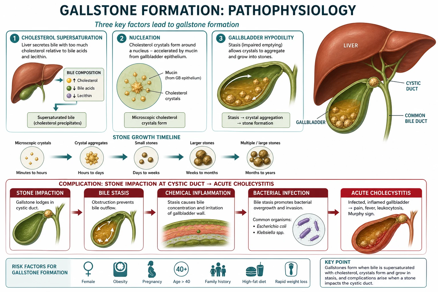
From a stone to acute cholecystitis — the pathophysiological cascade that examiners test most heavily:[1]
Stone lodges at cystic duct or Hartmann's pouch
Temporary obstruction in biliary colic; persistent obstruction in cholecystitis
Bile cannot exit the gallbladder
Gallbladder distends; mucus and bile accumulate (mucocele if sterile, empyema if infected)
Chemical inflammation
Bile salts and lysolecithin (formed from lecithin by phospholipase A) irritate the mucosa — this is why cholecystitis is initially sterile
Ischaemia and mural injury
Sustained distension reduces venous and then arterial supply, producing mural ischaemia (the route to gangrene and perforation)
Bacterial overgrowth
E. coli, Klebsiella, Enterococcus, and anaerobes seed the stagnant bile — pyrexia, leucocytosis, and systemic sepsis appear
Acute cholecystitis
The full syndrome of fever, RUQ pain, Murphy sign, and inflammatory markers
When the obstruction moves downstream into the CBD rather than the cystic duct, the same principles of stasis and infection play out in the biliary tree itself. Bile duct obstruction raises intraductal pressure, cholestasis produces jaundice, and bacterial ascent from the duodenum produces ascending cholangitis — a true biliary emergency. A stone wedged at the ampulla of Vater obstructs the pancreatic duct as well, triggering gallstone pancreatitis by reflux of bile into the pancreatic duct or by ductal hypertension. [1]
Clinical Presentation
Gallstone disease presents along the full severity ladder. The single most important discrimination the candidate must make at the bedside is between biliary colic (transient, self-limiting, no inflammation) and acute cholecystitis (persistent obstruction with inflammation, fever, and systemic upset). Confusing the two is the commonest management error.[1]
Biliary colic
The hallmark of biliary colic is episodic, postprandial RUQ or epigastric pain that builds to a plateau over minutes and persists for 2 to 6 hours before resolving spontaneously. The pain typically begins 2 to 6 hours after a fatty meal (CCK release causes the gallbladder to contract against an obstructing stone). It radiates to the right scapula or right shoulder tip (diaphragmatic peritoneal irritation via phrenic nerve roots C3 to C5) and is often associated with nausea and vomiting. Between episodes the patient is well, afebrile, and examined normally. The mechanism is temporary cystic duct obstruction — the stone impacts, the gallbladder contracts painfully, then the stone falls back into the fundus and the duct reopens. There is no fever, no leucocytosis, and no Murphy sign.[1]
Despite the name, biliary colic is not colicky — it is a sustained, constant, boring pain lasting hours rather than waxing-and-waning minutes like ureteric colic. This distinction is frequently tested. [1]
Acute cholecystitis
When the cystic duct obstruction becomes persistent, the gallbladder becomes inflamed and the pain stops resolving. Acute cholecystitis presents with RUQ pain lasting over 6 hours, fever (typically 38 to 39 degrees C), Murphy sign positive, anorexia, nausea, and vomiting. There is often a preceding history of biliary colic. Leucocytosis and raised inflammatory markers (CRP) distinguish it from simple colic. Approximately 10 to 15% of patients have associated jaundice, which signals coexistent choledocholithiasis and should trigger MRCP.[3]
Murphy sign — the named bedside manoeuvre that examiners reward — is performed as follows: place two fingers over the right subcostal margin at the midclavicular line (over the gallbladder), ask the patient to breathe in deeply, and watch for an inspiratory arrest as the inflamed gallbladder is driven down against the examining hand. A positive sign has a sensitivity of approximately 65% and specificity of approximately 87% for acute cholecystitis. The sonographic Murphy sign (maximum tenderness over the sonographically localised gallbladder) is even more specific.[1]
Other named signs worth knowing:
- Boas sign — hyperaesthesia of the skin below the right scapula, a referred phenomenon from diaphragmatic peritoneal irritation.
- Courvoisier sign (Courvoisier's law) — a palpable, non-tender, distended gallbladder in a jaundiced patient suggests malignant obstruction (pancreatic head carcinoma, cholangiocarcinoma), NOT gallstones. The reasoning: stones cause chronic inflammation that scars and contracts the gallbladder, so a chronically stone-diseased gallbladder cannot distend. There are exceptions (a stone coexisting with a tumour, or cystic duct obstruction by a stone in a previously healthy gallbladder), but the law holds as a clinical rule of thumb. [1]
Choledocholithiasis
A stone in the CBD may be silent (passed asymptomatically) or produce obstructive jaundice (painless or with biliary-type pain), pale stools and dark urine, and pruritus. LFTs show an obstructive pattern (raised bilirubin, ALP, and GGT). The danger is progression to cholangitis or pancreatitis, which is why choledocholithiasis mandates removal of the CBD stone even when symptoms are mild.[1]
Ascending cholangitis
This is the true emergency of gallstone disease. CBD obstruction with superimposed bacterial infection produces the classic Charcot triad — fever (with rigors), jaundice, and RUQ pain — present in roughly 50 to 70% of cases. When severe, the patient adds hypotension and confusion (or other altered mental state), forming Reynolds pentad and signalling suppurative cholangitis with septic shock — a condition with mortality of 10 to 30% if decompression is delayed. The bacteraemia is often polymicrobial (E. coli, Klebsiella, Enterococcus, anaerobes) and blood cultures are positive in 30 to 60%.[1]
Gallstone pancreatitis
A stone impacting at the ampulla of Vater obstructs the pancreatic duct and triggers acute pancreatitis. The patient presents with sudden severe epigastric pain radiating straight through to the back, nausea, vomiting, and raised serum amylase and lipase. Gallstone pancreatitis tends to run a more severe course than alcoholic pancreatitis and recurs in up to a third of patients unless the gallbladder is removed. Same-admission cholecystectomy (after recovery) prevents recurrence.[1]
Gallstone ileus
A chronic stone erodes through the gallbladder wall into the duodenum, forming a cholecystoenteric (usually cholecystoduodenal) fistula. A large stone passes down the small bowel and impacts at the terminal ileum (the narrowest segment), producing a mechanical small bowel obstruction in an elderly woman with no surgical scars and no obvious cause. The classic imaging triad of Rigler is: (1) pneumobilia (air in the biliary tree from the fistula), (2) small bowel obstruction, and (3) an ectopic gallstone (often in the right iliac fossa). Treatment is laparotomy with enterolithotomy — a longitudinal enterotomy proximal to the stone, removal of the stone, and transverse closure; the fistula is left to close spontaneously because cholecystectomy at the same sitting carries high mortality in these elderly patients.[1]
Empyema, mucocele, and porcelain gallbladder
Three less common but examinable presentations deserve explicit mention:[1]
- Empyema of the gallbladder — suppurative cholecystitis, with pus filling the gallbladder. The patient is septic with a tender RUQ mass. Treatment is IV antibiotics plus percutaneous cholecystostomy (drainage) in the first instance; definitive cholecystectomy is performed once sepsis has settled.
- Mucocele (hydrops) of the gallbladder — sterile obstruction of the cystic duct causes the gallbladder to distend with clear mucus secreted by goblet cells. The patient has a palpable, non-tender RUQ mass but is not septic. Treatment is elective cholecystectomy.
- Porcelain gallbladder — calcification of the gallbladder wall seen on plain X-ray or CT. Historically associated with a 15 to 60% risk of adenocarcinoma; modern series place the risk closer to 2 to 3%, but prophylactic cholecystectomy is still recommended, especially for the patchy (selective) calcification pattern that carries higher malignant potential than the continuous pattern.[1]
Gallbladder carcinoma
A late and dismal complication of long-standing gallstones. Adenocarcinoma (90%) arises in a background of chronic inflammation, typically in a gallbladder containing a large stone over 3 cm. It presents with weight loss, jaundice (from direct CBD invasion), and a hard RUQ mass, and is often found incidentally at cholecystectomy. Prognosis is poor (5-year survival under 10% for advanced disease) because symptoms appear late. Prophylactic cholecystectomy for stones over 3 cm and for porcelain gallbladder is justified by this risk.[2]
Differential Diagnosis
The RUQ is anatomically crowded, and the differential of gallstone presentations is correspondingly broad. The candidate must distinguish gallstone syndromes from the mimics using a focused, mechanistic approach.[1]
Peptic ulcer disease
mimics biliary colic
- Epigastric pain related to meals (DU relieved by food, GU worsened)
- Relief with antacids or PPI; no radiation to scapula
- Diagnosis: OGD; H. pylori testing
- Normal LFTs, normal USS
Acute pancreatitis
mimics cholecystitis or its cause
- Severe epigastric pain radiating straight through to back
- Relieved by sitting forward; raised **amylase and lipase** (over 3 times upper limit)
- May itself be caused by gallstones — check USS and LFTs
- Diagnosis: clinical plus lipase; CT if severe or diagnostic doubt
Acute hepatitis
mimics cholecystitis with jaundice
- Jaundice with **transaminases predominantly raised (ALT/AST)** rather than ALP
- Viral prodrome; risk factors for hepatitis A, B, C, E
- Tender hepatomegaly; viral serology confirms
- USS shows normal biliary tree
Inferior myocardial infarction
mimics biliary colic
- Epigastric pain, sweating, dyspnoea; may be painless in diabetics/elderly
- **ECG changes** (ST elevation in II, III, aVF; reciprocal in I, aVL)
- Raised troponin; normal USS and LFTs
- Always do an ECG in older patients with epigastric pain
Right lower lobe pneumonia
referred pain to RUQ
- Fever, cough, pleuritic pain, crackles on auscultation
- CXR shows right lower lobe infiltrate
- No Murphy sign; normal LFTs
- Pleural rub may be present
Renal colic
different radiation pattern
- Pain radiates **to the groin/testicle/labia**, not the scapula
- Truly colicky (waxing and waning); haematuria on dipstick
- CT KUB shows ureteric stone; normal USS of gallbladder
- No fever unless pyelonephritis supervenes
Retrocaecal appendicitis
atypical position
- Pain may be felt in RUQ or flank rather than RIF
- Usually migratory from central abdomen; anorexia, nausea
- CT is diagnostic; raised WCC and CRP
- Pregnancy shifts the appendix cephalad, mimicking RUQ pain
Clinical & Bedside Assessment
A focused examination in suspected gallstone disease serves three purposes: to confirm localisation to the RUQ, to stratify severity (colic vs cholecystitis vs cholangitis vs pancreatitis), and to screen for the complications that change management (sepsis, perforation, obstructive jaundice).[1]
General assessment comes first: is the patient well, systemically unwell, or in septic shock? Look for fever, tachycardia, hypotension, and confusion — these last two define Reynolds pentad in a jaundiced patient and demand immediate resuscitation and emergency ERCP. Jaundice is the single most important sign to elicit because it transforms a "cholecystitis" pathway into a "CBD stone or cholangitis" pathway.[1]
Abdominal examination in acute cholecystitis classically reveals RUQ tenderness, voluntary then involuntary guarding, a positive Murphy sign, and occasionally a palpable tender gallbladder mass (a distended, inflamed gallbladder wrapped in omentum). Fever is typically 38 to 39 degrees C. In cholangitis the same RUQ signs are present but the patient is more systemically unwell, with rigors and jaundice. In gallstone ileus the findings are those of distal small bowel obstruction (distension, tinkling bowel sounds, empty rectum) rather than RUQ peritonism. In pancreatitis the abdomen is tender across the epigastrium and may show Grey Turner or Cullen signs in severe haemorrhagic disease.[1]
A rectal examination (pale stool suggests obstructive jaundice) and a urinalysis (dark urine, bilirubinuria) complete the bedside screen. Always repeat observations in any patient in whom cholangitis is suspected, because the trajectory of vital signs predicts the need for ICU and emergency decompression better than any single value. [1]
Investigations
Investigations serve two distinct purposes: to confirm gallstones (USS is first-line) and to stage the clinical syndrome (LFTs, inflammatory markers, amylase). The two are performed together because management depends on both.[1]
[1]Blood tests — the focused panel and what each reveals:[1]
Full blood count
FBC
- Leucocytosis (over 12) supports cholecystitis or cholangitis
- Neutrophilia suggests bacterial infection
- Anaemia may point to haemolysis as the cause of pigment stones
Liver function tests
LFTs
- Normal in simple biliary colic
- **Obstructive pattern** (raised bilirubin, ALP, GGT) in CBD stone or cholangitis
- **Transient ALT/AST spike** (over 400 IU/L) suggests a stone at the ampulla
- Persistent elevation mandates MRCP before cholecystectomy
Inflammatory markers
CRP
- Raised CRP supports acute cholecystitis over biliary colic
- Tracks severity and response to treatment
- Normal CRP makes ongoing inflammation unlikely
Pancreatic enzymes
amylase/lipase
- Lipase over 3 times the upper limit diagnoses acute pancreatitis
- Always check in any severe epigastric pain
- May be normal in late or severely necrotic pancreatitis
Blood cultures
before antibiotics
- Positive in 30 to 60% of cholangitis
- Take two sets before starting antibiotics in any febrile jaundiced patient
- Guide subsequent antibiotic de-escalation
Advanced imaging is reserved for cases where USS is non-diagnostic, the CBD is dilated, or a complication is suspected:[1]
- MRCP (magnetic resonance cholangiopancreatography): the non-invasive gold standard for CBD stones, with sensitivity over 90% and specificity over 95%. Used when USS shows a dilated CBD, when LFTs are obstructive, or when the clinical risk of a CBD stone is intermediate. MRCP avoids the 5 to 10% complication rate of diagnostic ERCP and allows ERCP to be reserved for confirmed stones.
- ERCP (endoscopic retrograde cholangiopancreatography): the only modality that is both diagnostic and therapeutic. Sphincterotomy, stone extraction with a balloon or Dormia basket, stent placement, or nasobiliary drainage are all performed in the same sitting. Complications (pancreatitis in 5%, bleeding, perforation, cholangitis) mean ERCP should be reserved for confirmed or highly suspected CBD stones — never used purely as a diagnostic test when MRCP is available.
- HIDA scan (cholescintigraphy): the most sensitive test for acute cholecystitis when USS is equivocal. A technetium-99m-labelled HIDA analogue is injected intravenously, taken up by hepatocytes, and excreted into bile. Non-visualisation of the gallbladder at 1 hour indicates cystic duct obstruction and acute cholecystitis, with sensitivity over 95% and specificity over 90%.
- CT abdomen: less sensitive than USS for gallstones themselves, but invaluable for complications — perforation, empyema, gangrene, abscess, pancreatitis, and gallstone ileus (showing pneumobilia and the ectopic stone).
- EUS (endoscopic ultrasound): highly sensitive for CBD stones (over 95%) and useful to exclude a stone before ERCP in patients with intermediate-risk findings, sparing them an unnecessary therapeutic procedure.
- Plain abdominal X-ray: largely obsolete for gallstones (only 10 to 15% of cholesterol stones are radiopaque), but invaluable for gallstone ileus — showing pneumobilia, small bowel obstruction, and an ectopic calcified stone (Rigler triad). [1]
A practical risk stratification for the probability of a CBD stone guides whether MRCP or ERCP comes first. High risk (dilated CBD on USS plus jaundice, and a stone seen on USS) goes straight to ERCP. Intermediate risk (dilated CBD alone, abnormal LFTs alone, or age over 55 with gallstone pancreatitis) goes to MRCP first, with ERCP only if a stone is confirmed. Low risk (normal CBD, normal LFTs) proceeds straight to laparoscopic cholecystectomy without further imaging.[1]
Management — Resuscitation
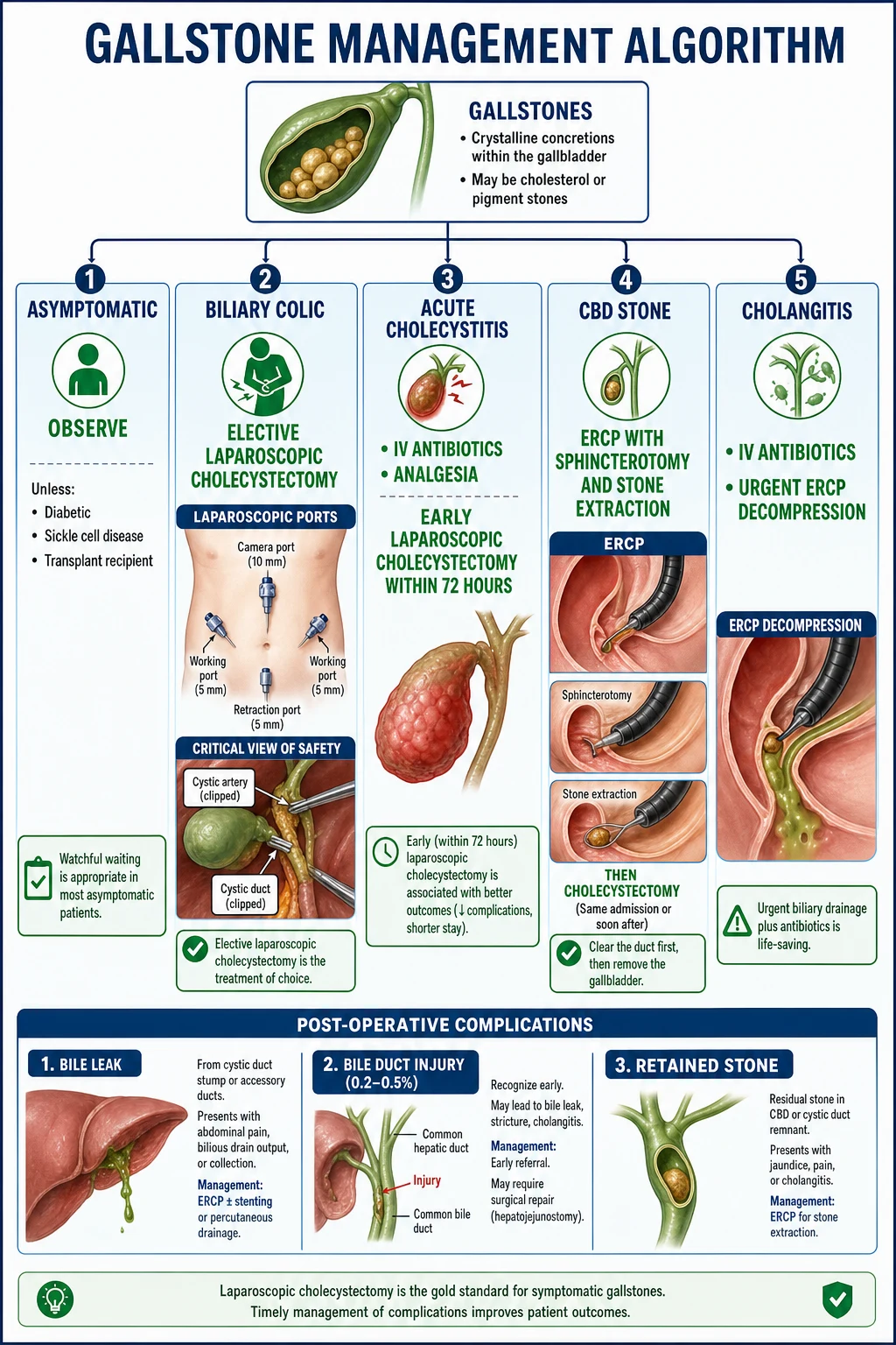
The immediate priority depends on the clinical syndrome. Biliary colic needs analgesia and outpatient referral; acute cholecystitis needs admission, antibiotics, and analgesia with early surgery; cholangitis and pancreatitis are time-critical emergencies requiring resuscitation, broad-spectrum antibiotics, and (for cholangitis) emergency biliary decompression.[1]
Biliary colic — managed in the emergency department with analgesia and discharge on oral NSAIDs, with referral for elective outpatient cholecystectomy. NSAIDs (diclofenac 75 mg intramuscularly) relieve biliary pain by reducing prostaglandin-mediated gallbladder contraction; opioids (morphine 0.1 mg/kg IV, or fentanyl) are added for severe pain. The historical fear of morphine causing sphincter of Oddi spasm is not supported by modern evidence.[1]
Acute cholecystitis — the standard resuscitation bundle:[1]
Co-amoxiclav (amoxicillin-clavulanate)
Dose
1.2 g IV
Plus NPO (nil by mouth), IV fluid resuscitation with crystalloid (Hartmann's or normal saline) to correct dehydration, IV analgesia (morphine, fentanyl, or NSAIDs), and antiemetics. Most patients improve within 24 to 48 hours and proceed to early laparoscopic cholecystectomy. [1]
Ascending cholangitis — a true surgical emergency:[1]
Resuscitate
Oxygen, two large-bore cannulae, IV crystalloid boluses; vasopressors (noradrenaline) for septic shock; ICU if Reynolds pentad or organ dysfunction
Broad-spectrum antibiotics
Piperacillin-tazobactam 4.5 g IV TDS (or ceftriaxone plus metronidazole); take blood cultures first
Urgent biliary decompression
ERCP with sphincterotomy and stone extraction within 24 to 48 hours; if severe, within 12 hours. Stent or nasobiliary drain if stone cannot be removed
Fallback if ERCP fails
Percutaneous transhepatic biliary drainage (PTBD) by interventional radiology; or surgical CBD exploration
Definitive management
Laparoscopic cholecystectomy once sepsis has resolved and the patient is fit
Management — Definitive & Stepwise
Definitive management of gallstone disease is dictated entirely by the clinical syndrome. The unifying principle is that the gallbladder is the source of recurrent stones, so any patient with symptomatic disease — colic, cholecystitis, CBD stone, or pancreatitis — ultimately needs cholecystectomy to prevent recurrence. The exception is gallstone ileus, where the priority is relieving the obstruction and the fistula is allowed to close in its own time.[1]
Asymptomatic gallstones
The default is observation — no surgery, no dietary restriction beyond sensible healthy eating. Only 1 to 4% per year of asymptomatic carriers develop symptoms, and the lifetime risk of serious complications is low. Once symptoms develop, the complication rate rises sharply, which is why symptomatic disease is always treated. A small group has prophylactic cholecystectomy recommended despite being asymptomatic:[1]
- Porcelain gallbladder — cancer risk.
- Stones over 3 cm — increased risk of gallbladder carcinoma.
- Sickle cell disease and other chronic haemolysis — pigment stones; high risk of acute crises if emergency surgery.
- Transplant candidates and immunosuppressed patients — emergency cholecystitis carries high mortality.
- Diabetes mellitus with asymptomatic stones — controversial; many units offer surgery because diabetics tolerate emergency cholecystitis poorly.
- Native American / Pima ancestry — very high progression to symptomatic disease. [1]
Biliary colic
Elective laparoscopic cholecystectomy is the gold standard. It removes the gallbladder and all its stones, abolishing both the source of recurrent stones and the risk of progression to cholecystitis. Surgery is scheduled electively — not as an emergency — because the obstruction is by definition transient and self-limiting. The laparoscopic approach uses four ports (umbilical camera, epigastric working port, right subcostal and lateral retraction ports). The Critical View of Safety — Calot's triangle dissected so that the cystic duct and cystic artery are clearly identified and the hepatocystic triangle is cleared of all tissue down to the liver bed — must be achieved before any clip is applied; this is the single most important safeguard against bile duct injury.[4]
Conversion to open cholecystectomy occurs in 2 to 10% of elective cases (higher in acute cholecystitis, obesity, and previous upper abdominal surgery) and should be regarded as a mark of safe surgical judgement rather than a failure. [1]
Acute cholecystitis
Early laparoscopic cholecystectomy within 72 hours of symptom onset is now the standard of care, supported by the Tokyo Guidelines and multiple meta-analyses. The evidence shows equivalent or better outcomes versus delayed (interval) surgery, with shorter hospital stay, lower readmission rates, no increase in conversion or complication rates, and abolition of the risk of recurrent cholecystitis during the waiting period.[3][5]
If presentation is delayed beyond 72 hours (typically because the patient presented late), the inflamed gallbladder is more difficult to dissect safely and the standard approach shifts to conservative management with IV antibiotics followed by interval laparoscopic cholecystectomy at 6 to 8 weeks, after inflammation has settled. Severe cholecystitis with complications (gangrene, perforation, empyema) may require open cholecystectomy or, in the unfit patient, percutaneous cholecystostomy as a bridge to later surgery.[6]
Percutaneous cholecystostomy
Dose
8 to 12 Fr pigtail catheter
Choledocholithiasis (CBD stones)
The principle is clear the duct, then remove the gallbladder.[1]
- ERCP with sphincterotomy — the sphincter of Oddi is cut with a sphincterotome, the CBD stone is extracted with a balloon catheter or Dormia basket, and patency is confirmed with cholangiography. A plastic stent may be placed if stone clearance is incomplete, with a plan for a second ERCP.
- Laparoscopic cholecystectomy during the same admission or within 2 weeks — to prevent the gallbladder generating further stones that re-obstruct the CBD.
- Laparoscopic CBD exploration (LCBDE) — a one-stage alternative available in specialist centres, where the CBD is opened (choledochotomy) or the cystic duct is dilated and the stone retrieved transcystically at the same operation as the cholecystectomy. Avoids ERCP and its complications in selected patients. [1]
Ascending cholangitis
Management follows the resuscitation bundle above: IV fluids and vasopressors, broad-spectrum antibiotics, and urgent ERCP for biliary decompression. The decompression must achieve drainage of the infected bile — removing the stone if possible, or placing a stent or nasobiliary drain if the stone cannot be retrieved at the first attempt. Definitive cholecystectomy follows once the patient has recovered.[1]
Gallstone pancreatitis
Most episodes are mild and self-limiting, managed conservatively with aggressive IV fluid resuscitation, analgesia, and monitoring. Early ERCP within 24 hours is indicated only if there is coexistent cholangitis or ongoing biliary obstruction (persistent jaundice and dilated CBD); routine ERCP in mild pancreatitis without obstruction is harmful and not recommended. Same-admission cholecystectomy (after recovery from the acute pancreatitis, before discharge) prevents recurrence — without it, recurrence rates of 25 to 30% are reported within weeks.[1]
Gallstone ileus
Laparotomy with enterolithotomy is the standard treatment: a longitudinal enterotomy is made in the ileum proximal to the impacted stone, the stone is milked out, and the enterotomy is closed transversely to avoid stricture. Run the entire small bowel to exclude a second stone (present in up to 5%). The cholecystoenteric fistula is left alone at the index operation because cholecystectomy with fistula repair in these elderly, septic patients carries prohibitive mortality; the fistula closes spontaneously in most, and only a minority develop recurrent biliary symptoms requiring later cholecystectomy.[1]
Empyema, mucocele, porcelain gallbladder
Empyema — IV antibiotics plus percutaneous cholecystostomy to drain the pus, followed by interval cholecystectomy once sepsis resolves.[6]
Mucocele (hydrops) — elective cholecystectomy, as the sterile obstruction will progress to infection or gangrene if left.[1]
Porcelain gallbladder — prophylactic cholecystectomy recommended, ideally laparoscopic; the patchy pattern of calcification carries the highest cancer risk and warrants a careful intraoperative frozen section, with conversion to an extended (radical) resection if adenocarcinoma is found.[1]
Specific Subtypes & Scenarios
-
Mirizzi syndrome: a stone impacted in Hartmann's pouch (the infundibulum of the gallbladder) externally compresses the common hepatic duct, producing obstructive jaundice with a gallbladder rather than a CBD stone. Classified Type I (external compression only) through Type V (cholecystoenteric fistula); Type II to IV involve a cholecystocholedochal fistula. Mirizzi mimics a CBD stone and is a classic trap for laparoscopic surgery because the distorted anatomy sharply raises the risk of CBD injury. Diagnosis is by MRCP; treatment is open or laparoscopic (in expert hands) cholecystectomy with careful duct identification, or cholecystoduodenostomy/choledochoenterostomy for the fistula subtypes.[1]
-
Biliary sludge: microcrystals of cholesterol and calcium bilirubinate in mucus, seen on USS as low-level echoes that layer. Causes the same symptoms as stones, may progress to stones, and resolves on its own in pregnancy or after withdrawal of the offending drug (ceftriaxone, octreotide). Symptomatic sludge is treated like symptomatic stones — cholecystectomy.[2]
-
Post-cholecystectomy syndrome: persistence or recurrence of RUQ pain after cholecystectomy, affecting 5 to 40% of patients. Causes include sphincter of Oddi dysfunction, retained or recurrent CBD stones, a long cystic duct stump, bile salt malabsorption, and functional (irritable bowel-like) disease. Investigation is with LFTs, MRCP, and EUS, and management targets the underlying cause.[1]
-
Acalculous cholecystitis: acute cholecystitis without gallstones, accounting for 5 to 10% of cases. Typically affects the critically ill (ICU patients, severe burns, trauma, sepsis, prolonged fasting, TPN). The pathophysiology is bile stasis and gallbladder ischaemia rather than mechanical obstruction. Diagnosis is challenging (the patient is often intubated and cannot report pain); USS shows a distended, thick-walled gallbladder with sludge but no stones. Treatment is broad-spectrum antibiotics and percutaneous cholecystostomy because these patients tolerate surgery poorly. Mortality is high (around 30%).[1]
Complications & Pitfalls
Gallstone complications divide into disease-related (from untreated or progressive disease) and procedure-related (from laparoscopic cholecystectomy and ERCP). Both are heavily examined.[2]
Disease complications
if untreated
- Acute cholecystitis (20 to 30% of symptomatic patients)
- Empyema, gangrene, perforation (complications of untreated cholecystitis)
- Choledocholithiasis (10 to 15% of patients with gallstones)
- Ascending cholangitis (mortality up to 30% if untreated)
- Gallstone pancreatitis (mortality approximately 5%)
- Gallstone ileus (rare; elderly women)
- Mirizzi syndrome, gallbladder carcinoma
Cholecystectomy complications
procedure-related
- **Bile duct injury (0.2 to 0.5%)** — most feared; prevented by Critical View of Safety; may need hepaticojejunostomy
- Bile leak (1 to 2%) — from cystic duct stump or duct of Luschka; managed by ERCP plus stent
- Retained CBD stone (1 to 5%) — postoperative jaundice or pancreatitis; ERCP extraction
- Post-cholecystectomy syndrome (5 to 40%) — persistent pain
- Bleeding (0.5%), infection (1 to 2%), port-site hernia
- Conversion to open (2 to 10%)
ERCP complications
5 to 10% overall
- **Post-ERCP pancreatitis (around 5%)** — commonest; reduced by rectal diclofenac and prophylactic pancreatic stent
- Bleeding (2%) — from sphincterotomy
- Perforation (0.3%) — of the duodenum or CBD
- Cholangitis (1%) — from incomplete drainage
Classic pitfalls — the errors examiners test:[1]
- Not distinguishing biliary colic from cholecystitis. Colic resolves within 6 hours and has no fever; cholecystitis persists beyond 6 hours and has fever plus Murphy sign. Colic gets elective surgery; cholecystitis gets antibiotics plus early surgery.
- Not checking LFTs. Abnormal LFTs suggest a CBD stone; MRCP is needed before cholecystectomy, or ERCP first if obstructive jaundice.
- Missing Courvoisier's law. Jaundice plus a palpable gallbladder is malignant obstruction, not stones.
- Performing cholecystectomy before clearing a known CBD stone. The CBD stone should be removed first (ERCP) or at the same operation (LCBDE), then cholecystectomy.
- Delaying cholecystectomy in acute cholecystitis. Early surgery within 72 hours is safe and superior to a prolonged conservative course.
- Delaying ERCP in cholangitis. Decompression cannot wait for further imaging in a septic, jaundiced patient — resuscitate in parallel and call the endoscopist.
- Inadequate Critical View of Safety. The single preventable cause of bile duct injury during laparoscopic cholecystectomy. If the view cannot be obtained, convert to open or call a senior colleague. [1]
Prognosis & Disposition
Asymptomatic gallstones — benign natural history. Only 1 to 4% per year become symptomatic; most carriers never require surgery. Once symptoms develop, however, the annual risk of complications rises to over 5%, which is why symptomatic disease is treated.[2]
After laparoscopic cholecystectomy — excellent outcome. Most patients return to normal activity within 1 to 2 weeks (compared with 4 to 6 weeks after open cholecystectomy). Over 90% are permanently cured of their biliary symptoms. Post-cholecystectomy syndrome affects 5 to 10% and should be investigated if pain persists beyond the recovery period. Mild diarrhoea (bile salt malabsorption) affects up to 10% and usually settles; persistent cases respond to bile acid sequestrants such as cholestyramine.[1]
Acute cholecystitis — mortality 0.5% in uncomplicated disease in the fit adult, rising to 10% or more in the elderly, diabetic, or those with gangrenous or perforated gallbladders. Early laparoscopic cholecystectomy has transformed outcomes.[1]
Ascending cholangitis — mortality 5 to 30%, driven by severity at presentation (Reynolds pentad), delay to decompression, and comorbidity. The single biggest determinant of survival is time to biliary drainage.[1]
Gallstone pancreatitis — overall mortality around 5%, rising to over 30% in severe necrotising disease. Same-admission cholecystectomy prevents the high recurrence rate seen when the gallbladder is left in situ.[1]
Gallbladder carcinoma — dismal; 5-year survival under 10% for advanced disease, but over 80% for an incidental T1a tumour found at cholecystectomy. This is why large stones (over 3 cm) and porcelain gallbladder warrant prophylactic cholecystectomy.[2]
Special Populations
-
Diabetes mellitus: diabetics have a higher incidence of gallstones (impaired motility) and a markedly higher rate of complications when cholecystitis develops — empyema, gangrene, perforation, and sepsis. Prophylactic cholecystectomy for asymptomatic stones is offered in many units, and any diabetic with acute cholecystitis is treated aggressively and early.[1]
-
Pregnancy: biliary colic is common (progesterone slows gallbladder motility; oestrogen raises bile cholesterol). The safest time for elective cholecystectomy is the second trimester, after organogenesis (first trimester) and before the uterus obstructs the operative field (third trimester). Acute cholecystitis in pregnancy is treated surgically rather than conservatively because delay risks preterm labour and recurrent admissions. ERCP is performed with lead shielding of the uterus and minimal fluoroscopy when essential for a CBD stone; MRCP is preferred for diagnosis because it avoids radiation entirely.[1]
-
Sickle cell disease and other haemolytic disorders: chronic haemolysis produces pigment stones from infancy. Prophylactic cholecystectomy is recommended once stones are detected, because emergency cholecystectomy during an acute pain crisis carries a high risk of precipitating a sickle cell crisis and multi-organ failure. Pre-operative transfusion to maintain haemoglobin S below 30% reduces peri-operative risk.[1]
-
Children: gallstones are uncommon and usually pigment stones from haemolytic disease (spherocytosis, sickle cell, thalassaemia) or from total parenteral nutrition in premature neonates. Prophylactic cholecystectomy is offered for haemolytic disorders; cholesterol stones in obese adolescents are an emerging problem mirroring the obesity epidemic.[1]
-
The elderly: atypical presentations are the rule rather than the exception — confusion rather than localised pain, acalculous cholecystitis, higher rates of gangrene and perforation, and Courvoisier-type malignant presentations. A low threshold for imaging and a low threshold for early surgery (where fit) improves outcomes; percutaneous cholecystostomy is a valuable bridge in the frail.[6]
India: gallstones are extremely common, driven by dietary change (refined carbohydrates, high-fat diet), rising urban obesity, and parasitic infestation. In endemic rural areas, Ascaris lumbricoides and Clonorchis sinensis migrate into the biliary tree, where they cause brown pigment stones, biliary stasis, and ascariasis-induced cholangitis — a presentation rare in the West. Sickle cell disease in tribal populations (Odisha, Chhattisgarh, Maharashtra) produces pigment stones in children. Access to ERCP may be limited in rural settings, in which case open cholecystectomy with on-table cholangiography and CBD exploration remains the only option. The clinical principles and the severity ladder are identical everywhere; only the infrastructure and the dominant aetiology differ.[1]
Evidence, Guidelines & Regional Differences
The Tokyo Guidelines 2018 (TG18) are the international standard for the diagnosis, severity grading, and management of acute cholecystitis and cholangitis.[3][4]
Tokyo Guidelines 2018 (TG18)
PMID 29032636, 29095575
International consensus guideline (Japan, 2007 first; updated 2013, 2018)
Population: Acute cholecystitis and acute cholangitis
Key finding
Standardised diagnostic criteria (one local sign plus systemic inflammation plus imaging) and a three-tier severity grading: Grade I (mild) suitable for early laparoscopic cholecystectomy; Grade II (moderate) with marked local inflammation or systemic compromise; Grade III (severe) with organ dysfunction requiring critical care and delayed surgery
TG18 severity grading of acute cholecystitis:[3]
- Grade I (mild): no organ dysfunction and no severe local inflammation. Early laparoscopic cholecystectomy is the treatment of choice.
- Grade II (moderate): marked local inflammation (gangrenous, pericholecystic abscess, hepatic abscess) or systemic compromise (WCC over 18,000, palpable RUQ mass, duration over 72 hours). Early or delayed surgery depending on context; percutaneous drainage considered if unfit.
- Grade III (severe): organ dysfunction (cardiovascular, neurological, respiratory, renal, hepatic, haematological) requiring critical care. Emergency biliary drainage (cholecystostomy) and intensive care; cholecystectomy is deferred until organ failure resolves. [1]
Early versus delayed cholecystectomy for acute cholecystitis — multiple systematic reviews and meta-analyses confirm that laparoscopic cholecystectomy within 72 hours of symptom onset is safe, has equivalent complication and bile duct injury rates, shorter hospital stay, lower readmission rates, and lower total cost compared to delayed (interval) surgery at 6 to 8 weeks. This has displaced the older "cool down" strategy as the global standard.[5]
Percutaneous cholecystostomy for high-risk acute cholecystitis — cohort data and meta-analyses show that drainage controls sepsis in 80 to 90% of unfit patients and is a safe bridge to interval cholecystectomy or, in the very frail, a definitive treatment. Mortality remains substantial (10 to 20%) reflecting the underlying comorbidity rather than the procedure itself.[6]
Ursodeoxycholic acid (UDCA) for gallstone dissolution — for carefully selected patients with small (under 10 mm), floating, cholesterol stones in a functioning gallbladder (confirmed by HIDA or oral cholecystography), UDCA at 10 to 15 mg/kg/day orally in divided doses can dissolve stones over 6 to 24 months in up to 60%. Recurrence is common (50% within 5 years) and patient selection is narrow, so UDCA is now reserved for patients unfit for surgery or who decline it. It is not a substitute for cholecystectomy in symptomatic disease.[1]
Prevention — beyond treating symptomatic disease, prevention centres on addressing modifiable risk factors: gradual weight loss (no more than 1.5 kg per week) after bariatric surgery, avoiding prolonged fasting in patients on TPN (intermittent CCK stimulation or UDCA prophylaxis), and lifestyle measures (regular meals, high-fibre diet, regular exercise). In patients on octreotide long-term, prophylactic UDCA reduces sludge and stone formation.[2]
Exam Pearls
- 5 Fs: Female, Fat, Forty, Fertile, Family. Cholesterol stones (80%, radiolucent). Pigment stones (20%, from haemolysis or infection, radiopaque).[1]
- Biliary colic: pain 2 to 6 h, resolves spontaneously, no fever. Elective lap chole. Cholecystitis: pain over 6 h plus fever plus Murphy. Early lap chole within 72 h.[1]
- Charcot triad (fever plus jaundice plus RUQ pain) = cholangitis. Reynolds pentad adds hypotension plus confusion. Urgent ERCP.[1]
- Courvoisier law: palpable GB plus jaundice = NOT stones (malignant obstruction — pancreatic head, cholangiocarcinoma).[1]
- CBD stone: ERCP plus sphincterotomy plus extraction. Then cholecystectomy. MRCP before ERCP to confirm if intermediate risk.[1]
- Gallstone ileus: Rigler triad (pneumobilia plus SBO plus ectopic stone). Obstruction at terminal ileum. Enterolithotomy; leave the fistula.[1]
- Murphy sign: inspiratory arrest on RUQ palpation. Sensitivity 65%, specificity 87% for cholecystitis.[1]
- Boas sign: hyperaesthesia below the right scapula.[1]
- Bile duct injury 0.2 to 0.5%. Prevented by Critical View of Safety. Mirizzi: open or expert laparoscopic; high risk of CBD injury.[4]
- Porcelain gallbladder: prophylactic cholecystectomy (cancer risk 2 to 3%). Stones over 3 cm: prophylactic cholecystectomy (carcinoma risk).[1]
- Empyema: percutaneous cholecystostomy. Mucocele (hydrops): elective cholecystectomy. Acalculous cholecystitis: ICU, burns, sepsis — cholecystostomy.[6]
- Gallstone pancreatitis: same-admission cholecystectomy prevents recurrence. Early ERCP within 24 h only if cholangitis or ongoing obstruction.[1]
- Ursodeoxycholic acid: only for small, floating, cholesterol stones in a functioning gallbladder, in the unfit. 50% recurrence.[1]
Quick self-test — cover the answers
- Charcot triad? — Fever, jaundice, RUQ pain.
- Reynolds pentad? — Charcot plus hypotension plus confusion.
- Rigler triad? — Pneumobilia plus small bowel obstruction plus ectopic gallstone.
- The 5 Fs? — Female, Fat, Forty, Fertile, Family.
- Courvoisier law? — Palpable gallbladder plus jaundice is NOT stones — it is malignant obstruction.
- First-line imaging? — Ultrasound (95% sensitive for stones over 2 mm).
- Critical View of Safety prevents what? — Bile duct injury during laparoscopic cholecystectomy.
- Empyema — first step? — Percutaneous cholecystostomy plus IV antibiotics.
Spectrum map and definitive procedures (exam detail)
Gallstone disease is a spectrum, not a single operation: [1]
| Syndrome | Key clinical cue | First-line definitive step |
|---|---|---|
| Asymptomatic stones | Incidental US | Observe (exceptions: porcelain GB, large polyps, selected high-risk) |
| Biliary colic | Severe RUQ/epigastric pain under 6 h, afebrile | Elective lap chole |
| Acute cholecystitis | Prolonged pain, fever, Murphy+ | Early lap chole (see acute-cholecystitis topic) |
| Choledocholithiasis | Abnormal LFT ± duct dilatation ± pancreatitis/cholangitis | ERCP stone extraction ± sphincterotomy, then cholecystectomy |
| Ascending cholangitis | Charcot triad (± Reynolds pentad) | Resuscitate + urgent biliary drainage (ERCP) |
| Gallstone pancreatitis | Pain + raised lipase/amylase + stones | Supportive care; cholecystectomy this admission once settled (mild); ERCP if cholangitis/obstruction |
| Gallstone ileus | SBO + pneumobilia + ectopic stone (Rigler triad) | Resuscitate; enterolithotomy ± fistula management strategy |
| Mirizzi | Stone compressing CHD/CBD | Careful imaging; expert surgery / endoscopic options |
Stone types (pathophysiology viva):
- Cholesterol stones (~80% West): supersaturated bile, gallbladder hypomotility — 5 Fs risk.
- Black pigment: haemolysis, cirrhosis — unconjugated bilirubin.
- Brown pigment: infection/stasis in ducts — more common in Asia for primary duct stones. [1]
ERCP essentials:
- Indications: cholangitis with obstruction, retained CBD stones, selected severe gallstone pancreatitis with ongoing obstruction.
- Risks: pancreatitis, bleeding, perforation, cholangitis — consent must name them.
- MRCP is non-invasive duct imaging when intermediate probability of CBD stone; EUS alternative. [1]
Laparoscopic cholecystectomy remains definitive for symptomatic gallbladder stones; IOC selective. Never claim medical dissolution is standard first-line for typical symptomatic calculi. [1]
Worked clinical stems (answer these without another book)
Stem A — Colic. 40 F, 3 h severe RUQ pain after pizza, resolved, normal WBC, US multiple stones.
Biliary colic; analgesia; elective lap chole; lifestyle advice meantime. [1]
Stem B — Charcot. 70 M fever, jaundice, RUQ pain, BP 85 systolic, confused.
Reynolds pentad = severe cholangitis. ABC, cultures, IV piperacillin-tazobactam 4.5 g TDS, urgent ERCP decompression, later cholecystectomy if GB stones. [1]
Stem C — Pancreatitis. 55 F epigastric pain to back, lipase 12× ULN, US stones, mild course.
Gallstone pancreatitis; supportive care (fluids, analgesia); index-admission cholecystectomy when pancreatitis settled if mild; ERCP only if cholangitis/persistent obstruction. [1]
Stem D — Ileus. 78 F SBO, pneumobilia on CT, stone in ileum.
Gallstone ileus via cholecystoenteric fistula; enterolithotomy; address fistula selectively (one- vs two-stage debate). [1]
Stem E — Post-chole pain + abnormal LFT.
Retained CBD stone vs bile leak vs non-biliary; LFTs, US/MRCP; ERCP if confirmed duct stone/leak management. [1]
Stem F — Courvoisier. Painless jaundice, palpable non-tender GB.
Not typical stone disease — periampullary/pancreatic head malignancy until proven otherwise. [1]
OSCE / short-case performance script
- Abdominal exam with Murphy technique; look for jaundice/scratch marks.
- State spectrum diagnosis aloud.
- Request US first; explain when MRCP/ERCP.
- Explain Charcot vs cholecystitis management difference (ERCP vs chole).
- Consent for lap chole: conversion, BDI, retained stones, diarrhoea.
- Safety-net for cholangitis and pancreatitis symptoms. [1]
Extended viva bank (model outlines)
- 5 Fs and stone composition.
- Charcot vs Reynolds.
- ASGE predictors of choledocholithiasis (very strong / strong / moderate).
- Timing of chole after gallstone pancreatitis.
- Rigler triad of gallstone ileus.
- Mirizzi types outline.
- Why asymptomatic stones usually observed.
- Complications of ERCP.
- Bile acid diarrhoea after cholecystectomy.
- Gallbladder cancer and stone disease association (north India). [1]
Common exam traps (fail patterns)
- Cholecystectomy alone for cholangitis without duct drainage.
- Delayed chole after mild gallstone pancreatitis → readmission.
- Assuming Courvoisier GB is stones.
- Dissolution therapy as default.
- Missing gallstone ileus as cause of SBO in elderly. [1]
Self-check coverage map
| Examiner dimension | Covered? |
|---|---|
| Definitions across spectrum | Yes |
| Epidemiology & stone types | Yes |
| Pathophysiology of formation | Yes |
| Clinical syndromes | Yes |
| Differentials | Yes |
| Bedside signs | Yes |
| Imaging ladder US→MRCP→ERCP | Yes |
| Cholangitis resuscitation + ERCP | Yes |
| Cholecystectomy timing | Yes |
| Pancreatitis / ileus / Mirizzi | Yes |
| Complications | Yes |
| Special populations | Yes |
| Guidelines & regional | Yes |
| Exam pearls | Yes |
References
- [1]Demehri FR, Alam HB Evidence-Based Management of Common Gallstone-Related Emergencies J Intensive Care Med, 2016.PMID 25320159
- [2]Gupta V, Abhinav A, Vuthaluru S, et al. The Multifaceted Impact of Gallstones: Understanding Complications and Management Strategies Cureus, 2024.PMID 39022477
- [3]Yokoe M, Hata J, Takada T, et al. Tokyo Guidelines 2018: diagnostic criteria and severity grading of acute cholecystitis (with videos) J Hepatobiliary Pancreat Sci, 2018.PMID 29032636
- [4]Wakabayashi G, Iwashita Y, Hibi T, et al. Tokyo Guidelines 2018: surgical management of acute cholecystitis: safe steps in laparoscopic cholecystectomy for acute cholecystitis (with videos) J Hepatobiliary Pancreat Sci, 2018.PMID 29095575
- [5]Nassar A, Ng HJ, Cao Y, et al. Outcome of early cholecystectomy compared to percutaneous drainage of gallbladder and delayed cholecystectomy for patients with acute cholecystitis: systematic review and meta-analysis HPB (Oxford), 2022.PMID 35597717
- [6]Turino SY, Barreda C, Grande J, et al. Percutaneous Cholecystostomy Versus Conservative Treatment for Acute Cholecystitis: a Cohort Study J Gastrointest Surg, 2019.PMID 30390182