General Surgery · General Surgery
Gastric Carcinoma
Also known as Stomach cancer · Gastric cancer · Carcinoma stomach · Linitis plastica
Gastric carcinoma is an adenocarcinoma arising from the gastric epithelium. It is the fifth most commonly diagnosed cancer worldwide and the fourth leading cause of cancer death, with the highest incidence in East Asia (Japan, Korea, China), Eastern Europe, and parts of South America. The dominant risk factor is Helicobacter pylori (a WHO Class I carcinogen), followed by diet (salted/smoked/pickled foods, N-nitroso compounds, low fruit and vegetables), smoking, chronic atrophic gastritis, pernicious anaemia, and the hereditary diffuse gastric cancer syndrome (CDH1/E-cadherin mutation). Most tumours arise through the Correa cascade (chronic gastritis to atrophic gastritis to intestinal metaplasia to dysplasia to adenocarcinoma) over decades. Presentation is characteristically late and insidious — epigastric pain, weight loss, early satiety, dysphagia (proximal), vomiting (distal), and anaemia — with eponymous metastatic signs (Virchow's node, Sister Mary Joseph nodule, Krukenberg tumour, Blumer's shelf). Diagnosis is by OGD with biopsy; staging combines CT, EUS, and staging laparoscopy (for occult peritoneal disease). Surgery — gastrectomy with D2 lymphadenectomy — is the only cure; perioperative FLOT chemotherapy (built on the MAGIC trial) is standard for resectable locally advanced disease; trastuzumab is added for HER2-positive and nivolumab/pembrolizumab for PD-L1-positive or MSI-H metastatic disease. Prognosis is poor overall (20 to 30 per cent five-year survival) because of late presentation, though Japan and Korea achieve far better outcomes through national endoscopic screening.
On this page & tools
Your progress
Saved locally on this device.
Exam tags
Red flags
Overview & Definition
Gastric carcinoma is a malignant epithelial tumour arising from the glandular epithelium of the stomach, of which adenocarcinoma accounts for about 95 per cent of all gastric malignancies. The remainder are lymphoma (predominantly MALT), gastrointestinal stromal tumour (GIST), and neuroendocrine tumours. Most gastric adenocarcinomas arise through a slow, multistep inflammatory pathway first described by Pelayo Correa in 1975 — the Correa cascade — in which chronic mucosal injury (most often driven by Helicobacter pylori) progresses over 20 to 40 years from chronic gastritis, to atrophic gastritis, to intestinal metaplasia, to dysplasia, and finally to invasive adenocarcinoma.[2]
The central clinical problem is lateness. The stomach is a capacious, highly distensible organ; an early tumour is asymptomatic or produces dyspepsia indistinguishable from benign peptic ulcer disease, so patients present only when the tumour has invaded the muscular wall, obstructed the lumen, or metastasised. This is why the global five-year survival sits at only 20 to 30 per cent, even though a tumour confined to the mucosa or submucosa (early gastric cancer) is curable in over 90 per cent of cases. The striking difference in outcome between Japan and Korea (where national endoscopic screening detects early disease) and the West and South Asia (where presentation is advanced) is one of the most examined facts in surgical oncology and is the core justification for screening in high-incidence populations.[1][4]
Classification
Gastric cancer is classified along four axes that the examiner expects you to hold simultaneously: histology (Lauren), anatomical site, depth of invasion (early versus advanced), and TNM stage (AJCC 8th edition). Each axis drives a different decision — prognosis, operative approach, and adjuvant therapy — so a candidate who can place a tumour on all four axes can answer almost any classification question. [1]
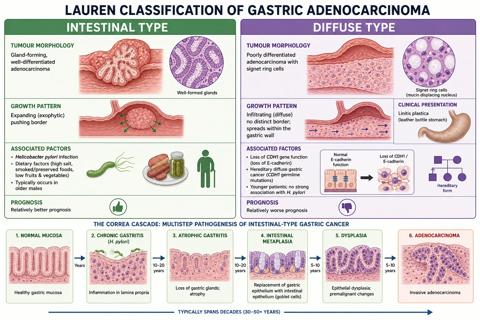
By histology — the Lauren classification (1965) is the exam favourite:[1]
- Intestinal type (Lauren type 1) — forms glandular structures, is usually well to moderately differentiated, and grows with an expanding, pushing margin. It is strongly associated with environmental factors — H. pylori, salted and smoked diet, and chronic atrophic gastritis — and follows the Correa cascade. It predominates in high-incidence regions and in older males, and carries the better prognosis of the two.
- Diffuse type (Lauren type 2) — composed of poorly differentiated, discohesive cells, many of which are signet ring cells (a mucin-laden cytoplasm that displaces the nucleus to the periphery). It grows by infiltration rather than expansion, is not clearly linked to H. pylori or diet, predominates in younger patients and women, and is driven in the hereditary form by loss of E-cadherin (CDH1 mutation). When it infiltrates the entire gastric wall it produces linitis plastica — the rigid, thickened, non-distensible "leather bottle" stomach. Prognosis is poor.
- Mixed type — contains both glandular and signet ring components. [1]
The WHO histological classification (tubular, papillary, mucinous, poorly cohesive including signet ring) is more granular and is what the pathologist reports, but the Lauren classification is what the examiner asks for because it separates the two biologically distinct diseases with different epidemiology, genetics, and prognosis. [1]
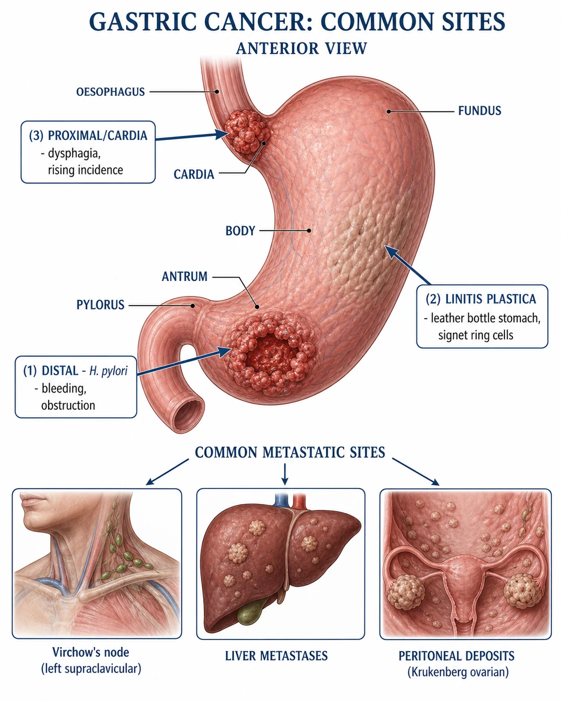
By anatomical site: the antrum and pylorus (distal stomach) is the commonest site worldwide and is the site most strongly associated with H. pylori; its incidence is falling globally. The cardia and proximal stomach at the gastro-oesophageal junction is rising rapidly in Western countries, paralleling obesity and reflux disease, and behaves biologically like a distal oesophageal adenocarcinoma. The body is the classic intermediate location. Diffuse involvement of the whole stomach is the signature of linitis plastica. [1]
By depth of invasion: early gastric cancer (EGC) is confined to the mucosa or submucosa regardless of lymph-node status; it carries a cure rate above 90 per cent and is the target of screening and of endoscopic submucosal dissection. Advanced gastric cancer invades the muscularis propria or beyond and has a much poorer outlook. The Japanese also recognise a macroscopic (Borrmann) classification for advanced tumours: type I polypoid, type II ulcerating with raised edges, type III ulcerating with infiltration, and type IV diffusely infiltrating (linitis plastica). [1]
By TNM stage (AJCC Cancer Staging Manual, 8th edition):[1]
| Stage | TNM (simplified) | Approximate 5-year survival |
|---|---|---|
| I | T1 to T2, N0, M0 | 70 to 90% |
| II | T2 to T3, N0 to N1, M0 | 45 to 55% |
| III | T3 to T4 or N2 to N3, M0 | 15 to 30% |
| IV | Any T, Any N, M1 | under 5% |
The T stage is defined by depth — T1 mucosa or submucosa, T2 muscularis propria, T3 subserosa, T4a serosa, T4b adjacent organs. N staging counts involved nodes (N1 one to two, N2 three to six, N3 seven or more), which is why an adequate lymphadenectomy (at least 16 nodes, ideally 25 or more) is needed both to stage accurately and to clear disease. M1 is any distant metastasis including non-regional nodes and peritoneal deposits. [1]
Intestinal type
Lauren type 1
- **Gland-forming**, well-differentiated; pushing margin
- Driven by **H. pylori, salted/smoked diet**, Correa cascade
- **Older males**, high-incidence regions; distal stomach
- **Better prognosis**
Diffuse type
Lauren type 2
- **Signet ring cells**, poorly cohesive; infiltrating margin
- **CDH1/E-cadherin loss**; hereditary diffuse gastric cancer
- **Younger patients, women**; causes **linitis plastica**
- **Poorer prognosis**
Epidemiology & Risk Factors
Gastric cancer is the fifth most commonly diagnosed cancer and the fourth leading cause of cancer death worldwide, with over one million new cases each year and a case fatality that still exceeds 70 per cent globally. Its epidemiology is defined by two features the examiner rewards: a dramatic geographic gradient, and a single dominant, modifiable cause. [1]
Geography is striking. The highest incidence is in East Asia (Japan, Korea, and China), Eastern Europe, and the Andean region of South America, where age-standardised rates are up to ten times those of the lowest-incidence countries. North America, Western Europe, much of Africa, and South Asia have the lowest rates. Men are affected roughly twice as often as women, and the distal (non-cardia) tumours that dominate high-incidence regions are declining worldwide, while proximal/cardia tumours are rising in the West in parallel with obesity and gastro-oesophageal reflux.[1]
Helicobacter pylori is the single most important risk factor and the reason this is now considered a largely preventable cancer. The WHO International Agency for Research on Cancer classifies H. pylori as a Group 1 (definite) human carcinogen. Chronic colonisation drives the Correa cascade over decades; only a minority of infected individuals ever develop cancer, but eradication of H. pylori reduces the subsequent gastric cancer risk, which underpins population-level test-and-treat strategies in high-prevalence countries.[2]
Gastric cancer at a glance
Non-modifiable risk factors: blood group A confers a modest increase in intestinal-type cancer; a personal history of chronic atrophic gastritis, intestinal metaplasia, or pernicious anaemia marks a mucosa already moving along the Correa cascade; prior partial gastrectomy (especially Billroth II) raises risk after 15 to 20 years; and several hereditary syndromes carry a markedly elevated lifetime risk — most importantly hereditary diffuse gastric cancer (HDGC) from a germline CDH1 (E-cadherin) mutation (autosomal dominant, 70 to 80 per cent lifetime risk of diffuse gastric cancer and increased lobular breast cancer), Lynch syndrome (especially MLH1/MSH2), familial adenomatous polyposis, and the hamartomatous polyposes (Peutz-Jeghers, juvenile polyposis). [1]
Modifiable risk factors: the diet that drives intestinal-type cancer is one rich in salted, smoked, and pickled foods (which deliver N-nitroso compounds and salt that damages the mucosal barrier) and poor in fresh fruit and vegetables (removing the antioxidant protection of vitamin C and beta-carotene). Tobacco smoking approximately doubles the risk. Obesity is linked mainly to proximal/cardia tumours. Epstein-Barr virus is found in about 8 to 10 per cent of gastric cancers (a distinct molecular subtype with a better-than-average prognosis), and occupational exposure to coal, metal, and rubber industries carries a small excess risk.[1][2]
Pathophysiology
The pathogenesis of gastric cancer divides cleanly along the Lauren line: the intestinal type is the endpoint of a long inflammatory cascade, while the diffuse type is driven by loss of cell-cell adhesion. Examiners test both mechanisms. [1]
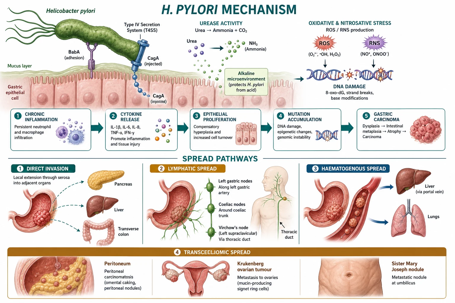
The Correa cascade — intestinal-type carcinogenesis. The cascade begins when H. pylori colonises the gastric mucus layer and adheres to surface epithelial cells. The bacterium expresses virulence factors that amplify injury: BabA binds the Lewis blood-group antigen on epithelium, the CagA protein is injected into the host cell through a type IV secretion system (where it is phosphorylated and disrupts cell signalling, stimulating proliferation and resisting apoptosis), VacA induces vacuolation and suppresses immune killing, and urease generates ammonia that neutralises luminal acid. The resulting chronic neutrophilic and monocytic inflammation generates reactive oxygen and nitrogen species that damage DNA. As glands are destroyed, the mucosa progresses to atrophic gastritis (loss of specialised glands, falling acid output), then to intestinal metaplasia (gastric epithelium replaced by goblet-cell intestinal-type epithelium), then to dysplasia (cytological atypia and disordered architecture), and finally to invasive adenocarcinoma when atypical cells breach the basement membrane. The whole sequence unfolds over 20 to 40 years, which is why cancer peaks in the sixth and seventh decades. As acid falls with advancing atrophy, bacterial overgrowth of the stomach produces further N-nitroso compounds that are themselves direct carcinogens, accelerating the late steps.[2]
The diffuse type bypasses the glandular cascade. The defining event is loss of E-cadherin (encoded by CDH1), the transmembrane protein that forms the adherens junctions binding epithelial cells together. Without E-cadherin the cells lose cohesion and polarity, become motile, and infiltrate the wall singly as signet ring cells laden with intracellular mucin. In the hereditary form the CDH1 mutation is germline; in sporadic diffuse tumours E-cadherin is lost by mutation, promoter hypermethylation, or aberrant cleavage. Diffuse infiltration of all layers produces linitis plastica — a thick, rigid, leather-bottle stomach with a characteristically shrunken lumen. [1]
Patterns of spread dictate the clinical and surgical approach: [1]
- Direct extension through the gastric wall to adjacent organs — pancreas, transverse colon and mesocolon, spleen, left lobe of liver, and the diaphragm.
- Lymphatic spread follows the perigastric and then the named arterial chains — left gastric, common hepatic, splenic, coeliac, hepatic-duodenal, and ultimately para-aortic nodes. The eponymous terminus is Virchow's node (the left supraclavicular node, reached via the thoracic duct); its palpability is Troisier's sign.
- Haematogenous spread seeds the liver (commonest solid-organ site) and the lungs; bone and brain are later.
- Transcoelomic spread across the peritoneal cavity produces peritoneal carcinomatosis with ascites, the Krukenberg tumour (a metastatic deposit in the ovary, classically bilateral and signet-ring, reached retrogradely through the peritoneum), the Sister Mary Joseph nodule (an umbilical metastasis along the ligamentum teres and paraumbilical vessels), and Blumer's shelf (a palpable anterior rectal-wall mass from a pelvic peritoneal deposit). [1]
The Correa cascade — intestinal gastric carcinogenesis
CAMDIA
H. pylori colonises mucosa, neutrophilic inflammation
loss of specialised glands, falling acid output
intestinal-type epithelium replaces gastric mucosa
cytological atypia, disordered architecture
atypical cells breach basement membrane
established intestinal-type malignancy
Clinical Presentation
The hallmark of gastric cancer is delay. Early disease is silent or mimics functional dyspepsia; the classic "cancer symptoms" appear only with advanced local disease or metastases. The examiner will probe both the common symptoms and the eponymous metastatic signs. [1]
Early disease is often asymptomatic and detected only by screening in endemic regions, or it produces vague postprandial fullness, bloating, or dyspepsia that is indistinguishable from benign ulcer disease. This is why any new dyspepsia in a patient over 55, or dyspepsia with an alarm feature at any age, mandates an OGD.[1]
Symptoms of established disease reflect tumour site, obstruction, and systemic effects: [1]
- Weight loss is the commonest presenting feature (over 60 per cent) and is often marked.
- Epigastric pain or discomfort — vague, non-specific, may mimic peptic ulcer and is classically unrelieved by food or antacids.
- Early satiety — a small-capacity sensation, especially marked in linitis plastica, where the stomach is rigid and non-distensible.
- Dysphagia — localises the tumour to the cardia or gastro-oesophageal junction.
- Nausea and vomiting — indicate a distal/antral tumour causing gastric outlet obstruction; the vomitus may contain undigested food.
- Anaemia — from chronic occult blood loss (iron deficiency) or, after total gastrectomy or with pernicious anaemia, from loss of intrinsic factor (B12 deficiency).
- Haematemesis or melaena — from tumour erosion; massive bleeding is an emergency. [1]
Signs of advanced or metastatic disease are the eponymous pearls the examiner loves: [1]
- Virchow's node (Troisier's sign) — a palpable left supraclavicular lymph node, the sentinel of abdominal malignancy reached via the thoracic duct.
- Sister Mary Joseph nodule — a firm metastatic deposit at the umbilicus.
- Krukenberg tumour — a bilateral ovarian metastasis, classically signet-ring, presenting in a woman with abdominal distension and a pelvic mass from an occult gastric primary.
- Blumer's shelf — a firm, anterior rectal-wall mass felt on digital rectal examination, from a pelvic peritoneal deposit.
- Ascites and an omentum-cake from peritoneal carcinomatosis.
- Hepatomegaly from liver metastases; a hard, nodular edge.
- An epigastric mass — the primary tumour itself, fixed and irregular when advanced. [1]
Paraneoplastic phenomena are rare but examinable: acanthosis nigricans (velvety hyperpigmentation in flexures) and the sign of Leser-Trélat (a sudden eruption of seborrhoeic keratoses) both herald gastric cancer, as may Trousseau's syndrome (migratory venous thrombophlebitis) and polymyositis (a proximal myopathy with raised creatine kinase and the anti-Mi2 antibody). [1]
Atypical presentations: the pernicious anaemia patient with new dyspepsia; the young woman with abdominal distension harbouring a Krukenberg tumour from an occult gastric primary; iron-deficiency anaemia in any man or postmenopausal woman, which demands OGD and colonoscopy; and the CDH1 carrier who may harbour an advanced diffuse cancer at a young age despite few symptoms. [1]
The natural history of an untreated gastric cancer
Differential Diagnosis
A patient with epigastric symptoms and weight loss generates a focused differential. Each mimic is distinguished by its pattern, its OGD appearance, and its histology.[1]
| Condition | Key distinguishing feature |
|---|---|
| Peptic ulcer disease | Episodic epigastric pain related to meals, relief with antacids or a PPI; OGD shows a benign-appearing ulcer with a clean base and biopsy negative for malignancy; significant weight loss is absent |
| Functional dyspepsia | Chronic symptoms for at least three months but no weight loss, no anaemia, no dysphagia, no vomiting and a normal OGD; a diagnosis of exclusion made only after endoscopy |
| Gastric MALT lymphoma | Associated with H. pylori; OGD shows diffuse thickening or a shallow ulcer; biopsy reveals a lymphoid infiltrate; low-grade disease may regress completely after H. pylori eradication |
| Gastrointestinal stromal tumour (GIST) | A submucosal mass on OGD with smooth, intact overlying mucosa; histology shows spindle cells positive for CD117 (c-KIT); treated with surgical resection and imatinib |
| Pancreatic cancer | Boring epigastric pain radiating to the back, obstructive (painless) jaundice when in the head, and Courvoisier's sign (a palpable, non-tender gallbladder); CT localises the lesion |
| Benign gastric outlet obstruction | Long-standing PUD with pyloric stenosis; vomiting of old food; OGD shows a benign fibrotic stricture, not a tumour |
| Oesophageal cancer | Progressive dysphagia first to solids then liquids, with weight loss; OGD and biopsy confirm a GOJ or oesophageal lesion |
Clinical & Bedside Assessment
Examination is often normal in early disease, which is precisely why alarm symptoms — not physical signs — trigger the endoscopy. A focused examination seeks the stigmata of advanced or metastatic disease.[1]
- General: pallor (anaemia), cachexia and muscle wasting, jaundice (liver failure or extensive liver replacement), and cervical lymphadenopathy — specifically palpate the left supraclavicular fossa for Virchow's node.
- Abdomen: inspect for distension (ascites); palpate for a hard, irregular, fixed epigastric mass, hepatomegaly (metastases), a palpable omentum-cake, and shifting dullness or a fluid thrill (ascites).
- Digital rectal examination: feel the anterior rectal wall for Blumer's shelf (a pelvic peritoneal deposit).
- Umbilicus and skin: inspect for a Sister Mary Joseph nodule and for acanthosis nigricans or a sudden crop of seborrhoeic keratoses (sign of Leser-Trélat). [1]
Investigations
Investigation has two goals — to make the diagnosis and to stage the disease accurately, because operability and the choice of perioperative therapy hinge on the T, N, and M status. [1]
Diagnostic — OGD with biopsy is the gold standard. Oesophagogastroduodenoscopy directly visualises the tumour, defines its site, size, and macroscopic morphology (ulcerative, polypoid, or infiltrative), and obtains tissue. Because gastric cancers may be submucosal and surrounded by inflammation or necrosis, a minimum of six to eight biopsies should be taken from the edge and base of the lesion (the edge holds viable tumour; the base often shows only necrosis). For an early, flat lesion, chromoendoscopy with indigo carmine or narrow-band imaging highlights the abnormal mucosal pattern.[1]
Locoregional staging — endoscopic ultrasound (EUS) assesses the depth of wall invasion (T stage) and the perigastric nodes (N stage) and is most useful for deciding whether an early lesion is amenable to endoscopic resection or for evaluating a GOJ tumour before oesophagogastrectomy. [1]
Distant staging — CT chest, abdomen, and pelvis with intravenous contrast is the backbone, assessing local invasion, nodal enlargement, and solid-organ metastases (liver, lung). PET-CT is more sensitive for distant nodal and soft-tissue disease but is less reliable for diffuse, mucinous, or signet-ring histology and for low-grade mucinous deposits. Crucially, CT misses peritoneal disease in 10 to 30 per cent of patients deemed resectable, which is why staging laparoscopy is mandatory before any attempt at curative resection. [1]
The staging pathway for newly diagnosed gastric cancer
OGD + biopsy
Diagnosis; 6 to 8 biopsies from edge and base; EUS for T stage if early
CT chest/abdomen/pelvis
Distant metastases (liver, lung), nodal and local invasion
Staging laparoscopy
Detects occult peritoneal deposits missed on CT; peritoneal washings for cytology
Multidisciplinary decision
Resectable: surgery with perioperative FLOT. Unresectable/metastatic: palliative therapy
Staging laparoscopy directly inspects the peritoneum, gastric surface, and liver, and obtains peritoneal washings for cytology; positive cytology upstages the patient to M1 and changes management from curative to palliative. [1]
Pre-operative bloods: full blood count (anaemia), iron studies (iron deficiency), liver function tests (metastases), renal function and electrolytes, coagulation, and a group and save. [1]
Tumour markers — CEA, CA 19-9, and CA 72-4 — may be elevated and are most useful for monitoring response and detecting recurrence, not for primary diagnosis (they lack sensitivity and specificity). [1]
Molecular testing (essential in advanced/metastatic disease) guides targeted and immunotherapy: HER2 status by immunohistochemistry and in-situ hybridisation (overexpressed or amplified in 15 to 20 per cent, and the gatekeeper for trastuzumab); MSI or mismatch-rerepair (MMR) status and PD-L1 combined positive score (for immunotherapy with pembrolizumab or nivolumab); and EBV status. This molecular layer is now a standard part of staging.[7][8]
Management — Resuscitation
A patient with gastric cancer may present acutely with gastric outlet obstruction, upper gastrointestinal bleeding, or perforation. Each is a surgical emergency handled in its own right before definitive cancer management is contemplated.[1]
Gastric outlet obstruction from a distal tumour presents with persistent vomiting of undigested food, dehydration, and a hypokalaemic, hypochloraemic metabolic alkalosis from loss of gastric acid. Resuscitate with intravenous fluids (normal saline with potassium chloride correction), decompress the stomach with a wide-bore nasogastric tube, start a proton-pump inhibitor, and correct electrolytes before planning definitive surgery or a palliative bypass or stent. [1]
Acute upper GI bleeding (haematemesis or melaena from tumour erosion) is managed with the ABCDE approach: secure two large-bore intravenous cannulae, resuscitate with crystalloid, crossmatch four units, and transfuse to a haemoglobin of 70 g/L or above (80 g/L or above in cardiovascular disease). Perform an urgent OGD for diagnosis and endoscopic control with adrenaline injection plus thermal coagulation or clips. Give a high-dose intravenous proton-pump inhibitor infusion. If bleeding is uncontrolled, escalate to interventional radiology (embolisation) or emergency surgery (usually a limited, life-saving procedure rather than a definitive cancer resection). [1]
Perforation of a gastric tumour is rare but catastrophic, presenting with sudden severe epigastric pain and peritonism and free gas on an erect chest X-ray or CT. It requires emergency laparotomy — in advanced disease the procedure is usually palliative (biopsy and closure or resection as the situation allows), because a perforated tumour generally indicates advanced, incurable disease. [1]
Management — Definitive & Stepwise
The governing principle is that surgery is the only curative treatment for localised gastric cancer, integrated with perioperative chemotherapy in the West and adjuvant therapy in Asia. For metastatic disease, treatment is palliative and increasingly personalised by molecular profile.[1][4]
Surgery
Extent of gastrectomy depends on tumour site and the need for a microscopically clear (R0) margin:[3][4]
- Distal tumours (antrum and pylorus): distal (subtotal) gastrectomy removing about two-thirds of the stomach with a 5 to 6 cm distal margin, reconstructed by Billroth I (gastroduodenostomy) or, more commonly, Roux-en-Y gastrojejunostomy (which diverts bile away from the remnant and reduces bile-reflux gastritis).
- Proximal tumours (cardia and fundus): proximal (subtotal) gastrectomy or a total gastrectomy, depending on the distal margin achievable.
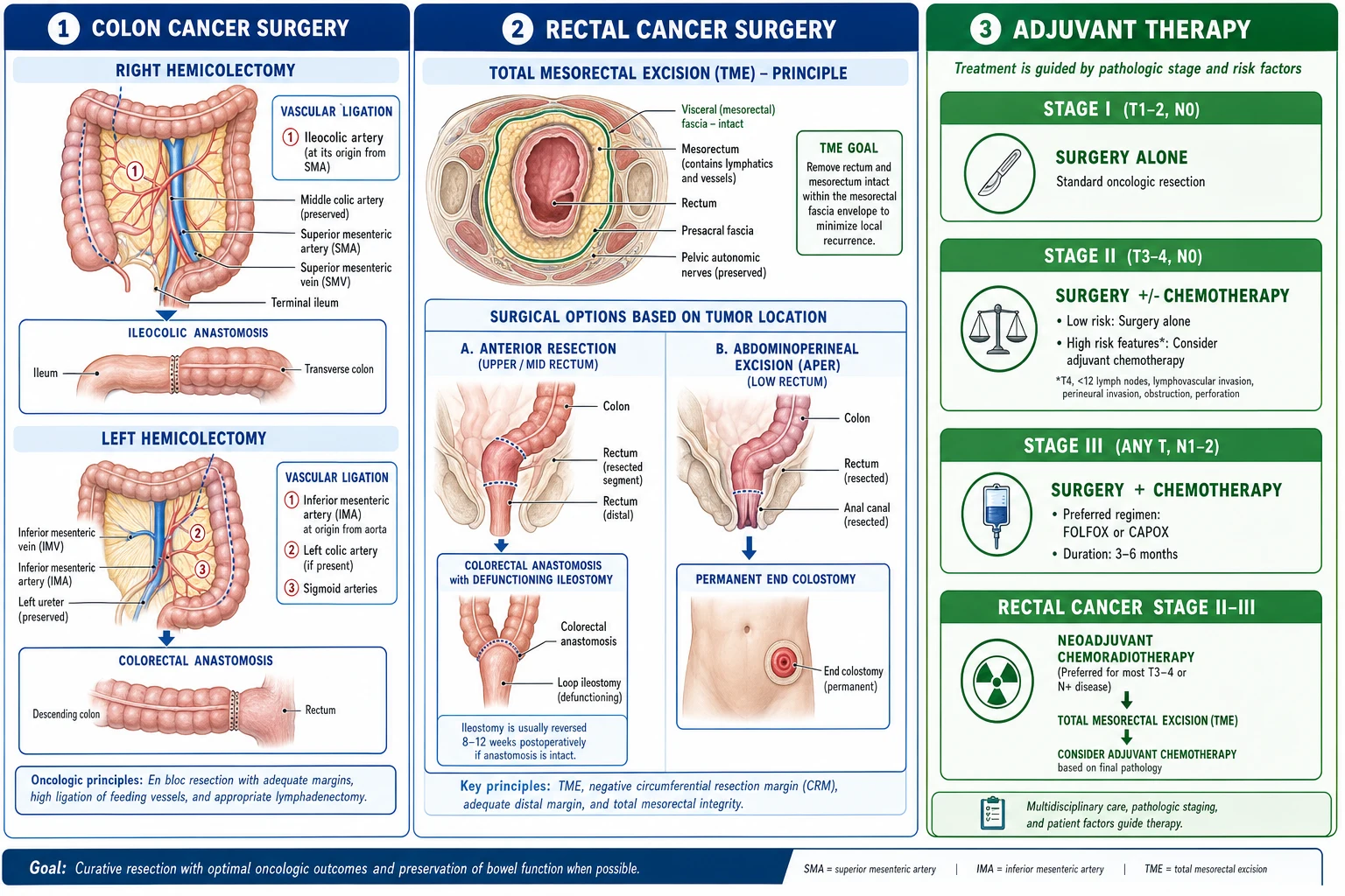
- Body of stomach and diffuse tumours: total gastrectomy with a Roux-en-Y oesophagojejunostomy.
- Linitis plastica: total gastrectomy if resectable, but the disease is often irresectable at presentation and managed palliatively. [1]
Lymphadenectomy is where the major surgical controversy — and the exam question — lives.[3]
- D1 dissection removes only the perigastric nodes (stations 1 to 6). It carries lower morbidity but higher locoregional recurrence.
- D2 dissection removes the perigastric nodes plus the nodes along the left gastric, common hepatic, splenic, and coeliac arteries, and the hepatoduodenal ligament (stations 7 to 12). It is the standard of care in East Asia and increasingly in the West. [1]
The landmark Dutch D1D2 trial randomised patients to D1 versus D2. Early on, D2 carried higher postoperative mortality because the original protocol mandated routine splenectomy and distal pancreatectomy; the modern spleen-preserving, pancreas-sparing D2 avoids this excess morbidity and is now preferred. The 15-year follow-up showed that D2 produced significantly lower gastric-cancer-related death (35 per cent versus 48 per cent D1) and lower locoregional recurrence (12 per cent versus 22 per cent), confirming D2 as the oncological standard. Routine splenectomy and distal pancreatectomy are not recommended in a modern D2 unless the tumour directly invades the spleen or pancreas. [1]
Oesophagogastrectomy (with a thoracic or transhiatal approach) is reserved for tumours at the gastro-oesophageal junction, classified by the Siewert system (type I distal oesophageal, type II true cardia, type III subcardial gastric) which dictates whether an oesophageal or gastric operative strategy is used. [1]
Endoscopic resection — endoscopic submucosal dissection (ESD), or endoscopic mucosal resection for very small lesions — is potentially curative for early gastric cancer that meets strict criteria: differentiated histology, confined to the mucosa, two centimetres or less in diameter, and without ulceration. Lesions with these features have negligible nodal metastasis and can be removed en bloc, avoiding gastrectomy entirely.[4]
Perioperative and adjuvant therapy
Three landmark strategies have defined modern multimodal treatment, and each is examinable. [1]
MAGIC trial (Cunningham et al.)
New England Journal of Medicine, 2006
Phase III: perioperative ECF (epirubicin, cisplatin, 5-fluorouracil) for three cycles before and three after surgery, versus surgery alone, in resectable gastric and lower-oesophageal adenocarcinoma.
Key finding
Perioperative chemotherapy improved five-year survival from 23% to 36% and reduced tumour size and stage at resection.
Practice change
Established perioperative (neoadjuvant) chemotherapy as standard in the West for resectable locally advanced gastric cancer.
INT-0116 / SWOG 9008 (Macdonald et al.)
New England Journal of Medicine, 2001
Phase III: postoperative chemoradiotherapy (5-FU/leucovorin plus 45 Gy radiotherapy) versus surgery alone after R0 resection.
Key finding
Adjuvant chemoradiotherapy improved median survival from 27 to 36 months and five-year survival from 41% to 50%.
Practice change
Adopted as the US standard when perioperative chemotherapy is not given; less used in Asia where D2 dissection is universal.
- Perioperative chemotherapy (MAGIC, then FLOT4). The MAGIC trial established that perioperative ECF (epirubicin, cisplatin, 5-FU) given as three preoperative and three postoperative cycles improved five-year survival from 23 to 36 per cent. The FLOT4 trial subsequently showed that FLOT (5-FU, leucovorin, oxaliplatin, docetaxel) outperformed ECF/ECX, so perioperative FLOT is now the preferred regimen for resectable locally advanced gastric and GOJ adenocarcinoma in the West.[5]
- Adjuvant chemoradiotherapy (US, INT-0116). Where neoadjuvant chemotherapy is not given, postoperative 5-FU/leucovorin plus radiotherapy improves survival; this strategy dominated US practice at a time when D2 dissection was uncommon.[6]
- Adjuvant chemotherapy (Asia, ACTS-GC/CLASSIC). After a D2 gastrectomy, Asian practice uses adjuvant S-1 (an oral fluoropyrimidine) alone or capecitabine plus oxaliplatin (XELOX/CAPOX) for one year, reflecting the high quality of D2 surgery in Japan and Korea.[4]
Metastatic and advanced disease
For unresectable or metastatic cancer, treatment is palliative and aims to prolong survival and control symptoms. [1]
ToGA trial (Bang et al.)
Lancet, 2010
Phase III: trastuzumab (anti-HER2) added to first-line chemotherapy versus chemotherapy alone in HER2-positive advanced gastric/GOJ cancer.
Key finding
Adding trastuzumab improved median overall survival from 11.1 to 13.8 months in HER2-positive disease.
Practice change
Made HER2 testing mandatory in advanced disease and trastuzumab the standard first-line addition for HER2-positive tumours.
CheckMate 649 (Janjigian et al.)
Lancet, 2021
Phase III: first-line nivolumab (anti-PD-1) plus chemotherapy versus chemotherapy alone in untreated advanced gastric/GOJ/oesophageal adenocarcinoma.
Key finding
Nivolumab plus chemotherapy improved median overall survival, most markedly in PD-L1 combined-positive-score (CPS) high tumours.
Practice change
Established first-line nivolumab plus chemotherapy for PD-L1 CPS-positive advanced gastric cancer.
- First-line palliative chemotherapy: a fluoropyrimidine plus platinum doublet — FOLFOX (5-FU, leucovorin, oxaliplatin) or CAPOX (capecitabine plus oxaliplatin) — often with a taxane in a triplet for fit patients.
- Trastuzumab (anti-HER2) is added to first-line chemotherapy for HER2-positive tumours (ToGA), the first targeted therapy to improve survival in this disease.[7]
- Immunotherapy: nivolumab plus chemotherapy is now first-line for PD-L1-positive advanced disease (CheckMate 649), and pembrolizumab is used for MSI-H or dMMR tumours, which are uniquely responsive to checkpoint blockade.[8]
- Second-line options: ramucirumab (anti-VEGFR2), alone or with paclitaxel, and paclitaxel.
- Palliative procedures: self-expanding metal stenting for gastro-oesophageal junction or gastric outlet obstruction; surgical bypass or feeding jejunostomy/gastrostomy for obstruction not amenable to stenting; radiotherapy for a bleeding tumour or painful metastasis; and paracentesis for symptomatic ascites.
Specific Subtypes & Scenarios
Each subtype carries a distinct management question, and each is examinable.[1][4]
- Early gastric cancer (EGC) — confined to mucosa or submucosa regardless of nodes. Lesions meeting the absolute criteria (differentiated, mucosal, two centimetres or less, no ulceration) are treated curatively by endoscopic submucosal dissection; anything beyond these criteria is managed by surgical gastrectomy with D1-plus or D2 dissection. Five-year survival exceeds 90 per cent, which is the entire rationale for screening in endemic countries.
- Linitis plastica — the diffuse, infiltrating signet-ring carcinoma that converts the stomach into a thickened, rigid, non-distensible "leather bottle." Endoscopy can be deceptively normal because the mucosa is intact and the disease is submucosal, so biopsies are often negative and diagnosis may require endoscopic ultrasound with deeper biopsies or CT. The prognosis is poor; if resectable a total gastrectomy is performed, but many cases are palliative.
- Hereditary diffuse gastric cancer (HDGC, CDH1) — autosomal dominant, with a 70 to 80 per cent lifetime risk of diffuse gastric cancer and increased lobular breast cancer. After genetic counselling, prophylactic total gastrectomy is recommended, typically between ages 18 and 40, because surveillance endoscopy misses the multifocal, submucosal signet-ring foci.
- Gastric MALT lymphoma — not an adenocarcinoma but a vital differential, tightly linked to H. pylori. Low-grade MALT often regresses completely after H. pylori eradication; the t(11;18) translocation predicts non-response to eradication and a need for chemoimmunotherapy or radiotherapy.
- Gastrointestinal stromal tumour (GIST) — a submucosal spindle-cell tumour driven by KIT or PDGFRA mutations, positive on CD117 (c-KIT) immunostain. Localised disease is resected; unresectable or metastatic disease responds dramatically to the tyrosine-kinase inhibitor imatinib.
- Gastric neuroendocrine tumours — type I (autoimmune chronic atrophic gastritis, indolent), type II (Zollinger-Ellison/MEN1), type III (sporadic, aggressive); management varies by type and grade. [1]
Complications & Pitfalls
Complications divide into disease-related and treatment-related, and the surgical complications after gastrectomy are among the most tested in general surgery. [1]
Disease-related: gastric outlet obstruction (distal tumours), upper GI bleeding (haematemesis or melaena), perforation (rare but catastrophic), fistulation into the colon or pancreas, and the metastatic burden — liver (commonest solid organ), peritoneum (ascites, Krukenberg, Blumer's shelf), lung, bone, and brain. [1]
Surgical complications: [1]
- Anastomotic leak — the oesophagojejunal anastomosis after total gastrectomy is the highest-risk join. It presents around days 5 to 7 with fever, abdominal or pleuritic pain, and tachycardia, and may progress to sepsis. A water-soluble contrast swallow or CT confirms it; management ranges from nil-by-mouth, drainage, and parenteral nutrition for a contained leak to stenting or re-operation for a free leak.
- Dumping syndrome — the consequence of losing the pyloric sphincter and the reservoir function of the stomach. Early dumping occurs 15 to 30 minutes after a meal: rapid emptying of hyperosmolar contents into the small bowel draws fluid into the lumen, producing vasomotor symptoms (palpitations, flushing, dizziness, diarrhoea, cramping). Late dumping occurs 1 to 3 hours later: exaggerated insulin release in response to the glucose load causes reactive hypoglycaemia. Management is dietary — small, frequent, low-carbohydrate, dry meals (fluids taken between meals), with octreotide for refractory cases.
- Bile-reflux (alkaline) gastritis — bile entering the stomach or oesophagus causes nausea, pain, and bilious vomiting; a Roux-en-Y reconstruction minimises it.
- Postgastrectomy nutritional deficiency — after total gastrectomy the patient loses intrinsic factor and needs lifelong intramuscular vitamin B12; iron, calcium, folate, and fat-soluble vitamins are also commonly deficient because of reduced acid and rapid transit, mandating supplementation and monitoring.
- Afferent-loop syndrome, postvagotomy diarrhoea, small-bowel obstruction, and incisional hernia are longer-term mechanical complications. [1]
Classic pitfalls in management: failing to perform an OGD in a patient over 55 with new dyspepsia; attributing weight loss to "age" or "depression" without investigation; missing Virchow's node on neck examination; omitting staging laparoscopy before curative surgery and so missing occult peritoneal disease; not testing HER2 and PD-L1/MSI in advanced disease, denying the patient trastuzumab or immunotherapy; and forgetting lifelong B12 after a total gastrectomy. [1]
Prognosis & Disposition
The overall five-year survival is 20 to 30 per cent, almost entirely a function of late presentation. Stage for stage, however, gastric cancer is curable when caught early.[1]
Gastric cancer — five-year survival by stage (AJCC 8th edition)
Locally advanced, nodal
Serosa or adjacent organs, multiple nodes; surgery plus adjuvant therapy
Favourable prognostic factors are intestinal histology (better than diffuse), early gastric cancer, distal location, an R0 (microscopically clear) resection, and fewer than four involved nodes. Unfavourable factors are diffuse and signet-ring histology, linitis plastica, proximal/cardia location, serosal penetration, peritoneal metastases, and elevated tumour markers. [1]
Disposition and follow-up: after a curative resection the patient is followed clinically with bloods (full blood count, liver function, B12) and CT as clinically indicated; there is no single universal surveillance protocol. After total gastrectomy, lifelong intramuscular vitamin B12 is mandatory. Patients should undergo H. pylori eradication postoperatively to lower the risk of a metachronous cancer. [1]
Special Populations
- H. pylori eradication after resection. Eradication is recommended for all patients after gastric cancer resection to reduce the risk of metachronous (new primary) tumours in the remnant. Standard first-line therapy is a triple regimen — a proton-pump inhibitor plus clarithromycin plus amoxicillin (or metronidazole) for 14 days; in areas of high clarithromycin resistance, a bismuth-based quadruple regimen is preferred.[2]
H. pylori eradication (post-resection / atrophic gastritis)
Reduce metachronous gastric cancer risk
Dose
PPI (e.g. omeprazole 20 mg PO BD) + clarithromycin 500 mg PO BD + amoxicillin 1 g PO BD for 14 days; bismuth quadruple if clarithromycin resistance
- CDH1 mutation carriers (hereditary diffuse gastric cancer). After genetic counselling and testing, prophylactic total gastrectomy is recommended between ages 18 and 40, with pre-operative counselling about the nutritional sequelae — dumping, weight loss, and lifelong B12 dependence.[1]
- Elderly and frail patients. For advanced disease, prioritise endoscopic stenting over surgery for obstruction, weigh the toxicity of neoadjuvant chemotherapy carefully, and adopt a palliative approach with early specialist-nurse and dietetic input.
- Screening populations (Japan and Korea). National programmes of barium meal photofluorography and/or direct OGD from around age 40 detect early gastric cancer and explain the markedly better survival in East Asia. In low-incidence countries (UK, US, India) population screening is not cost-effective; instead, endoscopic surveillance targets high-risk groups — those with atrophic gastritis or intestinal metaplasia, pernicious anaemia, prior gastrectomy, or a family history of HDGC.[4]
Evidence, Guidelines & Regional Differences
The evidence base for gastric cancer is unusually mature, and the four landmark trials below define modern practice. Regional differences arise from incidence, screening, and surgical tradition.[1][3]
The Dutch D1D2 trial settled the lymphadenectomy debate: although early D2 mortality was inflated by routine splenectomy and distal pancreatectomy, the modern spleen-preserving D2 delivers lower cancer-related death and recurrence at 15 years and is the global oncological standard.[3] The MAGIC trial brought perioperative chemotherapy to the West,[5] refined by FLOT4 to the docetaxel-based FLOT regimen that is now preferred. The INT-0116 trial underpins the US adjuvant chemoradiotherapy strategy used when neoadjuvant chemotherapy is not given.[6] In advanced disease, ToGA made HER2 testing mandatory and trastuzumab standard for HER2-positive tumours,[7] while CheckMate 649 brought immunotherapy into the first line for PD-L1-positive disease.[8]
- East Asia (Japan, Korea, China): national endoscopic screening from age 40 detects early gastric cancer; D2 dissection is universal; adjuvant S-1 or XELOX is standard after D2; and perioperative chemotherapy is added for locally advanced disease. Five-year survival is the highest in the world.
- United States and UK: no population screening; presentation at advanced stage; perioperative FLOT is the standard for resectable disease; D2 is increasingly adopted; adjuvant chemoradiotherapy (INT-0116) is used where neoadjuvant chemo was not given.
- Europe: perioperative FLOT with D2 gastrectomy mirrors UK practice; the Dutch D1D2 trial originated the modern D2 standard.
- India and South Asia: high H. pylori prevalence and high incidence in some regions (the Northeast); late presentation is the norm; in resource-limited settings a D1 or modified D2 dissection may be the pragmatic choice where high-volume D2 expertise is unavailable. [1]
Exam Pearls
- Correa cascade: normal mucosa, then chronic gastritis (H. pylori), then atrophic gastritis, then intestinal metaplasia, then dysplasia, then adenocarcinoma — over decades.[2]
- H. pylori is a WHO Class I carcinogen — the single most important risk factor; eradication reduces cancer risk.[2]
- Lauren classification: intestinal type (gland-forming, H. pylori-linked, older males, distal stomach, better prognosis) versus diffuse type (signet-ring, CDH1 loss, linitis plastica, younger, worse prognosis).[1]
- Eponymous metastatic signs: Virchow's node (left supraclavicular, Troisier's sign), Sister Mary Joseph nodule (umbilicus), Krukenberg (ovary), Blumer's shelf (rectal shelf), acanthosis nigricans / sign of Leser-Trélat (paraneoplastic).[1]
- D2 lymphadenectomy is the standard for curative gastrectomy (Dutch D1D2 15-year follow-up); routine splenectomy and distal pancreatectomy are not done.[3]
- Perioperative FLOT for resectable locally advanced disease (MAGIC → FLOT4). Adjuvant chemoradiotherapy (INT-0116) where neoadjuvant chemo not given.[5][6]
- HER2-positive metastatic disease gets trastuzumab (ToGA); PD-L1-positive or MSI-H gets nivolumab or pembrolizumab (CheckMate 649).[7][8]
- Lifelong intramuscular B12 after total gastrectomy (loss of intrinsic factor).[1]
- Dysphagia = urgent OGD; new dyspepsia over 55 = urgent OGD (NICE NG12).[1]
- CDH1 mutation = hereditary diffuse gastric cancer = prophylactic total gastrectomy between ages 18 and 40.[1]
- Linitis plastica = "leather bottle" stomach = diffuse signet-ring carcinoma; endoscopy may look deceptively normal.
- Early gastric cancer (mucosa/submucosa regardless of nodes) is curable by endoscopic submucosal dissection if it meets strict criteria.[4]
Exam application bank (NEET-PG / INICET)
One-line answer
Gastric carcinoma is an adenocarcinoma arising from the gastric epithelium. It is the fifth most commonly diagnosed cancer worldwide and the fourth leading cause of cancer death, with the highest incidence in East Asia (Japan, Korea, China), Eastern Europe, and parts of South America. The dominant risk factor is Helicobacter pylori (a WHO Class I carcinogen), followed by diet (salted/smoked/pickled foods, N-nitroso compounds, low fruit and vegetables), smoking, chronic atrophic gastritis, pernicious anaemia, and the hereditary diffuse gastric cancer syndrome (CDH1/E-cadherin mutation). Most tumours arise through the Correa cascade (chronic gastritis to atrophic gastritis to intestinal metaplasia to dysplasia to adenocarcinoma) over decades. Presentation is characteristically late and insidious — epigastric pain, weight loss, early satiety, dysphagia (proximal), vomiting (distal), and ana
Worked stems (answer without another resource)
Stem 1 — Classic presentation. Map symptoms to mechanism; name the first investigation and first treatment step with dose/route if drug therapy is standard. [1]
Stem 2 — Unstable / complicated. List red flags that force immediate resuscitation, theatre, ICU, antidote, or reperfusion — and what you do in the first 15 minutes. [1]
Stem 3 — Atypical group. Elderly, pregnancy, child, or immunocompromised: how presentation and thresholds change. [1]
Stem 4 — Differential trap. Name the three closest mimics and one discriminator for each. [1]
Stem 5 — Disposition. Who goes home with safety-netting, who is admitted, who needs HDU/ICU/theatre, and what follow-up is mandatory. [1]
Rapid viva checklist
- Definition + classification
- Pathophysiology chain
- Bedside signs / criteria
- Score with exact components (if any)
- Emergency bundle
- Definitive therapy with doses
- Complications of disease and of treatment
- Special populations
- Guideline/trial name if classic
- Three exam traps
Coverage self-check
If you cannot answer any stem above from this page alone, re-read the matching section — the page is intended to be self-sufficient for final-prof and NEET-PG/INICET questions on Gastric Carcinoma.
References
- [1]Sundar R, Nakayama I, Markar SR, et al. Gastric cancer Lancet, 2025.PMID 40319897
- [2]Duan Y, Xu Y, Dou Y, et al. Helicobacter pylori and gastric cancer: mechanisms and new perspectives J Hematol Oncol, 2025.PMID 39849657
- [3]Songun I, Putter H, Kranenbarg EM, et al. Surgical treatment of gastric cancer: 15-year follow-up results of the randomised nationwide Dutch D1D2 trial Lancet Oncol, 2010.PMID 20409751
- [4]Shen L, Shan YS, Hu HM, et al. Management of gastric cancer in Asia: resource-stratified guidelines Lancet Oncol, 2013.PMID 24176572
- [5]Cunningham D, Allum WH, Stenning SP, et al. Perioperative chemotherapy versus surgery alone for resectable gastroesophageal cancer N Engl J Med, 2006.PMID 16822992
- [6]Macdonald JS, Smalley SR, Benedetti J, et al. Chemoradiotherapy after surgery compared with surgery alone for adenocarcinoma of the stomach or gastroesophageal junction N Engl J Med, 2001.PMID 11547741
- [7]Bang YJ, Van Cutsem E, Feyereislova A, et al. Trastuzumab in combination with chemotherapy versus chemotherapy alone for treatment of HER2-positive advanced gastric or gastro-oesophageal junction cancer (ToGA): a phase 3, open-label, randomised controlled trial Lancet, 2010.PMID 20728210
- [8]Janjigian YY, Shitara K, Moehler M, et al. First-line nivolumab plus chemotherapy versus chemotherapy alone for advanced gastric, gastro-oesophageal junction, and oesophageal adenocarcinoma (CheckMate 649): a randomised, open-label, phase 3 trial Lancet, 2021.PMID 34102137