General Surgery · General Surgery
Haemorrhoids
Also known as Piles · Internal haemorrhoids · External haemorrhoids · Haemorrhoidal disease · Thrombosed external haemorrhoid
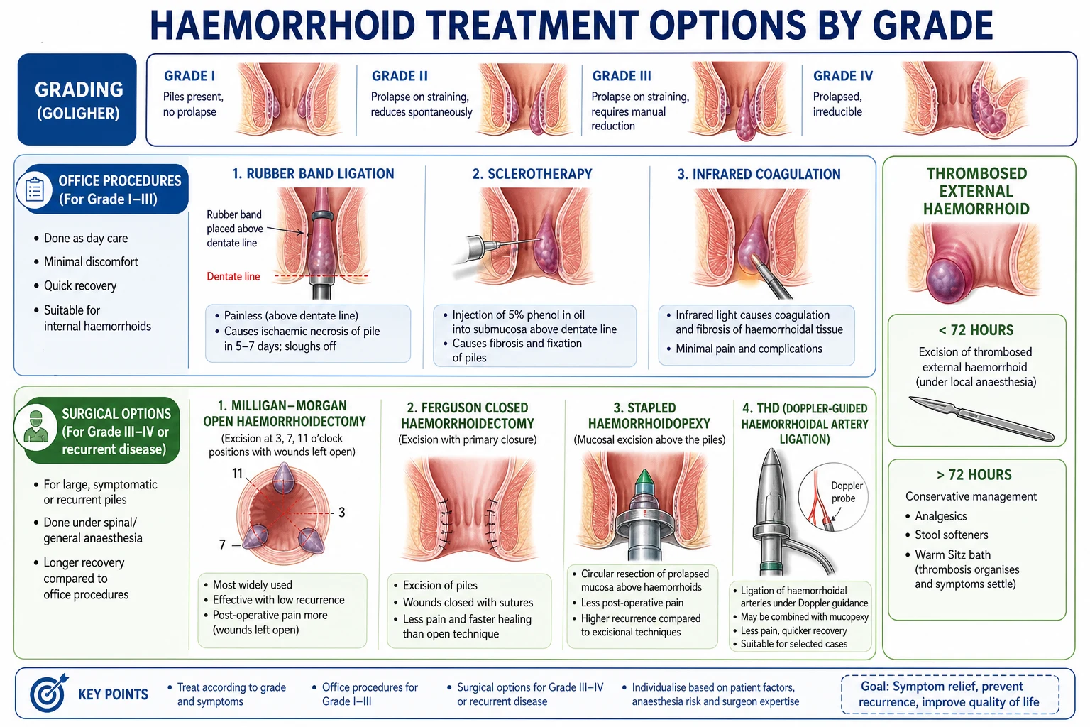
Haemorrhoids (piles) are engorged, displaced anal vascular cushions at the anorectal junction. Internal (above dentate line, painless bleeding) vs external (below dentate line, painful thrombosis). Goligher grading: Grade I (bleed, no prolapse), II (prolapse, reduce spontaneously), III (prolapse, need manual reduction), IV (prolapsed, irreducible). Risk: constipation, straining, pregnancy, prolonged sitting. Present with painless bright red bleeding per rectum (on toilet paper, dripping), prolapse, pruritus ani. Thrombosed external haemorrhoid = acutely painful perianal lump. Always exclude colorectal cancer in patients over 40 with rectal bleeding. Manage: Grade I to II (dietary, topical, rubber band ligation); Grade III to IV (surgical — open/closed haemorrhoidectomy, stapled haemorrhoidopexy, THD/HALO).
On this page & tools
Your progress
Saved locally on this device.
Exam tags
Red flags
Overview & Definition
Haemorrhoids (commonly called piles) are not, in their normal state, a disease at all. They are normal anatomical structures — three discrete anal vascular cushions composed of arteriovenous communications, connective tissue, and smooth muscle, located in the upper anal canal at the 3, 7, and 11 o'clock positions (left lateral, right posterior, right anterior) when the patient is examined in the lithotomy position. Thomson's landmark 1975 thesis established that these cushions are universal, physiological, and contribute roughly 15 to 20 per cent of resting anal continence by acting as compliant seals that fill the anal canal at rest.[4] They become pathological — what clinicians call haemorrhoidal disease — only when they engorge, enlarge, and prolapse as the supporting connective tissue degenerates under chronic straining, constipation, pregnancy, or prolonged sitting.[1]
The critical anatomical landmark is the dentate (pectinate) line, which lies approximately 2 cm proximal to the anal verge and marks the embryological junction of endoderm (proximal gut) and ectoderm (proctodeum). Above and below this line the epithelium, blood supply, lymphatic drainage, and innervation all differ — and these differences govern every clinical and therapeutic decision in haemorrhoidal disease:[1]
- Internal haemorrhoids arise above the dentate line, from the superior haemorrhoidal (rectal) vessels. The overlying mucosa carries visceral (autonomic) innervation and is therefore insensitive to cutting, burning, and banding — which is exactly why rubber band ligation is painless. They present with painless bright red bleeding and prolapse.
- External haemorrhoids arise below the dentate line, from the inferior haemorrhoidal vessels, and are covered by anoderm (modified squamous epithelum richly supplied by somatic inferior rectal nerves). They are exquisitely pain-sensitive, which is why a thrombosed external haemorrhoid hurts so intensely and why banding below the line is a cardinal surgical error. [1]
Applied anatomy — the vascular and innervation map
The blood supply of the anal canal follows the dentate line precisely, and remembering it explains both the symptoms and the surgical anatomy of every procedure. Above the dentate line the mucosa is supplied by the superior rectal artery, the terminal branch of the inferior mesenteric artery; its venous drainage is via the superior rectal vein into the inferior mesenteric vein and the portal system, which is why portal hypertension engorges these vessels. At the dentate line the middle rectal vessels (branches of the internal iliac artery and vein) provide an anastomotic link between the systemic and portal circulations — the classic portosystemic anastomosis. Below the dentate line the inferior rectal artery and vein, branches of the internal pudendal (and ultimately internal iliac) vessels, supply the anoderm and drain into the systemic circulation.[1]
This three-tier supply is what allows transanal haemorrhoidal dearterialisation (THD) to work: a Doppler probe locates the terminal branches of the superior rectal artery feeding each cushion, and suture ligation of these branches reduces inflow. Lymphatic drainage mirrors the arterial supply — above the dentate line to the inferior mesenteric and para-aortic nodes, below the line to the superficial inguinal nodes — which matters when staging anal-canal versus rectal malignancy, and when interpreting an enlarged inguinal node in a patient with perianal disease.[1]
The anal cushions themselves are not simply veins. They are discrete masses of submucosal tissue containing arteriovenous communications without an intervening capillary bed, supported by the muscularis submucosae ani (Treitz's muscle) and a fibroelastic scaffold. Because the feeding vessels are arteriovenous shunts at systemic arterial pressure, bleeding from an internal haemorrhoid is characteristically bright red and brisk — not the dark, sluggish ooze one would expect from a pure vein. This single anatomical fact — arterial blood at the anal verge — is the physiological basis of Thomson's vascular cushion theory and the reason haemorrhoidal bleeding can occasionally be dramatic.[4]
Haemorrhoidal disease is one of the most common surgical conditions encountered in clinical practice — it affects approximately 1 in 20 people at any given time, with a cumulative lifetime prevalence exceeding 50 per cent by the age of 50. The condition accounts for a substantial fraction of surgical outpatient attendances, and although benign, its hallmark symptom of painless rectal bleeding demands a disciplined, structured approach to exclude the serious mimics, above all colorectal cancer.[1]
Classification
The standard grading system, and the one every examiner expects, is Goligher's classification, which orders internal haemorrhoids by the degree of prolapse — because prolapse, more than bleeding, drives the choice between office and operating-room treatment.[1][2]
| Grade | Description | Treatment approach |
|---|---|---|
| I | Bleeding, no prolapse | Conservative: diet, topical agents, phlebotonics |
| II | Prolapse on straining, reduces spontaneously | Band ligation, dietary modification, sclerotherapy |
| III | Prolapse, requires manual reduction | Band ligation or surgery (THD, stapled, excisional) |
| IV | Prolapse, irreducible (permanently prolapsed) | Surgery (excisional haemorrhoidectomy) |
By anatomical origin, haemorrhoids are classified as internal (above the dentate line, mucosa-covered, painless, prolapsing), external (below the line, anoderm-covered, painful only when thrombosed), and mixed (interno-external) — a combination in which an internal component and an external perianal skin tag communicate across the dentate line and prolapse together. [1]
Several special clinical situations sit outside the Goligher grades and carry their own management logic:[1]
- Thrombosed external haemorrhoid — a sudden, acutely painful, firm, purple perianal lump caused by clot formation in an external cushion. Pain peaks at about 48 hours and resolves over 1 to 2 weeks as the thrombus organises.
- Strangulated internal haemorrhoid — a Grade IV prolapse that has become incarcerated outside the sphincter, with oedema, thrombosis, and a real risk of ulceration and necrosis. This is a surgical emergency.
- Interogoportal haemorrhoids in portal hypertension and pregnancy — engorged cushions driven by elevated venous pressure; in pregnancy they usually resolve postpartum. [1]
Epidemiology & Risk Factors
Haemorrhoidal disease is genuinely common. Cross-sectional surveys place the point prevalence at roughly 4 to 5 per cent of the general population, rising steeply with age so that more than half of those over 50 have some degree of symptomatic disease. The peak age is 45 to 65 years, and the sex distribution is approximately equal — although women more often date the onset to pregnancy. In surgical outpatient practice in India and South Asia the condition is one of the single most frequent reasons for attendance, often with patients presenting late, already at Grade III or IV.[1][1]
The unifying mechanism of every recognised risk factor is sustained elevation of intra-abdominal and intra-rectal pressure, which transmits to the anal cushions and, over time, fragments their anchoring connective tissue:[1]
- Constipation and chronic straining — the dominant factor; hard stool and prolonged toilet-sitting force the cushions down.
- Low-fibre diet — produces hard, low-bulk stools that demand straining; particularly relevant in urban Indian diets low in roughage.
- Prolonged sitting on the toilet (reading, phone use) — sustains the gravitational and pressure load on the cushions.
- Pregnancy — mechanical compression of pelvic veins by the gravid uterus plus progesterone-mediated vascular engorgement and laxity of supporting tissue.
- Obesity and raised intra-abdominal fat.
- Chronic diarrhoea (especially inflammatory bowel disease) — repeated straining against urgency.
- Heavy lifting as an occupational habit (porters, weightlifters, construction workers).
- Portal hypertension and cirrhosis — engorges the portosystemic anastomoses at the anorectal junction; note this produces rectal varices, a distinct entity, not haemorrhoids.
- Anal intercourse, chronic cough, benign prostatic hypertrophy with straining to void.
- Ageing — progressive loss and fragmentation of the connective-tissue support of the cushions.
- Family history — a weak familial tendency suggesting connective-tissue susceptibility. [1]
Pathophysiology
Two complementary theories explain how normal cushions become haemorrhoids, and the modern synthesis draws on both. Thomson's vascular cushion theory (1975) established that the anal cushions are rich arteriovenous communications — not dilated veins — fed at high flow by terminal branches of the superior rectal artery; this is why haemorrhoidal bleeding is bright red (arterial) rather than dark venous blood.[4] The sliding anal cushion theory adds the mechanical dimension: chronic straining and ageing degenerate the muscularis submucosae ani (Treitz's muscle) and the suspensory ligament of the anal cushions (Parks's ligament), so the cushions lose their mooring, slide distally, and prolapse.[1]
The cascade runs as follows. Repeated rises in intra-abdominal pressure (straining at stool, pregnancy, heavy lifting) raise hydrostatic pressure in the superior haemorrhoidal vessels at the portosystemic anastomosis of the anorectal junction. The cushions engorge with blood, their anchoring connective tissue fragments, and they slide downward (prolapse) during defecation. With repeated episodes the cushions fail to reduce spontaneously — the patient passes from Grade II to III to IV. Trauma from passing stool then tears the thinned, engorged overlying mucosa, producing the characteristic bright red bleeding seen on the toilet paper or dripping into the bowl.[1]
Thrombosis is the external-cushion parallel. When an external haemorrhoid's venous return is obstructed by acute straining, a thrombus forms within the cushion, producing the tender, firm, purple perianal lump of a thrombosed external haemorrhoid. As the thrombus organises it may erode through the overlying skin and discharge dark clot externally — a sign that spontaneous resolution is approaching. [1]
A distinction examiners repeatedly test: rectal varices (from portal hypertension) are submucosal portosystemic collaterals that extend proximally from the anorectal junction into the rectum and can cause massive, life-threatening bleeding. They are not haemorrhoids, do not originate from the anal cushions, and must not be treated with rubber band ligation or excisional haemorrhoidectomy — they require endoscopic variceal banding, TIPSS, or suture ligation.[1]
Goligher grading — the classification that drives treatment
Clinical Presentation
The clinical picture of haemorrhoidal disease is built from a small number of cardinal symptoms, and recognising the pattern — and especially what it is not — is what distinguishes a competent assessment from a dangerous assumption. [1]
Internal haemorrhoids produce the classical triad of painless bleeding, prolapse, and discharge:[1]
- Painless bright red bleeding per rectum is the hallmark. The blood is described as on the toilet paper after wiping, dripping into the toilet bowl during or after defecation, or on the surface of the stool — never mixed in with the stool. Mixing-in, dark blood, melaena, or clots all point away from haemorrhoids toward a more proximal source.
- Prolapse — tissue bulging from the anus during defecation. It may reduce spontaneously (Grade II), require the patient to push it back manually (Grade III), or remain permanently external and irreducible (Grade IV).
- Mucus discharge and pruritus ani — the prolapsed mucosa secretes mucus that soils the perianal skin and irritates it.
- Soiling of underclothes — from impaired continence when large prolapsing piles prevent full sphincter closure.
- Pain is NOT a feature of uncomplicated internal haemorrhoids (above the dentate line, visceral innervation). The presence of pain forces the question of thrombosis, strangulation, an associated fissure, or an abscess. [1]
External haemorrhoids are usually asymptomatic unless they thrombose. Acute thrombosis presents as sudden-onset severe perianal pain and a tender, firm, blue-purple lump at the anal verge. Pain peaks at about 48 hours and then gradually subsides over 1 to 2 weeks as the thrombus organises and is absorbed. The overlying skin may ulcerate and discharge dark clot, after which pain resolves. [1]
Strangulated internal haemorrhoid is the acute complication of a Grade IV prolapse: the prolapsed cushion is trapped outside the sphincter, venous return is occluded, and the cushion becomes oedematous, thrombosed, and — if untreated — ulcerated and necrotic. The patient is in severe pain, and the prolapsed, purplish, oedematous mass is visible at the anus; this needs urgent surgical assessment.[1]
Atypical presentations examiners probe deliberately: the elderly patient who attributes years of bleeding to "piles" while a caecal carcinoma grows silently proximally; the pregnant woman in whom bleeding is dismissed as a normal pregnancy complaint; the immunocompromised patient who presents with sepsis rather than pain; and the patient with iron-deficiency anaemia in whom haemorrhoids are an inadequate explanation until full colonic evaluation has excluded cancer.[2]
Internal (above dentate line)
visceral innervation
- **Painless** bleeding (bright red on toilet paper)
- **Prolapse** on straining (Grade I to IV)
- **Cannot feel** unless prolapsed
- Banding above the line is **painless**
External (below dentate line)
somatic innervation
- **Painful** if thrombosed
- Visible at anal verge
- **Purple/blue** firm lump if thrombosed
- Do NOT band below the line
Differential Diagnosis
Rectal bleeding has a long differential, and the disciplined habit is to never assume bleeding is haemorrhoidal until other causes have been actively excluded by history, examination, and — where indicated — endoscopy. Each mimic carries features that separate it from piles.[1][2]
| Condition | Key distinguishing feature |
|---|---|
| Colorectal cancer | Weight loss, change in bowel habit, blood mixed with stool (not on surface), tenesmus, iron-deficiency anaemia; age over 40; needs colonoscopy to exclude — never assume bleeding is haemorrhoids without examination |
| Anal fissure | Severe sharp pain on defecation; tiny amount of bright blood on toilet paper; visible linear tear at 6 o'clock (posterior midline); sphincter spasm; chronic fissure may show a sentinel tag and hypertrophied papilla |
| Perianal abscess / fistula | Severe throbbing pain, fever, fluctuant perianal swelling; not clearly related to defecation; needs incision and drainage; fistula shows discharge and recurrent abscess |
| Inflammatory bowel disease | Chronic bloody diarrhoea, tenesmus, mucus, abdominal pain, weight loss, extra-intestinal features (arthralgia, uveitis, erythema nodosum); colonoscopy with biopsy |
| Diverticular disease | Painless, often massive lower-GI bleeding in an older patient; typically self-limiting; diagnosed on colonoscopy or CT angiography |
| Angiodysplasia | Painless bleeding, often right-sided in the elderly; can be massive; diagnosed on colonoscopy or angiography |
| Rectal prolapse | Full-thickness rectal wall prolapses (concentric rings of mucosa, not discrete cushions); common in elderly women; associated with constipation and weak pelvic floor |
| Rectal varices | From portal hypertension (cirrhosis); can cause massive bleeding; do NOT band or excise like haemorrhoids — managed as portosystemic varices |
| Condylomata acuminata (anal warts) | Cauliflower-like perianal lesions; HPV-related; sexually transmitted |
| Anal cancer | Hard, irregular, non-tender perianal mass; biopsy essential for any atypical lesion |
| Solitary rectal ulcer syndrome | Passage of mucus and blood, tenesmus, sense of incomplete evacuation; anterior rectal ulcer at sigmoidoscopy; linked to internal rectal intussusception |
Clinical & Bedside Assessment
The focused assessment of suspected haemorrhoids is built on inspection, digital rectal examination, and proctoscopy, with endoscopy reserved for excluding proximal pathology. A candidate who skips any of these steps will miss cancer — and fail the station. [1]
Inspection of the perianal skin in the left lateral or lithotomy position looks for external tags, a visible thrombosed external haemorrhoid (the purple, tender lump), prolapsed internal piles, fissures (posterior midline), sentinel skin tags, fistula openings, condylomata, and any mass. Asking the patient to bear down may reveal a Grade II or III prolapse that is invisible at rest. [1]
Digital rectal examination (DRE):[1]
- Internal haemorrhoids are usually NOT palpable — they are soft, compressible, and collapse when the patient is not straining. A "palpable pile" is more often a tumour, a thrombosed external haemorrhoid, or a hypertrophied papilla.
- DRE is essential to exclude other pathology: anal fissure (check for the posterior midline tear — though if the fissure is exquisitely tender, DRE may be deferred and the patient referred for examination under anaesthesia), rectal mass (cancer polyp), prostate enlargement or malignancy, pelvic floor tone, and any anterior vaginal or pelvic mass.
- Assess sphincter tone and the anocutaneous (S4) reflex — a gentle scratch at the anal verge should contract the external sphincter. [1]
Proctoscopy (anoscopy) is the definitive bedside examination for internal haemorrhoids. The rigid proctoscope visualises the anal cushions at the 3, 7, and 11 o'clock positions, allowing direct assessment of their size, grade, and whether they bleed on contact. It permits rubber band ligation at the same sitting when an internal haemorrhoid is confirmed. A rigid sigmoidoscopy to 25 cm should accompany proctoscopy to inspect the rectum for proctitis, tumours, and polyps.[1]
Flexible sigmoidoscopy or colonoscopy is mandatory for any patient with rectal bleeding who is over 40 (some guidelines say over 45), or who carries alarm features — weight loss, change in bowel habit, iron-deficiency anaemia, family history of colorectal cancer, or atypical bleeding (mixed with stool, dark, or with mucus). A young patient with typical haemorrhoidal bleeding, a normal proctoscopy, and no alarm features can be managed without full colonic imaging.[2]
Investigations
Haemorrhoids are a clinical diagnosis — investigations exist to exclude other pathology and to quantify the consequences of chronic bleeding, not to confirm the diagnosis itself. [1]
- Full blood count (FBC) — check for iron-deficiency anaemia from chronic blood loss. Anaemia in a patient with "haemorrhoids" is a red flag that mandates full colonic evaluation, because haemorrhoids alone rarely produce enough blood loss to drop the haemoglobin; colorectal cancer must be excluded.
- Coagulation screen and platelets — if the patient is anticoagulated, has liver disease, or has a bleeding history, before any invasive intervention.
- Colonoscopy — for any patient over 40 to 45 (or younger with alarm features) presenting with rectal bleeding, to exclude proximal pathology — above all colorectal cancer, polyps, and inflammatory bowel disease.[2]
- Flexible sigmoidoscopy — an acceptable alternative when colonoscopy is unavailable or contraindicated, and the patient is young with typical haemorrhoidal bleeding.
- STI screen — if sexually transmitted anal pathology (gonococcus, syphilis, HPV, herpes) is suspected.
No imaging is routinely required. In the rare case of massive lower-GI bleeding where the source is unclear, CT angiography localises the bleeding point and guides intervention. [1]
Management — Resuscitation
Haemorrhoids rarely present as a true emergency, but three scenarios demand urgent, structured management.[1]
Massive haemorrhoidal bleeding causing haemodynamic instability is uncommon but real. Resuscitate first — two large-bore cannulae, intravenous crystalloid, blood sent for group and crossmatch, transfuse if haemoglobin is low or the patient is shocked. Then take the patient to theatre for proctoscopy under anaesthesia to identify the bleeding cushion; definitive control is by suture ligation of the bleeding pedicle or balloon tamponade with a Foley catheter inflated in the rectum as a temporising measure pending definitive surgery. [1]
Strangulated haemorrhoid — a prolapsed, incarcerated, oedematous, thrombosed Grade IV internal haemorrhoid. Give adequate analgesia (NSAIDs, opioids if needed), apply ice, and seek urgent surgical assessment. The preferred treatment, if the patient is fit, is urgent haemorrhoidectomy within 72 hours of onset; this relieves pain definitively and removes the necrotic tissue. Some surgeons favour an initial conservative approach (analgesia, antibiotics, stool softeners) with surgery reserved for established complications, but early excision generally produces better outcomes and a shorter illness.[1]
Thrombosed external haemorrhoid — if the patient presents within 72 hours of onset, excision under local anaesthetic (en bloc removal of the thrombosed vein with overlying skin ellipse) provides dramatic and immediate pain relief. If presentation is after 72 hours, the pain is already on the wane and conservative management (oral analgesia, stool softeners, warm sitz baths, topical agents) is preferred, because surgery at this stage offers little benefit over natural resolution.[1]
Management — Definitive & Stepwise
Definitive management follows a ladder that maps onto the Goligher grade — conservative and office measures for Grade I to II, surgery for Grade III to IV — and every patient, regardless of grade, benefits from baseline dietary and bowel-habit measures.[1][2]
General measures (all grades)
- Dietary modification: high-fibre diet, 25 to 30 g of fibre per day, and adequate fluid intake (about 2 L/day), to produce soft, bulky stool that passes without straining. Avoid straining and prolonged sitting on the toilet.
- Bulk-forming laxatives — ispaghula husk 3.5 g sachet, one to two sachets daily in water, or methylcellulose, for constipated patients; the goal is type 3 to 4 stool on the Bristol scale.
- Topical agents — creams and suppositories combining a local anaesthetic (lignocaine 2 to 5 per cent), an astringent (hamamelis, zinc oxide), or a corticosteroid (hydrocortisone 1 per cent — Anusol HC, Germoloids, Scheriproct) for short-term symptomatic relief. Steroid-containing preparations should not be used for more than 5 to 7 days because of skin thinning and contact dermatitis.
- Phlebotonics — micronised purified flavonoid fraction (diosmin 450 mg plus hesperidin 50 mg), two tablets daily, may reduce bleeding and symptoms by improving venous tone; the evidence is modest but consistent.[1]
Grade I to II — office-based
For Grade I and Grade II haemorrhoids, the first-line definitive treatments are office procedures that do not require anaesthesia or admission.[1][2]
- Rubber band ligation (Barron's banding) — the standard office procedure for Grade II and III (and Grade I with troublesome bleeding). A rubber band is placed at the base of the internal haemorrhoid pedicle, above the dentate line (painless), causing ischaemic necrosis; the banded tissue and band slough off at 5 to 7 days, leaving a small ulcer that heals by fibrosis. Success rate is about 70 to 80 per cent for Grade II. Band no more than one or two piles per session. Complications: pain (if the band is placed too low, below the dentate line — remove the band immediately), secondary bleeding at day 5 to 7 when the tissue sloughs (usually self-limiting), and the rare but serious pelvic sepsis / Fournier's gangrene — any severe pain or fever after banding needs urgent review.[1]
- Injection sclerotherapy — 5 per cent phenol in almond (arachis) oil, 2 to 3 mL injected into the submucosa just above the haemorrhoid pedicle. Phenol in oily solution causes a slow chemical fibrosis that tethers the mucosa to the underlying muscle and fixes the cushion in place. Used for Grade I and early Grade II with bleeding; less effective than banding for prolapse, and may need repeated sessions. Avoid in patients with nut allergy (use a non-arachis vehicle).
- Infrared coagulation — a tungsten-halogen lamp applies infrared heat to coagulate and fix the cushion; for Grade I and II; less effective than banding but less painful and suitable for small bleeding piles.
- Cryotherapy (freezing with liquid nitrogen) and bipolar diathermy — largely historical or niche; less used in modern practice.
Grade III to IV — surgical
For Grade III and IV haemorrhoids, or Grade I to II that have failed office treatment, surgery is the answer. The choice of operation balances efficacy, postoperative pain, recurrence, and complications.[1][2][3]
- Open (Milligan-Morgan) haemorrhoidectomy — the gold standard, especially in the UK, India, and much of the world. The three primary pedicles at 3, 7, and 11 o'clock are identified at proctoscopy; each is excised with diathermy down to the internal sphincter, the vascular pedicle is ligated or coagulated, and the mucocutaneous wounds are left open to heal by secondary intention, preserving skin bridges between wounds to avoid anal stenosis. Effective for Grade III and IV with recurrence under 5 per cent. The main drawback is significant postoperative pain for 2 to 3 weeks.[1][2]
- Closed (Ferguson) haemorrhoidectomy — the same excisional principle, but the mucocutaneous defect is closed primarily with a running absorbable suture. Less postoperative pain and faster healing than the open technique; popular in the US. Wound breakdown may convert it to an open technique.
- Stapled haemorrhoidopexy (Procedure for Prolapse and Haemorrhoids, PPH / MIII) — a circular stapler excises a ring of rectal mucosa 2 to 4 cm above the dentate line, lifting the prolapsed cushions back up into the anal canal and interrupting their arterial inflow. There is no perianal wound, so postoperative pain is markedly less than after excisional surgery, and recovery is faster. The trade-off, established by the eTHoS trial (Watson et al., 2017), is a higher symptom-recurrence rate (more patients returned with symptomatic piles over the long term) and specific complications: staple-line bleeding, chronic pain, faecal urgency, rectovaginal fistula, and — rarely — full-thickness rectal wall excision or pelvic sepsis. Best suited to circumferential Grade III disease.[3]
- Transanal haemorrhoidal dearterialisation (THD / HALO) — a Doppler probe in a specialised proctoscope locates the terminal branches of the superior rectal artery feeding each cushion; these are suture-ligated to reduce inflow, and the prolapsing cushion is hitched up by mucopexy. Minimal postoperative pain, no excisional wound, and a recurrence rate intermediate between stapling and excision; suitable for Grade II to IV. A systematic review found THD broadly comparable to stapled haemorrhoidopexy in symptom control with a favourable safety profile.[5]
- Laser haemorrhoidectomy — Nd:YAG or diode laser coagulation of the pedicle; niche, expensive, limited evidence.
The choice between these operations is best understood as a trade-off between postoperative pain, speed of recovery, recurrence rate, and specific complications. Excisional surgery (Milligan-Morgan, Ferguson) is the most durable but the most painful; stapled haemorrhoidopexy and THD spare the perianal skin at the cost of higher recurrence. The table distils the decision:[2][3]
| Procedure | Wound | Postoperative pain | Recurrence | Best for |
|---|---|---|---|---|
| Milligan-Morgan (open) | Perianal wounds left open | High (2 to 3 weeks) | under 5 per cent | Grade III to IV; circumferential disease |
| Ferguson (closed) | Mucocutaneous defect closed primarily | Moderate | 5 to 10 per cent | Grade III to IV; popular in the US |
| Stapled haemorrhoidopexy (PPH) | No perianal wound; mucosal staple line | Low | 15 to 20 per cent | Circumferential Grade III |
| THD / HALO | No excisional wound; suture mucopexy | Low to moderate | 8 to 12 per cent | Grade II to IV; pain-averse patient |
| Rubber band ligation | Ischaemic ulcer (painless, above line) | Minimal | 30 to 50 per cent at 5 years | Grade I to III |
Thrombosed external haemorrhoid
- Within 72 hours of onset: excision under local anaesthetic (1 to 2 per cent lignocaine, with or without adrenaline), removing an ellipse of overlying skin and the thrombosed vein en bloc. Pain relief is dramatic.
- After 72 hours: conservative — oral NSAIDs (ibuprofen 400 mg three times daily, or naproxen 500 mg twice daily), warm sitz baths two to three times daily, stool softeners, topical lignocaine 2 per cent gel, and topical glyceryl trinitrate 0.2 to 0.4 per cent ointment if anal spasm is contributing. The thrombus organises and absorbs over 2 to 4 weeks; the skin may ulcerate and discharge the clot. A residual skin tag may remain and require later cosmetic excision.[1]
Treatment ladder by Goligher grade
All grades
Fibre 25 to 30 g/day, fluids 2 L/day, bulk-forming laxatives (ispaghula 3.5 g BD), avoid straining
Grade I to II
Topical anaesthetic + short-course steroid; rubber band ligation first-line; injection sclerotherapy (5 per cent phenol in oil) or infrared coagulation if bleeding
Grade III
Rubber band ligation, or THD / stapled haemorrhoidopexy; excisional haemorrhoidectomy if circumferential or failed office treatment
Grade IV
Excisional haemorrhoidectomy (Milligan-Morgan open or Ferguson closed) — gold standard
Thrombosed external
Excision under LA if under 72 h; conservative (NSAIDs, sitz baths, topical GTN/lignocaine) if over 72 h
Specific Subtypes & Scenarios
- Pregnancy-related haemorrhoids — common in the third trimester from mechanical and hormonal factors. Manage conservatively (high-fibre diet, fluids, stool softeners, topical agents); most resolve or markedly improve within weeks of delivery. Avoid banding and excisional surgery during pregnancy unless the haemorrhoid is strangulated; if surgery is unavoidable, the second trimester is safest, with obstetric and anaesthetic input.[1]
- Portal hypertension and cirrhosis — distinguish rectal varices (submucosal portosystemic collaterals that may bleed massively) from coincidental haemorrhoids. Rectal varices are managed with endoscopic variceal banding, TIPSS, or suture ligation under anaesthesia — not by haemorrhoidectomy or routine banding. The two entities have different origins, different bleeding potential, and different treatment.[1]
- Inflammatory bowel disease — haemorrhoids are common in IBD, but excisional haemorrhoidectomy is contraindicated in active Crohn's disease because of a high risk of non-healing wounds, fistula formation, and incontinence. Manage conservatively until the IBD is quiescent; only then consider surgery, and with great caution.[1]
- Immunocompromised patients (HIV, chemotherapy, transplant) — avoid surgery where possible because of a high risk of sepsis and poor wound healing; favour conservative and office measures. If surgery is essential, optimise the immune status first, use antibiotic prophylaxis, and expect slower healing.
- Paediatric haemorrhoids — rare in children; their presence should trigger a search for an underlying cause — portal hypertension (chronic liver disease, portal vein thrombosis) or chronic constipation with prolonged straining.
- Circumferential (rosette) prolapse — when all three primary cushions plus secondary cushions prolapse circumferentially, stapled haemorrhoidopexy or a tailored Milligan-Morgan with careful preservation of skin bridges is preferred to avoid anal stenosis.
- Anticoagulated patients — continuing anticoagulants does not significantly increase bleeding after rubber band ligation for most patients, but warfarin/DOACs should be reviewed; consider injection sclerotherapy or surgical excision with cautery, and warn the patient about delayed bleeding.
Complications & Pitfalls
Complications of the disease itself: [1]
- Iron-deficiency anaemia from chronic blood loss — uncommon from haemorrhoids alone; its presence mandates colonoscopy to exclude colorectal cancer.
- Thrombosis of an external cushion — acutely painful.
- Strangulation and gangrene of a Grade IV prolapse — may progress to sepsis.
- Incontinence and soiling from large prolapsing piles disrupting sphincter function. [1]
Complications of rubber band ligation: [1]
- Severe pain — band placed too low, below the dentate line; remove the band immediately.
- Secondary bleeding at day 5 to 7 when the tissue sloughs — usually self-limiting; severe bleeding may need suture ligation under anaesthesia.
- Pelvic sepsis and Fournier's gangrene — rare but life-threatening; any fever, severe pain, or urinary retention after banding is an emergency.[1]
Complications of haemorrhoidectomy: [1]
- Pain — the dominant drawback; typically 2 to 3 weeks after open (Milligan-Morgan) and somewhat less after closed (Ferguson), stapled, or THD.
- Urinary retention — common in the immediate postoperative period from pain, sphincter spasm, and fluid overload; managed by analgesia and, if needed, temporary catheterisation.
- Acute urinary retention is the most common reason for overnight admission after day-case haemorrhoidectomy.
- Bleeding — primary (intra-operative, from an inadequately secured pedicle) and secondary (day 7 to 10, from infection or sloughing of the pedicle). Secondary bleeding can be torrential; the patient must be warned to attend urgently.
- Anal stenosis — from excessive excision of anoderm leaving insufficient skin bridges ("whitehead deformity"); prevented by always preserving skin bridges between excision wounds.
- Incontinence — from inadvertent injury to the internal sphincter, especially in repeated or aggressive surgery.
- Recurrence — about 5 to 10 per cent after excisional haemorrhoidectomy; higher (15 to 20 per cent) after stapled haemorrhoidopexy.[3]
- Infection, fissure, and fistula — less common but recognised.
Classic pitfalls (the errors examiners and the GMC care about):[1][2]
- Assuming all rectal bleeding is haemorrhoids without DRE or proctoscopy — the single most dangerous error; it misses colorectal cancer.
- Not performing colonoscopy in patients over 40 to 45 with new rectal bleeding, or in any patient with iron-deficiency anaemia.
- Banding an external haemorrhoid (below the dentate line) — causes severe pain. Only band internal haemorrhoids.
- Excising haemorrhoids in active Crohn's disease — catastrophic risk of non-healing wounds and fistula.
- Confusing rectal varices with haemorrhoids in portal hypertension — entirely different management; banding or excising a varix risks massive bleeding.
- Over-resection at haemorrhoidectomy leading to anal stenosis.
- Discharging a patient with "piles" without warning about red flags — change in bowel habit, weight loss, dark blood, or persistent bleeding all demand re-assessment. [1]
Prognosis & Disposition
Haemorrhoids are a benign, chronic, relapsing condition. Most patients are managed as outpatients with dietary advice, phlebotonics, and office procedures; only about 10 to 20 per cent of symptomatic patients eventually need surgery (Grade III to IV, or those who have failed conservative and office treatment).[1]
Recurrence shapes long-term follow-up: after excisional haemorrhoidectomy the long-term recurrence rate is the lowest, under 5 to 10 per cent; after rubber band ligation roughly 30 to 50 per cent of patients re-present over 5 years and may need repeat banding; after stapled haemorrhoidopexy recurrence of symptomatic prolapse is higher, around 15 to 20 per cent, as the eTHoS trial confirmed.[3] THD sits between the two.
Prevention is the often-neglected half of management and is the message every patient should leave with: a high-fibre diet (25 to 30 g/day), adequate fluid (2 L/day), avoiding straining and prolonged toilet-sitting, treating constipation promptly with bulk-forming laxatives, regular exercise, and responding to the call to stool rather than delaying. [1]
Special Populations
- Pregnancy — conservative only; most resolve or improve postpartum. Defer banding and excisional surgery; if surgery is unavoidable, second trimester with obstetric and anaesthetic input.[1]
- Portal hypertension — rectal varices, not piles; different management (endoscopic banding, TIPSS).
- Inflammatory bowel disease — avoid surgery in active disease; conservative measures until quiescent.
- Elderly — always exclude colorectal cancer and other causes of bleeding; balance surgical benefit against anaesthetic and comorbidity risk; favour office procedures where possible.
- Immunocompromised (HIV, transplant, chemotherapy) — avoid surgery if possible; high sepsis and poor wound healing.
- Children — rare; investigate for underlying cause (constipation, portal hypertension).
- Anticoagulated patients — review anticoagulation; warn about delayed bleeding; favour techniques with cautery.
Evidence, Guidelines & Regional Differences
ASCRS Clinical Practice Guidelines for the Management of Hemorrhoids (Davis et al., 2018) are the most widely cited consensus and recommend:[2]
- Dietary modification (adequate fluid and fibre) as first-line for all grades.
- Rubber band ligation as the preferred office procedure for Grade I to III.
- Excisional haemorrhoidectomy (open or closed) for Grade III to IV, or failed banding — the most effective long-term option.
- Stapled haemorrhoidopexy for circumferential Grade II to III, accepting less postoperative pain but higher recurrence and specific complications.
- THD as an alternative for Grade II to III with a favourable pain profile. [1]
eTHoS trial (Watson et al., 2017) — the definitive pragmatic multicentre RCT comparing stapled haemorrhoidopexy with traditional excisional surgery. It confirmed that stapling produces less short-term pain but a higher rate of symptomatic recurrence over follow-up, shifting the equipoise back toward excisional surgery for durable cure, especially for Grade IV.[3]
THD vs stapled (Sajid et al., 2012) — a systematic review comparing transanal haemorrhoidal dearterialisation with stapled haemorrhoidopexy found broadly comparable symptom control, with THD offering a favourable safety and pain profile.[5]
eTHoS — stapled haemorrhoidopexy vs excisional haemorrhoidectomy
Health Technology Assessment, 2017
Pragmatic multicentre randomised controlled trial; 777 adults with grade II to IV haemorrhoids across 32 UK hospitals; stapled haemorrhoidopexy vs traditional excisional surgery.
Key finding
Stapling caused less pain in the short term but higher symptomatic recurrence over follow-up; excisional surgery remains the durable standard for Grade IV.
Practice change
Excisional haemorrhoidectomy remains first-line for Grade IV; stapled haemorrhoidopexy reserved for circumferential Grade II to III.
Regional and guideline deltas: [1]
- India (ASCRS-aligned / ICMR practice) — very high prevalence due to low-fibre diets and prolonged squatting toilet use; many patients present late at Grade III or IV; open (Milligan-Morgan) haemorrhoidectomy is the dominant surgical technique; Ayurvedic Kshara Sutra (a medicated alkaline thread ligature) is used in some centres for grade III to IV disease with reported success. Antimicrobial prophylaxis follows ICMR AMR guidance — routine prophylactic antibiotics are NOT recommended for uncomplicated haemorrhoidectomy or banding; a single dose of co-amoxiclav or cefuroxime plus metronidazole may be used for high-risk patients before excisional surgery.[1]
- ACPGBI (UK) — rubber band ligation as first-line office procedure for Grade II to III; Milligan-Morgan open haemorrhoidectomy remains the standard for Grade IV; stapled haemorrhoidopexy offered selectively for circumferential Grade III.
- ASCRS (US) — Ferguson closed technique more common; stapling and THD widely adopted; colonoscopy recommended for any patient over 45 with rectal bleeding (lowered from 50 to align with screening guidance).
- Europe (ESCP) — broadly ASCRS-aligned; THD and laser techniques more widely adopted in Italy and France.
- Far East — THD and stapling more widely adopted; rubber band ligation standard for Grade II.
Anal cushion positions (lithotomy position)
3-7-11
left lateral cushion (left lateral position)
right posterior cushion
right anterior cushion
Exam Pearls
- Anal cushions at 3, 7, 11 o'clock. Dentate line separates internal (painless) from external (painful).[1]
- Goligher grades: I bleed only; II prolapse + self-reduce; III manual reduction; IV irreducible.[1]
- Painless bright red bleeding per rectum on toilet paper / dripping = internal haemorrhoids. Severe pain + purple perianal lump = thrombosed external haemorrhoid.[1]
- Always exclude CRC in over 40s with rectal bleeding — never assume bleeding is haemorrhoids without DRE + proctoscopy (+/- colonoscopy).[2]
- Grade I to II: dietary, topical, rubber band ligation (painless — above dentate line). Grade III to IV: haemorrhoidectomy.[1]
- Thrombosed external: excise if under 72 h; conservative if over 72 h. Strangulated internal: urgent haemorrhoidectomy within 72 h.[1]
- Do NOT band external haemorrhoids (below dentate line = severe pain). Do NOT excise in Crohn's disease. Rectal varices (portal HTN) are NOT haemorrhoids.[1]
- Milligan-Morgan = open (wounds left open). Ferguson = closed (primary closure). Stapled = mucosal excision above the piles (less pain, higher recurrence). THD = Doppler-guided arterial ligation + mucopexy.[2][5]
- Thomson (1975) defined the anal cushions as arteriovenous communications — that is why haemorrhoidal bleeding is bright red (arterial), not venous.[4]
- Most common complication of haemorrhoidectomy = pain; most common cause of readmission = secondary (day 7 to 10) bleeding; most common cause of overnight admission = urinary retention.[1]
- Anal fissure: severe sharp pain on defecation + tiny blood on paper + posterior midline tear. NOT a haemorrhoid.[1]
Exam application bank (NEET-PG / INICET)
One-line answer
Haemorrhoids (piles) are engorged, displaced anal vascular cushions at the anorectal junction. Internal (above dentate line, painless bleeding) vs external (below dentate line, painful thrombosis). Goligher grading: Grade I (bleed, no prolapse), II (prolapse, reduce spontaneously), III (prolapse, need manual reduction), IV (prolapsed, irreducible). Risk: constipation, straining, pregnancy, prolonged sitting. Present with painless bright red bleeding per rectum (on toilet paper, dripping), prolapse, pruritus ani. Thrombosed external haemorrhoid = acutely painful perianal lump. Always exclude colorectal cancer in patients over 40 with rectal bleeding. Manage: Grade I to II (dietary, topical, rubber band ligation); Grade III to IV (surgical — open/closed haemorrhoidectomy, stapled haemorrhoidopexy, THD/HALO).
Worked stems (answer without another resource)
Stem 1 — Classic presentation. Map symptoms to mechanism; name the first investigation and first treatment step with dose/route if drug therapy is standard. [1]
Stem 2 — Unstable / complicated. List red flags that force immediate resuscitation, theatre, ICU, antidote, or reperfusion — and what you do in the first 15 minutes. [1]
Stem 3 — Atypical group. Elderly, pregnancy, child, or immunocompromised: how presentation and thresholds change. [1]
Stem 4 — Differential trap. Name the three closest mimics and one discriminator for each. [1]
Stem 5 — Disposition. Who goes home with safety-netting, who is admitted, who needs HDU/ICU/theatre, and what follow-up is mandatory. [1]
Rapid viva checklist
- Definition + classification
- Pathophysiology chain
- Bedside signs / criteria
- Score with exact components (if any)
- Emergency bundle
- Definitive therapy with doses
- Complications of disease and of treatment
- Special populations
- Guideline/trial name if classic
- Three exam traps
Coverage self-check
If you cannot answer any stem above from this page alone, re-read the matching section — the page is intended to be self-sufficient for final-prof and NEET-PG/INICET questions on Haemorrhoids.
References
- [1]Lohsiriwat V Hemorrhoids: from basic pathophysiology to clinical management World J Gastroenterol, 2012.PMID 22563187
- [2]Davis BR, Lee-Kong SA, Migaly J, Feingold DL, Steele SR The American Society of Colon and Rectal Surgeons Clinical Practice Guidelines for the Management of Hemorrhoids Dis Colon Rectum, 2018.PMID 29420423
- [3]Watson AJ, Cook JA, Hudson NC, et al. A pragmatic multicentre randomised controlled trial comparing stapled haemorrhoidopexy with traditional excisional surgery for haemorrhoidal disease: the eTHoS study Health Technol Assess, 2017.PMID 29205150
- [4]Thomson WH The nature of haemorrhoids Br J Surg, 1975.PMID 1174785
- [5]Sajid MS, Parampalli U, Whitehouse P, Sains P, McFall MR, Baig MK A systematic review comparing transanal haemorrhoidal de-arterialisation to stapled haemorrhoidopexy in the management of haemorrhoidal disease Tech Coloproctol, 2012.PMID 22183450