General Surgery · General Surgery
Hydrocele
Also known as Hydrocoele · Vaginal hydrocele · Congenital hydrocele · Filarial hydrocele · Processus vaginalis cyst
Hydrocele is an abnormal collection of serous fluid within the tunica vaginalis of the testis (vaginal type) or along a patent processus vaginalis (congenital type). It presents as painless, gradually enlarging scrotal swelling that is positive on transillumination and that the examiner can get above (confirming it is scrotal, not inguinal). Congenital hydrocele (patent processus vaginalis) is common in infants and resolves by age 2 in over 90 percent of cases. Adult hydrocele is idiopathic or secondary to infection, trauma, tumour, or filariasis. The clinical skill is excluding the dangerous mimics — inguinal hernia (cough impulse positive, reducible, cannot get above), testicular tumour (heavy, opaque to transillumination), and testicular torsion (painful). Diagnosis is clinical, confirmed by ultrasound scrotum (excludes underlying testicular pathology). Treatment: Lord plication, Jaboulay eversion, or sac excision for adults; observe until age 2 then herniotomy for congenital. Aspiration is not recommended (recurs and risks infection); sclerotherapy is an alternative for unfit patients.
On this page & tools
Your progress
Saved locally on this device.
Red flags
Overview & Definition
A hydrocele is an abnormal collection of serous fluid within the tunica vaginalis — the double-layered (visceral and parietal) serous sac that invests the testis and lines the scrotum. The visceral layer is adherent to the testis and epididymis; the parietal layer lines the scrotal wall. Between them, a few millilitres of serous fluid normally lubricate testicular movement. A hydrocele forms when this fluid accumulates in excess.[1]
The clinical importance of hydrocele is not the diagnosis itself — that is clinical and straightforward — but rather the discipline of excluding the dangerous conditions it mimics. A testicular tumour can present behind a reactive hydrocele; an incarcerated inguinal hernia can be mislabelled as a hydrocele; and a tense hydrocele can obscure an underlying malignancy. The single most reliable discriminator at the bedside is whether the examiner can palpate above the upper limit of the swelling — if yes, the lesion is confined to the scrotum; if no, it extends into the inguinal canal and is most likely a hernia.[2][1]
A hydrocele is the most common cause of painless scrotal swelling across all age groups. The aetiology divides cleanly by age: in infants it is almost always congenital (a patent processus vaginalis); in adults it is primary (idiopathic) or secondary to infection, trauma, tumour, irradiation, or filarial lymphatic obstruction. Understanding the embryology of testicular descent is the key to understanding every anatomical subtype.[4][6]
Classification
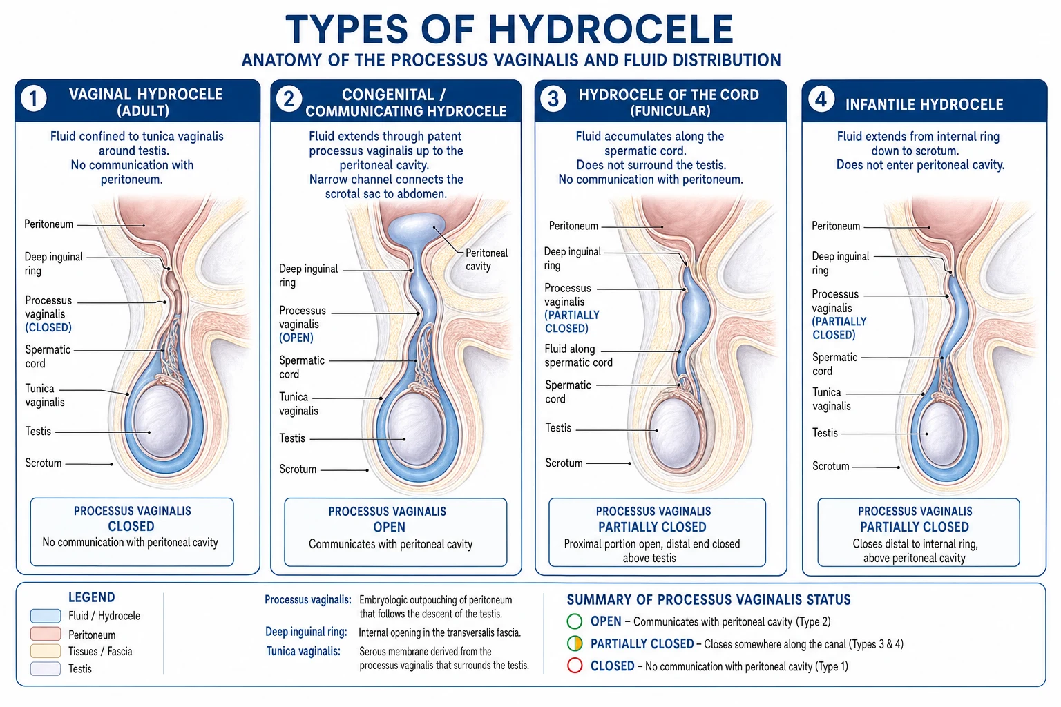
Hydroceles are classified two ways — by anatomical relationship to the peritoneal cavity (the classification an examiner asks for and the one that determines management), and by aetiology.[1][6]
Vaginal (adult)
commonest type
- Tunica vaginalis is a **closed sac** (no peritoneal communication)
- **Testis impalpable** within fluid, displaced posteriorly
- **Cannot reduce**; cough impulse absent
- **Can get above** the swelling
- Adult onset; idiopathic or secondary
Congenital (communicating)
infant type
- **Patent processus vaginalis** connects to peritoneum
- **Cough impulse positive**; reducible on lying
- Size **fluctuates** through the day (smaller in morning)
- Due to **failure of obliteration** of processus vaginalis
- Often associated with **indirect inguinal hernia**
Infantile
sac to deep ring only
- Processus closed at **internal ring** but patent down to scrotum
- Fluid extends **up to deep ring** but **not** into peritoneum
- Does **not** communicate with peritoneum
- **Not reducible** (no peritoneal connection)
- A closed sac shaped like an hour-glass
Encysted
rare, loculated
- Loculated fluid **around the cord** or testis
- No communication with peritoneum or tunica
- May be **intrascrotal** or **along the cord**
- Can mimic a solid mass — ultrasound confirms
- **Encysted hydrocele of the cord** = fluid in a segment of unobliterated processus
By aetiology: [1]
- Primary (idiopathic) — imbalanced secretion and absorption by the tunica vaginalis; no underlying cause found; the commonest adult type.[1]
- Secondary — reactive hydrocele due to an identifiable cause: epididymo-orchitis, testicular tumour, trauma, post-surgical (e.g. after varicocelectomy or renal transplant), irradiation, or tuberculosis. A secondary hydrocele is usually small-to-moderate and the underlying cause must be sought and treated.[2][5]
- Filarial — endemic in tropical regions; caused by Wuchereria bancrofti lymphatic obstruction producing a chylocele (lymph-rich, milky fluid). A major public-health problem in parts of India, Africa, and South-East Asia.[1][3]
Two further terms examiners use: [1]
- Funicular hydrocele — fluid along the spermatic cord in a sac that is patent at the internal ring but closed distally above the testis; the testicular tunica is separate. It lies above and separate from the testis, transilluminates, and mimics a cord swelling.
- Hydrocele of the cord — a loculated collection along a segment of unobliterated processus vaginalis, with the sac closed both proximally and distally; it forms a transilluminable swelling along the line of the cord that is separate from the testis.[1]
Epidemiology & Risk Factors
Hydrocele is common worldwide but the aetiology and demographics shift markedly with geography.[1][4]
- Congenital hydrocele — detectable in approximately 6 percent of newborn males because the processus vaginalis is still patent at birth. Over 90 percent close spontaneously by 12 to 24 months. Persistent patency after two years warrants surgical referral.[6]
- Adult primary hydrocele — typically presents in the fourth to sixth decades, unilateral in about two-thirds, right-sided slightly more often than left. The lifetime risk in temperate climates is modest; most are idiopathic.
- Filarial hydrocele — the dominant form in the tropics. The Global Programme to Eliminate Lymphatic Filariasis (GPELF) estimated approximately 19 million prevalent hydrocele cases worldwide, the great majority caused by Wuchereria bancrofti in sub-Saharan Africa, India, South-East Asia, and the Pacific. In endemic Indian states (Bihar, Uttar Pradesh, Jharkhand, West Bengal, Odisha) hydrocele is the most common surgical condition in adult males.[1][4]
Hydrocele — the numbers
Risk factors: [1]
- Infancy — a patent processus vaginalis is a normal developmental variant that usually closes.
- Endemic filarial exposure — residence in or travel to a lymphatic-filariasis zone; repeated mosquito bites (Culex, Anopheles, Aedes).
- Scrotal trauma or surgery — injury to the tunica or lymphatics (e.g. after varicocelectomy, inguinal hernia repair, renal transplant).
- Epididymo-orchitis or testicular tumour — inflammatory or neoplastic transudation produces a secondary hydrocele.
- Nephrotic syndrome or heart failure — rare cause of bilateral hydroceles through anasarca in severe fluid overload. [1]
Pathophysiology
Embryology — the key to every anatomical subtype
The testis develops in the retroperitoneum near the kidney and descends through the inguinal canal into the scrotum during the seventh to eighth month of gestation. As it descends, it carries a finger-like projection of peritoneum — the processus vaginalis — ahead of it into the scrotum. After descent, the portion of the processus vaginalis lying within the inguinal canal normally obliterates, while the portion surrounding the testis persists as the tunica vaginalis (the serous covering of the testis).[1][6]
The fate of the processus vaginalis determines the clinical picture: [1]
- Complete obliteration (normal) — only the tunica vaginalis remains; no communication with the peritoneum.
- Complete patency — peritoneal fluid drains freely into the scrotum: a congenital (communicating) hydrocele. Because the channel is open, the same defect can admit bowel as an indirect inguinal hernia; the two conditions share a common anatomical origin.
- Obliteration at the internal ring only — fluid tracks down but cannot return to the peritoneum: an infantile hydrocele (a closed sac extending to the deep ring).
- Segmental obliteration with a locule remaining along the cord — an encysted hydrocele of the cord.[1]
The processus vaginalis fails to obliterate in roughly 20 percent of adults on post-mortem study, explaining why a proportion of adult hydroceles are actually communicating, and why a clinically apparent hernia can appear de novo later in life. [1]
Adult hydrocele mechanism — secretion versus absorption
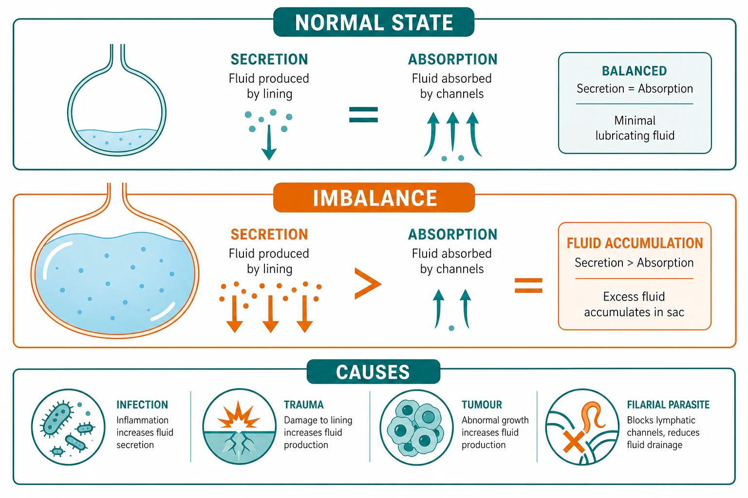
The healthy tunica vaginalis secretes and reabsorbs serous fluid in equilibrium. When this balance is disrupted — either by excessive secretion (inflammation, infection, trauma) or impaired absorption (lymphatic obstruction) — fluid accumulates. Primary idiopathic hydrocele is thought to arise from a subtle imbalance of this transport, though the precise trigger is usually unidentifiable.[5]
Filarial hydrocele — lymphatic obstruction
In lymphatic filariasis, adult Wuchereria bancrofti worms reside in human lymphatics and lymph nodes, where they live for years causing lymphangitis, lymphatic dilation, and progressive obstruction. Obstruction of the scrotal, inguinal, and retroperitoneal lymphatics prevents reabsorption of tunical fluid; the fluid becomes milky and lymph-rich (chylocele) and may contain microfilariae. Repeated inflammatory episodes drive thickening and fibrosis of the tunica and over time produce elephantiasis of the scrotum. The hydrocele in filariasis is therefore a chronic lymphatic disease, not a simple fluid imbalance — which is why surgery alone, without anti-parasitic treatment, risks recurrence.[1][3]
Secondary hydrocele
Any insult that inflames the tunica vaginalis or distorts local fluid dynamics can produce a secondary hydrocele: epididymo-orchitis, testicular torsion (the reactive hydrocele is an important trap), testicular tumour (up to 10 percent of testicular tumours present with a secondary hydrocele), trauma, post-surgical lymphatic disruption, and tuberculosis. The fluid is usually small-to-moderate in volume and the priority is to find and treat the cause rather than the hydrocele itself.[2][5]
Clinical Presentation
Typical presentation
The hallmark of a vaginal hydrocele is painless, gradually enlarging scrotal swelling noticed over weeks to months. The swelling is unilateral in most cases, soft and fluctuant on palpation, and the patient is systemically well — no fever, no urinary symptoms, no weight loss. The overlying skin is normal. Because the testis is suspended posteriorly within the fluid, the patient often cannot feel the testis separately and may report a sense of heaviness rather than pain. Very large or bilateral hydroceles can interfere with walking, clothing, and sexual function, and the cosmetic deformity is often the presenting complaint.[1]
Congenital hydrocele in infants
The mother reports a scrotal swelling that changes size — typically larger in the evening or when the infant cries and strains, smaller in the morning after a night lying flat. This diurnal variation reflects peritoneal fluid draining down the patent processus when intra-abdominal pressure rises. The swelling is soft, transilluminant, and reducible with gentle pressure. The testis is often palpable separately. Bilateral congenital hydroceles are common.[6]
Clinical examination — the four named signs
Examine the patient both standing and lying down. The four bedside signs that confirm a hydrocele and distinguish it from a hernia are:[2][1]
- Can get above the swelling. The examiner's two index fingers can be brought together and palpate above the upper limit of the swelling — confirming it is confined to the scrotum. If the swelling extends up into the inguinal canal, you cannot get above it, and it is an inguinal hernia. This is the single most important discriminator.
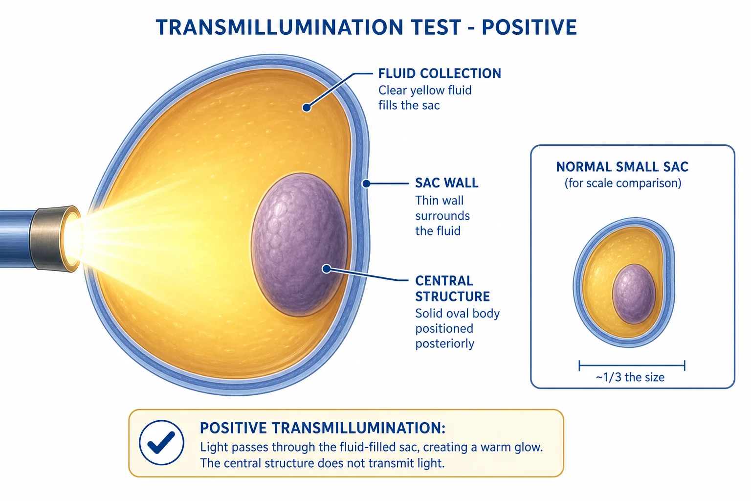
- Transillumination positive. In a darkened room, a torch held against the swelling transilluminates the serous fluid as a diffuse red glow. (A bowel-containing hernia or a solid tumour does not transilluminate; a chylocele may transilluminate poorly because of its lipid content.)
- Cough impulse absent in a vaginal hydrocele (the sac is closed). In a congenital/communicating hydrocele or a hernia, the cough impulse is positive because the peritoneal connection transmits raised intra-abdominal pressure.
- Cannot feel the testis separately — it is buried posteriorly within the fluid. In a hernia, the testis is palpable in its normal position separate from the swelling. [1]
Atypical and red-flag presentations
Exam application bank (NEET-PG / INICET)
One-line answer
Hydrocele is an abnormal collection of serous fluid within the tunica vaginalis of the testis (vaginal type) or along a patent processus vaginalis (congenital type). It presents as painless, gradually enlarging scrotal swelling that is positive on transillumination and that the examiner can get above (confirming it is scrotal, not inguinal). Congenital hydrocele (patent processus vaginalis) is common in infants and resolves by age 2 in over 90 percent of cases. Adult hydrocele is idiopathic or secondary to infection, trauma, tumour, or filariasis. The clinical skill is excluding the dangerous mimics — inguinal hernia (cough impulse positive, reducible, cannot get above), testicular tumour (heavy, opaque to transillumination), and testicular torsion (painful). Diagnosis is clinical, confirmed by ultrasound scrotum (excludes underlying testicular pathology). Treatment: Lord plication, Jabo
Worked stems (answer without another resource)
Stem 1 — Classic presentation. Map symptoms to mechanism; name the first investigation and first treatment step with dose/route if drug therapy is standard. [1]
Stem 2 — Unstable / complicated. List red flags that force immediate resuscitation, theatre, ICU, antidote, or reperfusion — and what you do in the first 15 minutes. [1]
Stem 3 — Atypical group. Elderly, pregnancy, child, or immunocompromised: how presentation and thresholds change. [1]
Stem 4 — Differential trap. Name the three closest mimics and one discriminator for each. [1]
Stem 5 — Disposition. Who goes home with safety-netting, who is admitted, who needs HDU/ICU/theatre, and what follow-up is mandatory. [1]
Rapid viva checklist
- Definition + classification
- Pathophysiology chain
- Bedside signs / criteria
- Score with exact components (if any)
- Emergency bundle
- Definitive therapy with doses
- Complications of disease and of treatment
- Special populations
- Guideline/trial name if classic
- Three exam traps
Coverage self-check
If you cannot answer any stem above from this page alone, re-read the matching section — the page is intended to be self-sufficient for final-prof and NEET-PG/INICET questions on Hydrocele.
[1]Atypical scenarios to flag: a rapidly enlarging hydrocele in an older man (underlying malignancy until proven otherwise); bilateral huge hydroceles in an adult (consider filariasis or anasarca from nephrotic syndrome or heart failure); a tense hydrocele after scrotal trauma (haematocele — fluid blood-stained, does not transilluminate); and a secondary hydrocele with fever (epididymo-orchitis or pyocele). [1]
Differential Diagnosis
The differential of painless scrotal swelling is short but every member must be confidently distinguished, because the management diverges sharply.[2][1]
| Condition | Distinguishing feature from hydrocele |
|---|---|
| Inguinal hernia (indirect, scrotal) | Cannot get above the swelling; cough impulse positive; reducible with a gurgling sensation; may hear bowel sounds over it; transillumination negative |
| Testicular tumour | Heavy, hard testis; does not transilluminate (solid); no cough impulse; check AFP, beta-hCG, LDH; ultrasound shows a solid intratesticular lesion; up to 10 percent have an associated reactive hydrocele |
| Epididymal cyst / spermatocele | Lies above and behind the testis (testis palpable separately); transilluminates brightly; smaller, often multiple; spermatocele fluid is opalescent (contains sperm) |
| Varicocele | Bag of worms feel; left side in 90 percent; prominent on standing, disappears lying down; does not transilluminate; cough impulse present; may impair fertility |
| Haematocele | After trauma; blood-stained, does not transilluminate; tender; testis may be ruptured (urgent ultrasound) |
| Pyocele | Infected hydrocele; tender, hot, systemically unwell; does not transilluminate (pus); surgical drainage and antibiotics |
| Epididymo-orchitis | Tender, fever, dysuria; hydrocele is reactive; urine dipstick positive; usually gradual onset |
| Inguinal lymphadenopathy / lipoma of cord | Solid, non-transilluminant; separate from testis |
Clinical & Bedside Assessment
A focused scrotal examination answered in under two minutes confirms the diagnosis and excludes the dangerous mimics.[1]
Position. Examine standing first (to reveal a varicocele and to test the cough impulse), then lying down (to test reducibility and re-palpate). [1]
Inspection: [1]
- Size, symmetry, and overlying skin of the scrotum (erythema implies infection; blue discoloration implies torsion or torsion of appendage).
- Visible swelling on standing; does it extend into the groin?
- Look for elephantiasis of the scrotum or limbs in endemic areas (filariasis). [1]
Palpation: [1]
- Can you get above it? — The pivotal question. Yes = scrotal. No = inguinal hernia.
- Feel the testis separately. In a vaginal hydrocele you cannot — it lies posteriorly within the fluid. Palpate the epididymis above and behind (epididymal cyst lies here).
- Cough impulse. Place two fingers over the swelling and ask the patient to cough. A vaginal hydrocele has no impulse; a communicating hydrocele or hernia gives a palpable expansile impulse.
- Reducibility. Gentle sustained pressure on a communicating hydrocele reduces it slowly with a gurgling feel; a vaginal hydrocele cannot be reduced.
- Transillumination. Darken the room, place a pen-torch against the swelling: a diffuse red glow = fluid (hydrocele); no transmission = solid (tumour, hernia, haematocele).
- Examine the contralateral side and do a general examination for lymphoedema, groin lymph nodes, and signs of systemic disease (heart failure, nephrotic syndrome). [1]
Transillumination — correct technique: Use a cold-light source or pen-torch in a darkened room. Place it flat against the posterior aspect of the swelling so light passes through the fluid-filled sac and testis. A clear hydrocele gives a homogeneous red glow. A chylocele or haematocele transilluminates poorly. Always interpret transillumination alongside the other signs — a fluid-filled hernia (rare) can transilluminate, and a tense hydrocele in a young child may not. [1]
Hydrocele
fluid in tunica vaginalis
- **Can get above** the swelling
- **Transillumination positive**
- **Cough impulse absent** (vaginal type)
- Testis buried posteriorly in fluid
Inguinal hernia
bowel/omentum in sac
- **Cannot get above** (extends into canal)
- **Transillumination negative**
- **Cough impulse positive**
- **Reducible**, may have bowel sounds
Testicular tumour
solid intratesticular mass
- **Heavy** testis
- **Transillumination negative** (solid)
- Check **AFP, beta-hCG, LDH**
- Urgent ultrasound; up to 10 percent have reactive hydrocele
Investigations
Hydrocele is essentially a clinical diagnosis. Imaging and blood tests are reserved for confirming the diagnosis when atypical, excluding an underlying testicular tumour, and investigating a suspected secondary or filarial cause.[2][5]
Ultrasound scrotum — the single essential investigation
A scrotal ultrasound is mandatory in any adult hydrocele before surgery, because it visualises the underlying testis that the fluid hides. It confirms: [1]
- An anechoic fluid collection around the testis with the testis displaced posteriorly (classic vaginal hydrocele).
- The testicular parenchyma — excluding a solid intratesticular tumour that would otherwise be missed behind the fluid. This is the critical safety step.
- Internal echoes, septations, or a solid component within the fluid — suggests haematocele, pyocele, or tumour.
- Testicular blood flow on Doppler — excludes torsion when the presentation is acute.[2]
In infants and children with a clear, transilluminant, reducing swelling, ultrasound is not routinely required unless the diagnosis is in doubt.[6]
Blood tests
- No bloods needed for a straightforward primary hydrocele.
- AFP, beta-hCG, and LDH — if a testicular tumour is suspected (solid mass on ultrasound, or a hydrocele that does not transilluminate or is rapidly enlarging). These are germ-cell tumour markers; their elevation mandates urgent urology referral and staging CT.
- Full blood count and CRP — if epididymo-orchitis or pyocele is suspected (leucocytosis, raised inflammatory markers).
- Filarial serology (antigen test) and peripheral blood smear for microfilariae (nocturnal, as Wuchereria is nocturnally subperiodic) — in endemic areas or in a patient with a chylocele or elephantiasis. [1]
Aspiration (diagnostic — rarely needed)
Hydrocele fluid is clear, straw-coloured and transudative. Diagnostic aspiration is reserved for an atypical fluid collection where the diagnosis is uncertain after ultrasound — for example to distinguish a chylocele (milky, lymph-rich) or a pyocele (purulent) from a simple hydrocele. Therapeutic aspiration is not a treatment (see Management).[7]
Management — Resuscitation
A hydrocele is not an emergency. The resuscitation question arises only when the diagnosis is wrong:[1]
- Strangulated inguinal hernia misdiagnosed as hydrocele — if a scrotal swelling is tender, irreducible, with systemic upset, vomiting, and abdominal distension, it is a strangulated hernia. This is a surgical emergency: resuscitate with intravenous fluids, analgesia, broad-spectrum antibiotics, and emergency surgery.
- Testicular torsion — sudden painful swelling in a young male with an absent cremasteric reflex is torsion regardless of any associated fluid. Immediate surgical exploration within the 6-hour salvage window; do not wait for imaging.
- Pyocele — an infected hydrocele presents with fever, severe tenderness, and systemic upset. Urgent ultrasound, intravenous antibiotics, and surgical drainage.
- Ruptured hydrocele / haematocele after trauma — urgent ultrasound to assess testicular integrity; surgical exploration if the tunica albuginea is torn. [1]
The rest of hydrocele management is elective and structured by age and aetiology. [1]
Management — Definitive & Stepwise
Congenital hydrocele (infants and children)
Congenital hydrocele — stepwise management
Reassure and observe
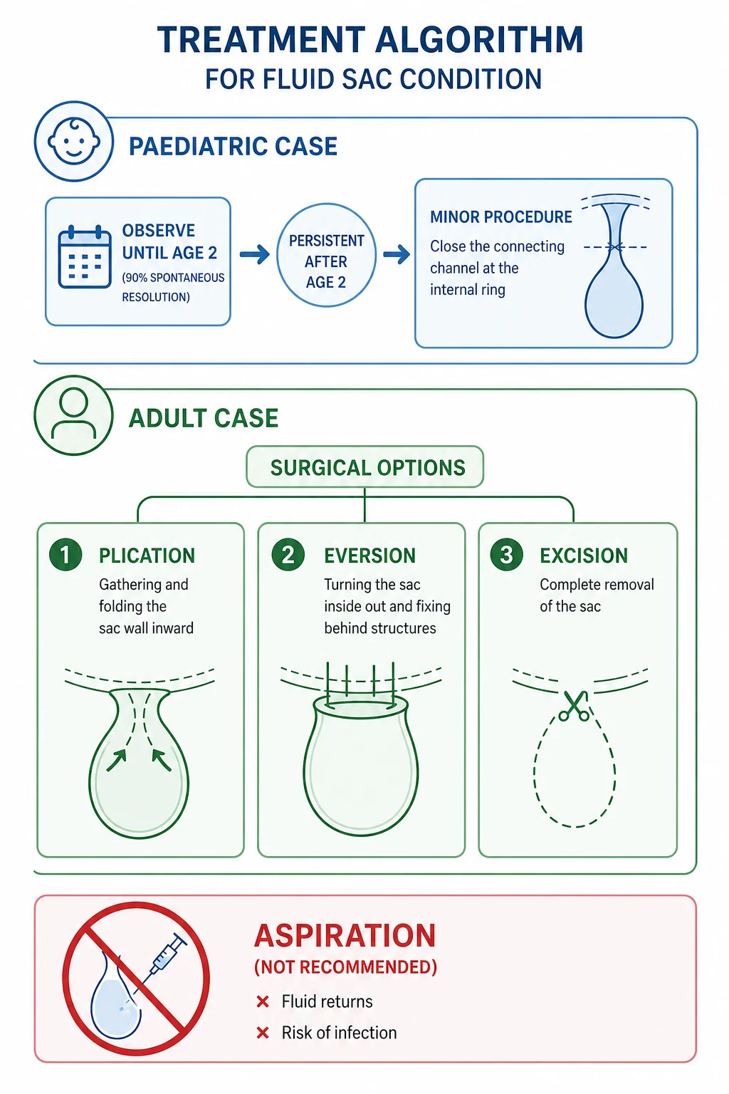
Over 90 percent of congenital hydroceles resolve spontaneously by 12 to 24 months as the processus vaginalis closes. Reassure the parents; review at intervals.
Threshold for surgery
If the hydrocele persists beyond age 2 years, is clearly communicating (changes size, has a cough impulse), or is associated with a hernia, refer for surgery.
Operation: herniotomy
**Inguinal approach**. Ligate the patent processus vaginalis at the **internal (deep) ring** and divide it. The distal hydrocele sac is left open or drained. This addresses the anatomical defect, not the fluid.
Do NOT aspirate
Aspiration in infants is condemned — it recurs immediately (the processus is open), risks introducing infection, and does not address the patent processus vaginalis.
The key principle in children: the operation is on the processus vaginalis (high ligation at the internal ring via an inguinal incision), not on the tunica itself. This is the same operation as for an indirect inguinal hernia in a child, because the two share a common anatomical defect.[6]
Adult hydrocele — surgical options
Three open surgical procedures dominate adult practice; all are curative with recurrence under 5 percent. Choice depends on the thickness of the sac and surgeon preference.[1][5]
- Jaboulay's procedure (eversion of the sac). A scrotal incision opens the tunica vaginalis, fluid is evacuated, and the redundant sac is everted (turned inside out) and sutured behind the spermatic cord and epididymis. The serosal surface now faces outward so any further secretion is absorbed by the surrounding subcutaneous tissues. This is the standard procedure for a moderate-to-large hydrocele with a thin sac.[1]
- Lord's plication. No sac excision; instead the redundant tunica is pleated (gathered) with a running suture around its circumference and sutured to the epididymis/testicular border, so the sac collapses onto the testis. Because there is minimal dissection, the haematoma rate is lower and it suits thin-walled sacs and small-to-moderate hydroceles.[1]
- Excision of the sac (subtotal excision). The tunica is dissected free and excised leaving a 1 to 2 cm rim sutured behind the cord. Reserved for thick-walled, fibrous sacs (common in longstanding or filarial hydrocele) where plication or eversion is impractical. Higher risk of haematoma due to more dissection.[1][5]
Surgery versus aspiration-sclerotherapy — Shakiba 2023 meta-analysis
PMID 37277518
Systematic review and meta-analysis, 5 RCTs, 335 patients / 342 hydroceles
Key finding
Sclerotherapy had a **significantly higher recurrence** than hydrocelectomy (RR 9.43, 95 percent CI 1.82 to 48.77) but no significant difference in fever, infection, or haematoma. Clinical cure rates were similar.
Practice change
Surgery is the durable curative option. Aspiration-sclerotherapy is a reasonable alternative for patients at high surgical risk or who decline surgery, accepting a higher recurrence rate.
Aspiration and sclerotherapy
Aspiration alone always recurs (within days to weeks) and risks introducing infection (pyocele) — it is not a treatment. Aspiration followed by sclerotherapy (instilling a sclerosant — phenol, tetracycline, ethanolamine oleate, or polidocanol — to obliterate the sac) is an alternative for the medically unfit patient who cannot tolerate surgery, or for those who decline an operation. The 2023 meta-analysis confirms it is effective but carries a substantially higher recurrence rate than surgery (recurrence risk ratio 9.43), so it is second-line.[7]
Filarial hydrocele
Surgery follows the same principles (Lord, Jaboulay, or excision — often excision because the sac is thickened and fibrous), plus anti-parasitic therapy to clear the worms and prevent recurrence and progression to lymphoedema: [1]
- Diethylcarbamazine (DEC) 6 mg/kg orally for 12 days (the standard anti-filarial regimen).[1]
- Doxycycline 100 to 200 mg daily for 4 to 6 weeks targets the Wolbachia endosymbiont and reduces worm burden and inflammation.[3]
- In India's Mass Drug Administration programme, DEC plus albendazole is given annually to endemic districts to interrupt transmission.[3]
Secondary hydrocele
Treat the underlying cause. Antibiotics for epididymo-orchitis; treat torsion or trauma surgically; excise a testicular tumour (radical inguinal orchidectomy with staging). The secondary hydrocele often resolves once the cause is treated; surgery on the hydrocele itself is reserved for large, persistent collections.[2]
Specific Subtypes & Scenarios
- Hydrocele of the cord (funicular). Fluid accumulates along the spermatic cord without scrotal involvement, in a segment of unobliterated processus vaginalis. It presents as a transilluminable swelling along the line of the cord, separate from and above the testis. It may mimic an inguinal hernia but, unlike a hernia, has no cough impulse (the sac is closed) and you can often get above it. Treatment is surgical excision.[1]
- Encysted hydrocele of the cord. A loculated collection trapped within a short segment of processus vaginalis, closed both above and below — a discrete, mobile, transilluminant scrotal or inguinal lump separate from the testis. Excision is curative.
- Infantile hydrocele. The processus is closed at the internal ring but patent down to the scrotum, so fluid extends up to the deep ring but does not communicate with the peritoneum. It does not reduce and has no cough impulse. Most resolve by age 2; persistent cases need surgery.[6]
- Filarial hydrocele / chylocele. Milky, lymph-rich fluid in an endemic-area patient, often with scrotal elephantiasis and lymphoedema of the limbs. Surgery plus DEC and doxycycline. A major cause of disability in the tropics.[1][4]
- Post-surgical hydrocele. Occurs after varicocelectomy, inguinal hernia repair, or renal transplantation due to lymphatic disruption. Often resolves spontaneously; persistence warrants ultrasound and possible surgery.[5]
- Hydrocele of the canal of Nuck — the female analogue (a patent processus vaginalis along the round ligament) presents as a swelling in the inguinal canal or labium majus in females; management parallels the male condition.[1]
Complications & Pitfalls
Complications of the hydrocele itself
- Testicular atrophy — long-standing, large hydroceles exert chronic pressure on the testicular vessels, impairing blood supply and causing atrophy. Bilateral large hydroceles may impair fertility.[1]
- Infertility — from pressure atrophy (bilateral), or from an underlying cause (filariasis, tumour).
- Infection / pyocele — secondary infection of the hydrocele fluid produces a tender, hot, non-transilluminant swelling with systemic upset; needs drainage and antibiotics.
- Haematocele — bleeding into the sac after trauma (or, rarely, spontaneous) produces a tender, non-transilluminant swelling; the underlying testis must be assessed for rupture by ultrasound.
- Rupture — rare; sudden reduction of a large hydrocele with scrotal oedema.
- Calcification — longstanding hydroceles may develop a calcified tunica (a hard, irregular scrotal mass on examination).[1]
- Malignant mesothelioma of the tunica vaginalis — a very rare but recognised association with longstanding hydrocele; any hydrocele with a nodular sac, bloody fluid, or persistent recurrence after surgery should raise suspicion.[5]
Complications of surgery
- Haematoma — the commonest surgical complication; reduced by meticulous haemostasis and the Lord technique (less dissection). A scrotal haematoma can be large and painful and may need drainage.[5]
- Recurrence — under 5 percent for all three open procedures; higher for aspiration-sclerotherapy.
- Infection — wound or scrotal sepsis.
- Injury to the spermatic cord — the vas deferens or testicular vessels lie within the cord; careless dissection risks ischaemic atrophy or infertility. Particularly relevant in eversion and excision.
- Fistula — rare; persistent leakage of fluid.
- Chronic pain — a small proportion of patients report chronic scrotal discomfort postoperatively.[5]
Classic pitfalls
- Misdiagnosing an inguinal hernia as a hydrocele — failing to test whether you can get above it or whether there is a cough impulse. A strangulated hernia is a surgical emergency; a hydrocele is not.
- Missing a testicular tumour behind a hydrocele — every adult hydrocele needs an ultrasound before surgery; a reactive hydrocele may be the only sign of an underlying germ-cell tumour.
- Aspirating an infant's hydrocele — it recurs immediately and risks infection; observe until age 2.
- Operating on a filarial hydrocele without DEC — recurrence and progression of lymphatic disease are likely.
- Calling torsion a hydrocele — hydrocele is painless; torsion is excruciating. Mislabelling costs the testis. [1]
Prognosis & Disposition
Excellent prognosis after surgical treatment. The recurrence rate is under 5 percent for all three open procedures, and testicular function is preserved in the vast majority. Most patients are discharged the same day or after an overnight stay and return to normal activity within 2 to 4 weeks (avoiding heavy lifting for 6 weeks).[1]
Congenital hydrocele: over 90 percent resolve by age 2; those requiring surgery (herniotomy) have an excellent outcome with minimal recurrence.[6]
Filarial hydrocele: good surgical outcome, but the patient needs ongoing anti-filarial therapy (DEC, doxycycline) to prevent recurrence and progression to lymphoedema and elephantiasis. Surgical camps in endemic districts provide hydrocele surgery as part of the WHO morbidity-management strategy.[1][4]
Secondary hydrocele: prognosis is that of the underlying cause. A testicular tumour has its own staging and treatment algorithm; epididymo-orchitis usually settles with antibiotics and the hydrocele resolves. [1]
Disposition: primary and congenital hydroceles are managed electively in the outpatient setting with surgery as a day case. Admission is reserved for complications (pyocele, haematocele, strangulated hernia, torsion) or for extensive filarial surgery requiring inpatient postoperative care. [1]
Special Populations
- Infants and children. Most congenital hydroceles resolve by age 2. Do not aspirate. Observe with periodic review. If persistent at age 2, clearly communicating, or associated with a hernia, perform herniotomy (inguinal approach, high ligation of the processus at the internal ring). Bilateral congenital hydroceles are common and bilateral repair is performed if both persist.[6]
- Elderly men. An adult hydrocele in an older man mandates a careful search for an underlying testicular tumour — ultrasound before any surgery. Large, longstanding hydroceles may cause testicular atrophy; surgery preserves residual function. Comorbidity may favour aspiration-sclerotherapy over surgery.[5]
- Filarial-endemic populations. Surgery is combined with anti-parasitic therapy (DEC, doxycycline) and embedded in the national MDA programme (India: annual DEC plus albendazole). Hydrocele surgery is delivered through camps in endemic districts and is among the most cost-effective surgical interventions globally.[1][4]
- Females (canal of Nuck). The rare female equivalent presents as an inguinal/labial swelling; management parallels the male condition.[1]
- Post-renal-transplant patients. Hydrocele and lymphocele can develop from lymphatic disruption; ultrasound and conservative management first, surgery for persistent collections.[5]
Evidence, Guidelines & Regional Differences
Surgical technique — the evidence base
No single open technique is demonstrably superior. Comparative series and reviews report similar recurrence rates (under 5 percent) for Lord plication, Jaboulay eversion, and sac excision. Lord plication has fewer haematomas (less dissection) and suits thin-walled sacs; excision is preferred for thick, fibrous sacs (common in filariasis). Jaboulay eversion is the standard for moderate-to-large thin sacs. Choice is largely by surgeon preference and sac characteristics.[1][5]
Laparoscopic approaches (e.g. the internal-ring Jaboulay or endoscopic techniques) have been described with comparable results but no clear advantage over open surgery for routine vaginal hydrocele; they have a role in selected cases and in specialist centres.[5]
Aspiration-sclerotherapy versus surgery
The 2023 meta-analysis by Shakiba and colleagues pooled five small RCTs (335 patients) and found that aspiration-sclerotherapy carried a markedly higher recurrence rate (RR 9.43, 95 percent CI 1.82 to 48.77) than surgery, with similar rates of fever, infection, and haematoma. Clinical cure rates were comparable, supporting sclerotherapy as a reasonable second-line option for the surgically unfit or those declining surgery, but not as first-line where surgery is feasible.[7]
Regional guideline differences
India-specific filarial burden: India bears a large share of global filarial hydrocele. The National Centre for Disease Control (NCDC) runs the Mass Drug Administration (MDA) programme — annual DEC plus albendazole to endemic districts — alongside hydrocele surgical camps. Bihar, Uttar Pradesh, Jharkhand, West Bengal, and Odisha are the highest-burden states. Hydrocelectomy in this setting is one of the most cost-effective surgical interventions in global health.[1][4]
Surgery vs sclerotherapy — the meta-analysis numbers
Exam Pearls
- Can get above a hydrocele but NOT above an inguinal hernia. The single most important distinguishing sign.[1]
- Transillumination positive in hydrocele; negative in tumour, haematocele, and most hernias.[2]
- Cough impulse: absent in vaginal hydrocele; present in hernia and in congenital/communicating hydrocele.[1]
- Congenital hydrocele = patent processus vaginalis. Resolves by age 2 in over 90 percent. If persistent: herniotomy at the internal ring (inguinal incision).[6]
- Adult hydrocele: always ultrasound first to exclude an underlying testicular tumour. Surgery: Lord plication or Jaboulay eversion (thin sac) or excision (thick sac). All under 5 percent recurrence.[2][5]
- Aspiration is not treatment — recurs and risks infection. Sclerotherapy is second-line for the unfit (recurrence RR 9.43 vs surgery).[7]
- Jaboulay = eversion of the sac; Lord = plication (pleating); excision = for thick sacs.[1]
- Filarial hydrocele: Wuchereria bancrofti. Milky chylocele fluid. Treat with DEC 6 mg/kg for 12 days plus doxycycline for Wolbachia. India's MDA: DEC plus albendazole annually.[1][3]
- Up to 10 percent of testicular tumours present with a reactive hydrocele — never assume a hydrocele is simple without ultrasound.[2]
- Infantile hydrocele extends to the deep ring but does not communicate with the peritoneum (closed at the internal ring). Funicular hydrocele is fluid along the cord separate from the testis.[1]
Clinical triad distinguishing hydrocele from hernia
GTC
can get above the upper limit = hydrocele; cannot = inguinal hernia
positive (red glow) = hydrocele; negative = tumour, haematocele, hernia
absent = vaginal hydrocele; present = hernia or communicating hydrocele
The three adult surgical procedures
JLE
eversion of the sac — sutured behind the cord; standard for moderate-to-large thin sacs
plication (pleating) of the sac to the testis border — less dissection, fewer haematomas; thin sacs
subtotal excision — for thick, fibrous sacs (filariasis); more dissection, higher haematoma risk
A 1-year-old boy has a soft, transilluminant scrotal swelling that is larger in the evening. What is the diagnosis and the management?
Congenital (communicating) hydrocele due to a patent processus vaginalis. The diurnal size change (larger when crying/straining) confirms communication with the peritoneum. Management: reassure and observe — over 90 percent close spontaneously by age 2. Do NOT aspirate. If still present after age 2, refer for herniotomy (high ligation of the processus vaginalis at the internal ring via an inguinal incision).
A 45-year-old man has a large, tense, non-tender left scrotal swelling that transilluminates. The testis cannot be felt separately. What is the next step before surgery?
Ultrasound scrotum to evaluate the underlying testis and exclude a testicular tumour — up to 10 percent of testicular tumours present with a reactive hydrocele. If ultrasound is suspicious (solid intratesticular lesion), check AFP, beta-hCG, and LDH and refer urgently to urology. If ultrasound confirms a simple hydrocele with a normal testis, proceed to elective surgery (Lord plication or Jaboulay eversion; excision if the sac is thick).
References
- [1]Rajasekaram S, Anuradha R, Manokaran G, Bethunaickan R. An overview of lymphatic filariasis lymphedema Lymphology, 2017.PMID 30248721
- [2]Gratzke C, Seitz M, Zaak D, Reich O, Schlenker B, Stief CG. [Painless enlargement of the scrotum] MMW Fortschr Med, 2006.PMID 16875378
- [3]Goldin J, Juergens AL. Filariasis 2026.PMID 32310472
- [4]Beard JH, Ohene-Yeboah M, Devas CR, Schecter WP. Hernia and Hydrocele 2015.PMID 26741001
- [5]Filmar S, Gross AJ, Hook S, Rosenbaum CM, Netsch C, Becker B. [Hydrocele] Urologie, 2024.PMID 38780784
- [6]Patoulias I, Koutsogiannis E, Panopoulos I, Michou P, Feidantsis T, Patoulias D. Hydrocele in Pediatric Population Acta Medica (Hradec Kralove), 2020.PMID 32771069
- [7]Shakiba B, Heidari K, Afshar K, Faegh A, Salehi-Pourmehr H. Aspiration and sclerotherapy versus hydrocelectomy for treating hydroceles: a systematic review and meta-analyses Surg Endosc, 2023.PMID 37277518