General Surgery · General Surgery
Inflammatory Bowel Disease — Surgical Management
Also known as Inflammatory Bowel Disease — Surgical Management
Surgical management of IBD differs between Crohn's disease (surgery is not curative — recurrence is expected) and ulcerative colitis (proctocolectomy is curative). UC: indications include acute severe colitis (failure of IV steroids), toxic megacolon, perforation, dysplasia/cancer. Definitive: proctocolectomy with ileal pouch-anal anastomosis (IPAA/J-pouch). Crohn's: strictureplasty (Heineke-Mikulicz, Finney, Michelassi), resection with anastomosis, abscess drainage, fistula management.
On this page & tools
Your progress
Saved locally on this device.
Exam tags
Red flags
Overview & Definition
Inflammatory bowel disease (IBD) comprises two chronic, immune-mediated inflammatory disorders of the gastrointestinal tract: ulcerative colitis (UC) and Crohn's disease (CD). IBD is a global disease of rising incidence; in newly industrialised countries of Asia, the Middle East and South America, incidence is climbing fastest while it has stabilised at high levels in the West.[1]
Although both diseases share the umbrella term "IBD" and many medical therapies, their surgical management is diametrically opposite, and this is the single most examined concept in the topic: [1]
- In ulcerative colitis, disease is confined to the colon and rectum (continuous, mucosal, begins at the rectum and extends proximally to a variable extent). Because the disease is anatomically limited to an organ that can be entirely removed, surgery is curative — proctocolectomy eliminates all at-risk mucosa.[2]
- In Crohn's disease, disease can affect any segment from the mouth to the anus (skip lesions, transmural inflammation). Because no surgeon can remove the entire GI tract, surgery can never cure — it treats the complications (obstruction, fistula, abscess, perforation) and recurrence is expected.[2]
This philosophical divide dictates every other principle: UC surgery aims to remove all colonic mucosa while restoring intestinal continuity (IPAA); Crohn's surgery aims to conserve as much bowel as possible (strictureplasty over resection, limited resection over colectomy) to prevent the catastrophic end-state of short bowel syndrome.[5]
Approximately 30–40% of UC patients and 70–80% of Crohn's patients ultimately require surgery during their lifetime, making IBD surgery one of the most common indications for elective colorectal resection in younger adults. [1]
Ulcerative Colitis
surgery is CURATIVE
- Disease **confined to colon + rectum** (continuous, mucosal, rectal-sparing rare)
- **Proctocolectomy removes all at-risk mucosa** → disease eliminated
- Definitive operation: **proctocolectomy + IPAA (J-pouch)**
- Indications: acute severe colitis failing steroids, toxic megacolon, perforation, dysplasia/cancer, refractory disease
- Sphincter-preserving; aim is stoma-free survival with good continence
Crohn's Disease
surgery is NEVER curative
- Disease **mouth to anus** (skip lesions, transmural, fissuring)
- **Bowel conservation is paramount** — recurrence is expected
- Strictureplasty preferred to resection where possible (preserves length)
- Limited resection with primary anastomosis; avoid panproctocolectomy if possible
- Surgery treats **complications**: obstruction, fistula, abscess, perforation, medically refractory disease
- **Perianal disease** managed by setons, fistulotomy, advancement flap — drain sepsis first
IBD surgery — the numbers that matter
Disease Classification (Montreal) — the Framework Examiners Test
Surgical decisions in IBD are anchored on the Montreal classification, which stratifies both diseases by extent and behaviour — the two axes that change operative strategy. Every IBD surgery question is answerable only if the Montreal class is known.[7]
Ulcerative colitis — Montreal extent (E1–E3)
| Class | Extent | Surgical implication |
|---|---|---|
| E1 | Proctitis (rectum only) | Rarely needs surgery; topical therapy; cancer risk near baseline |
| E2 | Left-sided (distal to splenic flexure) | Intermediate cancer risk; subtotal colectomy sufficient if operated |
| E3 | Extensive / pancolitis (proximal to splenic flexure) | Highest cancer risk; proctocolectomy is the standard definitive operation |
Crohn's disease — Montreal age, location, behaviour
Age at diagnosis: A1 (under 16), A2 (17–40), A3 (over 40). Location (L1–L4): [1]
| Class | Location | Typical surgery |
|---|---|---|
| L1 | Ileal (terminal ileum) | Ileocaecal resection (commonest Crohn's operation) |
| L2 | Colonic | Segmental colectomy; cancer risk if extensive |
| L3 | Ileocolonic | Ileocolic resection + strictureplasty |
| L4 | Upper GI (added modifier) | Gastric/duodenal strictureplasty, bypass |
Behaviour (B1–B3, plus perianal modifier "p"): [1]
| Class | Behaviour | Surgical implication |
|---|---|---|
| B1 | Non-stricturing, non-penetrating (inflammatory) | Medical therapy first; surgery for refractory disease |
| B2 | Stricturing | Strictureplasty or resection — obstruction is the indication |
| B3 | Penetrating (fistulising) | Drain abscess, manage fistula, resect fistula-bearing segment |
| p | Perianal disease modifier | Seton, fistulotomy, advancement flap — perianal surgery algorithm |
The behaviour modifier is the key: B2 stricturing disease drives the strictureplasty algorithm, while B3 penetrating disease drives abscess and fistula management. Smoking (a Crohn's risk factor and recurrence amplifier) and perianal disease are the two strongest predictors of post-operative recurrence.[2]
UC — Surgical Indications
UC surgery is divided into emergency (life-saving), urgent (steroid/ rescue failure), and elective (quality-of-life, dysplasia) indications. Recognising which applies changes the operation performed.[2]
Emergency indications
- Toxic megacolon — transverse colon diameter over 6 cm on plain AXR with systemic toxicity (fever, tachycardia, leucocytosis, altered mental status). Risk of perforation is high; emergency subtotal colectomy if deterioration despite medical therapy.
- Free perforation — emergency laparotomy.
- Uncontrolled massive colonic haemorrhage — rare; emergency colectomy.
Urgent indication — acute severe UC (ASUC)
- ASUC (modified Truelove & Witts: ≥6 bloody stools/day PLUS any systemic toxicity marker — fever, tachycardia, ESR over 30, haemoglobin under 10, albumin under 30). Admit, IV hydrocortisone 100 mg IV QDS (400 mg/day) or methylprednisolone 60 mg/day.
- If Travis/Oxford day-3 criteria met (see below) → rescue therapy (ciclosporin or infliximab). If rescue fails → subtotal colectomy. [1]
Elective indications
- Medically refractory disease — chronic steroid dependence, failure of biologics, poor quality of life.
- Dysplasia or colorectal cancer on surveillance biopsy — low-grade dysplasia warrants colectomy discussion; high-grade dysplasia or cancer mandates colectomy.
- Growth failure / delayed puberty in paediatric UC.
Acute Severe UC — Predicting Colectomy & Rescue Therapy
The decision to escalate from steroids to rescue therapy or colectomy is governed by two validated prediction scores, both examining the response to IV steroids by day 3–4.[3]
Travis/Oxford criteria (Day 3 of IV steroids)
Colectomy predicted (≈85% need colectomy) if either present on Day 3:
- Stool frequency over 8/day, OR
- Stool frequency 3–8/day AND CRP over 45 mg/L [1]
If met → start rescue therapy (ciclosporin or infliximab). If no improvement at day 5–7 of rescue → colectomy.[3]
St Mark's criteria (Day 4)
- Stool frequency over 8/day AND CRP over 45 mg/L on Day 4 → 75% colectomy rate within that admission. [1]
Rescue therapy protocols
Ciclosporin — 2 mg/kg/day IV continuous infusion for 24 h; trough 150–250 ng/mL; monitor renal function, BP, magnesium, cholesterol; oral 4 mg/kg/day for 3 months after; Pneumocystis prophylaxis with co-trimoxazole 960 mg three times weekly. Side effects: nephrotoxicity, hypertension, gingival hyperplasia, tremor, hyperkalaemia. [1]
Infliximab — 5 mg/kg IV at weeks 0, 2, 6 then 8-weekly. Pre-medicate with hydrocortisone 100 mg IV + chlorphenamine 10 mg IV. Screen for TB (IGRA), HBV, HCV, HIV before starting. The CONSTRUCT trial found infliximab and ciclosporin similarly effective for ASUC rescue; infliximab is generally preferred for ease and lower toxicity.[2]
UC Surgical Procedures — The 3-Stage Approach
The strategy in UC is to remove all colonic and rectal mucosa while preserving the anal sphincters and restoring continuity. In a healthy, well-nourished, elective patient this is achievable in one or two stages; in a sick, malnourished, immunosuppressed patient with ASUC it is staged to minimise morbidity.[5]
The 3-stage approach (why and when)
Stage 1
Subtotal colectomy + end ileostomy
- Removes the inflamed colon (the source of sepsis, bleeding, perforation risk)
- **Preserves the rectum** for future pouch surgery (rectal stump as Hartmann's or mucous fistula)
- Emergency/urgent setting: sick, malnourished, high-dose steroids — minimally invasive, fast recovery
- Allows steroids to be weaned and nutrition restored before definitive surgery
Stage 2
Completion proctectomy + IPAA + loop ileostomy
- Elective, weeks-months later when patient optimised
- Removes the rectum, constructs the ileal pouch, anastomoses to anal canal
- **Diverting loop ileostomy** protects the fresh ileoanal anastomosis from leakage
Stage 3
Loop ileostomy closure
- 8–12 weeks after Stage 2
- **Pouchogram** (water-soluble contrast enema) confirms an intact anastomosis and pouch before closure
- Pouch starts functioning: 4–8 stools/day, 1–2 at night
When to use 3-stage: ASUC on high-dose steroids/biologics, malnutrition (albumin under 30), obesity, elderly, or any factor raising anastomotic leak risk. 2-stage (IPAA + loop ileostomy, then closure) suits the well, elective patient. 1-stage (IPAA without diverting ileostomy) is selected only in meticulously selected low-risk patients and is uncommon.[5]
Definitive operation — IPAA construction step by step
The ileal pouch-anal anastomosis (IPAA) is the gold-standard definitive operation for UC: it cures the disease (removes all colonic and rectal mucosa) while restoring intestinal continuity and preserving continence.[2]
- Total proctocolectomy — mobilise the entire colon and rectum to the pelvic floor in the mesorectal plane, carefully preserving the pelvic autonomic nerves (hypogastric and pelvic splanchnic nerves — injury causes bladder and sexual dysfunction). The rectum is dissected to the anorectal ring.
- Mucosectomy (optional) or stapled transection — either the rectal mucosa is stripped to the dentate line (hand-sewn anastomosis, removes all mucosa) or the rectum is stapled across just above the anal transitional zone (ATZ) (double-stapled, preserves a 1–2 cm cuff of ATZ mucosa).
- Pouch construction from the terminal 15–20 cm of ileum — see pouch configurations below.
- Ileoanal anastomosis — the pouch outlet is anastomosed to the anal canal, either hand-sewn to the dentate line (after mucosectomy) or double-stapled to the ATZ (stapled technique).
- Diverting loop ileostomy — protects the anastomosis; closed 8–12 weeks later after a normal pouchogram. [1]
Pouch configurations — J, S, and W
| Configuration | Construction | Volume | Outlet length | Use |
|---|---|---|---|---|
| J-pouch | Terminal 15–20 cm ileum folded in two, stapled along antimesenteric border (two GIA firings) | 150–300 mL | Short (the apex) | Commonest by far — easy to construct, reliable emptying, lowest leak rate |
| S-pouch | Three limbs of ileum in an S; longer efferent limb | Larger | Long efferent limb (2 cm) | Used when reach to anal canal is difficult (tall patients, fatty mesentery); risk of outlet obstruction if efferent limb over 2 cm |
| W-pouch | Four limbs of ileum | Largest (~400 mL) | Short | Better capacity, less frequent stools; more complex to construct, now rarely used |
The J-pouch is the workhorse configuration (>90% of cases) because it is simple, reproducible, and empties well. The S-pouch is reserved for the difficult "reach" where the ileum will not comfortably reach the anal canal — but its efferent limb must be kept under 2 cm to avoid obstructed defaecation / pouch outlet syndrome.[2]
Alternative operations when IPAA is unsuitable
- Total proctocolectomy with end (Brooke) ileostomy — the fail-safe option for patients unfit for a pouch (ageing sphincter, low rectal cancer, Crohn's of the colon confirmed intra-operatively, obese with no reach). Cures UC, no pouch complications, but permanent stoma.
- Ileorectal anastomosis (IRA) — preserves the rectum; only if rectum is spared and compliant (rare in UC; more relevant in Crohn's colitis). Carries ongoing rectal cancer surveillance burden.
- Kock pouch (continent ileostomy) — an internal ileal reservoir with a nipple valve, emptied by intermittent intubation; largely superseded by IPAA and used now only in selected revision cases. [1]
Pouchitis and Pouch Complications — Management Algorithm
The IPAA is not a normal colon: patients live with a neorectum that stores and reabsorbs ileal effluent, which predisposes to a characteristic spectrum of complications. Pouchitis is the dominant long-term problem.[2]
Pouchitis — diagnosis and treatment ladder
Pouchitis (non-specific inflammation of the ileal pouch) has a lifetime incidence of 40–50% after IPAA, presenting with increased stool frequency, urgency, abdominal cramping, tenesmus, low-grade fever, and sometimes arthralgia. Diagnosis combines clinical symptoms + pouchoscopy with biopsy (oedema, granularity, friability, ulceration).[2]
Treatment algorithm: [1]
- Acute pouchitis (first episode, under 4 weeks) — ciprofloxacin 500 mg BD + metronidazole 400 mg TDS for 14 days. Most respond within days.
- Acute relapsing / chronic antibiotic-dependent pouchitis — rotate antibiotics (ciprofloxacin, metronidazole, rifaximin, tinidazole); add probiotics (VSL#3, 1 sachet BD) for maintenance; consider budesonide 9 mg OD.
- Chronic antibiotic-refractory pouchitis — investigate for Crohn's of the pouch (fistulae, prepouch ileitis, granulomata on biopsy). Treat with oral budesonide, then immunosuppression (azathioprine 2–2.5 mg/kg/day, or anti-TNF infliximab/adalimumab, or ustekinumab/vedolizumab).
- Pouch failure (5–10%) — refractory dysfunction, fistulae, sepsis, or confirmed Crohn's of the pouch → pouch excision + permanent end ileostomy. [1]
Other pouch complications
| Complication | Incidence | Management |
|---|---|---|
| Cuffitis | 10–15% | Inflammation of the retained rectal cuff (after stapled anastomosis). Topical mesalazine or steroid foam; rarely revision. |
| Anastomotic stricture | 10–15% | Endoscopic balloon dilatation; repeated as needed; surgery if refractory. |
| Pelvic sepsis / anastomotic leak | 5–10% | Drainage (radiological or surgical), antibiotics, prolonged diversion; may require pouch excision. Strongest predictor of later pouch failure. |
| Pouch-vaginal fistula | 3–10% | Advancement flap, seton, pouch advancement; if Crohn's confirmed → medical therapy; pouch failure if refractory. |
| Pouch outlet obstruction (S-pouch) | Variable | Shorten efferent limb; revision surgery. |
| ATZ dysplasia / cancer | Rare | Mucosectomy and pouch revision; pouchoscopy surveillance every 1–3 years. |
J-POUCH
Crohn's Disease Surgery — Principles
Surgery in Crohn's is governed by one overriding principle: conserve bowel. Because disease recurs and patients face multiple operations over a lifetime, every centimetre of small bowel preserved today is absorptive capacity preserved against future short bowel syndrome.[5]
The cardinal principles
- Bowel conservation is paramount — prefer strictureplasty over resection, limited resection over segmental, segmental over subtotal colectomy.
- Limited resection to grossly disease-free margins (2 cm) — wide margins do not reduce recurrence, so there is no oncological-style clearance. Frozen section is not routinely required.
- Primary anastomosis whenever possible — avoid stomas in the elective, well-prepared patient. If the patient is septic, malnourished, or on high-dose steroids, form a stoma.
- Surgery treats COMPLICATIONS, not the disease — operate for obstruction, fistula, abscess, perforation, or medically refractory disease, not "to remove Crohn's."
- Drain sepsis before definitive surgery — abscess drained percutaneously and antibiotics given before elective resection.
- Address nutrition first — resection in a malnourished patient carries 2–3× the complication rate.
Crohn's Surgical Procedures
1. Strictureplasty — the bowel-conserving operation
Strictureplasty widens a narrowed segment without removing it, preserving absorptive length. It is the procedure of choice for fibrotic (B2) strictures where resection would sacrifice excessive bowel, particularly for multiple short strictures (skip lesions) and in patients with prior resections at risk of short bowel.[5]
Three techniques, selected by stricture length: [1]
Heineke-Mikulicz strictureplasty (strictures under 10 cm) — the workhorse, analogous to a pyloroplasty:
- Longitudinal incision along the antimesenteric border through the full stricture, extending 1–2 cm onto normal bowel either end.
- Send biopsy of the strictured wall for frozen section — exclude malignancy (essential in isolated strictures or long-standing disease).
- The longitudinal opening is closed transversely (perpendicular) in a single or double layer — this widens the lumen. [1]
Finney strictureplasty (strictures 10–25 cm):
- The bowel is folded at the stricture site into a U-shape (antimesenteric borders apposed).
- A long longitudinal incision is made along the antimesenteric border of the folded loop.
- The two opened limbs are sewn together as a side-to-side anastomosis, creating a wide, shortened, diverted segment. [1]
Side-to-side isoperistaltic strictureplasty (Michelassi) (strictures over 25 cm, or long diseased segments where resection would cause short bowel):
- The diseased loop is divided and the two ends are overlapped isoperistaltically.
- A long longitudinal enterotomy on each limb, then the two are sewn side-to-side, creating a single wide channel through the diseased segment.
- Used for extensive jejunoileal disease — preserves maximal absorptive surface. [1]
Advantages of strictureplasty over resection: preserves bowel length (prevents short bowel syndrome), equivalent or lower recurrence rates at the strictureplasty site, maintains absorptive capacity, no stoma required, and repeatable. Contraindication: stricture with fistula or phlegmon (resect), or malignancy on biopsy. [1]
2. Resection — when to remove bowel
| Indication | Procedure |
|---|---|
| Ileocaecal disease (L1/L3, commonest operation) | Ileocaecal resection with primary stapled/side-to-side anastomosis — removes terminal ileum and caecum |
| Segmental colitis (limited Crohn's colitis) | Segmental colectomy with anastomosis — avoid subtotal/total colectomy to preserve absorptive colon |
| Extensive colitis with rectal sparing | Subtotal colectomy + ileorectal anastomosis |
| Extensive colitis + rectal involvement / dysplasia | Panproctocolectomy + end ileostomy (the only case of a stoma being the planned outcome in Crohn's) |
| Perforation | Limited resection + stoma (avoid anastomosis in contaminated field) |
| Severe haemorrhage | Segmental resection of bleeding segment |
Resection margins need only be 2 cm of grossly normal bowel — wider margins do not reduce recurrence, because Crohn's recurs regardless of margin status. Anastomotic configuration (side-to-side stapled vs end-to-end hand-sewn) does not consistently affect recurrence; side-to-side stapled has lower leak rates and is preferred. [1]
3. Perianal Crohn's disease — the management algorithm
Perianal disease (the "p" modifier) affects up to 40% of Crohn's patients and is the most challenging manifestation. The overriding rule: drain sepsis first, then manage the fistula, then treat the underlying disease medically.[2]
Step 1 — control sepsis:
- Examination under anaesthetic (EUA) is both diagnostic and therapeutic. Loose draining seton placement is the cornerstone — the seton keeps the tract open, preventing abscess re-accumulation, while medical therapy takes effect.
- Antibiotics — ciprofloxacin 500 mg BD + metronidazole 400 mg TDS for 4–8 weeks. [1]
Step 2 — classify the fistula (by Parks classification and by simple vs complex):
- Simple fistula — single, low, superficial or low intersphincteric, no sphincter involvement → fistulotomy (lay open) is safe and curative.
- Complex fistula — high transsphincteric/suprasphincteric, multiple tracts, associated with abscess, or recurrent → avoid fistulotomy (would transect too much sphincter → incontinence). Manage with long-term draining seton, endorectal advancement flap, or LIFT (ligation of intersphincteric fistula tract). [1]
Step 3 — definitive management:
- Advancement flap — rectal mucosal flap advanced over the internal fistula opening; closure rates 60–70%; preserves sphincter.
- LIFT — ligate the fistula in the intersphincteric space; success 70–80%; minimal sphincter division.
- Fistula plug / fibrin glue — lower success (30–50%) but sphincter-sparing.
- Anti-TNF (infliximab/adalimumab) — combined with seton drainage, closes complex fistulae in 50–70%; remove seton once tract has closed clinically/radiologically. EUtrasound/MRI (pelvic MRI is gold standard) assesses response and detects occult collections. [1]
Iron rule of perianal Crohn's: never perform a fistulotomy on a complex or high fistula — the sphincter damage causes incontinence that is irreversible. Setons and advancement flaps are always preferred in Crohn's. [1]
Pre-operative Optimisation — Detailed Protocol
Optimising the IBD patient before surgery halves the complication rate. The ECCO peri-operative roadmap organises this into five domains: nutrition, medication, sepsis, thromboprophylaxis, and stoma planning.[5]
Nutritional optimisation
- Screen: BMI, weight loss trajectory (over 10% in 6 months = severe), serum albumin (under 30 g/L = high risk), haemoglobin, iron studies, vitamin D, B12.
- Treat malnutrition: 7–14 days of intensive oral nutritional supplements or enteral (NG) feeding before elective surgery reduces complications. TPN reserved for those who cannot tolerate enteral feeding or have short bowel.
- Correct iron-deficiency anaemia: IV ferric carboxymaltose (preferred over oral for speed and tolerance) to bring haemoglobin to acceptable range.
- Vitamin D supplementation (deficiency linked to worse surgical outcomes); B12 replacement if terminal ileum diseased/resected. [1]
Medication management (the viva question)
- 5-ASA (mesalazine): continue — no peri-operative harm.
- Steroids: taper to lowest effective dose. Over 20 mg prednisolone/day roughly doubles anastomotic leak, wound infection, and peptic ulcer bleeding. Do not stop abruptly — give peri-operative IV hydrocortisone (100 mg at induction) for patients on long-term steroids to prevent adrenal crisis.
- Thiopurines (azathioprine, 6-MP): continue peri-operatively — no convincing increase in complications.
- Methotrexate: continue.
- Anti-TNF (infliximab, adalimumab): traditionally held 4–8 weeks (a half-life washout), but modern evidence shows holding is not clearly necessary and may precipitate disease flare. Current ECCO guidance: continue if needed for disease control; the small increase in infectious complications does not justify postponing necessary surgery.
- Vedolizumab: gut-selective, minimal systemic immunosuppression — generally continued; PREPARE-style data suggest no increased surgical complications.
- Ustekinumab: long half-life; consider holding ~12 weeks if surgery is elective and disease stable. [1]
VTE prophylaxis
IBD patients are in a 2–3× heightened prothrombotic state (systemic inflammation, dehydration, steroids, immobility, post-operative). LMWH (enoxaparin 40 mg SC OD) is given from admission, continued through surgery (omit the morning dose, restart 6–12 h post-op), and extended for 28 days post-discharge after major colorectal surgery (per ACCP/ACG guidance). Add mechanical thromboprophylaxis (compression stockings, intermittent pneumatic compression). [1]
Stoma planning and MDT
- Stoma nurse marks the site pre-operatively, considering the patient's waistline, bony prominences, scars, and creases; both an ileostomy and a permanent colostomy site are marked if either is possible.
- MDT discussion (surgeon, gastroenterologist, IBD nurse, dietitian, stoma therapist, anaesthetist) aligns the operative plan with the medical strategy — particularly important when considering IPAA in a patient who might have Crohn's.
Post-operative Crohn's Recurrence — Rutgeerts Score & Prevention
Crohn's recurs after surgery with near-certainty at the neoterminal ileum (just proximal to the anastomosis). Without prophylaxis, endoscopic recurrence is 70–80% at 1 year and clinical recurrence reaches 50% by 5 years. The goal of post-operative management is to detect and treat endoscopic recurrence early, before it becomes symptomatic and causes re-operation.[4]
Rutgeerts endoscopic score (the standard since 1990)
Ileocolonoscopy at 6–12 months after resection grades the neoterminal ileum:[4]
| Score | Endoscopic finding | Clinical recurrence risk |
|---|---|---|
| i0 | No lesions | Low |
| i1 | Under 5 aphthous lesions | Low |
| i2 | Over 5 aphthous lesions, skip areas, or larger lesions (not circumferential) | Intermediate |
| i3 | Diffuse aphthous ileitis with diffusely inflamed mucosa | High |
| i4 | Diffuse inflammation with large ulcers, nodules, cobblestoning | High |
i0–i1 = remission (continue current therapy). i2–i4 = recurrence (escalate therapy). A newer modified Rutgeerts (i2a/i2b) subdivides i2 but the original i0–i4 remains the examinable standard. [1]
Risk stratification — who needs aggressive prophylaxis?
High-risk patients (need immediate post-operative anti-TNF): current smoker, two or more prior resections, penetrating/fistulising (B3) disease, perianal disease, extensive small-bowel resection (short bowel risk), and early first recurrence. Low-risk patients (single first resection, non-smoker, non-penetrating) can be managed expectantly with step-up therapy guided by the 6-month colonoscopy. [1]
Medical prophylaxis algorithms
| Patient risk | Post-operative strategy |
|---|---|
| Low risk | No immediate medication; ileocolonoscopy at 6 months (Rutgeerts score). If i0–i1, observe; if i2–i4, start thiopurine or anti-TNF. |
| Moderate risk | Thiopurine (azathioprine 2–2.5 mg/kg/day) post-op; reassess at 6 months; escalate to anti-TNF if recurrence. |
| High risk | Anti-TNF immediately post-op (infliximab 5 mg/kg at weeks 0, 2, 6 then 8-weekly, or adalimumab 160/80/40 mg every other week). This is the most effective prevention — POCER trial showed anti-TNF beats thiopurines for preventing recurrence. |
| Adjunct in all | Metronidazole 20 mg/kg/day for 3 months reduces early recurrence (Rutgeerts); modest effect. Smoking cessation halves recurrence risk — the single most powerful modifiable factor. |
Newer agents (ustekinumab, vedolizumab) also reduce post-operative recurrence and are options for patients intolerant of or failing anti-TNF. [1]
Colorectal Cancer Surveillance in IBD
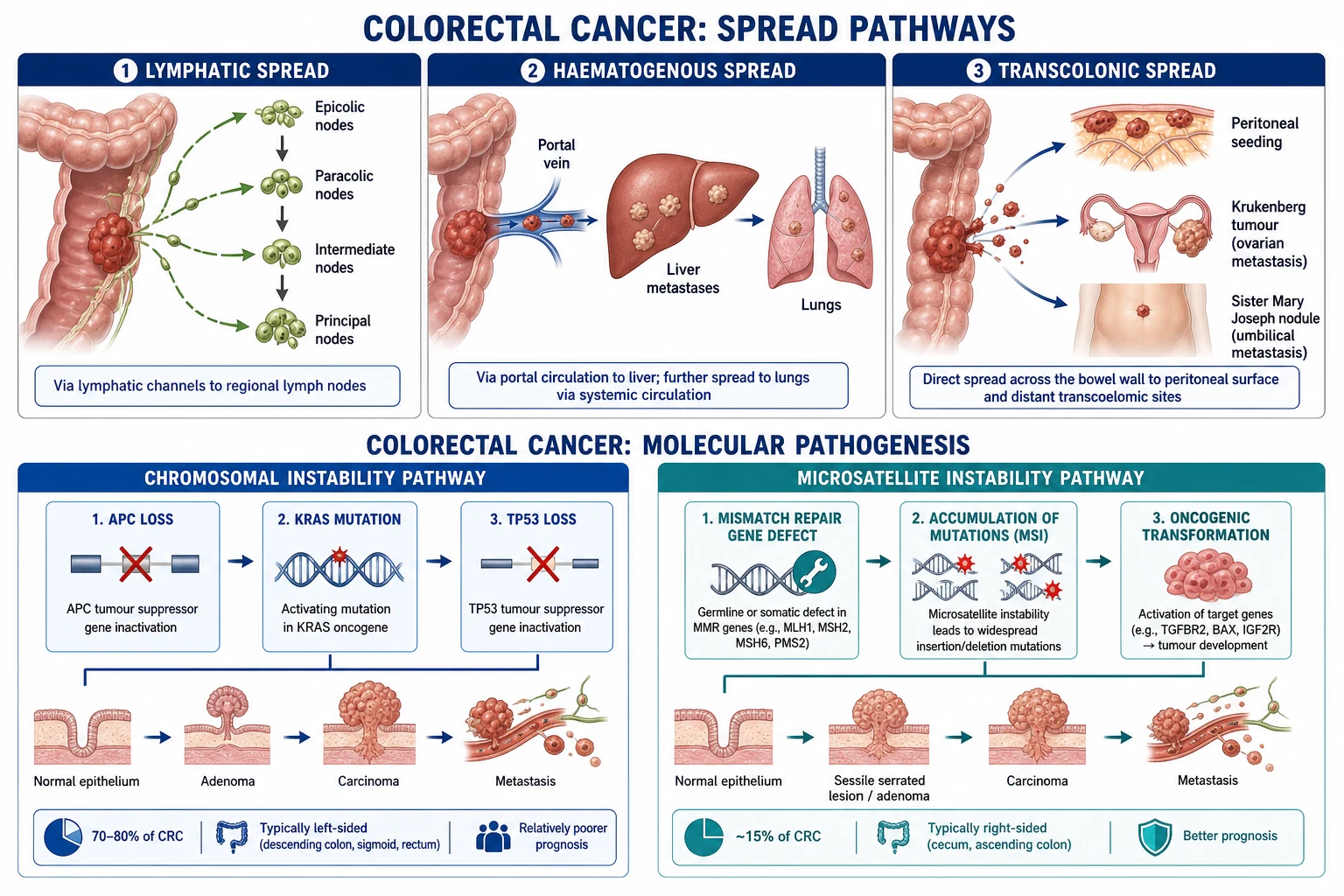
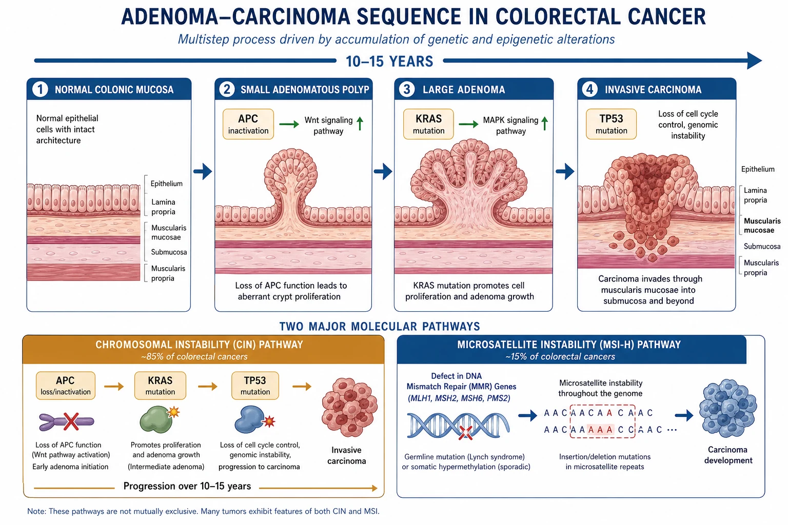
Long-standing IBD carries a well-defined colorectal cancer (CRC) risk driven by chronic inflammation. After 8–10 years of pancolitis (E3 UC or extensive colonic Crohn's), CRC risk rises approximately 0.5–1% per year, reaching a cumulative risk of 8–10% after 20 years of pancolitis. Left-sided disease (E2) has a lower but still elevated risk after 10–15 years; isolated proctitis (E1) has near-baseline risk.[6]
Risk amplifiers
- Duration of disease over 8–10 years.
- Extensive disease (pancolitis E3).
- Active ongoing inflammation (endoscopic/histological activity).
- Primary sclerosing cholangitis (PSC) — coexistence multiplies CRC risk several-fold; these patients start surveillance at diagnosis (not after 8 years) and have annual surveillance.
- Family history of CRC in a first-degree relative.
- Strictures and pseudopolyps — make surveillance harder and harbour higher dysplasia rates.
Surveillance protocol (BSG/SCENIC)[6]
- Start at 8–10 years from diagnosis (or at IBD diagnosis in PSC patients) for extensive/pancolitis.
- Interval: every 1–2 years (annual for PSC, prior dysplasia, or severe active inflammation).
- Technique: colonoscopy with chromoendoscopy (methylene blue 0.1% or indigo carmine 0.03–0.4% dye-spray) — SCENIC consensus finds chromoendoscopy detects significantly more dysplasia than standard white-light with random biopsies. Targeted biopsies of any visible lesion (using the modified Paris classification) have replaced the old "32 random biopsies" protocol in most centres, though some still combine both.
- Stricture: any colonic stricture in chronic colitis is malignant until proven otherwise — biopsy and image; often an indication for colectomy.
Dysplasia management
- No dysplasia: continue routine surveillance.
- Visible dysplasia (DALM — dysplasia-associated lesion or mass), completely resectable: endoscopic resection + intensified surveillance; colectomy if not resectable en bloc or if high-grade dysplasia.
- Invisible low-grade dysplasia: colectomy discussion — risk of concurrent cancer or progression is significant; many centres recommend colectomy, others intensified surveillance with chromoendoscopy.
- High-grade dysplasia or cancer: colectomy (proctocolectomy with IPAA for UC; segmental or panproctocolectomy for Crohn's depending on extent).
Complications of IBD Surgery
| Complication | Setting | Management |
|---|---|---|
| Anastomotic leak | IPAA, ileocolic anastomosis | Higher with steroids, malnutrition, anti-TNF; managed by drainage, diversion, antibiotics; pouch failure if severe |
| Pelvic sepsis | IPAA | Radiological drainage, antibiotics; strongest predictor of pouch failure |
| Small bowel obstruction | Adhesions, IPAA | Initial conservative (NGT); surgery if persistent or strangulation |
| Pouch-vaginal fistula | IPAA | Advancement flap, seton; if Crohn's of pouch → medical therapy ± pouch excision |
| Urinary/sexual dysfunction | Proctectomy (nerve injury) | Hypogastric/pelvic nerve damage; careful mesorectal plane dissection prevents it |
| Infertility | IPAA in women | Adhesions around tubes; 6× higher infertility after IPAA — counsel women pre-operatively |
| Short bowel syndrome | Multiple Crohn's resections | Avoid by strictureplasty; manage with nutritional support (TPN, intestinal rehabilitation) |
| Stoma complications | Any ileostomy/colostomy | Parastomal hernia, retraction, necrosis, skin excoriation; stoma nurse input |
| VTE | All IBD surgery | Extended LMWH 28 days; high baseline risk |
Long-term Outcomes & Surveillance
UC after IPAA
- CRC risk very low after proctocolectomy (all colonic mucosa removed), though the ATZ retains a small risk — pouchoscopy every 1–3 years.
- ATZ dysplasia → mucosectomy or pouch revision.
- Fertility: IPAA reduces female fertility ~6-fold (pelvic adhesions); counsel women of reproductive age. IVF is often needed. Pregnancy after IPAA: vaginal delivery usually possible; caesarean may be advised for perianal Crohn's or pouch dysfunction to protect the sphincter.
Crohn's after surgery
- Ileocolonoscopy at 6–12 months for Rutgeerts scoring (see above).
- Smoking cessation halves recurrence — the single most important patient action.
- Re-operation in ~50% within 10 years without effective medical prophylaxis; anti-TNF in high-risk patients reduces this substantially.
Exam Tips & Mnemonics
[1]STRICTURE
POUCH-IT
Exam application bank (NEET-PG / INICET)
One-line answer
Surgical management of IBD differs between Crohn's disease (surgery is not curative — recurrence is expected) and ulcerative colitis (proctocolectomy is curative). UC: indications include acute severe colitis (failure of IV steroids), toxic megacolon, perforation, dysplasia/cancer. Definitive: proctocolectomy with ileal pouch-anal anastomosis (IPAA/J-pouch). Crohn's: strictureplasty (Heineke-Mikulicz, Finney, Michelassi), resection with anastomosis, abscess drainage, fistula management.
Worked stems (answer without another resource)
Stem 1 — Classic presentation. Map symptoms to mechanism; name the first investigation and first treatment step with dose/route if drug therapy is standard. [1]
Stem 2 — Unstable / complicated. List red flags that force immediate resuscitation, theatre, ICU, antidote, or reperfusion — and what you do in the first 15 minutes. [1]
Stem 3 — Atypical group. Elderly, pregnancy, child, or immunocompromised: how presentation and thresholds change. [1]
Stem 4 — Differential trap. Name the three closest mimics and one discriminator for each. [1]
Stem 5 — Disposition. Who goes home with safety-netting, who is admitted, who needs HDU/ICU/theatre, and what follow-up is mandatory. [1]
Rapid viva checklist
- Definition + classification
- Pathophysiology chain
- Bedside signs / criteria
- Score with exact components (if any)
- Emergency bundle
- Definitive therapy with doses
- Complications of disease and of treatment
- Special populations
- Guideline/trial name if classic
- Three exam traps
Coverage self-check
If you cannot answer any stem above from this page alone, re-read the matching section — the page is intended to be self-sufficient for final-prof and NEET-PG/INICET questions on Inflammatory Bowel Disease — Surgical Management.
[1]Viva Scenarios — Model Answers
"A 30-year-old with pancolitis has 10 bloody stools/day on day 3 of IV hydrocortisone, CRP 50. What now?" Apply Travis/Oxford criteria — stool frequency over 8/day AND CRP over 45 means ~85% will need colectomy. Start rescue therapy: ciclosporin 2 mg/kg/day IV OR infliximab 5 mg/kg. If no response by day 5–7, proceed to subtotal colectomy with end ileostomy (stage 1 of a 3-stage approach). Counsel that rescue is a bridge, not a cure — many will need colectomy within the year.[3]
"Why is a J-pouch not offered in Crohn's disease?" Because Crohn's recurs in the pouch in a high proportion (up to 50%), causing pouchitis, fistulae, and pouch failure. IPAA in confirmed Crohn's is avoided unless the diagnosis is genuinely uncertain and the patient desperately wants to avoid a permanent stoma — even then with extensive counselling that pouch failure rates are much higher. A subtotal colectomy + end ileostomy, or an ileorectal anastomosis if the rectum is spared, is preferred. [1]
"How do you prevent recurrence after an ileocaecal resection for Crohn's?" First, risk-stratify: smoker, prior resections, penetrating B3 disease, perianal disease, short-bowel risk = high risk. Stop smoking — halves recurrence. High-risk patients get anti-TNF (infliximab/adalimumab) immediately post-op. All patients get an ileocolonoscopy at 6–12 months for Rutgeerts score; i2–i4 triggers escalation to anti-TNF. Metronidazole for 3 months is a modest adjunct.[4]
"A patient 8 years after IPAA presents with increased stool frequency. Differential?" Pouchitis (commonest — 40–50%), cuffitis, anastomotic stricture, Crohn's of the pouch (if fistulae, prepouch ileitis, granulomata), pelvic sepsis, Clostridioides difficile or CMV infection, or — rarely — pouch/ATZ dysplasia. Pouchoscopy with biopsy and cultures is the key investigation. Treat pouchitis with ciprofloxacin + metronidazole; if antibiotic-refractory, investigate and treat Crohn's of the pouch. [1]
Region-specific notes. In India and South Asia, IBD incidence is rising rapidly with urbanisation and Westernised diet; ulcerative colitis predominates over Crohn's (unlike the West), and Crohn's is frequently confused with intestinal tuberculosis — a critical differential since anti-TNF therapy reactivates TB. Always exclude TB (chest X-ray, IGRA, colonoscopic biopsy with caseating granulomata and AFB) before biologics or surgery. Intestinal TB mimics Crohn's ileocaecal disease; intra-operative appearance (transverse ulcers, thickened caecum, caseation) and histology (caseating granulomata) distinguish them. Malnutrition is more prevalent and worsens surgical outcomes — aggressive pre-operative nutritional support is even more critical. Access to biologics and biologics cost limit post-operative anti-TNF prophylaxis in resource-constrained settings, making expectant management with thiopurines and close Rutgeerts surveillance a pragmatic alternative.
Summary
IBD surgery is one of the most rewarding and most examined domains in general surgery because it tests clinical judgement as much as operative skill. The single axis on which everything turns is UC (curative, mucosal, colon-limited) versus Crohn's (palliative, transmural, whole-gut). From it flows every decision: the 3-stage IPAA in sick UC versus strictureplasty and limited resection in Crohn's; the pouchitis algorithm versus the perianal seton-first rule; the Travis day-3 rescue versus Rutgeerts 6-month prophylaxis. Mastery of the drug doses (ciclosporin 2 mg/kg, infliximab 5 mg/kg, ciprofloxacin + metronidazole for pouchitis, extended LMWH), the classification (Montreal), and the cancer surveillance intervals (8–10 years, chromoendoscopy) is what converts a candidate into a confident responder in the viva and a safe surgeon at the table.[1][2][5]
References
- [1]Ng SC, Shi HY, Hamidi N, et al. Worldwide incidence and prevalence of inflammatory bowel disease in the 21st century: a systematic review of population-based studies Lancet, 2017.PMID 29050646
- [2]Lamb CA, Kennedy NA, Raine T, et al. British Society of Gastroenterology consensus guidelines on the management of inflammatory bowel disease in adults Gut, 2019.PMID 31562236
- [3]Travis SP, Farrant JM, Ricketts C, et al. Predicting outcome in severe ulcerative colitis Gut, 1996.PMID 8984031
- [4]Rutgeerts P, Geboes K, Vantrappen G, et al. Predictability of the postoperative course of Crohn's disease Gastroenterology, 1990.PMID 2394349
- [5]Adamina M, Bonovas S, Domingues T, et al. ECCO Topical Review: Roadmap to Optimal Peri-Operative Care in IBD J Crohns Colitis, 2023.PMID 36055337
- [6]SCENIC International Consensus Statement (Subramanian V, et al.) SCENIC international consensus statement on surveillance and management of dysplasia in inflammatory bowel disease Gastroenterology, 2015.PMID 25702852
- [7]Magro F, Gionchetti P, Eliakim R, et al. Third European Evidence-based Consensus on Diagnosis and Management of Ulcerative Colitis. Part 1: Definitions, Diagnosis, Extra-intestinal Manifestations, Pregnancy, Cancer Surveillance, Surgery, and Ileo-anal Pouch Disorders J Crohns Colitis, 2017.PMID 28158501