General Surgery · General Surgery
Intestinal Obstruction
Also known as Bowel obstruction · Small bowel obstruction · SBO · Large bowel obstruction · LBO · Adhesive obstruction · Volvulus · Paralytic ileus
Intestinal obstruction is the partial or complete blockage of the bowel that prevents normal transit of intestinal contents. Small bowel obstruction (SBO) is caused by adhesions (60 to 75%), hernias (15 to 20%) and malignancy (10 to 15%); large bowel obstruction (LBO) by colorectal cancer (60%), volvulus (10 to 15%) and diverticulitis (10%). The classic tetrad is colicky abdominal pain, vomiting, distension and absolute constipation (obstipation). CT is the gold standard. The single decision that saves life is recognising strangulation (constant severe pain, tachycardia, fever, peritonism, raised lactate) which mandates emergency laparotomy. Adhesive SBO without strangulation resolves with conservative drip-and-suck management (NBM + NG tube + IV fluids) in 70 to 80% within 48 to 72 hours. Bologna guidelines (2018) and WSES volvulus guidelines (2023) standardise management.
On this page & tools
Your progress
Saved locally on this device.
Exam tags
Red flags
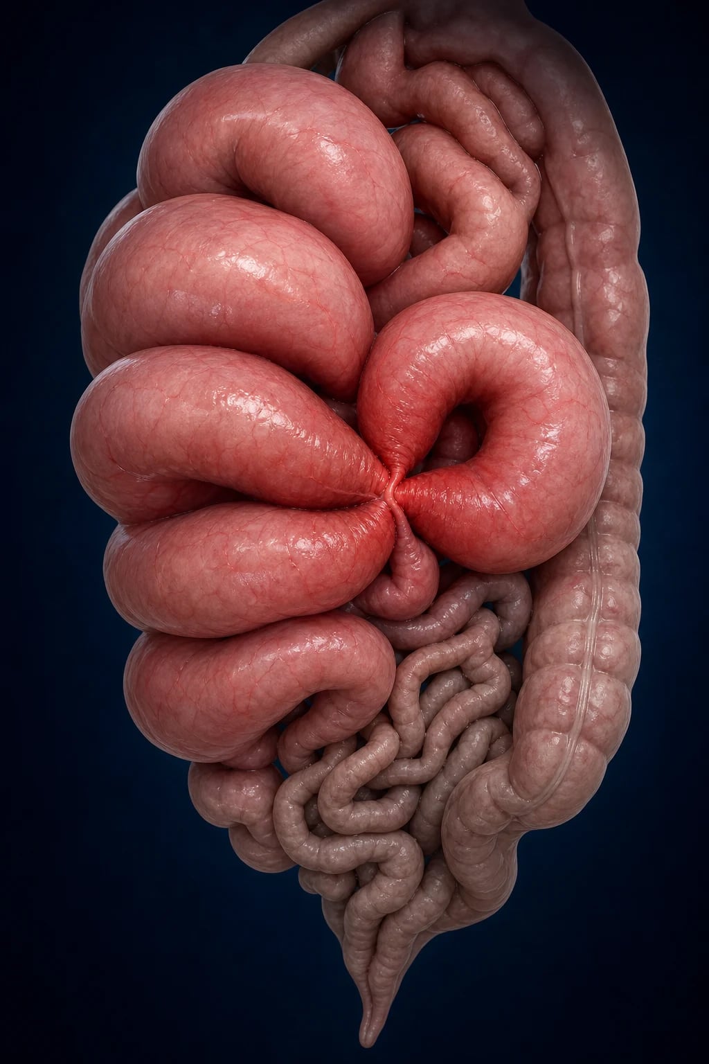
Overview & Definition
Intestinal obstruction is the partial or complete impairment of the normal aboral transit of intestinal contents. It is one of the commonest surgical emergencies, accounting for roughly 15 to 20% of all emergency surgical admissions for acute abdominal pain, and it generates a disproportionate share of medicolegal claims because the diagnosis is missed most often when the bowel is still viable and conservative management is inappropriately prolonged.[2]
Three properties must be established at the bedside before any investigation, because each drives a different management branch: [1]
- Mechanism — mechanical (a physical barrier: adhesion, hernia, tumour, volvulus, intussusception, gallstone) versus functional (no physical barrier: paralytic ileus, pseudo-obstruction). Mechanical obstruction is a surgical problem until proven otherwise; functional obstruction is managed medically by correcting the underlying cause.
- Vascular status — simple (bowel wall perfusion preserved) versus strangulated (blood supply compromised). Strangulation converts a tolerable problem into a time-critical emergency, because ischaemia progresses to transmural gangrene, perforation and faecal peritonitis within hours.
- Completeness — partial (some gas and fluid still passes, high chance of conservative resolution) versus complete (no transit at all; high chance of needing surgery). The clinical surrogate of complete obstruction is obstipation — the passage of neither faeces nor flatus. [1]
The single most important clinical judgement is therefore not what is the cause? but is the bowel strangulating? A strangulated loop is a surgical emergency regardless of aetiology, and the examiner's first question after the tetrad is almost always aimed at this fork.[1][1]
Classification
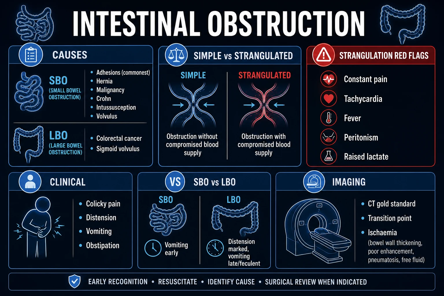
Obstruction is classified along four largely independent axes — anatomical level, mechanism, completeness, and vascular status — and each axis carries a different implication for management. [1]
Small Bowel Obstruction
SBO — 80% of all obstruction
- **Adhesions** (post-surgical) — 60 to 75%
- **Hernias** (incarcerated) — 15 to 20%
- **Malignancy** — 10 to 15% (primary small bowel or metastatic)
- Crohn strictures, intussusception, gallstone ileus
- AXR: **central** dilated loops greater than 3 cm with **valvulae conniventes** crossing the full width
- High SBO: early vomiting, minimal distension; low SBO: prominent distension
Large Bowel Obstruction
LBO — 20% of all obstruction
- **Colorectal cancer** — 60%
- **Volvulus** (sigmoid 75%, caecal) — 10 to 15%
- **Diverticulitis** (stricture) — 10%
- Faecal impaction, ischaemic stricture
- AXR: **peripheral** loops greater than 6 cm with **haustra** that do NOT cross full width
- Caecum greater than 9 cm or colon greater than 6 cm = imminent perforation
Functional Obstruction
non-mechanical
- **Paralytic ileus**: post-operative, electrolytes (low K+, Mg, Na), opioids, peritonitis, retroperitoneal bleed
- **Pseudo-obstruction (Ogilvie syndrome)**: massive colonic dilatation with no mechanical cause
- No transition point on imaging
- **Silent abdomen** on auscultation
- Managed conservatively — correct the cause; neostigmine or decompression for Ogilvie
A useful additional term is closed-loop obstruction, in which a segment of bowel is blocked at two points so that gas cannot escape either way — the classic example being a strangulated hernia or a volvulus. Closed-loop obstruction is dangerous because the trapped segment distends rapidly and ischaemia develops early, even before the patient looks septic.[1]
Epidemiology & Risk Factors
The single best clue to aetiology is the surgical history. A patient with a previous laparotomy has adhesive SBO until proven otherwise; a patient with no surgical history and obstruction at older age has a malignant or volvular cause until proven otherwise. [1]
Small bowel obstruction — causes and proportions (developed countries):[1]
- Adhesions (60 to 75%) — overwhelmingly the leading cause. Risk rises with the number and type of previous operations: lower midline laparotomy, pelvic surgery, colorectal and gynaecological procedures generate the densest adhesions.
- Hernias (15 to 20%) — inguinal, femoral, umbilical, incisional, parastomal. Femoral hernias strangulate most often and present in elderly women with a small, easily missed lump below and lateral to the pubic tubercle.
- Malignancy (10 to 15%) — primary small bowel tumours (adenocarcinoma, carcinoid, lymphoma, GIST) or metastatic deposits (melanoma, ovarian, gastric).
- Crohn disease — fibrotic strictures, typically terminal ileum.
- Intussusception — adults (rare; pathological lead point) and children (idiopathic ileocolic, peak 6 months to 2 years).[7]
- Gallstone ileus — an uncommon (1 to 4%) cause of SBO, but disproportionately affects elderly women; a gallstone erodes through a cholecysto-enteric fistula and impacts at the ileocaecal valve.[5]
- Volvulus of midgut — rare in adults.
Large bowel obstruction — causes and proportions:[2]
- Colorectal cancer (60%) — left-sided tumours obstruct more often than right; sigmoid is the commonest site.
- Volvulus (10 to 15%) — sigmoid in roughly 75% of volvulus cases (twists on its mesentery around a narrow, elongated pedicle), caecal in the remainder.[3]
- Diverticular stricture (10%) — chronic recurrent diverticulitis producing a fibrotic narrow segment.
- Faecal impaction / constipation — common in the elderly and institutionalised.
- Ischaemic stricture, anastomotic stricture, endometriosis — less common.
Headline numbers
India and the developing world: the cause profile shifts. Intestinal tuberculosis with ileocaecal strictures is an important cause of SBO in endemic regions and presents with weight loss, evening fever, night sweats and a doughy mass in the right iliac fossa; it mimics Crohn disease. Ascariasis (roundworm infestation) causes bolus obstruction of the ileum in children in endemic areas and may present with a worm bolus palpable through the abdominal wall. Volvulus is geographically clustered — the "volvulus belt" of Africa, the Middle East, India and parts of South America — driven by high-residue vegetarian diets, chronic constipation and a long, redundant sigmoid colon. Access to CT may be limited, so the diagnosis is often made on AXR and clinical findings, and surgical exploration is undertaken earlier when signs of strangulation appear.
Developed world: adhesions from prior surgery overwhelmingly dominate the SBO cause list, CT is universally available, and laparoscopic adhesiolysis is increasingly used for single-band obstruction. Ogilvie syndrome (acute colonic pseudo-obstruction) clusters in elderly, bed-bound, post-orthopaedic and critically ill patients rather than in surgical obstructions.[4]
Pathophysiology
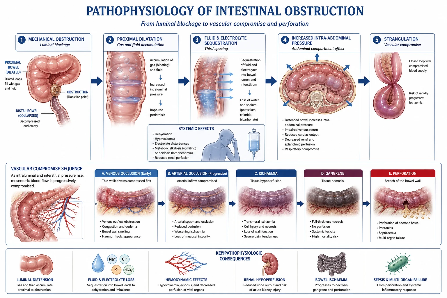
The pathophysiology of mechanical obstruction is a predictable and sequential cascade. Understanding it is what allows the examiner to be answered on why a patient becomes hypovolaemic without any external fluid loss, and why strangulation is so lethal. [1]
1. Mechanical block and the transition point. Once contents cannot pass, swallowed air (mostly nitrogen, poorly absorbed) and digestive secretions (saliva, gastric, biliary, pancreatic and up to 8 litres of small bowel secretion per day) accumulate proximal to the block. The proximal bowel dilates; the bowel distal to the block, having evacuated its contents, collapses. The point at which dilated meets collapsed bowel is the transition point, and CT hunts for precisely this anatomical sign.[1]
2. Fluid and electrolyte sequestration. Dilatation reflexly stimulates further secretion while simultaneously blocking reabsorption, so large volumes of fluid — often several litres — pool within the lumen of the obstructed loop (the "third space"). Vomiting compounds the loss. The result is hypovolaemia, dehydration, hyponatraemia, hypokalaemia, hypochloraemia and a hypokalaemic, hypochloraemic metabolic alkalosis from the loss of gastric acid. A patient with distal SBO can lose 3 to 4 litres of fluid into the gut lumen within 24 hours, which is why aggressive crystalloid resuscitation is the first intervention, not an afterthought.[1]
3. Increased intra-abdominal pressure. Massively dilated loops raise intra-abdominal pressure, which impairs venous return, reduces cardiac output, splints the diaphragm (basal atelectasis and reduced functional residual capacity) and limits renal perfusion. This is the mechanism by which a "simple" obstruction tips a patient into oliguric, hypotensive shock. [1]
4. Bacterial overgrowth and translocation. Stasis within the dilated loop promotes proliferation of gut flora — Gram-negatives and anaerobes — and the congested, oedematous bowel wall loses its mucosal barrier. Bacterial translocation to mesenteric nodes and the systemic circulation is the bridge between simple obstruction and sepsis; it is also the rationale for giving broad-spectrum antibiotics whenever strangulation is suspected or surgery is planned.[2]
5. Strangulation — the cascade that kills. In a closed-loop or high-grade obstruction, rising intraluminal pressure first compresses the thin-walled venous return, producing congestion, mural oedema and a haemorrhagic, dusky bowel. As pressure mounts, arterial inflow is also interrupted, the bowel wall becomes ischaemic and then frankly gangrenous, and organisms translocate in huge numbers. The ischaemic bowel switches to anaerobic metabolism and lactate rises — a venous or arterial lactate above 2 mmol/L in the obstructed patient is a red flag, and a rising lactate is one of the most specific bedside markers of strangulation. Transmural gangrene progresses to perforation, releasing faeculent contents into the peritoneal cavity and producing faecal peritonitis, septic shock and, without urgent surgery, death.[1][1]
Clinical Presentation
The four cardinal features form the classic tetrad of obstruction, and any candidate who cannot reproduce it cleanly will fail the question. Each feature varies predictably with the level of the obstruction. [1]
[1]How the tetrad varies with level:[1]
| Level | Pain | Vomiting | Distension | Constipation |
|---|---|---|---|---|
| High SBO (jejunum) | Colicky, frequent | Early, profuse, bilious | Minimal | Variable |
| Low SBO (ileum) | Colicky, every few minutes | Later, faeculent | Marked | Progressive |
| Large bowel | Colicky, every 10 to 15 min | Late, faeculent | Marked, gross | Prominent, early |
| Strangulated loop | Constant, severe (not colicky) | May vomit | Variable | Variable |
Signs of strangulation — the emergency pivot. Once the bowel is ischaemic, the character of the pain changes from intermittent cramping to constant, unremitting, severe pain disproportionate to the clinical findings. This is accompanied by systemic signs — tachycardia, fever, tachypnoea, hypotension — and by local signs of peritonism: guarding, rigidity and rebound tenderness. A venous or arterial lactate above 2 mmol/L, a metabolic acidosis, or a rising leucocytosis with left shift all point to ischaemia. The presence of any of these is an absolute indication for emergency laparotomy, and continuing conservative management in the face of them is the classic, examinable, and dangerous error.[1]
Atypical presentations that examiners test deliberately. The elderly patient may present with confusion alone and a vague, minimally tender abdomen (the "painless obstruction"). The diabetic may have autonomic neuropathy that blunts pain. The post-operative patient with paralytic ileus may be entirely comfortable with a silent abdomen but distended, and the trap is to operate on an ileus rather than wait for it to resolve. The pregnant patient with obstruction is rare but the diagnosis is delayed because the symptoms overlap with normal pregnancy; ultrasound is first-line and MRI is preferred to CT where feasible.[1]
Differential Diagnosis
The differential diagnosis of the obstructed patient splits into (a) other causes of an acute distended abdomen, and (b) the non-surgical mimics that should not be operated upon. [1]
| Condition | Key distinguishing feature |
|---|---|
| Paralytic ileus | No mechanical cause on imaging; silent abdomen; post-operative, electrolyte disturbance (low K+), opioids, retroperitoneal bleed, peritonitis; resolves in 2 to 5 days with conservative management |
| Pseudo-obstruction (Ogilvie) | Massive caecal dilatation with no mechanical transition point on CT; elderly, bed-bound, post-orthopaedic or critically ill; neostigmine or colonoscopic decompression if caecum greater than 9 to 12 cm[4] |
| Gastroenteritis | Vomiting precedes pain; watery diarrhoea (not constipation); diffuse, non-colicky cramping; self-limiting |
| Acute mesenteric ischaemia | Pain grossly out of proportion to examination; history of atrial fibrillation, vascular disease or recent embolus; raised lactate, metabolic acidosis; may have frank peritonism late |
| Perforated viscus (other cause) | Sudden constant pain, rigid board-like abdomen, free gas under the diaphragm on erect CXR; often duodenal ulcer or diverticular perforation |
| Renal colic / biliary colic | Pain radiates to groin or back; no distension; haematuria (renal) or Murphy sign (biliary) |
| Pancreatitis | Constant epigastric pain radiating to back; raised amylase and lipase; no obstipation |
| Lower lobe pneumonia / MI | Referred upper abdominal pain; chest signs and ECG abnormalities; no distension |
The can't-miss mimics are paralytic ileus and Ogilvie syndrome (operating on them is harmful), and acute mesenteric ischaemia (missing it is fatal). All three are excluded — or confirmed — by CT with IV contrast, which is why CT is performed before, not after, a decision to operate whenever the patient is stable enough.[2]
Clinical & Bedside Assessment
A focused, systematic abdominal examination is high-yield and answers two questions in a single pass: is this obstruction? and is the bowel strangulating? [1]
1. General inspection. Look for dehydration (dry tongue, sunken eyes, loss of skin turgor), signs of recent weight loss (malignancy, tuberculosis), and chronic disease. Note the patient's posture — a still, motionless patient has peritonitis; a restless, writhing patient has colic or ischaemia out of proportion to findings. [1]
2. Inspect the abdomen. Distension is graded qualitatively and is more pronounced in distal SBO and LBO. Look for visible peristalsis — sinuous loops of bowel visibly moving under the abdominal wall — which is pathognomonic of chronic or subacute mechanical obstruction. Map every surgical scar (midline laparotomy points to adhesions; Pfannenstiel to gynaecological or urological surgery; gridiron or Lanz to appendicectomy). Inspect all hernial orifices. [1]
3. Auscultate before palpation (so as not to stimulate artefactual sounds). Early obstruction produces hyperactive, high-pitched, tinkling bowel sounds and audible borborygmi, as the proximal bowel hyper-peristalses against the block. Late obstruction, strangulation or established peritonitis produces a silent abdomen — the bowel has given up. A silent abdomen in a patient who is not yet post-operative is a sinister sign. [1]
4. Deep palpation. Localised tenderness over the obstructed loop, or generalised tenderness with guarding and rigidity, indicates peritonism and shifts the decision to operate. A palpable mass may represent an intussusception (sausage-shaped in a child), a tumour or a distended volvular sigmoid (resonant, drum-like). [1]
5. CHECK THE HERNIAL ORIFICES — non-negotiable. Examine every orifice: inguinal, femoral, umbilical, incisional, parastomal. An incarcerated femoral hernia in an elderly woman is small, lies below and lateral to the pubic tubercle, and is the single most easily missed and most easily fixed cause of SBO. Failing to examine the hernial orifices is the classic and examinable bedside error.[1]
6. Rectal examination. An empty rectum supports obstruction. A palpable mass suggests rectal cancer. Blood on the glove suggests ischaemia, intussusception (redcurrant jelly stool in children) or a malignant ulcer. [1]
Signs of dehydration and shock: dry mucous membranes, loss of skin turgor, sunken eyes, oliguria, tachycardia, postural drop, and in advanced cases hypotension with cold peripheries. Quantifying dehydration drives the fluid resuscitation. [1]
Investigations
Investigation serves three purposes: confirm obstruction, localise the level and cause, and detect strangulation. Plain films are cheap and fast; CT is the gold standard because it answers all three questions at once. [1]
Plain radiographs. [1]
- Erect chest X-ray — looks for free subdiaphragmatic gas (perforation) and incidentally surveys the lung bases.
- Supine abdominal X-ray — the workhorse. In SBO it shows central dilated small bowel loops greater than 3 cm with valvulae conniventes (plicae circulares) that cross the full width of the bowel, often in a stepladder arrangement with multiple air-fluid levels on an erect film. In LBO it shows peripheral dilated large bowel loops greater than 6 cm with haustra that do not cross the full width, plus a paucity of rectal gas. The coffee-bean sign — a hugely dilated loop projecting from the pelvis toward the right or left upper quadrant with a central dense stripe — is the classic radiographic signature of sigmoid volvulus.[3] A caecum greater than 9 cm, or a colon greater than 6 cm, is a radiological warning of imminent perforation.
CT abdomen and pelvis with IV contrast — the gold standard. CT has a sensitivity above 95% for obstruction and answers the three critical questions in a single study: it localises the transition point, identifies the cause (adhesions by exclusion, hernia, tumour, volvulus, intussusception, gallstone), and — crucially — detects the CT signs of strangulation and ischaemia. The high-yield CT signs of ischaemia are bowel wall thickening, mesenteric oedema and haziness, increased or absent mural enhancement, free intraperitoneal fluid, pneumatosis intestinalis (gas in the bowel wall) and portal venous gas. A swirl or whorl of mesenteric vessels indicates a volvulus or internal hernia. CT also separates true obstruction from paralytic ileus (no transition point) and from Ogilvie syndrome (no mechanical cause).[1][2]
Water-soluble contrast (Gastrografin) — diagnostic and therapeutic. In adhesive SBO without strangulation, the Bologna guidelines endorse giving 100 mL of water-soluble contrast via the NG tube. If contrast reaches the colon on a 4 to 24 hour film, the obstruction is partial and almost certain to resolve (negative predictive value above 95%); if it does not, surgery is indicated. The hyperosmolar Gastrografin also draws fluid into the lumen and oedematous bowel, which itself helps resolve partial obstruction.[1]
Radiological clues on AXR
Laboratory tests. [1]
- Full blood count — leukocytosis with a left shift suggests strangulation or sepsis; anaemia points to malignancy or chronic disease.
- Urea and electrolytes — hypokalaemia, hyponatraemia, raised urea (dehydration); guides replacement.
- Venous blood gas — a metabolic alkalosis reflects vomiting; a metabolic acidosis with raised lactate (greater than 2 mmol/L) is the most specific bedside marker of bowel ischaemia.
- C-reactive protein — rising trend suggests strangulation or sepsis.
- Group and save / crossmatch — before any operation.
- Tumour markers (CEA) — if colorectal cancer is suspected.
- Amylase / lipase — to exclude pancreatitis as a mimic.
- Beta-hCG — in any woman of reproductive age before imaging or surgery. [1]
Management — Resuscitation
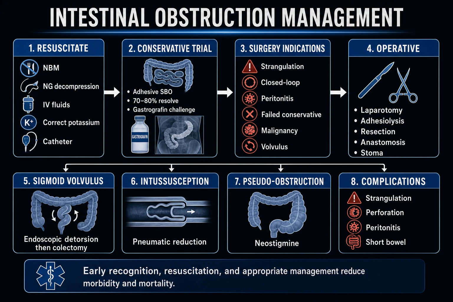
Every patient with obstruction — whether mechanical or functional, whether they will ultimately go to theatre or not — receives the same immediate resuscitation bundle. The classic mnemonic is "drip and suck": intravenous fluids and nasogastric decompression. Resuscitation is begun at the same time as investigation, not after it. [1]
| Step | Intervention | Why |
|---|---|---|
| 1. NBM | Nil by mouth, immediately | Rests the bowel; aspirate prevention; allows theatre without delay |
| 2. Suck | Large-bore nasogastric tube (Salem sump) | Decompresses the stomach, relieves vomiting, removes swallowed air, monitors output |
| 3. Drip | IV crystalloid — Hartmann's or normal saline, 1 to 2 L bolus then replace deficit plus ongoing losses | Reverses hypovolaemia from sequestration and vomiting; titrate to urine output and venous lactate |
| 4. Electrolytes | Replace potassium (10 to 20 mmol/h, ECG-monitored) and magnesium | Hypokalaemia both results from and perpetuates ileus; correct before theatre |
| 5. Catheter | Urinary catheter; aim for urine output greater than 0.5 mL/kg/h | Tracks resuscitation and renal perfusion in real time |
| 6. Antibiotics | IV broad-spectrum — cefuroxime 1.5 g TDS plus metronidazole 500 mg TDS, or co-amoxiclav 1.2 g TDS | If strangulation suspected, before surgery, or for established sepsis; covers Gram-negatives and anaerobes |
| 7. Analgesia | Morphine titrated to pain; antiemetic (ondansetron 4 mg IV) | Relief of severe colic; symptomatic nausea control |
| 8. Thromboprophylaxis | LMWH (enoxaparin 40 mg SC OD) plus TEDS | Obstructed patients are high VTE risk; essential before and after any surgery |
Resuscitation is reassessed continuously: serial abdominal examinations (every 2 to 4 hours in the conservative phase), hourly urine output, and a repeat venous lactate. Any deterioration — worsening pain, rising lactate, new peritonism, falling urine output despite fluid — is an absolute trigger to abandon conservative management and operate.[1][1]
Management — Definitive & Stepwise
Definitive management is dictated by the cause, the presence or absence of strangulation, and the response to the conservative trial. The decision tree has three main branches: adhesive SBO, other causes of SBO, and large bowel obstruction. [1]
Small bowel obstruction — adhesive (the commonest scenario)
Per the Bologna guidelines (2018, WSES):[1]
- No signs of strangulation or peritonism: undertake a conservative trial for 48 to 72 hours — NBM, NG tube, IV fluids, electrolyte correction, and serial examination. Roughly 70 to 80% of adhesive SBO resolves without surgery. Water-soluble contrast (Gastrografin 100 mL via NG) is both diagnostic and therapeutic: if contrast reaches the colon within 24 hours, resolution is almost certain; if not, surgery is indicated.
- Signs of strangulation, peritonism, or no resolution by 48 to 72 hours: proceed to laparotomy (or laparoscopy in selected patients with single-band obstruction, no severe distension, and no peritonism). The operation comprises adhesiolysis (division of the obstructing band or bands), inspection of the entire bowel for viability, and resection with primary anastomosis of any non-viable segment. If the patient is unstable, grossly contaminated, or the viability is in doubt, a stoma (defunctioning ileostomy or jejunostomy) is safer than an anastomosis under tension. [1]
Small bowel obstruction — other causes
- Incarcerated hernia: emergency hernia repair, with resection of non-viable bowel. Femoral hernias must never be reduced by taxis — the risk of reducing a strangulated loop into the peritoneum ("en masse" reduction) is real and fatal.[1]
- Gallstone ileus: enterolithotomy — a longitudinal enterotomy proximal to the impacted stone, milking the stone out, and closure transversely. The cholecysto-enteric fistula usually closes spontaneously; a one-stage cholecystectomy with fistula repair is reserved for fit patients with ongoing biliary symptoms.[5]
- Crohn stricture: conservative trial first (Crohn obstruction is often inflammatory); if recurrent, stricturoplasty or resection.
- Malignancy: resection with anastomosis (curative intent), or palliative bypass if advanced.
Large bowel obstruction
- Colorectal cancer. The options depend on stability and the site of the tumour. In the unstable or perforated patient, a Hartmann's procedure (resection of the obstructing sigmoid tumour, end colostomy, and rectal stump) is safest. In the stable patient without perforation, one-stage resection with primary anastomosis (with or without a defunctioning ileostomy) is increasingly preferred. For left-sided obstructing tumours in fit patients, a self-expanding metal stent (SEMS) can be placed endoscopically either as a bridge to elective surgery (allowing stabilisation and a staged one-stage resection) or as palliation in advanced disease; meta-analysis of randomised trials shows SEMS as bridge reduces stoma rates compared with emergency resection, at a small cost in perforation risk.[6]
- Sigmoid volvulus. Initial endoscopic decompression — gentle insertion of a flexible sigmoidoscope and leaving a flatus tube in situ — succeeds in 70 to 80% of cases. Deflation relieves the immediate emergency; the tube is left in for 48 to 72 hours. Because the recurrence rate is 50 to 70%, the WSES guidelines recommend elective sigmoid colectomy with primary anastomosis after the first resolved episode in fit patients.[3]
- Caecal volvulus. Unlike sigmoid volvulus, endoscopic decompression is NOT appropriate (the colonoscope cannot reliably reach and detorse a caecal volvulus, and the risk of iatrogenic perforation of an ischaemic caecum is high). Treat with right hemicolectomy (primary anastomosis or ileostomy depending on stability).[1][3]
- Faecal impaction. Manual evacuation, enemas, and stool softeners; exclude an underlying obstructing lesion with colonoscopy once resolved.
Paralytic ileus and pseudo-obstruction
- Paralytic ileus is managed conservatively — NBM, NG tube, IV fluids, aggressive potassium and magnesium correction, minimisation of opioids, early mobilisation, and treatment of any underlying sepsis. It typically resolves in 2 to 5 days. Persistent ileus beyond 5 days prompts a search for a missed cause (abscess, electrolyte derangement, drug).[1]
- Acute colonic pseudo-obstruction (Ogilvie syndrome): conservative measures first (correction of electrolytes, withdrawal of offending drugs, mobilisation). If the caecum is greater than 9 to 12 cm or there is no improvement at 48 to 72 hours, IV neostigmine (2 to 2.5 mg slow bolus with ECG monitoring — be prepared for bradycardia, give atropine if needed) induces decompression in the majority. Colonoscopic decompression is second-line; surgery (caecostomy or resection) is reserved for refractory cases or perforation.[4]
The classic tetrad of obstruction
PVDA
colicky, intermittent (peristalsis against the obstruction)
early and bilious in high SBO; late and faeculent in LBO
more pronounced in distal SBO and in LBO
no faeces AND no flatus — a late sign
SBO versus LBO on the plain film
DISH
central loops = SBO; peripheral frame = LBO
valvulae conniventes cross the full width (SBO); haustra do not (LBO)
small bowel greater than 3 cm; large bowel greater than 6 cm; caecum greater than 9 cm = danger
always check — an incarcerated hernia is the most easily missed and the most easily fixed SBO cause
Specific Subtypes & Scenarios
Gallstone ileus. An elderly woman presents with intermittent, fluctuating SBO over days (the stone tumbles and intermittently impacts). The diagnostic triad is SBO + gallstones + air in the biliary tree (pneumobilia) on imaging, sometimes with a stone visible in the right iliac fossa. The mechanism is erosion of a large gallstone (usually greater than 2.5 cm) through a cholecystoduodenal fistula; the stone impacts at the narrowest point of the small bowel, the terminal ileum and ileocaecal valve. Treatment is enterolithotomy via a longitudinal enterotomy proximal to the stone; the fistula usually closes spontaneously and is not repaired at the same operation in the elderly, frail patient.[5]
Intussusception. In infants (peak 6 months to 2 years) it is idiopathic ileocolic, probably triggered by lymphoid hyperplasia after a viral illness. The classic triad is intermittent colicky abdominal pain with drawing up of the legs, vomiting, and redcurrant-jelly stool; a sausage-shaped mass may be palpable in the right upper quadrant with emptiness in the right lower quadrant (Dance sign). In adults intussusception is rare and almost always has a pathological lead point (polyp, tumour, Meckel diverticulum) demanding resection. In children the first-line treatment is pneumatic (air) or hydrostatic (saline) enema reduction under fluoroscopic or ultrasound guidance; surgery — usually manual reduction or resection — is reserved for failed reduction, peritonitis, or recurrent intussusception.[7]
Sigmoid volvulus. A twisted sigmoid colon on its mesentery, classically in an elderly, constipated, institutionalised patient or in the geographic "volvulus belt." Presentation is of subacute LBO with gross distension. The coffee-bean sign on AXR — a hugely dilated omega-shaped loop arising from the pelvis and pointing toward the right upper quadrant — is characteristic; CT confirms a whirl sign (twisted mesentery). Endoscopic detorsion with a flatus tube is first-line; the tube is left in for 48 to 72 hours and the patient is offered elective sigmoid colectomy because of the high (50 to 70%) recurrence.[3]
Caecal volvulus. A mobile caecum twists on its mesentery. Unlike sigmoid volvulus, it presents more acutely and is not amenable to endoscopic decompression. The risk of ischaemic perforation of the thinned caecal wall is high, so the treatment is right hemicolectomy.[1][3]
Malignant large bowel obstruction. An obstructing colorectal cancer is a surgical emergency but also a stage of disease. The choice between Hartmann's, primary resection with anastomosis, defunctioning stoma, and SEMS as bridge depends on the patient's physiological state, the tumour site, the presence of perforation, and the likelihood of cure. SEMS as a bridge to elective surgery is most useful in fit patients with left-sided tumours without perforation.[6]
Internal hernia. A loop of bowel herniates through a normal or abnormal mesenteric defect — congenital (paraduodenal) or acquired (after Roux-en-Y gastric bypass). Presentation is of SBO that may be intermittent; CT shows a cluster of dilated loops and a mesenteric swirl. At laparotomy the hernia is reduced, the defect closed, and any non-viable bowel resected. [1]
Complications & Pitfalls
Complications of the disease: [1]
- Strangulation progressing to bowel ischaemia, gangrene and perforation — the central danger. Perforation produces faecal peritonitis, with mortality of 5 to 15% even with prompt surgery.[1]
- Sepsis and septic shock from bacterial translocation and peritonitis.
- Short bowel syndrome if an extensive ischaemic segment must be resected — a devastating, lifelong consequence of late presentation.
- Recurrence — adhesive SBO recurs in roughly 30% of patients over their lifetime after a first episode; recurrence is higher still after a second episode, which is why some surgeons offer elective adhesiolysis or even a Noble-type plication after multiple recurrences (controversial).
- Intra-abdominal compartment syndrome from massive bowel distension, impairing ventilation, renal and cardiac output.
Complications of surgery: [1]
- Anastomotic leak — the most feared; higher in emergency, contaminated, ischaemic or shocked settings, which is why a stoma is often the safer choice.
- Intra-abdominal abscess and wound infection.
- Adhesion formation — the operation that cures today's obstruction is itself the cause of tomorrow's.
- Stoma complications — necrosis, retraction, parastomal hernia, high output and skin excoriation.
- Enterocutaneous fistula.
- Incisional hernia. [1]
Classic and examinable pitfalls: [1]
- Missing strangulation by continuing conservative management when the bowel is already ischaemic — the cardinal error. Any constant pain, peritonism, raised lactate or rising leucocytosis is an absolute trigger to operate.
- Not checking the hernial orifices and missing an incarcerated femoral hernia in an elderly woman that could have been fixed in 30 minutes.
- Treating caecal volvulus like sigmoid volvulus — caecal volvulus needs surgery; endoscopic decompression risks perforating an ischaemic caecum.
- Operating on paralytic ileus or Ogilvie syndrome — these are functional obstructions; the correct treatment is conservative, with neostigmine or colonoscopic decompression for Ogilvie.[4]
- Delaying surgery in complete obstruction — the risk of perforation rises sharply with time.
- Under-resuscitating before theatre — the patient arrives in theatre hypovolaemic, hypokalaemic and acidotic, and dies on induction.
Prognosis & Disposition
Prognosis tracks the vascular status of the bowel and the comorbidities of the patient far more closely than the anatomical level of obstruction. [1]
- Simple adhesive SBO managed conservatively: mortality under 2%; 70 to 80% resolve without surgery.[1]
- Adhesive SBO requiring surgery: mortality 2 to 5%, driven by comorbidity.
- Strangulated SBO: mortality 5 to 15%, rising to over 20% in the elderly and comorbid and in those presenting late with established peritonitis.[1]
- Large bowel obstruction from cancer: mortality 5 to 10% for the emergency admission, and long-term prognosis depends on cancer stage.
- Volvulus decompressed endoscopically: recurrence 50 to 70% without definitive sigmoid colectomy.[3]
Disposition. All obstructed patients are admitted. Those managed conservatively stay in a monitored surgical ward with serial examinations every 2 to 4 hours, hourly urine output, and serial lactate; any deterioration goes to theatre. Post-operative patients go to HDU or ICU if they were shocked, had extensive resection, or have significant comorbidity. A stoma or a short bowel mandates enterostomal nurse input, dietetic support, and patient education before discharge. [1]
Special Populations
- Children. The dominant cause is intussusception (6 months to 2 years); the second is inguinal hernia and, in endemic regions, ascaris bolus obstruction. Redcurrant jelly stool, sausage-shaped mass, and intermittent drawing up of the legs are the classic features. Pneumatic or hydrostatic enema reduction is first-line; surgery is for failed reduction or peritonitis. Appendicitis may also obstruct in a child and should not be forgotten.[7]
- The elderly with no prior surgery. Suspect malignancy (colorectal cancer) until proven otherwise, and also consider gallstone ileus, sigmoid volvulus, incarcerated femoral hernia, and faecal impaction. Presentations are often atypical — confusion, anorexia, or a vague, minimally tender abdomen — and the diagnosis is delayed, which is why mortality in this group is the highest.
- Post-operative patients. Paralytic ileus is physiological and resolves in 2 to 5 days. An ileus persisting beyond 5 days, or one accompanied by fever, rising lactate or peritonism, is not a simple ileus — consider a leak, abscess, or early mechanical obstruction from an internal hernia (especially after laparoscopic surgery or bypass).
- Pregnancy. Obstruction in pregnancy is rare but dangerous; diagnosis is delayed because the symptoms overlap with normal pregnancy, and the enlarging uterus displaces bowel and masks signs. Ultrasound is first-line, and MRI is preferred to CT where feasible to limit radiation. Adhesions and volvulus (particularly caecal, from displacement by the gravid uterus) are the commonest causes.
- The immunocompromised. A blunted inflammatory response may mask peritonism; CT is relied upon earlier, and a low threshold for surgery is maintained.
- The anticoagulated. Reverse warfarin (vitamin K, prothrombin complex concentrate) pre-operatively; balance bleeding against thrombotic risk. Patients with obstruction are high VTE risk, so thromboprophylaxis is essential once surgical bleeding is controlled.
Evidence, Guidelines & Regional Differences
Bologna guidelines for adhesive SBO (2018, WSES).[1] The cornerstone document for the commonest scenario. Key recommendations: a conservative trial of 48 to 72 hours for adhesive SBO without signs of strangulation; water-soluble contrast (Gastrografin) as both a diagnostic and therapeutic agent — contrast reaching the colon within 24 hours predicts resolution with a negative predictive value above 95% and the osmotic effect itself aids resolution; early laparoscopy is safe and effective in selected patients (single band, no severe distension, no peritonism, no previous multiple adhesiolysis); and adhesion barriers (hyaluronic acid-carboxymethylcellulose films) are recommended at the index operation to reduce the future burden of adhesion-related obstruction.
WSES consensus guidelines on sigmoid volvulus (2023).[3] Endoscopic detorsion with a flatus tube is first-line for sigmoid volvulus without peritonitis, succeeding in 70 to 80%; elective sigmoid colectomy with primary anastomosis after the first resolved episode is recommended because recurrence without it is 50 to 70%. Caecal volvulus is a surgical problem from the outset.
Acute colonic pseudo-obstruction (Ogilvie).[4] Conservative measures first; IV neostigmine 2 to 2.5 mg slow bolus (with atropine available for bradycardia) is the pharmacological mainstay for refractory dilatation; colonoscopic decompression is second-line; surgery (caecostomy or resection) is reserved for failure or perforation.
Colonic stenting (SEMS) for malignant LBO.[6] Meta-analysis of randomised trials confirms that SEMS as a bridge to elective surgery in suitable left-sided obstructing tumours reduces the rate of stoma formation compared with emergency resection, at the cost of a small perforation risk. SEMS is also valuable for palliation in advanced disease.
Paediatric intussusception.[7] Systematic review evidence supports ultrasound-guided pneumatic or hydrostatic enema reduction as first-line in stable children, with surgery reserved for failed reduction, peritonitis, or the pathological lead point.
Regional differences. In endemic regions of India, Africa, the Middle East and South America, intestinal tuberculosis (ileocaecal strictures mimicking Crohn), ascaris bolus obstruction in children, and geographically clustered sigmoid volvulus (the "volvulus belt") shift the cause profile away from adhesions. CT may not be universally available, so the diagnosis rests more heavily on AXR and clinical findings, and surgical exploration is undertaken earlier when signs of strangulation appear. In developed countries, adhesions from prior surgery dominate, CT is universal, and laparoscopic adhesiolysis is increasingly common; Ogilvie syndrome clusters in elderly, bed-bound, post-orthopaedic and critically ill patients.
Exam Pearls
- SBO: central loops + valvulae conniventes (cross the full width). LBO: peripheral loops + haustra (do NOT cross the full width).[1]
- Adhesions = commonest cause of SBO (60 to 75%). Colorectal cancer = commonest cause of LBO (60%).[2]
- Absolute constipation (obstipation) = no faeces AND no flatus — a late sign; its absence does not exclude obstruction.
- Strangulation = constant pain, tachycardia, fever, peritonism, raised lactate — emergency laparotomy.[1]
- ALWAYS check the hernial orifices — an incarcerated femoral hernia in an elderly woman is the most easily missed and most easily fixed cause of SBO.
- Bologna guidelines: conservative trial for adhesive SBO without strangulation for 48 to 72 hours; 70 to 80% resolve.[1]
- Gastrografin is diagnostic AND therapeutic — contrast in the colon within 24 hours predicts resolution.
- Gallstone ileus triad: SBO + gallstones + pneumobilia (air in the biliary tree).[5]
- Sigmoid volvulus: coffee-bean sign; decompress with flatus tube or sigmoidoscope; recurrence 50 to 70% — elective colectomy.[3]
- Caecal volvulus: needs surgery (right hemicolectomy). Do NOT attempt endoscopic decompression.[3]
- Intussusception in a child: sausage-shaped mass, redcurrant jelly stool — pneumatic or hydrostatic enema reduction first-line.[7]
- Paralytic ileus: no mechanical cause, silent abdomen, post-op — conservative.[1]
- Ogilvie syndrome: dilated colon, no mechanical cause — neostigmine or colonoscopic decompression if caecum greater than 9 to 12 cm.[4]
- Drip and suck: NBM + NG tube + IV fluids + correct K+/Mg2+ + catheter — the universal first response.[1]
Mechanical vs functional — rapid discrimination
| Feature | Mechanical SBO/LBO | Paralytic ileus |
|---|---|---|
| Pain | Colicky then continuous if ischaemia | Diffuse discomfort, less colic |
| Bowel sounds | High-pitched tinkling → silent if infarction | Absent or sparse |
| Imaging gas pattern | Transition point, dilated proximal loops | Uniform dilation, no transition |
| Peritonism | Late red flag | Usually absent unless secondary |
| First action | Resuscitate + decompress + urgent surgery if closed-loop/ischaemia | Correct cause (electrolytes, opioids, sepsis, retroperitoneal) |
Closed-loop and ischaemia red flags (operate now)
- Continuous pain replacing colic
- Fever, tachycardia, raised lactate, metabolic acidosis
- Peritonism, guarding
- CT: closed loop, whirl sign, reduced wall enhancement, pneumatosis, portal venous gas
- Bloody PR stool (late) [1]
Do not chase endless serial observation once these are present. [1]
SBO management algorithm (learnable)
- ABC, IV access, crystalloid, catheter, NBM, NG tube (drip and suck).
- Labs: FBC, U&E, lactate, VBG/ABG, group and save.
- Imaging: CT abdomen/pelvis with IV contrast when stable (transition point, cause, complications).
- Adhesive SBO without ischaemia: trial of non-operative management 24–72 h with serial review if improving.
- Failing trial, virgin abdomen with hernia, closed loop, ischaemia, peritonitis: urgent laparotomy/laparoscopy.
- Hernia: reduce if possible when appropriate; emergency repair if strangulated.
- Malignant LBO: decompress, stage, stent vs emergency resection/stoma MDT-dependent. [1]
Worked stems
Stem 1 — Adhesive SBO. Prior laparotomy, colicky pain, bilious vomiting, high-pitched sounds, CT transition mid-ileum, no ischaemia.
NBM, NG, IV fluids, analgesia; trial non-op with serial exams; operate if no resolution or red flags. [1]
Stem 2 — Closed-loop. Continuous pain, lactate 4.2, CT whirl/closed loop.
Resuscitate and emergency surgery — do not wait for further “conservative trial.” [1]
Stem 3 — Large bowel obstruction + caecal tenderness.
Risk of caecal perforation if competent ileocaecal valve; urgent decompression/resection planning. [1]
Stem 4 — Gallstone ileus. Pneumobilia, SBO, ectopic stone.
Enterolithotomy ± later cholecystectomy discussion. [1]
Extended viva outlines
- Rule of 3/6/9 bowel diameters (SI/colon/caecum teaching thresholds — state unit numbers carefully).
- Causes of SBO — adhesions, hernia, malignancy, intussusception, gallstone, foreign body.
- Causes of LBO — cancer, volvulus, diverticular stricture, faecal impaction.
- Water-soluble contrast challenge in adhesive SBO — prognostic, not for ischaemia.
- When laparoscopy is unsafe — massive distension, dense adhesions, haemodynamic instability.
- Ileus after surgery — opioids, electrolytes (K⁺/Mg²⁺), sepsis, haematoma. [1]
Coverage self-check
Pathophysiology of closed loop · drip-and-suck · CT transition · ischaemia markers · hernia · malignancy · volvulus · ileus vs mechanical · special populations (elderly, pregnancy) · exam traps (treating ileus as obstruction and vice versa). [1]
Exam application bank (NEET-PG / INICET)
Worked stems
Adhesive SBO — drip and suck, CT transition, trial vs operate.
Closed-loop / ischaemia — continuous pain, lactate, emergency surgery.
LBO + tender caecum — risk of perforation; decompress.
Ileus vs mechanical — sounds, transition point, correct cause.
Rapid viva checklist
Definition · causes SBO/LBO · closed loop · drip-and-suck · CT signs · red flags for ischaemia · hernia · malignancy · special populations · pitfalls.
References
- [1]Ten Broek RPG, Krielen P, Di Saverio S, et al. Bologna guidelines for diagnosis and management of adhesive small bowel obstruction (ASBO): 2017 update of the evidence-based guidelines from the world society of emergency surgery ASBO working group World J Emerg Surg, 2018.PMID 29946347
- [2]Catena F, De Simone B, Coccolini F, et al. Bowel obstruction: a narrative review for all physicians World J Emerg Surg, 2019.PMID 31168315
- [3]De U, Ghosh S, Ghosh P, et al. WSES consensus guidelines on sigmoid volvulus management World J Emerg Surg, 2023.PMID 37189134
- [4]Haj M, Haj M, Rockey DC. Update on the Diagnosis and Management of Acute Colonic Pseudo-obstruction (ACPO) Curr Gastroenterol Rep, 2023.PMID 37486594
- [5]Nuno-Guzman CM, Marin-Contreras ME, Figueroa-Moreno L, et al. Gallstone ileus: An overview of the literature Rev Gastroenterol Mex, 2017.PMID 28433486
- [6]Wang J, Liu Y, Li Y, et al. Colonic stent as a bridge to surgery versus emergency rection for malignant left-sided colorectal obstruction: A systematic review and meta-analysis of randomized controlled trials Medicine (Baltimore), 2023.PMID 38115371
- [7]Gray MP, Li SH, Hoffmann DF, Gorelik M. Management of intussusception in children: A systematic review J Pediatr Surg, 2021.PMID 33158508