General Surgery · General Surgery
Neck Swellings
Also known as Neck lump · Neck mass · Thyroglossal cyst · Branchial cyst · Cervical lymphadenopathy
Neck swellings are classified by location: midline (thyroid goitre moves on swallowing; thyroglossal duct cyst moves on swallowing AND tongue protrusion; dermoid does not move) versus lateral (cervical lymphadenopathy is the commonest cause; also branchial cyst in young adults, salivary gland tumour, cystic hygroma). In an adult over 40, a new neck lump is malignancy until proven otherwise. Ultrasound plus FNA is the standard workup. Sistrunk procedure (excise cyst + tract + central hyoid) for thyroglossal cyst; surgical excision for branchial cyst; superficial parotidectomy with facial nerve preservation for pleomorphic adenoma; do NOT biopsy a carotid body tumour. Metastatic SCC node mandates panendoscopy to find the head and neck primary.
On this page & tools
Your progress
Saved locally on this device.
Exam tags
Red flags
Overview & Definition
A neck swelling is any palpable or visible abnormal mass within the boundaries of the neck (lower border of mandible superiorly, suprasternal notch and clavicles inferiorly, anterior midline and posterior border of trapezius laterally). Neck swellings are among the most common surgical outpatient presentations, and the diagnostic discipline rests on two simple ideas: classify by anatomical location (midline versus lateral) and elicit clinical behaviour (does it move on swallowing, on tongue protrusion, transilluminate, or pulsate).[1]
The single most important clinical principle, repeated as a mantra by every surgical teacher, is this: a new neck lump in an adult over 40 is malignancy until proven otherwise. This is the explicit position of the AAO-HNS Clinical Practice Guideline on the Neck Mass in Adults, which states that malignant neoplasms far exceed any other aetiology of an adult neck mass and that an asymptomatic neck mass may be the initial or only manifestation of head and neck cancer, lymphoma, thyroid, or salivary gland malignancy.[1] Conversely, in children and young adults, neck lumps are most commonly developmental (thyroglossal cyst, branchial cyst, cystic hygroma) or reactive (lymphadenopathy from viral or bacterial upper respiratory infection).[1]
This topic covers the full differential of neck swellings, the neck-triangle and nodal-station anatomy an examiner expects, the workup of the thyroid nodule and the cervical lymph node, and the surgical principles of the Sistrunk procedure, parotidectomy with facial nerve preservation, and Shamblin-graded carotid body tumour resection. [1]
Anatomy of the Neck
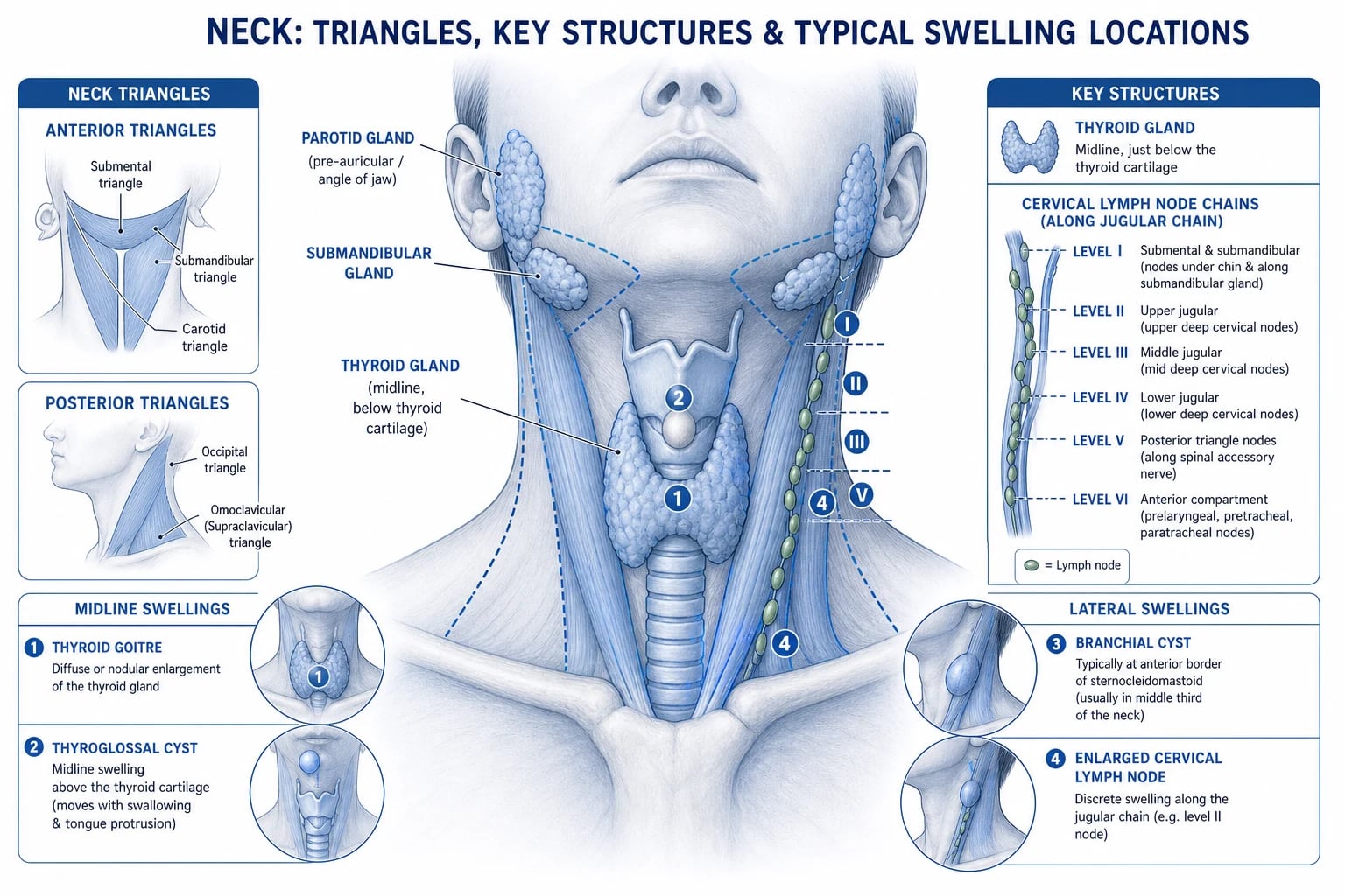
Diagnosing a neck swelling is impossible without a working command of the triangles of the neck and the nodal level stations, because the position of a swelling narrows the differential more sharply than any single bedside feature.[1]
Anterior triangle
bounded by midline, SCM, mandible
- **Boundaries**: anterior border of SCM (posterior limit), midline of neck (medial limit), lower border of mandible (superior limit); apex at manubrium
- **Contents**: thyroid, larynx/trachea, carotid sheath (ICArV + vagus), submandibular gland, cervical nodes levels I to VI
- **Swellings here**: thyroid goitre/nodule (midline), thyroglossal duct cyst (midline), branchial cyst (anterior border SCM), cervical adenopathy, carotid body tumour (carotid bifurcation)
- **Surface landmark**: SCM is the great landmark — feel it tense when patient turns head against resistance
Posterior triangle
bounded by SCM, trapezius, clavicle
- **Boundaries**: posterior border of SCM (anterior limit), anterior border of trapezius (posterior limit), middle third of clavicle (base, inferior limit); apex where SCM and trapezius meet at the occiput
- **Contents**: spinal accessory nerve (XI), cervical plexus roots, external jugular vein, supraclavicular and occipital nodes (level V)
- **Swellings here**: cystic hygroma (infants, transilluminates), supraclavicular lymphadenopathy (Virchow node — gastric/abdominal primary), brachial plexus neuroma
- **Caveat**: cervical rib may present as a hard supraclavicular mass with TOS features
Cervical lymph node levels — surgical stations
The carotid sheath runs deep to SCM through the anterior triangle and contains, from lateral to medial, the internal jugular vein, the vagus nerve, and the common then internal carotid artery (mnemonic "VA-N" within the sheath: Vein lateral, Artery medial, Nerve between). The carotid bifurcation — the seat of the carotid body tumour — sits at the upper border of the thyroid cartilage around the level of C3 to C4. The thyroid gland lies anterior to the second to fourth tracheal rings, draped around the larynx; this is why a thyroid swelling moves up with swallowing. The submandibular gland wraps around the posterior belly of digastric, with the facial vein superficial and the lingual nerve superior; the hypoglossal nerve runs in the floor of the submandibular triangle. [1]
Classification
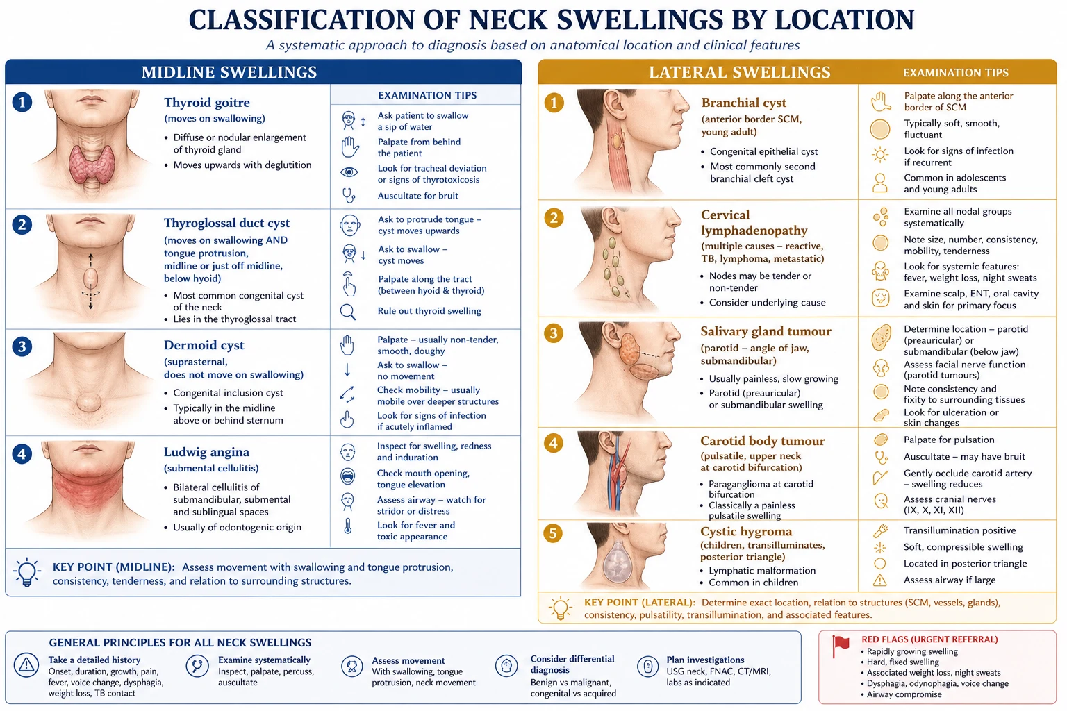
The first branch point is midline versus lateral, then cystic versus solid versus vascular within each.[1]
Midline swellings
thyroid region
- **Thyroid goitre or nodule** — moves on swallowing (tethered to trachea/larynx); isthmus over tracheal rings 2 to 4
- **Thyroglossal duct cyst** — moves on swallowing AND on tongue protrusion (tract to foramen caecum); usually infrahyoid (80%)
- **Dermoid or epidermoid cyst** — suprasternal or submental; does NOT move on swallowing or tongue protrusion
- **Submental lymphadenitis or Ludwig angina** — submental/abscess of dental origin; tender, warm
- **Enlarged prelaryngeal or pretracheal node (level VI)** — thyroid or subglottic primary
Lateral swellings
anterior or posterior triangle
- **Cervical lymphadenopathy** — the commonest lateral neck mass; reactive, tuberculous, lymphoma, metastatic SCC
- **Branchial cyst** — anterior border of SCM, junction of upper and middle third, young adult (20 to 40 years)
- **Salivary gland tumour** — parotid (angle of jaw, pre-auricular), submandibular
- **Cystic hygroma (lymphatic malformation)** — posterior triangle in infants and young children; brilliantly transilluminates
- **Pharyngeal pouch or laryngocele** — fluctuant, may gurgle (Boyd sign)
Pulsatile swellings
vascular — DO NOT biopsy blindly
- **Carotid body tumour (paraganglioma, chemodectoma)** — at carotid bifurcation; moves horizontally not vertically; bruit may be audible
- **Carotid artery aneurysm** — expansile pulsation; consider after trauma, dissection, or atherosclerosis
- **Arteriovenous malformation** — thrill and bruit, compressible, refills
- **Tortuous carotid or subclavian artery in the elderly** — a mimic, not a tumour
Neck swelling — the key diagnostic clues
Epidemiology and Risk Factors
The differential of a neck swelling is age-dependent, and forgetting this is a classic examination error. In children and young adults, neck lumps are most often benign: reactive lymphadenopathy from viral upper respiratory infection or bacterial tonsillitis is overwhelmingly the commonest, followed by developmental cysts (thyroglossal duct cyst is the most common congenital neck mass, with a prevalence of about 7 per cent in the population).[3] Branchial cysts present typically in the second to fourth decade.[4]
In adults over 40, the picture inverts. Malignancy becomes the dominant concern: metastatic cervical lymphadenopathy from head and neck squamous cell carcinoma, thyroid cancer, lymphoma, or salivary gland malignancy. The AAO-HNS guideline is explicit — most persistent neck masses in adults are neoplasms, and malignant neoplasms far exceed benign ones.[1]
[1]India and high-TB-burden settings. Tuberculous cervical lymphadenitis (scrofula) is the commonest form of extrapulmonary TB. A 20-year Indian rural series of 1019 cases confirmed cervical nodes as the dominant site, presenting as matted, non-tender nodes — often with systemic symptoms. Diagnosis relies on FNA for AFB, culture, or GeneXpert MTB/RIF (which also detects rifampicin resistance), with excision biopsy when cytology is equivocal; histology shows caseating granulomas. Co-infection with HIV must always be excluded. Head and neck squamous cell carcinoma from the oral cavity (tobacco and betel nut chewing, pan masala) is also highly prevalent in the Indian subcontinent and frequently presents with metastatic cervical lymphadenopathy — a high index of suspicion is essential.[1]
Pathophysiology
Neck swellings arise from three broad mechanisms: developmental rests, lymph node enlargement, and neoplastic or vascular lesions.[1]
Thyroglossal duct cyst. During embryogenesis the thyroid gland descends from the foramen caecum at the base of the tongue along the thyroglossal duct, passing through (or curling behind and in front of) the hyoid bone to reach its final pre-tracheal position. The duct normally obliterates. If a remnant persists, a cyst can form anywhere along this tract — about 80 per cent are infrahyoid (below the hyoid), 15 per cent suprahyoid, 5 per cent intralingual. Because the tract passes through the hyoid and connects to the tongue base, the cyst moves upward both on swallowing (the larynx rises) and on tongue protrusion (the tract is pulled up). This dual movement is the pathognomonic clinical sign.[3]
Branchial cyst. Arises from remnants of the second branchial cleft (the commonest branchial anomaly, accounting for around 95 per cent of branchial lesions). The cyst classically lies at the junction of the upper and middle thirds of the anterior border of SCM, deep to the muscle. The tract, if present, runs between the external and internal carotid arteries to the tonsillar fossa (second cleft) — surgical anatomy relevant when excising a fistula. Presents typically in the second to fourth decade, often after an upper respiratory infection that fills the dormant cyst with fluid. Contents are typically clear or milky fluid rich in cholesterol crystals.[4]
Cervical lymphadenopathy. Lymph nodes enlarge by one of four mechanisms. Reactive hyperplasia (viral or bacterial infection) produces soft, mobile, tender nodes that resolve over 2 to 4 weeks. Granulomatous inflammation (tuberculosis, atypical mycobacteria, sarcoid) produces matted, often non-tender nodes with caseating granulomas on histology; tuberculosis favours the posterior triangle and level V. Lymphoma (Hodgkin or non-Hodgkin) produces firm, rubbery, painless nodes that grow slowly and may become matted; nodal architecture on excision biopsy is essential for subtyping. Metastatic carcinoma produces hard, fixed, irregular nodes — classically from head and neck SCC, thyroid, or skin (scalp, melanoma); a hard supraclavicular node (Virchow-Troisier) points to a primary below the clavicle (stomach via the thoracic duct is the classic textbook association).[1]
Cystic hygroma (lymphatic malformation). A congenital malformation of the jugular lymphatics resulting in multiloculated cystic spaces in the posterior triangle, present at birth or appearing in early infancy. It is the commonest cause of a transilluminating neck mass in a neonate.[1]
Carotid body tumour (paraganglioma, chemodectoma). A neuroendocrine tumour arising from the carotid body chemoreceptor at the carotid bifurcation, derived from neural-crest chromaffin cells. It is highly vascular and typically splays the internal and external carotid arteries (the lyre sign on imaging). Most are sporadic; about 10 per cent are familial (SDHB, SDHD germline mutations) and roughly 5 to 10 per cent are bilateral. The vast majority are benign, but local invasion can make resection hazardous.[1]
Thyroid nodule. A discrete lesion within the thyroid, distinct from the surrounding parenchyma. Most are benign (colloid nodules, cysts, benign follicular adenomas); roughly 5 to 15 per cent of thyroid nodules that come to FNA are malignant — papillary carcinoma predominates (around 80 per cent), follicular (10 per cent), medullary (2 to 4 per cent), anaplastic (1 to 2 per cent). The risk of malignancy rises with a history of neck irradiation, family history of thyroid cancer, age under 20 or over 60, male sex, and a nodule that is hard, fixed, rapidly growing, or accompanied by cervical adenopathy.[2]
Clinical Presentation
Most neck swellings are painless lumps noticed by the patient while washing, shaving, or fastening a collar. Pain tends to indicate infection or rapid expansion (abscess, infected branchial cyst, haemorrhage into a cyst, anaplastic thyroid carcinoma). The history should capture onset, duration, growth rate, pain, and associated symptoms.[1]
[1]Red-flag symptoms that should escalate concern for malignancy include hoarseness (recurrent laryngeal nerve involvement — until proven otherwise, thyroid or laryngeal cancer), dysphagia or odynophagia, weight loss, night sweats, fever (lymphoma, TB), haemoptysis or epistaxis (head and neck primary), otalgia (referred pain from tongue base or larynx via the tympanic branch of the glossopharyngeal nerve), and a rapidly enlarging mass with stridor (anaplastic thyroid carcinoma, airway compromise — emergency). [1]
Atypical presentations examiners test deliberately. An elderly man with anaplastic thyroid carcinoma may present with a rapidly growing, painful, fixed neck mass and stridor — a surgical emergency. A young adult with cystic nodal metastasis from HPV-positive oropharyngeal SCC may be mislabelled as a branchial cyst; the AAO-HNS guideline is explicit that cystic neck masses in adults must not be assumed benign.[1] An immunocompromised patient may present with rapidly progressive lymphadenopathy from lymphoma or atypical mycobacterial infection. A child with cystic hygroma may have airway compromise from a mediastinal extension. Pregnant patients can have a rapidly enlarging thyroid nodule that is benign (gestational hyperplasia) but warrants USS to exclude carcinoma.
Differential Diagnosis
The full differential, with the features that distinguish each, is the highest-yield examination content on this topic. Three or more discriminators per entity are expected.[1][1]
Midline neck swellings — distinguishing features
Lateral neck swellings — distinguishing features
The can't-miss diagnoses are metastatic cervical lymphadenopathy (an asymptomatic hard node may be the only sign of a small head and neck primary), lymphoma (excision biopsy for architecture), anaplastic thyroid carcinoma (airway emergency), and cystic metastasis masquerading as a branchial cyst in an adult. The classic mimics are a tortuous carotid in the elderly (no further workup needed but commonly over-investigated), a cervical rib (hard supraclavicular mass with TOS), and an ectopic thyroid presenting as a thyroglossal cyst (must image before excision — removal causes hypothyroidism).[3]
Clinical and Bedside Assessment
A focused neck examination follows a fixed sequence and is a high-yield OSCE station.[1]
Systematic neck examination
INSPECT from the front with the neck comfortably extended: swelling position (midline or lateral, triangle, nodal level), size, overlying skin changes (erythema, sinus, scar, punctum), distended neck veins, scars from previous surgery.
PALPATE from BEHIND the patient with both hands: define site, size, shape, surface (smooth or irregular), consistency (soft, cystic, firm, rubbery, hard), temperature, tenderness, fixity to skin and to deep structures, and relationship to surrounding anatomy.
TEST MOVEMENT on swallowing — give a sip of water; thyroid and thyroglossal swellings rise. TEST MOVEMENT on tongue protrusion — thyroglossal cyst alone rises.
TRANSMILLUMINATE with a penlight in a darkened corner — cystic hygroma transilluminates brilliantly; sebaceous cyst rarely.
PALPATE the cervical lymph node chains in order: submental, submandibular, upper/mid/lower jugular (levels I to IV), posterior triangle and supraclavicular (level V), pre- and post-auricular, occipital.
EXAMINE the primary sites: oral cavity (including base of tongue and floor of mouth), oropharynx, larynx (indirect laryngoscopy if available), scalp, ears, thyroid, breasts, abdomen (for Virchow node primary), and testes (testicular cancer can present with supraclavicular node).
AUSCULTATE the swelling — a bruit over a lateral mass suggests carotid body tumour or AV malformation.
Named signs and manoeuvres. The dual movement sign (swallowing plus tongue protrusion) is pathognomonic for thyroglossal cyst. Transillumination of a soft post-triangle mass in an infant clinches cystic hygroma. Horner syndrome with a neck mass suggests a tumour involving the cervical sympathetic chain (Pancoast extending down, or sympathetic chain schwannoma). Recurrent laryngeal nerve palsy (hoarse, breathy voice, bovine cough) with a thyroid mass is malignancy until proven otherwise. Boyd sign (gurgling on compression of a pharyngeal pouch) is rarely elicited but a known viva stem. Stemmer sign (inability to pinch a skin fold at the base of the second toe) is for lymphoedema, not neck, but a tempting trap. [1]
Investigations
The investigation ladder depends on whether the swelling is cystic or solid, midline or lateral, and whether the patient is a child or an adult over 40. The AAO-HNS guideline is unambiguous that adults at increased risk of malignancy (mass present for more than 2 weeks without fluctuation, or fixity, firmness, size over 1.5 cm, or ulceration) require ultrasound plus fine needle aspiration, NOT a trial of antibiotics.[1]
Investigation ladder — sensitivity and role
First-line for every neck swelling.
- Ultrasound neck (high-frequency linear probe): cystic versus solid; thyroid nodule characterisation with TI-RADS scoring; lymph node morphology (hilum preserved, shape, cortical thickness, vascularity on Doppler); guides FNA. Operator-dependent but cheap, radiation-free, and the agreed first test.[1][2]
- Fine-needle aspiration cytology (FNA): for thyroid nodule, suspicious lymph node, and salivary gland mass. A 23 to 25 G needle with or without suction; complications are negligible; seeding along the track is essentially unheard of except for some salivary tumours. Thyroid cytology is reported by the Bethesda system (six categories): I non-diagnostic, II benign, III atypia of undetermined significance (AUS/FLUS), IV follicular neoplasm/suspicious, V suspicious for malignancy, VI malignant — each carries an escalating risk of malignancy from under 1 per cent to over 95 per cent.[2]
Specific investigations.
- Thyroid function tests (TSH, free T4): for any thyroid swelling. A suppressed TSH argues for an autonomous functioning nodule (lower malignancy risk); a normal or raised TSH does not exclude cancer. Calcitonin if medullary carcinoma suspected (and to screen for MEN2).[2]
- Contrast-enhanced CT (or MRI) of neck and chest: for staging once a malignant diagnosis is established; the AAO-HNS guideline strongly recommends contrast CT or MRI for any adult neck mass deemed at increased risk for malignancy. MRI is preferred for salivary gland tumours and soft-tissue detail; CT for nodal staging and bone invasion.[1]
- CT angiogram or MR angiogram for any pulsatile or vascular lesion suspected clinically; never biopsy a lesion that may be a carotid body tumour or aneurysm.
- PET-CT for staging head and neck SCC, lymphoma, and for an unknown primary with metastatic cervical node (sometimes the primary lights up in the palatine or lingual tonsil).[1]
- Panendoscopy (examination under anaesthesia): direct laryngoscopy, oesophagoscopy, nasopharyngoscopy — with directed biopsies of the tonsil, base of tongue, piriform fossa, and nasopharynx — when a metastatic SCC node is found and the primary is occult on imaging.
- Histology — excision biopsy when FNA is inconclusive or lymphoma is suspected; nodal architecture is essential for subtyping (Hodgkin Reed-Sternberg cells, follicular architecture in follicular lymphoma). Never incise a node suspected of lymphoma — it seeds the tract and distorts architecture.
- TB workup: FNA for AFB stain, culture (gold standard, takes weeks), and GeneXpert MTB/RIF (rapid PCR — diagnoses TB and rifampicin resistance within hours). Send for HIV testing whenever TB or lymphoma is on the cards.[1]
- Full blood count, LDH, ESR, CRP, HIV, EBV serology, and peripheral blood film when lymphoma is suspected; serum angiotensin-converting enzyme for sarcoid.
Neck lump workup — the SAFE mnemonic
SAFE
first-line for ALL neck swellings — cystic vs solid, thyroid nodule TI-RADS, node morphology, guides FNA
cytology for thyroid nodule (Bethesda), node, salivary gland; NEVER for a pulsatile mass
FBC, CRP, ESR, TFTs, LDH (lymphoma), HIV (TB risk), calcitonin if medullary suspected
if FNA is inconclusive OR lymphoma suspected — preserves nodal architecture for subtyping
Management — Resuscitation
Most neck swellings are not emergencies, but three scenarios are time-critical. [1]
[1]The remaining neck swellings are worked up electively on the principles above. Do not trial antibiotics in adults at increased malignancy risk — the AAO-HNS guideline recommends against routine antibiotics for a neck mass without signs of bacterial infection.[1]
Management — Definitive and Stepwise
The definitive treatment depends on the diagnosis. The AAO-HNS workup (history, examination, ultrasound, FNA, contrast CT/MRI, panendoscopy) delivers the diagnosis; the treatment then follows a cause-specific ladder.[1]
Thyroid nodule and thyroid cancer
Risk-stratify by TI-RADS on USS and Bethesda on FNA. Bethesda II (benign) nodules are observed with serial ultrasound; Bethesda III or IV trigger repeat FNA, molecular testing (Afirma, Thyroseq), or diagnostic lobectomy; Bethesda V or VI mandate surgery (lobectomy or total thyroidectomy based on size, bilateral disease, and histology).[2]
For confirmed thyroid cancer: papillary and follicular carcinomas (differentiated thyroid cancer) are treated with total thyroidectomy (with prophylactic central neck dissection for tumours over 4 cm or clinically involved nodes), adjuvant radioactive iodine for intermediate and high-risk disease, and TSH suppression with levothyroxine (titrated to risk). Medullary carcinoma requires total thyroidectomy plus central and lateral neck dissection (it does not take up radioiodine; calcitonin is the tumour marker); screen for MEN2 (pheochromocytoma first — exclude before surgery to avoid hypertensive crisis) and consider prophylactic thyroidectomy in RET-mutation carriers. Anaplastic carcinoma has near-uniform mortality; treatment is largely palliative (debulking, tracheostomy, chemoradiotherapy) with airway protection as the priority.[2]
Thyroglossal duct cyst
Sistrunk procedure — excision of the cyst, the entire thyroglossal tract, AND the central portion of the hyoid bone (because the tract passes through the hyoid). This reduces recurrence from about 50 per cent with simple excision to under 5 per cent with Sistrunk. Pre-operatively confirm a normally located and functioning thyroid (TFTs plus ultrasound) to avoid removing an ectopic thyroid that is the patient's only thyroid tissue.[3]
Branchial cyst
Surgical excision of the entire cyst and any tract (which may extend between the internal and external carotid arteries to the tonsillar fossa). Performed under general anaesthesia via a transverse cervical incision along skin creases over the cyst; avoid rupture (spillage causes infection and recurrence). In adults over 40 with a cystic lateral mass, exclude cystic metastasis with FNA first — a branchial cyst in this age group is a diagnosis of exclusion.[4]
Cervical lymphadenopathy
- Reactive: treat the underlying cause (viral URTI, bacterial tonsillitis — see primary source); observe for 2 to 4 weeks. Persistent or growing nodes mandate FNA; in adults the AAO-HNS threshold is a mass present for more than 2 weeks.[1]
- Tuberculous (scrofula): standard antitubercular therapy — the WHO revised regimen is 2 months of isoniazid, rifampicin, pyrazinamide, ethambutol followed by 4 months of isoniazid and rifampicin (2HRZE/4HR), all once daily, oral. Excision biopsy is reserved for nodes that fail to resolve, are large and unsightly, or where the diagnosis is in doubt after FNA and GeneXpert.[1]
- Lymphoma: excision biopsy for architecture (not FNA, not incision biopsy) — refer urgently to haematooncology for staging (CT neck, chest, abdomen, pelvis; PET-CT; marrow biopsy) and stage-driven chemotherapy (e.g. ABVD for Hodgkin; R-CHOP for diffuse large B-cell NHL).
- Metastatic SCC: the primary is sought by panendoscopy with directed biopsies of the tonsil, base of tongue, piriform fossa, and nasopharynx; PET-CT if the primary remains occult. Treatment depends on site and stage — early oral cavity SCC: surgical resection plus neck dissection; oropharynx, hypopharynx, larynx, nasopharynx: chemoradiotherapy is the mainstay; nasopharyngeal carcinoma is radiotherapy/chemotherapy only (EBV-driven).[1]
Salivary gland tumour
The 80 per cent rule of the parotid: roughly 80 per cent of parotid tumours are benign, of which roughly 80 per cent are pleomorphic adenomas. Warthin tumour (papillary cystadenoma lymphomatosum) is the next commonest benign — bilateral in 10 per cent, smoker, slow-growing at the tail of the parotid. Malignant parotid tumours include mucoepidermoid carcinoma (the commonest malignancy), adenoid cystic carcinoma (notable for perineural spread along the facial nerve — pain and skip lesions along the nerve, with a long, indolent course), and carcinoma ex-pleomorphic (transformation of a long-standing adenoma).[1]
Pleomorphic adenoma carries a 5 per cent risk of malignant transformation over years if left, so the treatment is superficial parotidectomy with facial nerve preservation (the nerve runs through the gland and is the keystone of safe parotid surgery). Malignant tumours may require total parotidectomy, possible facial nerve sacrifice, neck dissection, and adjuvant radiotherapy. Submandibular gland tumours are roughly 50 per cent malignant and treated by gland excision with level I dissection.[1]
Carotid body tumour
DO NOT biopsy. Diagnose with CT angiogram or MR angiogram, which shows the highly vascular mass splaying the internal and external carotid arteries — the lyre sign. Classify with the Shamblin classification: type I — small, easily dissected; type II — adherent to vessels, partially surrounds carotids; type III — deeply adherent, surrounds carotids (may require vessel resection and graft). Treatment is surgical resection by a vascular or head-and-neck surgeon with adequate exposure; pre-operative embolisation may reduce bleeding for large tumours. Radiotherapy is an alternative for the elderly, the surgically unfit, or for residual/recurrent disease. Screen for multiple paragangliomata and SDHx germline mutations in familial or bilateral cases.[1]
Cystic hygroma (lymphatic malformation)
Best managed by a multidisciplinary vascular anomalies team. Sclerotherapy (picibanil/OK-432, doxycycline, or bleomycin injected into the cysts) is first-line for macrocystic lesions, with surgical excision reserved for microcystic disease, residual masses, or compression of the airway and great vessels. Airway compromise in a neonate may require immediate intervention at delivery (EXIT procedure — ex-utero intrapartum treatment).[1]
Specific Subtypes and Scenarios
The clinical scenarios an examiner returns to are: the solitary thyroid nodule, the hard cervical node, the thyroglossal cyst in a child, the cystic lateral neck mass in an adult (the trap), and the pulsatile upper-neck mass.[1]
Solitary thyroid nodule
TFTs, USS, FNA
- Risk-stratify with TI-RADS on ultrasound and Bethesda on FNA
- Functional nodule (suppressed TSH, hot on scan) — usually benign; treat with radioiodine or lobectomy
- Cold nodule with benign cytology — observe with serial ultrasound
- Bethesda V or VI — surgery (lobectomy or total thyroidectomy)
- Cervical adenopathy or hoarseness in same patient — papillary carcinoma until proven
Hard cervical node over 40
malignancy until proven
- USS plus FNA — NOT antibiotics, NOT observation (AAO-HNS)
- If metastatic SCC — panendoscopy to find the head and neck primary
- HPV-positive oropharyngeal SCC (tonsil, base of tongue) — better prognosis
- If lymphoma — excision biopsy for architecture, then stage and treat
- Hard supraclavicular node (Virchow) — image chest, abdomen, pelvis for primary
Cystic lateral mass in an adult
the trap — exclude cystic metastasis
- Branchial cyst is a diagnosis of YOUNG adults; in the over-40s it is metastatic SCC until proven
- HPV-positive oropharyngeal SCC metastasises to level II as a cystic mass — easy to mislabel as branchial
- FNA cytology with p16 or HPV testing helps separate the two
- Imaging and panendoscopy before excision
- Excising a cystic metastasis compromises subsequent neck dissection planes
Pulsatile upper-neck mass
carotid body until proven
- Do NOT biopsy — catastrophic bleeding risk
- Move horizontally but NOT vertically (tethered to the carotid)
- Auscultate for bruit
- CT angiogram or MR angiogram — lyre sign
- Vascular or head-and-neck surgeon for resection; Shamblin grade plans difficulty
Complications and Pitfalls
Of the disease:
- Airway compromise from a large goitre, rapidly enlarging thyroid mass (anaplastic), retrosternal extension, or retropharyngeal abscess.
- Infection of a developmental cyst (branchial, thyroglossal) — pain, cellulitis, abscess.
- Fistula formation — a branchial cyst that has been incised and drained may form a chronic discharging branchial fistula.
- Malignant transformation — papillary carcinoma can arise within a long-standing thyroglossal duct cyst (around 1 per cent); carcinoma ex-pleomorphic adenoma in a long-standing parotid adenoma.
- Nerve involvement — recurrent laryngeal nerve (hoarseness) in thyroid cancer; facial nerve in malignant parotid tumour; hypoglossal nerve in carotid body tumour; sympathetic chain with Horner syndrome. [1]
Of surgery:
- Recurrence — most often from incomplete excision: thyroglossal cyst without Sistrunk (50 per cent recurrence; under 5 per cent with Sistrunk), branchial cyst with a residual tract.[3]
- Recurrent laryngeal nerve injury at thyroidectomy — hoarseness, aspiration; bilateral injury causes airway obstruction and tracheostomy. Intra-operative nerve monitoring is increasingly used.
- Facial nerve injury at parotidectomy — weakness of the ipsilateral face; transient neuropraxia common, permanent injury rare with meticulous dissection.
- Hypocalcaemia after total thyroidectomy (parathyroid devascularisation) — check corrected calcium day 1 post-op.
- Hypothyroidism after thyroidectomy, or after removal of an ectopic thyroid misdiagnosed as a thyroglossal cyst.
- Cervical chain nerve injury in neck dissection — spinal accessory nerve (shoulder droop), hypoglossal (tongue palsy), vagus (voice), phrenic (diaphragm).
- Stroke after carotid body tumour resection (carotid injury or embolism).
- Hypertrophic or keloid scarring in the neck, especially in darker skin types — plan incisions along skin creases.
Classic pitfalls.
- Biopsying a carotid body tumour — catastrophic bleeding; diagnose with imaging, never a needle.[1]
- Treating an adult over 40 with a neck lump with antibiotics — the AAO-HNS guideline explicitly recommends against this; you have lost diagnostic time.[1]
- Incision biopsy of a node that turns out to be lymphoma — distorts architecture and seeds the tract; excision biopsy is mandatory.
- Mistaking a cystic metastasis from HPV-positive oropharyngeal SCC for a branchial cyst in an adult — a classic trap.
- Removing an ectopic thyroid mislabelled as thyroglossal cyst — causes permanent hypothyroidism.
- Not screening for MEN2/pheochromocytoma before thyroidectomy for medullary carcinoma — anaesthetic hypertensive crisis.
Prognosis and Disposition
Benign developmental cysts have an excellent prognosis after complete excision (Sistrunk for thyroglossal — under 5 per cent recurrence; branchial — under 3 per cent).[3][4]
Thyroid nodules are mostly benign (over 90 per cent). Differentiated thyroid cancer (papillary, follicular) has an excellent prognosis — 5-year survival over 98 per cent for localised papillary disease in patients under 55. Medullary carcinoma prognosis depends on stage (5-year survival 40 to 90 per cent). Anaplastic carcinoma is one of the most aggressive human cancers, with median survival under 6 months.[2]
Metastatic cervical node prognosis depends on primary site, nodal stage (N1 versus N3), extra-nodal extension, and HPV status. HPV-positive oropharyngeal SCC has significantly better survival than HPV-negative disease (around 80 to 90 per cent versus 40 to 60 per cent 5-year).[1]
Lymphoma prognosis is subtype- and stage-dependent: Hodgkin lymphoma 5-year survival around 85 to 90 per cent; diffuse large B-cell non-Hodgkin around 60 to 70 per cent; follicular lymphoma indolent, 10-year survival common.[1]
Carotid body tumour is benign in over 90 per cent; the surgical morbidity (cranial nerve injury, stroke) is the main determinant of outcome and rises with Shamblin grade.[1]
Disposition. Patients with an unexplained neck mass at increased malignancy risk should be referred urgently (the UK 2-week-wait suspected cancer pathway, or equivalent) to a head-and-neck or surgical team with USS plus FNA on the same visit. Confirmed malignancy is managed in a head-and-neck MDT (surgery, oncology, radiation oncology, pathology, radiology, specialist nursing). Benign developmental cysts are managed electively by a surgical team with paediatric ENT input for children. [1]
Special Populations
- Children. The dominant causes are developmental cysts (thyroglossal, branchial, cystic hygroma) and reactive lymphadenopathy; malignancy is rare but lymphoma is the main concern — a node over 2 cm, supraclavicular, hard or fixed, growing, or persisting beyond 4 to 6 weeks is a red flag. USS first-line; lower threshold for observation in clearly reactive nodes; excision biopsy if persistent or suspicious. Cystic hygroma may compromise the airway at delivery and require EXIT procedure.[3]
- Elderly. Risk of malignancy rises with each decade; anaplastic thyroid carcinoma, salivary malignancy, and metastatic SCC dominate. A hard fixed node is metastatic until proven. Comorbidities make surgical decision-making harder; weigh fitness for surgery against the natural history of the disease.
- Pregnant patients. Physiological thyroid enlargement is common; a new nodule still warrants USS (no radiation). FNA is safe in pregnancy. Definitive surgery for cancer, if indicated, is generally deferred to the second trimester or post-partum unless aggressive (anaplastic). Radioactive iodine is absolutely contraindicated in pregnancy and breastfeeding.[2]
- Immunocompromised (HIV, post-transplant). Lymphoma and atypical infection (mycobacterium avium, cryptococcus, Kaposi sarcoma) rise up the differential. Test for HIV whenever TB or lymphoma is suspected. EBV-driven lymphoma (post-transplant lymphoproliferative disorder) can present as a neck mass.
- Anticoagulated. FNA is generally safe; risk of haematoma is small but real. Warfarin or DOAC may be continued for thyroid FNA; check local policy. Stop anticoagulation before any surgical biopsy or excision and bridge as appropriate.
Evidence, Guidelines and Regional Differences
The single most influential guideline is the AAO-HNS Clinical Practice Guideline: Evaluation of the Neck Mass in Adults (Pynnonen et al., 2017) — referenced throughout this topic. Its headline actionable statements:[1]
- Identify adults at increased malignancy risk: mass present for more than 2 weeks without fluctuation, OR fixity, firmness, size over 1.5 cm, or skin ulceration.
- Perform a targeted examination including visualisation of the mucosa of the larynx, base of tongue, and pharynx.
- Strong recommendation: contrast CT or MRI for any mass at increased malignancy risk.
- Perform FNA rather than open biopsy when the diagnosis is uncertain.
- Continue to evaluate a cystic neck mass until a diagnosis is obtained — do not assume benign.
- Examine the upper aerodigestive tract under anaesthesia before open biopsy when FNA, imaging, and ancillary tests have not yielded a diagnosis (i.e. suspected occult primary).
- Do NOT routinely prescribe antibiotics for a neck mass without signs of bacterial infection.
For thyroid nodules and thyroid cancer, the 2015 American Thyroid Association (Haugen) guidelines define the TI-RADS-derived risk of malignancy, Bethesda cytology thresholds, surgical extent, radioiodine use, and TSH suppression targets.[2]
Regional deltas.
- India (ICMR Guidelines for Management of Head and Neck Cancers). The ICMR algorithm emphasises the high prevalence of tobacco and betel-nut-related oral cavity SCC presenting with metastatic cervical lymphadenopathy; clinical oral examination plus USS plus FNA is the entry point. Tuberculous cervical lymphadenitis is endemic — FNA for GeneXpert MTB/RIF is essential in any persistent cervical node, with standard ATT (2HRZE/4HR) as first-line treatment. Access to PET-CT and panendoscopy may be limited in rural settings; clinical examination plus USS-guided FNA plus laryngoscopy remain the practical backbone.[1]
- UK (NICE / BAOLS / BAHNO). A neck lump in an adult is referred on the 2-week-wait suspected cancer pathway; USS plus FNA in a one-stop neck lump clinic is the diagnostic standard.
- ANZ. Neck lump clinics adopt the AAO-HNS algorithm; high incidence of skin SCC and melanoma metastasis to parotid and cervical nodes (sun exposure) — examine the scalp and ears.
- US (AAO-HNS, ATA). As above; molecular testing (Afirma, Thyroseq) of indeterminate thyroid cytology is more widely reimbursed than in other regions.
Where the evidence is weak. Optimal management of cystic nodal metastasis from HPV-positive oropharyngeal SCC continues to evolve (de-escalation trials of chemoradiation). The role of routine pre-operative embolisation for carotid body tumours is debated. The best sclerosant for cystic hygroma (picibanil versus doxycycline versus bleomycin) varies by centre and lesion type. [1]
Exam Pearls
- Midline plus moves on swallowing equals thyroid. Midline plus moves on swallowing AND tongue protrusion equals thyroglossal cyst.[1]
- Adult over 40 with a new neck lump equals malignancy until proven otherwise. US plus FNA, NOT antibiotics.[1]
- Sistrunk equals cyst plus tract plus central hyoid. Reduces thyroglossal recurrence from 50 per cent to under 5 per cent.[3]
- Branchial cyst is lateral, anterior border of SCM, young adult, second branchial cleft; treat by surgical excision. In adults over 40, exclude cystic metastasis from HPV-positive oropharyngeal SCC first.[4]
- Carotid body tumour is pulsatile, moves horizontally but NOT vertically, has a bruit — DO NOT biopsy; CT angiogram shows the lyre sign; vascular surgery for resection; Shamblin grades difficulty.[1]
- Cystic hygroma — infants, posterior triangle, brilliantly transilluminates; sclerotherapy first-line.[1]
- Pleomorphic adenoma — commonest parotid tumour; superficial parotidectomy with facial nerve preservation; 5 per cent malignant transformation if left.[1]
- Adenoid cystic carcinoma — perineural spread along the facial nerve; pain and skip lesions.[1]
- Medullary thyroid carcinoma — calcitonin-producing, parafollicular C cells, MEN2, screen for pheochromocytoma BEFORE thyroidectomy; total thyroidectomy plus central and lateral neck dissection.[2]
- Lymphoma needs excision biopsy for nodal architecture — never incision biopsy.[1]
- Metastatic SCC cervical node with occult primary — panendoscopy with directed biopsies of tonsil, base of tongue, piriform fossa, and nasopharynx; PET-CT to find a small primary.[1]
- Virchow node (Troisier sign) — left supraclavicular node from abdominal primary (gastric classic) via the thoracic duct.[1]
- Thyroid Bethesda cytology — six categories, rising risk of malignancy from under 1 per cent (Bethesda II) to over 95 per cent (Bethesda VI).[2]
Exam application bank (NEET-PG / INICET)
One-line answer
Neck swellings are classified by location: midline (thyroid goitre moves on swallowing; thyroglossal duct cyst moves on swallowing AND tongue protrusion; dermoid does not move) versus lateral (cervical lymphadenopathy is the commonest cause; also branchial cyst in young adults, salivary gland tumour, cystic hygroma). In an adult over 40, a new neck lump is malignancy until proven otherwise. Ultrasound plus FNA is the standard workup. Sistrunk procedure (excise cyst + tract + central hyoid) for thyroglossal cyst; surgical excision for branchial cyst; superficial parotidectomy with facial nerve preservation for pleomorphic adenoma; do NOT biopsy a carotid body tumour. Metastatic SCC node mandates panendoscopy to find the head and neck primary.
Worked stems (answer without another resource)
Stem 1 — Classic presentation. Map symptoms to mechanism; name the first investigation and first treatment step with dose/route if drug therapy is standard. [1]
Stem 2 — Unstable / complicated. List red flags that force immediate resuscitation, theatre, ICU, antidote, or reperfusion — and what you do in the first 15 minutes. [1]
Stem 3 — Atypical group. Elderly, pregnancy, child, or immunocompromised: how presentation and thresholds change. [1]
Stem 4 — Differential trap. Name the three closest mimics and one discriminator for each. [1]
Stem 5 — Disposition. Who goes home with safety-netting, who is admitted, who needs HDU/ICU/theatre, and what follow-up is mandatory. [1]
Rapid viva checklist
- Definition + classification
- Pathophysiology chain
- Bedside signs / criteria
- Score with exact components (if any)
- Emergency bundle
- Definitive therapy with doses
- Complications of disease and of treatment
- Special populations
- Guideline/trial name if classic
- Three exam traps
Coverage self-check
If you cannot answer any stem above from this page alone, re-read the matching section — the page is intended to be self-sufficient for final-prof and NEET-PG/INICET questions on Neck Swellings.
References
- [1]Pynnonen MA, Gillespie MB, Roman B, Rosenfeld RM, et al. Clinical Practice Guideline: Evaluation of the Neck Mass in Adults Otolaryngol Head Neck Surg, 2017.PMID 28891406
- [2]Haugen BR, Alexander EK, Bible KC, Doherty GM, Mandel SJ, et al. 2015 American Thyroid Association Management Guidelines for Adult Patients with Thyroid Nodules and Differentiated Thyroid Cancer: The American Thyroid Association Guidelines Task Force on Thyroid Nodules and Differentiated Thyroid Cancer Thyroid, 2016.PMID 26462967
- [3]Amos J, Sutton AE, Shermetaro C. Thyroglossal Duct Cyst 2026.PMID 30085599
- [4]Holt AC, Lofgren DH, Hohman MH, Shermetaro C. Branchial Cleft Anomalies 2026.PMID 29763089