General Surgery · General Surgery
Obstructive Jaundice
Also known as Surgical jaundice · Post-hepatic jaundice · Cholestatic jaundice · Biliary obstruction · Courvoisier positive
Obstructive jaundice is mechanical blockage of bile flow anywhere from the intrahepatic ducts to the ampulla of Vater, producing conjugated hyperbilirubinaemia with a cholestatic enzyme pattern (ALP and GGT raised, AST and ALT near-normal). Painless progressive jaundice with weight loss and a palpable gallbladder (Courvoisier's law) = pancreatic head cancer until proven otherwise. Fever, jaundice and right-upper-quadrant pain (Charcot's triad) = ascending cholangitis — an emergency needing IV antibiotics and urgent ERCP for biliary drainage. Ultrasound is first-line imaging; MRCP is the non-invasive gold standard for the biliary tree; ERCP is both diagnostic and therapeutic (stone extraction, stent, brush cytology). Vitamin K 10 mg IV corrects the coagulopathy of cholestasis before any procedure.
On this page & tools
Your progress
Saved locally on this device.
Practise this topic
Exam tags
Red flags
Overview & Definition
Obstructive (surgical, post-hepatic) jaundice results from mechanical interruption of bile flow at any point from the bile canaliculi through to the ampulla of Vater, preventing bile from reaching the duodenum. The cardinal biochemical signature is conjugated (direct) hyperbilirubinaemia — the direct fraction exceeds half the total bilirubin — accompanied by a cholestatic enzyme pattern: alkaline phosphatase (ALP) raised more than three times the upper limit of normal, gamma-glutamyl transferase (GGT) markedly elevated confirming the biliary origin of the ALP, and aspartate aminotransferase (AST) and alanine aminotransferase (ALT) only mildly deranged.[1]
The distinction from hepatocellular (medical) jaundice is the single most important triage decision in any jaundiced patient, because surgical causes demand a mechanical intervention (ERCP, surgery, or percutaneous drainage) and will not respond to medical therapy. The discriminator is the ALP-to-AST ratio: a high ALP with mild AST elevation points to obstruction, whereas transaminases greater than ten times the upper limit of normal point to hepatocellular injury. A simple rule of thumb — "dark urine, pale stools, raised ALP, conjugated bilirubin, itchy skin" — flags obstruction in any clinic.[1]
Anatomy of the biliary tree (know it for the viva)
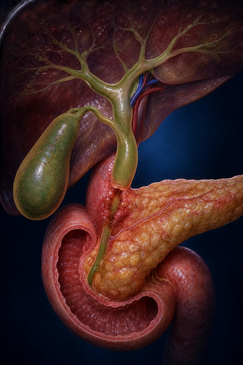
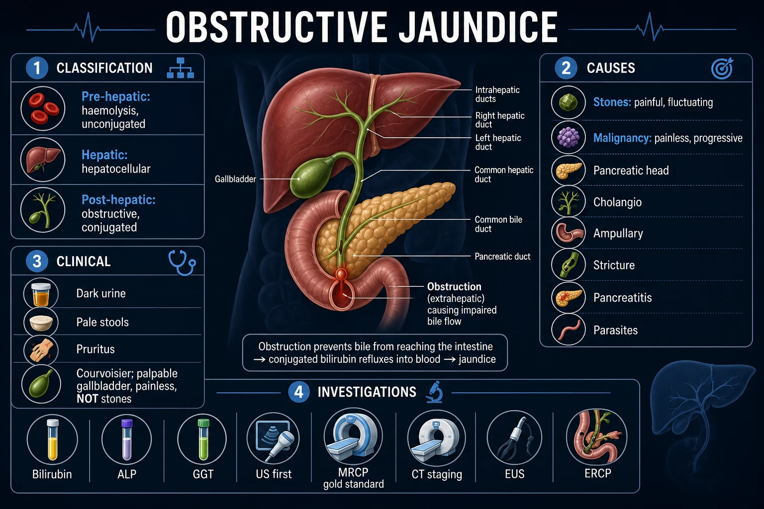
Bile is secreted by hepatocytes into bile canaliculi, which coalesce into segmental and area bile ducts, then the right and left hepatic ducts draining the two lobes. The right and left hepatic ducts join outside the liver at the porta hepatis to form the common hepatic duct (about 4 cm long). The cystic duct from the gallbladder joins the common hepatic duct at an angle to form the common bile duct (CBD). The CBD descends in the free edge of the lesser omentum, passes behind the first part of the duodenum, and runs in a groove on the posterior surface of the pancreatic head before joining the main pancreatic duct of Wirsung at the ampulla of Vater (hepatopancreatic ampulla). The ampulla opens into the medial wall of the second part of the duodenum at the major duodenal papilla, surrounded by the sphincter of Oddi (of Boyden), a circular smooth-muscle complex that regulates the flow of bile and pancreatic juice and prevents reflux of duodenal contents.[1]
The gallbladder lies in a fossa on the undersurface of the liver, stores and concentrates bile (5 to 10 times) between meals, and contracts in response to cholecystokinin when fat enters the duodenum. Normal common bile duct diameter on ultrasound is 6 mm or less in adults under 60, increasing by roughly 1 mm per decade thereafter, and up to 10 mm is acceptable after cholecystectomy. A duct that measures larger than 7 mm in a young patient or that increases in size on serial imaging is dilated and mandates search for a distal obstruction.[1]
Classification
Obstructive jaundice is most usefully classified by the anatomical level of obstruction (intrahepatic vs extrahepatic) and by the mechanism at that level (intraluminal, mural, extrinsic). Extrahepatic obstruction accounts for roughly 80 per cent of surgical jaundice; intrahepatic causes are predominantly medical but a few are managed by hepatologists alongside surgeons, so the boundary must be drawn explicitly.[1]
Intraluminal
occlusion from within the duct
- **Choledocholithiasis** — common bile duct stones; the commonest cause overall
- Mirizzi syndrome — gallstone impacted in Hartmann's pouch or cystic duct compressing the common hepatic duct
- Parasites — Ascaris lumbricoides (India, South-East Asia), liver flukes Clonorchis sinensis and Opisthorchis (cholangiocarcinoma risk)
- Haemobilia — blood clot from trauma, iatrogenic liver biopsy, or hepatocellular tumour
Mural
disease of the duct wall
- **Cholangiocarcinoma** — adenocarcinoma of the biliary epithelium (intrahepatic, perihilar Klatskin, or distal)
- Inflammatory stricture — post-cholecystectomy (most common), chronic pancreatitis, primary sclerosing cholangitis
- Choledochal cyst (Todani types I to V) with risk of malignancy
- Biliary atresia — neonatal, the surgical cause of conjugated jaundice in infants
Extrinsic
compression from outside the duct
- **Pancreatic head adenocarcinoma** — the commonest malignancy causing obstructive jaundice
- Periampullary carcinoma — ampulla of Vater, duodenum, distal bile duct (better prognosis than pancreas)
- Chronic pancreatitis — fibrotic stricture of the intrapancreatic CBD
- Enlarged lymph nodes — porta hepatis metastases or lymphoma
- Pancreatic pseudocyst compressing the duct
Intrahepatic (medical)
cholestasis at canaliculi
- **Drugs** — flucloxacillin, co-amoxiclav, chlorpromazine, anabolic steroids, oral contraceptives
- **Primary biliary cholangitis (PBC)** — anti-mitochondrial antibody positive, middle-aged women
- **Primary sclerosing cholangitis (PSC)** — association with ulcerative colitis; cholangiocarcinoma risk
- Viral hepatitis — cholestatic phase of hepatitis A, B, E
- **Sepsis** — cytokine-mediated cholestasis, especially gram-negative
Extrahepatic (surgical)
mechanical duct obstruction
- Stones — choledocholithiasis, Mirizzi
- Tumours — pancreas, cholangiocarcinoma, ampullary, gallbladder
- Strictures — post-surgical, chronic pancreatitis, PSC
- Parasites — Ascaris, flukes
- Congenital — choledochal cyst, biliary atresia
Epidemiology & Risk Factors
The single most discriminating fact in obstructive jaundice epidemiology is the bimodal age distribution of the cause. In patients under 50, the cause is overwhelmingly gallstones (choledocholithiasis). In patients over 60, the cause shifts sharply towards malignancy — pancreatic head cancer, cholangiocarcinoma, and ampullary carcinoma — and the burden of weight loss and painless progression rises accordingly.[1]
Causes by frequency and age
The classic risk factors for gallstones remain the 5 Fs — female, forty, fertile, fair, fat — supplemented by rapid weight loss, oral contraception, total parenteral nutrition, ileal Crohn's disease, and haemolytic anaemia (pigment stones). Pancreatic head cancer is driven by smoking (two-fold risk), long-standing type 2 diabetes, chronic pancreatitis, familial cancer syndromes (BRCA2, Peutz-Jeghers, Lynch, hereditary pancreatitis), and obesity. Cholangiocarcinoma risk is concentrated in primary sclerosing cholangitis (lifetime risk 5 to 15 per cent), liver-fluke infestation (Clonorchis, Opisthorchis in South-East Asia), choledochal cysts, and hepatolithiasis (recurrent pyogenic cholangitis).[4][13]
Pathophysiology
Obstruction of the biliary tree produces a predictable and clinically recognisable cascade, whatever the level of the block. The mechanism is best understood by tracing the consequences of failure of conjugated bilirubin and bile salts to reach the gut.[1]
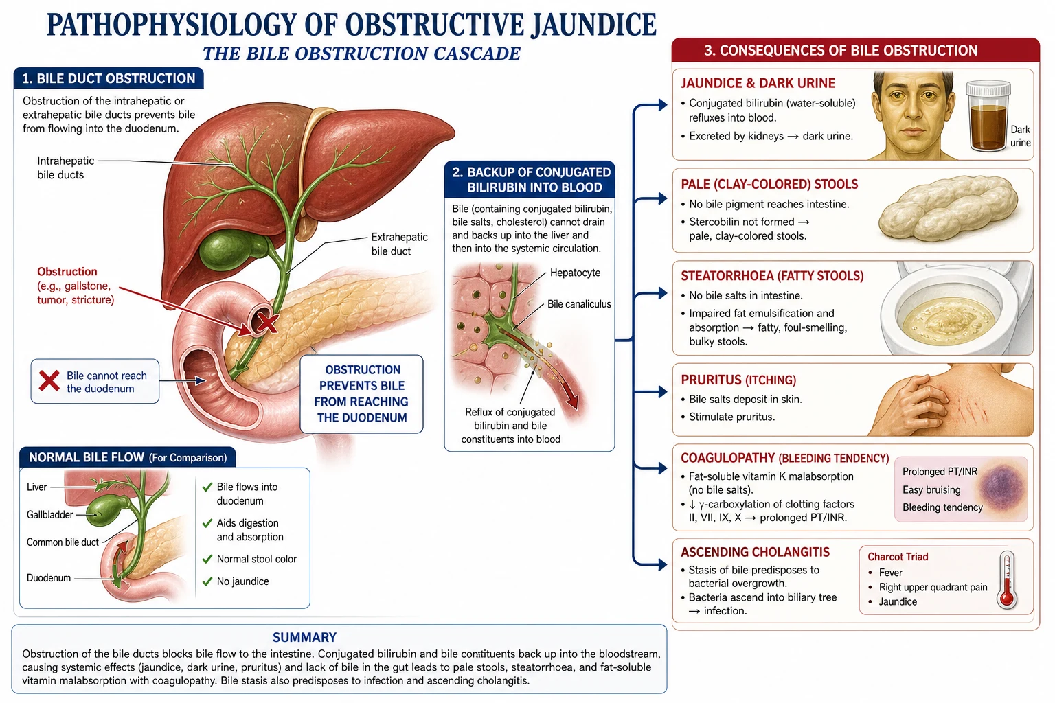
- Bile cannot reach the duodenum. Conjugated bilirubin, normally excreted in bile and converted to urobilinogen then stercobilin by gut bacteria (giving stool its brown colour), now backs up in the biliary tree. Rising intrabiliary pressure disrupts hepatocyte tight junctions; conjugated bilirubin regurgitates directly into plasma and, being water-soluble, is filtered at the glomerulus — producing the dark brown urine of the patient. The loss of stercobilin turns the stool pale and clay-coloured, the patient often volunteering that the stool is "putty-like" or floats (steatorrhoea).[1]
- Bile salts accumulate in plasma and deposit in skin — the cause of pruritus, which characteristically is worse at night, affects the palms and soles, and is partly central (opioid-receptor mediated in the brain) and partly peripheral (bile salt activation of a TGR5 receptor on sensory neurones). Scratch marks and excoriations may be the most striking physical sign.
- Fat maldigestion and malabsorption. With no bile in the gut, dietary triglyceride cannot be emulsified into micelles. Steatorrhoea ensues (bulky, foul, floating stools), and the fat-soluble vitamins A, D, E, and K are malabsorbed. Vitamin K deficiency is the most clinically acute consequence — factors II, VII, IX, X (the vitamin-K-dependent factors) fall, and the prothrombin time prolongs within one to two weeks of complete obstruction. This is reversible with parenteral vitamin K, distinguishing it from the irreversible coagulopathy of hepatocellular failure. Vitamin D deficiency eventually produces osteomalacia; long-standing cholestasis causes bone disease (hepatic osteodystrophy).[1]
- Biliary stasis predisposes to ascending infection. Bile is normally sterile, but obstruction raises intraductal pressure, disrupts the bile-canaliculus barrier, and permits gut organisms — classically Escherichia coli, Klebsiella, Enterococcus, and anaerobes — to ascend from the duodenum or to enter via the portal vein. Infected bile under pressure spills into the systemic circulation (cholangio-venous reflux), producing the bacteraemia, fever with rigors, and septic shock of ascending cholangitis.[8]
- Progressive hepatocellular dysfunction. Long-standing obstruction causes secondary biliary cirrhosis within months — a critical reason not to delay relief of a malignant obstruction even when the cancer itself is incurable.
Clinical Presentation
The patient with obstructive jaundice presents with a combination of the cholestatic triad — dark urine, pale stools, and pruritus — together with yellow discolouration of the sclera, sublingual mucosa, and skin. Jaundice becomes clinically visible when serum bilirubin exceeds roughly 50 micromol per litre (3 mg per decilitre). The tempo, the presence and character of pain, and the associated systemic features point to the underlying cause with remarkable accuracy.[1]
Courvoisier's law
Courvoisier's law (1890) states: in a jaundiced patient, a palpable, non-tender, distended gallbladder is unlikely to be caused by gallstones. The rationale is mechanical — repeated episodes of chronic cholecystitis from stones cause fibrosis and contraction of the gallbladder wall, so a gallbladder chronically scarred by stones cannot distend. A distended palpable gallbladder therefore implies a non-stone obstruction of the lower biliary tree — classically carcinoma of the pancreatic head, where the gallbladder has never been scarred and distends passively from backed-up bile.[1]
The law is statistical, not absolute: exceptions include a double impaction (stone at the cystic duct plus another at the ampulla), pancreatic pseudocyst, and a stone forming in a previously normal (unscarred) gallbladder. Nonetheless, when a jaundiced patient has a palpable gallbladder, malignancy is the working diagnosis until disproved. The mirror-image clinical pearl: most jaundice from stones is painful and the gallbladder is impalpable.[1]
Charcot's triad and Reynolds' pentad
Acute ascending cholangitis is characterised by Charcot's triad (1877): fever (often with rigors), jaundice, and right-upper-quadrant abdominal pain. Modern series report a sensitivity of only about 25 per cent for the full triad at first presentation — fever alone is the most sensitive single sign — so its absence does not exclude cholangitis in an obstructed patient. Reynolds' pentad (1959) adds hypotension (septic shock) and altered mental state (confusion or depressed consciousness) and indicates severe grade III disease with multi-organ dysfunction. Mortality of severe cholangitis without drainage approaches 50 per cent; the dictum of surgery is "pus under pressure" — the only definitive treatment is decompression.[8][10]
General and constitutional features
Yellow skin and sclera, scratch marks (the sign of pruritus, not jaundice itself), cachexia (suggests malignancy), palmar erythema and spider naevi (suggest chronic liver disease and therefore a medical cause), asterixis (hepatic encephalopathy — a danger sign in fulminant hepatocellular failure), and bruising (coagulopathy). Ask about pale floating stools and dark brown urine, travel, blood transfusion, drug history (especially flucloxacillin, co-amoxiclav, anabolic steroids, oral contraceptives), alcohol, sexual exposure, family history of cancer, and recent weight loss.[1]
Differential Diagnosis
A wide differential is the rule in any jaundiced patient, and the first task is to decide pre-hepatic, hepatic, or post-hepatic. The biochemical pattern (conjugated vs unconjugated, transaminase vs ALP dominant) refines the list, but the obstruction is then narrowed by imaging.[1]
Pattern recognition across the three categories of jaundice
The GGT parallels ALP in biliary disease and confirms the biliary origin of a raised ALP (an isolated high ALP of bone origin will have a normal GGT). The prothrombin time prolongs in cholestasis from vitamin K malabsorption and corrects with parenteral vitamin K — a useful bedside discriminator from the irreversible coagulopathy of hepatocellular failure.[1]
Clinical & Bedside Assessment
The focused examination of the jaundiced patient follows the standard inspection, palpation, percussion, auscultation sequence, but with specific high-yield manoeuvres. Examine for stigmata of chronic liver disease (palmar erythema, spider naevi, gynaecomastia, caput medusae, asterixis) — their presence argues for a hepatic rather than surgical cause. Look for lymphadenopathy, especially a Virchow's node (Troisier's sign — left supraclavicular node), which suggests metastatic intra-abdominal malignancy, typically gastric or pancreatic.[1]
Abdominal examination
- Inspection: surgical scars (previous cholecystectomy raises possibility of a retained or recurrent stone or post-surgical stricture), cachexia, visible peristalsis.
- Palpation: a palpable, non-tender gallbladder is Courvoisier-positive and mandates workup for malignancy. Hepatomegaly with a hard, irregular edge suggests metastases or hepatocellular carcinoma; a craggy, fixed mass in the right upper quadrant may be gallbladder carcinoma. Epigastric mass or Sister Mary Joseph nodule (umbilical metastatic deposit) suggests advanced pancreatic cancer. Ascites in a jaundiced patient is suspicious for peritoneal metastases or decompensated cirrhosis.
- Percussion: shifting dullness confirms ascites; a globular, dull-to-percussion, ballotable mass in the right upper quadrant in the jaundiced patient is the distended gallbladder of Courvoisier.
- Auscultation: an upper abdominal bruit is occasionally heard over a pancreatic tumour with vascular involvement; a succession splash suggests gastric outlet obstruction from an advanced periampullary tumour.[1]
Named signs worth knowing for the viva
| Sign | Finding | Interpretation |
|---|---|---|
| Courvoisier's sign | Palpable, non-tender, distended gallbladder in a jaundiced patient | Malignant distal obstruction (not stones) |
| Charcot's triad | Fever, jaundice, right-upper-quadrant pain | Ascending cholangitis (sensitivity about 25 per cent) |
| Reynolds' pentad | Charcot's triad plus hypotension plus confusion | Severe grade III cholangitis with organ dysfunction |
| Troisier's sign | Enlarged left supraclavicular (Virchow) node | Metastatic abdominal malignancy (gastric, pancreatic) |
| Murphy's sign | Inspiratory arrest on palpation of right upper quadrant | Acute cholecystitis (not specifically obstructive jaundice) |
| Boas' sign | Hyperaesthesia in right infrascapular region | Acute cholecystitis referral |
Investigations
Investigation of the jaundiced patient proceeds in three steps: confirm cholestasis biochemically, localise the level of obstruction by imaging, and establish the cause with targeted tests.[1]
Blood tests
Biochemical pattern of obstructive jaundice
- Liver function tests: conjugated (direct) bilirubin raised; ALP and GGT markedly elevated (typically greater than three times the upper limit of normal); AST and ALT usually less than two to three times normal, though transient rises above 1000 units per litre may occur in acute biliary obstruction (so-called "cholangitic hepatitis"), mimicking acute viral hepatitis.
- Coagulation: prothrombin time and INR prolonged due to vitamin K malabsorption; corrects within 24 to 48 hours of parenteral vitamin K 10 mg IV.
- Full blood count: leucocytosis and raised C-reactive protein suggest ascending cholangitis; anaemia raises suspicion of malignancy or haemolysis.
- Tumour markers: CA 19-9 elevated in pancreatic cancer (sensitivity about 80 per cent, specificity 70 to 90 per cent) but also raised in any cholestasis, including cholangitis and PSC — it must therefore be interpreted with imaging and not used as a stand-alone screen. CEA may support cholangiocarcinoma. Alpha-fetoprotein excludes hepatocellular carcinoma in cirrhotic patients. IgM anti-mitochondrial antibody confirms primary biliary cholangitis.
- Viral and autoimmune screen if hepatocellular causes are plausible: hepatitis B surface antigen, hepatitis C antibody, hepatitis A IgM, hepatitis E IgM, anti-smooth muscle antibody, anti-nuclear antibody, anti-LKM.[1]
Imaging — the diagnostic ladder
Imaging answers two questions in sequence: is the biliary tree dilated? and where and what is the obstruction?[1][3]
The diagnostic imaging ladder in obstructive jaundice
**Transabdominal ultrasound** — the first-line investigation in every jaundiced patient. Non-invasive, cheap, widely available, no ionising radiation. Identifies the **level of obstruction** (intrahepatic versus extrahepatic duct dilatation), gallstones in the gallbladder (sensitivity over 90 per cent) and CBD (sensitivity 50 to 75 per cent, limited by bowel gas over the pancreatic head), pancreatic head mass, gallbladder distension (Courvoisier), and liver metastases. A normal-calibre CBD is less than 6 mm; greater than 7 mm in a young adult or progressive dilatation on serial scans is significant.
**MRCP (magnetic resonance cholangiopancreatography)** — the **non-invasive gold standard** for biliary tree anatomy. Sensitivity and specificity greater than 95 per cent for choledocholithiasis and strictures; non-diagnostic ERCP avoided when MRCP is negative. No radiation, no contrast in most cases, no sedation. Defines stricture length, ductal anatomy variants, cholangiocarcinoma extent, and Klatskin tumour Bismuth-Corlette classification.
**Contrast-enhanced CT (pancreatic protocol)** — the staging investigation of choice when **malignancy is suspected or confirmed**. Multi-phase arterial, portal venous, and pancreatic-phase imaging defines tumour size, vascular invasion (superior mesenteric artery and vein, portal vein — the determinants of resectability), regional lymph nodes, liver and peritoneal metastases, and ascites. CT replaces ultrasound for staging but not for screening.
**EUS (endoscopic ultrasound)** — highest spatial resolution for small pancreatic head lesions, ampullary tumours, and distal CBD stones; allows **fine-needle aspiration** for tissue diagnosis. The investigation of choice when CT/MRCP show a small or equivocal pancreatic lesion, and for nodal staging.
**ERCP (endoscopic retrograde cholangiopancreatography)** — the only modality that is **both diagnostic and therapeutic**. Performed when obstruction is confirmed and intervention is planned. Allows **sphincterotomy, stone extraction with balloon or basket, biliary stent placement (plastic or metal), stricture dilatation, and brush cytology or biopsy** for tissue diagnosis. Complications include **pancreatitis (3 to 10 per cent), bleeding, perforation, and cholangitis**.
**PTC (percutaneous transhepatic cholangiography)** — when ERCP fails (post-surgical anatomy, duodenal obstruction, prior Roux-en-Y) or for proximal hilar obstruction. Provides a transhepatic route for **external or internal-external biliary drainage** and for placement of self-expanding metal stents. Bleeding, bile leak, and cholangitis are recognised risks.
**Intra-operative cholangiography (IOC)** — performed during cholecystectomy to confirm duct clearance or detect residual stones; increasingly replaced by laparoscopic ultrasound but still the gold standard at open surgery.
**HIDA scan / hepatobiliary scintigraphy** — limited role in adults; used to confirm cystic duct obstruction in acalculous cholecystitis and biliary leak post-cholecystectomy.
Diagnostic accuracy at a glance
The meta-analysis by Garrow and colleagues (2007) reported endoscopic ultrasound sensitivity 89 per cent and specificity 94 per cent for choledocholithiasis, superior to MRCP in the small-stone and pancreas-head scenarios. MRCP sensitivity is 90 to 95 per cent for stones greater than 5 mm but falls for smaller stones and for impacted distal CBD stones obscured by bowel gas.[3]
Management — Resuscitation
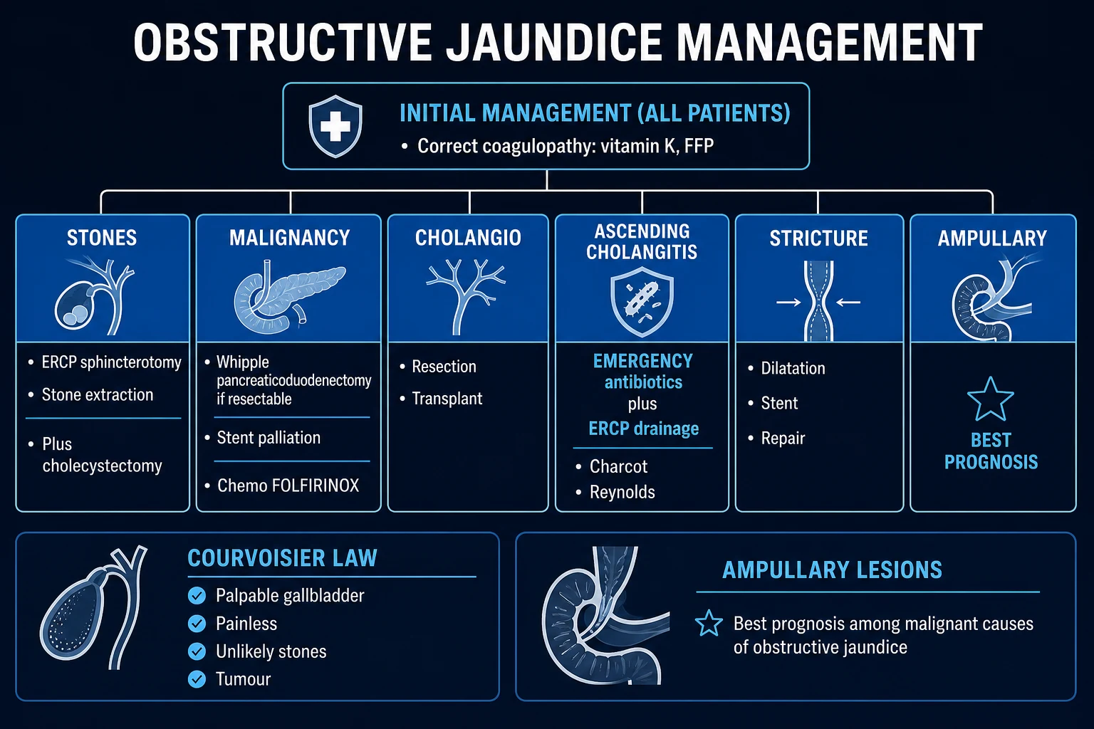
The immediate priorities in obstructive jaundice are to (a) recognise and treat cholangitis, (b) correct coagulopathy, and (c) relieve the obstruction. The first two are time-critical; the third defines the disease-specific phase that follows.[1][8]
Ascending cholangitis (Charcot's triad or Reynolds' pentad)
Acute cholangitis is the surgical emergency of biliary obstruction — infected bile under pressure that spills organisms into the systemic circulation. The Tokyo Guidelines 2018 provide the contemporary diagnostic and severity framework.[8][9]
Initial management bundle (TG18)
Definitive treatment is biliary drainage — antibiotics alone will not cure an obstructed, infected system. The dictum is "pus under pressure needs decompression." For most patients, ERCP within 24 to 48 hours with sphincterotomy, stone extraction, or stent placement is the procedure of choice. In severe grade III cholangitis with refractory shock, drainage may need to be same-shift emergent. Percutaneous transhepatic drainage or surgical drainage are second-line when ERCP fails or is unavailable.[8][10]
Tokyo Guidelines 2018 severity grading of acute cholangitis
TG18 severity grades and disposition
Coagulopathy
Vitamin K malabsorption causes the prothrombin time to prolong in any patient with weeks of complete obstruction. Phytomenadione (vitamin K1) 10 mg IV once daily for 3 days corrects the PT within 24 to 48 hours and must precede ERCP, percutaneous transhepatic procedures, and surgery. If an emergency procedure cannot wait, fresh frozen plasma 10 to 15 mL per kilogram is infused. Always recheck the INR immediately before any invasive procedure.[1]
Pruritus
Cholestyramine 4 g orally four times daily is first-line; rifampicin 150 to 300 mg orally twice daily is second-line (monitor liver enzymes). Sertraline 50 to 100 mg daily and the opioid antagonist naltrexone are third-line. Pruritus resolves within days of biliary decompression.[1]
Nutrition and analgesia
Steatorrhoea and fat-soluble vitamin malabsorption warrant low-fat diet, oral fat-soluble vitamin supplementation (A, D, E, K), and pancreatic enzyme replacement if pancreatic insufficiency coexists (chronic pancreatitis, after Whipple). Avoid morphine in undrained biliary obstruction — it increases sphincter of Oddi tone and may worsen pain; pethidine or fentanyl are preferred opioids.[1]
Management — Definitive & Stepwise
Choledocholithiasis
The ASGE 2019 guideline stratifies patients by the predicted probability of CBD stones.[6]
High risk (over 50%)
proceed to ERCP
- CBD stone on transabdominal ultrasound, MRCP, or EUS
- Total bilirubin over 4 mg per decilitre plus dilated CBD on ultrasound
Intermediate (10 to 50%) accent: amber
EUS or MRCP first
- Dilated CBD over 6 mm
- Bilirubin 1.8 to 4 mg per decilitre
- Abnormal liver tests or age over 55
Low risk (under 10%)
cholecystectomy only
- No predictors present
- Proceed directly to laparoscopic cholecystectomy without CBD imaging
Definitive therapy is ERCP with endoscopic sphincterotomy and stone extraction using a balloon or Dormia basket. For stones over 1.5 cm, mechanical lithotripsy, laser lithotripsy, or extracorporeal shock-wave lithotripsy may be needed. After clearance, the patient undergoes laparoscopic cholecystectomy during the same admission to prevent recurrent stone migration. In young, fit patients, single-stage laparoscopic CBD exploration at cholecystectomy is an alternative where ERCP is unavailable.[6][7]
Pancreatic head cancer
Pancreatic head cancer — resectability and intent
Curative intent — Whipple's procedure (pancreaticoduodenectomy) is offered when the tumour is resectable: no distant metastases, no encasement of the superior mesenteric artery or coeliac axis, and no unreconstructable superior mesenteric or portal vein involvement. The procedure removes the head of the pancreas, the second and third parts of the duodenum, the distal common bile duct, the gallbladder, and the distal stomach (the classical Whipple) or spares the pylorus (pylorus-preserving pancreaticoduodenectomy, the contemporary standard). Reconstruction is by pancreaticojejunostomy, hepaticojejunostomy, and gastrojejunostomy (or duodenojejunostomy).[1]
Operative mortality in high-volume centres is below 5 per cent; morbidity remains 30 to 40 per cent, dominated by post-operative pancreatic fistula, delayed gastric emptying, and post-pancreatectomy haemorrhage. Minimally invasive (laparoscopic or robotic) Whipple is increasingly performed in selected centres, with the 2025 DIPLOMA-2 multicentre trial showing non-inferiority for perioperative outcomes and equivalent R0 resection rates.[16]
Adjuvant chemotherapy with gemcitabine plus capecitabine (ESPAC-4, 28-month median survival vs 25 months for gemcitabine alone) is the standard of care after resection; modified FOLFIRINOX offers an alternative in fit patients. For unresectable locally advanced and metastatic disease, FOLFIRINOX or gemcitabine plus nab-paclitaxel extends median survival from 6 to 11 months.[11][12]
Palliation in unresectable disease aims to relieve jaundice, duodenal obstruction, and pain. Endoscopic biliary stenting at ERCP is the palliation of choice — a self-expanding metal stent patency of 9 to 12 months is preferred when survival exceeds 4 months, plastic stents otherwise. Duodenal obstruction (in 10 to 20 per cent) is palliated by endoscopic duodenal stenting or surgical gastric bypass. Coeliac plexus block relieves refractory back pain.[7]
Preoperative biliary drainage — the controversy
Routine preoperative biliary drainage (PBD) before Whipple was once thought to improve outcomes by reversing cholestasis and coagulopathy. The Dutch randomised trial by van der Gaag et al. (NEJM 2010) disproved this — PBD significantly increased serious complications (74 per cent vs 39 per cent, mostly cholangitis and pancreatitis from ERCP) without improving surgical outcomes or survival. PBD is now reserved for cholangitis, severe pruritus delaying surgery, neoadjuvant chemotherapy, or delayed surgery for logistical reasons.[5]
Cholangiocarcinoma
Prognosis and approach depend on location (Bismuth-Corlette for hilar,modified Bismuth for extrahepatic).[13][14]
- Distal cholangiocarcinoma — Whipple's procedure if resectable; 5-year survival 25 to 35 per cent.
- Perihilar (Klatskin) cholangiocarcinoma — extended right or left hepatectomy with caudate lobectomy and extrahepatic bile duct resection is the only curative option; 5-year survival 20 to 30 per cent in expert centres. Pre-operative portal vein embolisation to grow the future liver remnant is often used. Lymph node metastases, bilateral hepatic duct involvement to secondary radicals, and vascular invasion contraindicate resection.[14]
- Intrahepatic cholangiocarcinoma — anatomical hepatic resection; adjuvant capecitabine.
- Unresectable disease — biliary drainage (multi-stent, percutaneous, or endoscopically placed across the hilum), systemic chemotherapy (gemcitabine plus cisplatin), and selective radiotherapy.
Ampullary and periampullary carcinoma
Periampullary tumours (ampulla of Vater, distal CBD, second part of duodenum) share the surgical approach with pancreatic head cancer (Whipple) but have a markedly better prognosis — 5-year survival up to 40 per cent for ampullary carcinoma after R0 resection, because they obstruct early and present at smaller size.[1]
Benign biliary stricture
Causes include post-cholecystectomy (most common, from clip misplacement, thermal injury, or ischaemia), chronic pancreatitis, and primary sclerosing cholangitis. Endoscopic balloon dilatation with multiple plastic stents exchanged every 3 to 6 months for 1 to 2 years is the standard first-line therapy in expert centres; fully covered self-expanding metal stents (FCSEMS) are an alternative. Surgical Roux-en-Y hepaticojejunostomy is reserved for endoscopic failure, complex hilar strictures, or low CBD injuries.[7]
Specific Subtypes & Scenarios
Mirizzi syndrome
An impacted gallstone in Hartmann's pouch or the cystic duct compresses the common hepatic duct from outside, producing obstructive jaundice in the absence of a true CBD stone. Type I is external compression; type II to IV involve a cholecystocholedochal fistula eroding into the duct. MRCP demonstrates the level; cholecystectomy with careful dissection (often subtotal to avoid iatrogenic CBD injury) is the treatment. A Csendes type II or higher may require cholecystojejunostomy or biliary-enteric reconstruction.[4]
Biliary atresia
The neonatal surgical cause of conjugated jaundice. Presents in the first 2 to 6 weeks of life with pale stools, dark urine, and progressive conjugated hyperbilirubinaemia. Kasai portoenterostomy before 60 days of life restores bile flow in up to two-thirds; most surviving children ultimately need liver transplantation. Any jaundiced infant beyond 2 weeks of age must have conjugated bilirubin measured — a critical neonatal pearl.[1]
Post-cholecystectomy jaundice
A patient who becomes jaundiced within days of cholecystectomy has either a retained CBD stone (most common), an iatrogenic CBD injury (clip, transection, thermal stricture), or a bile leak with biloma compressing the duct. Urgent MRCP, ERCP, and surgical re-exploration in a tertiary HPB unit are the working algorithm. Early recognition and immediate referral to a specialist HPB surgeon dramatically improves outcome — primary repair at the index operation outperforms delayed reconstruction.[1][7]
Parasitic obstruction
Ascaris lumbricoides worms migrate up the papilla and obstruct the CBD; presentation is with biliary colic, cholangitis, or pancreatitis. Endoscopic extraction at ERCP with a Dormia basket or forceps is the treatment; mebendazole 100 mg orally twice daily for 3 days or albendazole 400 mg single dose eradicates residual worms. Liver flukes (Clonorchis, Opisthorchis) cause chronic cholangitis, hepatolithiasis, and cholangiocarcinoma; praziquantel 75 mg per kilogram in three divided doses is curative for the parasite, but the cancer risk persists.[4]
Complications & Pitfalls
Disease-related complications include ascending cholangitis and septic shock, secondary biliary cirrhosis (within months of unrelieved obstruction), hepatorenal syndrome from biliary sepsis, coagulopathy and bleeding, malnutrition and fat-soluble vitamin deficiency, and renal failure from bilirubin nephropathy or sepsis-associated acute kidney injury.[1]
Procedure-related complications (ERCP, after Cotton et al. 1991 consensus definitions):[2]
Cotton-consensus ERCP complications
Risk-stratify every ERCP — high-risk features for pancreatitis include SOD (sphincter of Oddi dysfunction), previous post-ERCP pancreatitis, young age, female sex, normal bilirubin, difficult cannulation, and repeated pancreatic injection. Prophylaxis with rectal indomethacin 100 mg immediately post-procedure and prophylactic pancreatic stent placement in high-risk patients reduces pancreatitis by 50 to 70 per cent.[2][15]
Classic pitfalls
- Missing pancreatic cancer — always investigate painless progressive jaundice with CT and MRCP; never attribute a Courvoisier-positive gallbladder to stones.
- Performing ERCP before MRCP in a patient with intermediate probability of stones — wastes resources and exposes the patient to pancreatitis when MRCP would have excluded obstruction.
- Operating with a raised INR — the patient with weeks of obstruction has vitamin K deficiency; check and correct before any procedure.
- Failing to drain an obstructed, infected system — antibiotics alone are insufficient in cholangitis; biliary decompression is definitive therapy.
- Misreading CA 19-9 in cholestasis — levels rise in any obstruction, including PSC and cholangitis; do not over-call malignancy on the basis of CA 19-9 alone.
- Lewis-negative patients (about 10 per cent of the population) cannot synthesise CA 19-9 — a normal value excludes nothing, and a falsely low value may mask pancreatic cancer.[1]
Prognosis & Disposition
Prognosis is wholly determined by cause.[1]
Prognosis by cause
Disposition: most obstructed patients require admission for imaging, drainage, or surgery. Ascending cholangitis is an immediate surgical admission with ICU input for grade III. Pancreatic and cholangiocarcinoma pathways belong in the multidisciplinary HPB tumour board. Discharge is appropriate once jaundice has been relieved, cholangitis treated, coagulopathy corrected, and a clear plan for definitive management communicated to the patient and general practitioner.[1]
Special Populations
- Elderly patients carry a higher probability of malignancy at any level of obstruction — always image with contrast CT and have a low threshold for MRCP. Atypical presentations are common: confusion without fever may be the only sign of cholangitis. Reserve major resection (Whipple) for physiologically fit patients over 80 — frailty scoring (CRS, mFI) guides shared decision-making.
- Pregnancy — obstructive jaundice is rare, mostly due to choledocholithiasis. Intrahepatic cholestasis of pregnancy is the principal differential (third trimester, pruritus, raised bile acids, normal GGT). ERCP is technically feasible in pregnancy with lead shielding, left lateral position, and minimal fluoroscopy; sphincterotomy and stone extraction are safe in the second and third trimester. Deferring cholecystectomy to the post-partum period is acceptable after stone clearance in asymptomatic patients; recurrent biliary colic or pancreatitis warrants same-admission cholecystectomy in the second trimester.[1]
- Cirrhosis and chronic liver disease — obstructive jaundice may be masked by underlying parenchymal dysfunction; the GGT and ALP pattern still discriminates. Cirrhosis is a relative contraindication to extended hepatectomy ( Klatskin resection); Child-Pugh C or MELD over 10 carries prohibitive mortality. Portal hypertension increases bleeding risk at ERCP.
- Post-cholecystectomy — a retained or recurrent CBD stone, or a post-surgical stricture, must always be considered; MRCP is the diagnostic test of choice.
- Immunocompromised patients (HIV, post-transplant) — consider AIDS cholangiopathy (cryptosporidium, CMV, microsporidium causing papillary stenosis and sclerosing cholangitis-like changes) and drug-induced cholestasis from antiretrovirals and immunosuppressants (azathioprine, ciclosporin, tacrolimus).
- Anticoagulated patients — bridge to heparin or withhold warfarin before ERCP with sphincterotomy; check the INR immediately pre-procedure. Direct oral anticoagulants are omitted for 24 to 48 hours pre-procedure; resumed once haemostasis is secured.
Evidence, Guidelines & Regional Differences
Landmark trials
van der Gaag et al., NEJM 2010 — preoperative biliary drainage in pancreatic head cancer
Randomised controlled trial, 238 patients with resectable pancreatic head cancer and bilirubin 40 to 250 micromol per litre, assigned to immediate surgery vs PBD followed by surgery.
Key finding
PBD significantly increased serious complications (74 per cent vs 39 per cent), driven by cholangitis and pancreatitis from ERCP, with no improvement in surgical outcome or survival. **PBD is not routine** — reserved for cholangitis, severe pruritus, or delayed surgery.
ESPAC-4 (Neoptolemos et al., Lancet 2017)
Multicentre randomised trial, 730 patients after resection of pancreatic ductal adenocarcinoma, adjuvant gemcitabine alone vs gemcitabine plus capecitabine.
Key finding
Combination adjuvant chemotherapy improved median survival from 25 to 28 months and 5-year survival from 17 to 29 per cent. **Gemcitabine plus capecitabine is the standard adjuvant regimen.**
PRODIGE 4 / ACCORD 11 (Conroy et al., NEJM 2011)
Randomised trial, 342 patients with metastatic pancreatic cancer, FOLFIRINOX vs gemcitabine.
Key finding
FOLFIRINOX nearly doubled median survival (11.1 vs 6.8 months) and improved 1-year survival (48 vs 20 per cent) at the cost of greater toxicity (neutropenia, diarrhoea). **FOLFIRINOX is the preferred regimen for fit patients with metastatic disease.**
Guidelines — ESGE and TG18
ESGE 2018 biliary stenting guidelines (Dumonceau) recommend an uncovered self-expanding metal stent for palliative drainage of malignant distal biliary obstruction when survival exceeds 4 months, plastic stents otherwise. Antibiotic prophylaxis is not routinely required for ERCP unless the biliary tree is incompletely drained, when a single dose is given.[7]
Tokyo Guidelines 2018 (TG18) standardised the diagnosis, severity grading, and management bundles for acute cholangitis. Severity-guided antimicrobial therapy is now universal: Grade I mild — community-acquired third-generation cephalosporin or piperacillin-tazobactam; Grade II moderate — piperacillin-tazobactam or cefepime with metronidazole; Grade III severe — broad-spectrum with anti-pseudomonal and anti-enterococcal cover (vancomycin if healthcare-associated).[8][9][10]
India and tropical practice. Open surgical biliary drainage (cholecystoduodenostomy, choledochoduodenostomy, Roux-en-Y hepaticojejunostomy) retains a role where ERCP services are scarce. Tropical calcific pancreatitis of young non-alcoholic patients, endemic in Kerala and Sri Lanka, produces obstructive jaundice by fibrotic stricture of the intrapancreatic CBD — managed by surgical lateral pancreaticojejunostomy (Partington-Rochelle) for ductal pain plus hepaticojejunostomy for jaundice. Recurrent pyogenic cholangitis (oriental cholangiohepatitis) is treated by cholecystectomy, CBD clearance, T-tube drainage, and biliary-enteric anastomosis for recurrent intrahepatic stones. Ascaris and liver-fluke (Clonorchis, Opisthorchis) are treated by endoscopic extraction plus anthelminthic (albendazole or praziquantel); the cholangiocarcinoma risk from chronic fluke infestation persists.[4]
Where the evidence is weak
- Pre-operative biliary drainage in resectable pancreatic cancer remains debated in the era of neoadjuvant FOLFIRINOX — most centres now stent for the 2 to 3 months of neoadjuvant therapy, but the Dutch trial's caution against routine stenting still applies to patients going directly to surgery.
- Covered vs uncovered SEMS for hilar cholangiocarcinoma, antibiotic prophylaxis in fully-drained ERCP, and adjuvant therapy for resected cholangiocarcinoma remain areas of active investigation.[5][7]
Exam Pearls
Causes of obstructive jaundice — STONE
STONE
choledocholithiasis — the commonest cause overall
pancreatic head (commonest malignancy), cholangiocarcinoma, ampullary
lymph nodes, chronic pancreatitis, pseudocyst
post-surgical, primary sclerosing cholangitis, inflammatory
Ascaris (India, South-East Asia), liver flukes Clonorchis and Opisthorchis
Charcot's triad and Reynolds' pentad
C-H-A-R-P
the underlying diagnosis — ascending infection of an obstructed biliary tree
the H of Charcot
right-upper-quadrant pain of Charcot
Reynolds' pentad adds hypotension and confusion to Charcot's triad
the third limb of Charcot's triad — yellow skin and sclera
The eleven one-liners that decide an obstructive jaundice answer:[1]
- Painless progressive jaundice plus weight loss = pancreatic head cancer until proven otherwise — urgent CT and MRCP.[1]
- Courvoisier's law: palpable, non-tender, distended gallbladder in a jaundiced patient = malignancy, NOT stones.[1]
- Charcot's triad: fever plus jaundice plus right-upper-quadrant pain = ascending cholangitis.[8]
- Reynolds' pentad: Charcot's triad plus hypotension plus confusion = severe cholangitis with organ dysfunction — emergency drainage.[8]
- Dark urine plus pale stools = obstructive (post-hepatic) jaundice.[1]
- Conjugated bilirubin raised, ALP and GGT raised, AST and ALT mildly raised = cholestatic (obstructive) pattern.[1]
- Ultrasound first-line, MRCP gold standard, ERCP therapeutic.[3]
- Whipple removes head of pancreas, duodenum, gallbladder, distal CBD, and distal stomach (classical) — reconstruction by pancreaticojejunostomy, hepaticojejunostomy, gastrojejunostomy.[1]
- Vitamin K deficiency causes coagulopathy — give phytomenadione 10 mg IV for 3 days before any procedure.[1]
- Preoperative biliary drainage is NOT routine — van der Gaag 2010 showed harm without benefit.[5]
- Rectal indomethacin and prophylactic pancreatic stent halve post-ERCP pancreatitis in high-risk patients.[15]
Exam application bank (NEET-PG / INICET)
One-line answer
Obstructive jaundice is mechanical blockage of bile flow anywhere from the intrahepatic ducts to the ampulla of Vater, producing conjugated hyperbilirubinaemia with a cholestatic enzyme pattern (ALP and GGT raised, AST and ALT near-normal). Painless progressive jaundice with weight loss and a palpable gallbladder (Courvoisier's law) = pancreatic head cancer until proven otherwise. Fever, jaundice and right-upper-quadrant pain (Charcot's triad) = ascending cholangitis — an emergency needing IV antibiotics and urgent ERCP for biliary drainage. Ultrasound is first-line imaging; MRCP is the non-invasive gold standard for the biliary tree; ERCP is both diagnostic and therapeutic (stone extraction, stent, brush cytology). Vitamin K 10 mg IV corrects the coagulopathy of cholestasis before any procedure. [1]
Worked stems (answer without another resource)
Stem 1 — Classic presentation. Map symptoms to mechanism; name the first investigation and first treatment step with dose/route if drug therapy is standard. [1]
Stem 2 — Unstable / complicated. List red flags that force immediate resuscitation, theatre, ICU, antidote, or reperfusion — and what you do in the first 15 minutes. [1]
Stem 3 — Atypical group. Elderly, pregnancy, child, or immunocompromised: how presentation and thresholds change. [1]
Stem 4 — Differential trap. Name the three closest mimics and one discriminator for each. [1]
Stem 5 — Disposition. Who goes home with safety-netting, who is admitted, who needs HDU/ICU/theatre, and what follow-up is mandatory. [1]
Rapid viva checklist
- Definition + classification
- Pathophysiology chain
- Bedside signs / criteria
- Score with exact components (if any)
- Emergency bundle
- Definitive therapy with doses
- Complications of disease and of treatment
- Special populations
- Guideline/trial name if classic
- Three exam traps
Coverage self-check
If you cannot answer any stem above from this page alone, re-read the matching section — the page is intended to be self-sufficient for final-prof and NEET-PG/INICET questions on Obstructive Jaundice.
References
- [1]Elmunzer BJ, Kumar R, Elta G, et al. ACG Clinical Guideline: Diagnosis and Management of Biliary Strictures Am J Gastroenterol, 2023.PMID 36863037
- [2]Cotton PB, Lehman G, Vennes J, et al. Endoscopic sphincterotomy complications and their management: an attempt at consensus Gastrointest Endosc, 1991.PMID 2070995
- [3]Garrow D, Miller S, Singh D, et al. Endoscopic ultrasound: a meta-analysis of test performance in suspected biliary obstruction Clin Gastroenterol Hepatol, 2007.PMID 17478348
- [4]Tazuma S, Unno M, Igarashi Y, et al. Evidence-based clinical practice guidelines for cholelithiasis 2016 J Gastroenterol, 2017.PMID 27942871
- [5]van der Gaag NA, Rauws EA, van Eijck CH, et al. Preoperative biliary drainage for cancer of the head of the pancreas N Engl J Med, 2010.PMID 20071702
- [6]ASGE Standards of Practice Committee; Buxbaum JL, Abbas Fehmi SM, et al. ASGE guideline on the role of endoscopy in the evaluation and management of choledocholithiasis Gastrointest Endosc, 2019.PMID 30979521
- [7]Dumonceau JM, Tringali A, Papanikolaou IS, et al. Endoscopic biliary stenting: indications, choice of stents, and results: European Society of Gastrointestinal Endoscopy (ESGE) Clinical Guideline - Updated October 2017 Endoscopy, 2018.PMID 30086596
- [8]Kiriyama S, Kozaka K, Takada T, et al. Tokyo Guidelines 2018: diagnostic criteria and severity grading of acute cholangitis (with videos) J Hepatobiliary Pancreat Sci, 2018.PMID 29032610
- [9]Gomi H, Solomkin JS, Takada T, et al. Tokyo Guidelines 2018: antimicrobial therapy for acute cholangitis and cholecystitis J Hepatobiliary Pancreat Sci, 2018.PMID 29090866
- [10]Mayumi T, Okada KI, Takada T, et al. Tokyo Guidelines 2018: management bundles for acute cholangitis and cholecystitis J Hepatobiliary Pancreat Sci, 2018.PMID 29090868
- [11]Conroy T, Desseigne F, Ychou M, et al. FOLFIRINOX versus gemcitabine for metastatic pancreatic cancer N Engl J Med, 2011.PMID 21561347
- [12]Neoptolemos JP, Palmer DH, Ghaneh P, et al. Comparison of adjuvant gemcitabine and capecitabine with gemcitabine monotherapy in patients with resected pancreatic cancer (ESPAC-4): a multicentre, open-label, randomised, phase 3 trial Lancet, 2017.PMID 28129987
- [13]Brindley PJ, Bachini M, Ilyas SI, et al. Cholangiocarcinoma Nat Rev Dis Primers, 2021.PMID 34504109
- [14]Mansour JC, Aloia TA, Crane CH, et al. Hilar cholangiocarcinoma: expert consensus statement HPB (Oxford), 2015.PMID 26172136
- [15]Freeman ML, Devereaux BM. Preventing Post-ERCP Pancreatitis: Update 2016 Curr Treat Options Gastroenterol, 2016.PMID 27349659
- [16]de Graaf N, van der Wilt AA, Busweiler LAD, et al. Minimally Invasive versus Open Pancreatoduodenectomy for Resectable Neoplasms NEJM Evid, 2025.PMID 41288428