General Surgery · General Surgery
Peptic Ulcer Disease
Also known as PUD · Gastric ulcer · Duodenal ulcer · Perforated peptic ulcer
Peptic ulcer disease (PUD) is a break in the gastric or duodenal mucosa caused by an imbalance between acid-peptic injury and mucosal defence. Two major causes: Helicobacter pylori (60 to 80%) and NSAIDs (20 to 30%). Duodenal ulcer: epigastric pain relieved by food/antacids, wakes at night. Gastric ulcer: pain worsened by food, weight loss. Diagnosis: OGD + biopsy (gastric ulcers are always biopsied to exclude malignancy); H. pylori testing (CLO test, urea breath, stool antigen). Treatment: H. pylori eradication triple therapy (PPI + clarithromycin + amoxicillin) 14 days; PPI for 4 to 8 weeks; stop NSAIDs. Surgical complications: perforation (Graham omental patch), haemorrhage (under-run bleeding vessel), gastric outlet obstruction (scarring from chronic DU).
On this page & tools
Your progress
Saved locally on this device.
Exam tags
Red flags
Overview & Definition
Peptic ulcer disease (PUD) is a break in the mucosal lining of the stomach (gastric ulcer) or proximal duodenum (duodenal ulcer) that extends through the muscularis mucosae. It results from an imbalance between aggressive factors (acid, pepsin, H. pylori, NSAIDs) and defensive factors (mucus-bicarbonate barrier, prostaglandins, mucosal blood flow, rapid cell turnover).[1][1]
The discovery of H. pylori (Marshall and Warren, 1982) revolutionised PUD management — what was once a surgical disease (vagotomy, antrectomy, gastrectomy) is now primarily medical (eradication therapy + PPI). Surgery is now reserved for complications: perforation, haemorrhage, and gastric outlet obstruction.[1][2]
Classification
By anatomical site:[1]
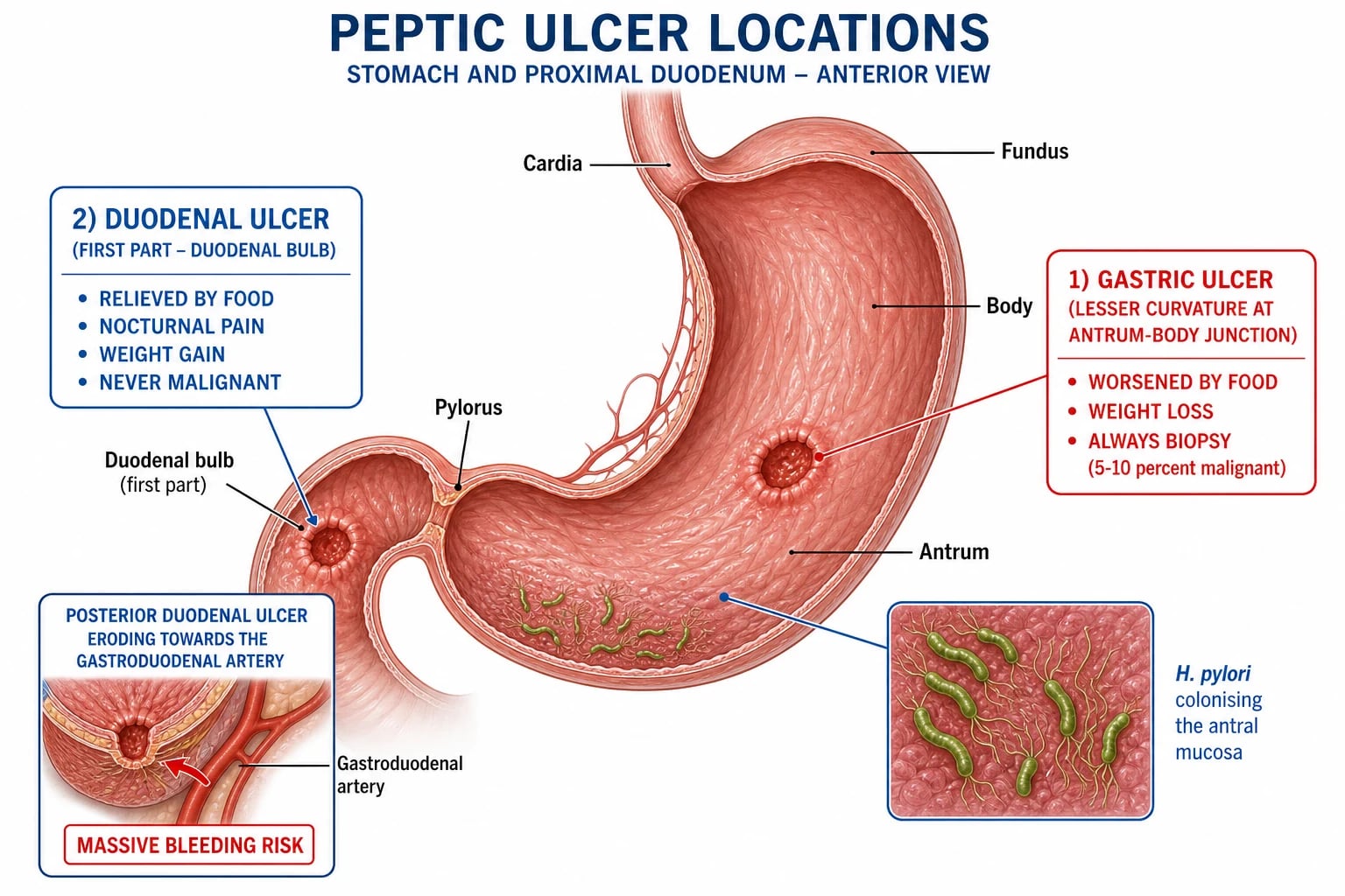
- Duodenal ulcer (DU) — occurs in the proximal duodenum (usually the first part, the duodenal bulb). Far more common than gastric ulcer (3:1). Almost never malignant. Associated with H. pylori (90%) and acid hypersecretion.
- Gastric ulcer (GU) — occurs in the stomach (commonly along the lesser curvature at the antrum-body junction). Can be malignant (5 to 10% of gastric ulcers are gastric carcinoma). Classified by location (Johnson types I to V).
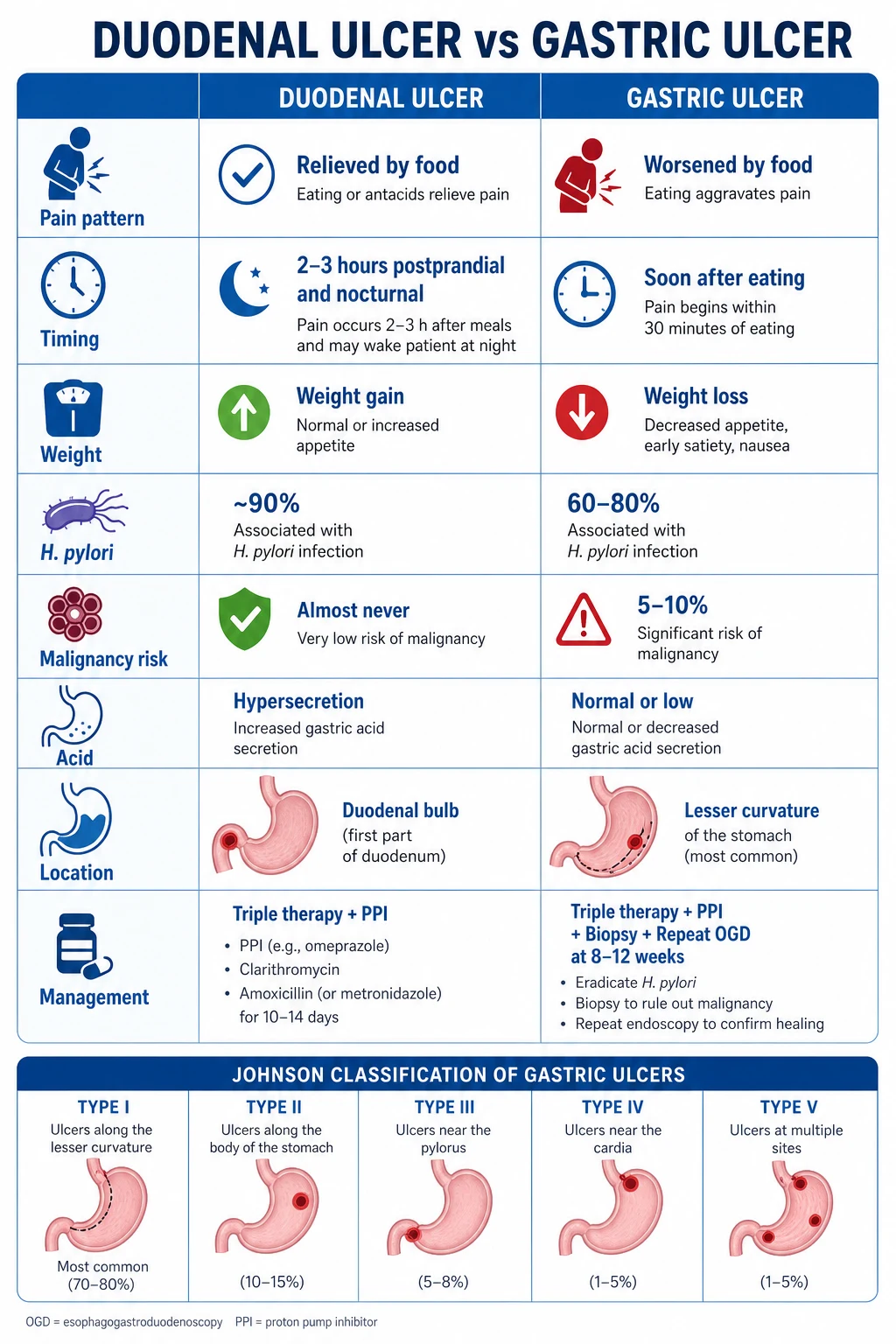
- Type I — along lesser curvature (most common).
- Type II — gastric + duodenal ulcer (combined).
- Type III — prepyloric.
- Type IV — near the gastro-oesophageal junction (cardia).
- Type V — anywhere (NSAID-induced). [1]
By cause:
- H. pylori-associated (60 to 80%) — duodenal (90% H. pylori-positive) and gastric (60 to 80% H. pylori-positive).
- NSAID-induced (20 to 30%) — direct mucosal injury + inhibition of protective prostaglandins.
- Stress-related mucosal disease (ICU patients, burns [Curling's ulcer], CNS injury [Cushing's ulcer]).
- Zollinger-Ellison syndrome (gastrinoma) — multiple, recurrent, atypical ulcers.
- Idiopathic (no H. pylori, no NSAIDs) — increasing proportion. [1]
Epidemiology & Risk Factors
Lifetime prevalence approximately 5 to 10% (declining due to H. pylori eradication and safer NSAIDs). Peak age 40 to 60 years. Males slightly more affected.[1]
Risk factors:
- H. pylori infection — present in 90% of DU and 60 to 80% of GU. Transmitted via the faecal-oral route; acquired in childhood (especially in developing countries where prevalence exceeds 50%). Only 10 to 15% of infected individuals develop PUD.[1]
- NSAIDs and aspirin — inhibit cyclo-oxygenase (COX), reducing protective mucosal prostaglandins. Risk proportional to dose, duration, age over 60, and concurrent corticosteroid use. COX-2 selective inhibitors (celecoxib) have lower GI risk but higher cardiovascular risk.[1]
- Smoking — impairs mucosal healing; synergises with H. pylori.
- Alcohol — direct mucosal irritant (but no proven causal link at moderate intake).
- Stress — psychological stress may worsen but does not directly cause PUD.
- Diet — spicy food does NOT cause PUD (a myth); coffee/caffeine may increase acid secretion.
- Genetics — family history; blood group O (DU); HLA-B5; familial peptic ulcer.
- Zollinger-Ellison syndrome — gastrinoma (pancreas/duodenum) causing massive acid hypersecretion and multiple/recurrent ulcers.
- Comorbidity — COPD, cirrhosis, chronic renal failure (increased PUD risk).
Pathophysiology
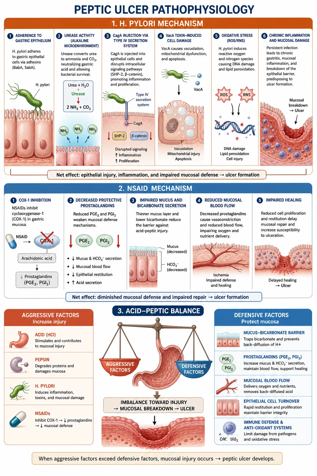
H. pylori pathogenesis:[1]
- H. pylori is a spiral, microaerophilic, urease-producing Gram-negative bacterium that colonises the gastric mucus layer.
- Urease converts urea to ammonia, creating an alkaline microenvironment that protects the bacterium from gastric acid.
- CagA and VacA virulence factors cause epithelial cell damage, inflammation, and ulceration.
- In the antrum, H. pylori causes gastritis with reduced somatostatin (normally inhibits gastrin) → hypergastrinaemia → increased acid secretion → duodenal ulcer (acid overload overwhelms the duodenal mucosal defence).
- In the body/fundus, H. pylori causes atrophic gastritis → reduced acid → gastric ulcer (and increased gastric cancer risk).
NSAID pathogenesis:
- NSAIDs inhibit COX-1 (constitutive, produces protective prostaglandins in gastric mucosa) and COX-2 (inducible, inflammation).
- Loss of prostaglandin E2 and I2 reduces mucus and bicarbonate secretion, reduces mucosal blood flow, and impairs healing — making the mucosa vulnerable to acid injury.
- NSAIDs may also cause direct topical injury to the mucosa (especially acidic NSAIDs like aspirin). [1]
Duodenal vs gastric ulcer key differences (exam table): [1]
| Feature | Duodenal ulcer | Gastric ulcer |
|---|---|---|
| Pain-food relationship | Relieved by food (and antacids) | Worsened by food |
| Timing | 2 to 3 h after meals; nocturnal (wakes at 2 to 3 am) | Soon after eating |
| Weight | Normal or increased | Weight loss |
| H. pylori | 90% | 60 to 80% |
| Malignancy risk | Almost never | 5 to 10% — always biopsy |
| Acid | Hypersecretion | Normal or hyposecretion |
| Location | Duodenal bulb (first part) | Lesser curvature (antrum) |
PUD management — the key drugs
Clinical Presentation
Typical presentation:[1]
- Epigastric pain — burning or gnawing; the hallmark symptom.
- DU: pain 2 to 3 hours after meals, relieved by food, milk, or antacids, often nocturnal (wakes patient at 2 to 3 am — pathognomonic).
- GU: pain worsened by food, nausea, vomiting, weight loss.
- Water brash (excess salivation), heartburn (acid reflux).
- Nausea, bloating, early satiety.
- Periodicity — episodes of pain lasting days to weeks with symptom-free intervals.
Signs — usually normal between episodes. Epigastric tenderness on deep palpation. May have iron-deficiency anaemia (chronic occult blood loss). [1]
Presentation with complications:
- Perforation — sudden, severe, generalised abdominal pain (like being "stabbed"); rigid, board-like abdomen; guarding and rebound; free gas under the diaphragm on erect CXR. Classical: "mummified" patient lying still (any movement worsens pain).
- Haemorrhage — haematemesis (coffee-ground or fresh blood) and/or melaena (black, tarry, foul-smelling stool). May cause hypovolaemic shock.
- Gastric outlet obstruction — non-bilious vomiting of undigested food eaten hours/days earlier; succussion splash on examination (splashing sound on shaking the abdomen); visible gastric peristalsis; hypokalaemic, hypochloraemic metabolic alkalosis (loss of HCl and K+ from vomiting). [1]
Duodenal ulcer
90% H. pylori
- Pain **relieved by food** and antacids
- **Nocturnal** pain (wakes at 2-3 AM)
- **Weight gain**
- Almost **never malignant**
Gastric ulcer
60-80% H. pylori
- Pain **worsened by food**
- **Weight loss**
- **5-10% are malignant** — always biopsy
- Located on lesser curvature
Differential Diagnosis
| Condition | Key distinguishing feature |
|---|---|
| Gastric carcinoma | Weight loss, dysphagia, mass; GU must be biopsied at OGD to exclude |
| GORD | Heartburn, regurgitation; no epigastric pain pattern related to meals; relieved by PPI |
| Non-ulcer dyspepsia | Dyspeptic symptoms but normal OGD; diagnosis of exclusion; very common |
| Biliary colic / acute cholecystitis | Right upper quadrant pain, worse after fatty food; fever; positive Murphy's sign; US shows gallstones |
| Acute pancreatitis | Severe epigastric pain radiating to back; elevated amylase/lipase; CT |
| Myocardial infarction | Chest/epigastric pain, dyspnoea; ECG changes, troponin |
| Mesenteric ischaemia | Pain out of proportion to examination; post-prandial pain (chronic); elevated lactate |
Clinical & Bedside Assessment
History is key — the pain-food relationship, nocturnal symptoms, NSAID/aspirin use, corticosteroids, anticoagulants/antiplatelets, smoking, alcohol, prior ulcer or H. pylori treatment, and red-flag systemic symptoms. Ask specifically about over-the-counter ibuprofen/diclofenac — patients often omit them unless prompted. [1]
Examination:
- Epigastric tenderness — often the only sign between uncomplicated episodes.
- Complications: board-like rigidity and silence of bowel sounds (perforation); tachycardia, postural hypotension, melaena on DRE, haematemesis (bleeding); succussion splash, visible gastric peristalsis, dehydration (GOO).
- Chronic disease: pallor (iron-deficiency anaemia), cachexia (think malignancy if GU phenotype), lymphadenopathy (Virchow node if gastric cancer on differential). [1]
Alarm features requiring urgent OGD (do not treat empirically alone): age over 55 with new dyspepsia, unintentional weight loss, dysphagia/odynophagia, GI bleeding, persistent vomiting, palpable epigastric mass, iron-deficiency anaemia, progressive symptoms on PPI, family history of upper GI cancer. [1]
Worked bedside vignette — "is this still medical?"
A 60-year-old on aspirin for stents develops melaena without free air. This is a bleed pathway (resuscitate → risk score → OGD), not Graham-patch theatre. Conversely, sudden rigidity with free gas is a surgical perforation pathway even if the patient later needs H. pylori eradication after repair. [1]
Investigations
Diagnostic:
- OGD (oesophagogastroduodenoscopy) — the gold standard. Directly visualises the ulcer, assesses location, size, depth, and signs of malignancy (irregular borders, heaped-up edges). Gastric ulcers are ALWAYS biopsied (multiple biopsies from the edge and base) to exclude carcinoma — even if they look benign. Duodenal ulcers are rarely biopsied (almost never malignant).[1]
- H. pylori testing:
- CLO test (rapid urease test) — biopsy placed in urea-containing medium; colour change indicates urease (H. pylori). Done at OGD. Highly sensitive and specific.
- Urea breath test (13C or 14C) — non-invasive; patient ingests labelled urea; if H. pylori urease is present, labelled CO2 is exhaled. Used for confirmation of eradication (4 weeks after completing therapy). False-negative if on PPI or antibiotics.
- Stool antigen test — non-invasive; detects H. pylori antigen in stool. Also used for eradication confirmation.
- Serology — detects anti-H. pylori antibodies; cannot distinguish current vs past infection; not useful for eradication confirmation.
- Histology — biopsy stained (Giemsa or immunohistochemistry) shows H. pylori; gold standard but slower.
Risk scores for bleeding (reproduce purpose, not every rare variant):
- Glasgow-Blatchford score (pre-endoscopy): urea, haemoglobin, systolic BP, pulse, melaena, syncope, hepatic disease, cardiac failure — predicts need for intervention; score 0 may support outpatient pathways in selected units.[3]
- Rockall score (post-endoscopy): age, shock, comorbidity, diagnosis, major SRH — predicts mortality/rebleed risk.[3]
For complications:
- Erect CXR — free gas under the diaphragm (perforation); sensitivity approximately 75% (some perforations sealed by the omentum show no free gas). If clinical perforation but CXR negative → CT abdomen (more sensitive for free air/fluid).
- CT abdomen — for perforation (free gas/fluid) or exclusion of other pathology; do not delay theatre for CT when peritonitis is clear and patient is unstable.
- FBC, U&Es, group & save/crossmatch, coagulation, lactate, ABG as indicated — anaemia (bleeding), hypokalaemia/hypochloraemia/alkalosis (GOO from vomiting), raised urea out of proportion to creatinine (UGI bleed digestion of blood). [1]
Screening for Zollinger-Ellison syndrome — if multiple, recurrent, jejunal, or atypical ulcers, or ulcers with diarrhoea: fasting serum gastrin (markedly elevated) and gastric pH (hyperchlorhydria, pH under 2). Interpreting gastrin on PPI is complex — involve gastroenterology; seek MEN1 features (hyperparathyroidism, pituitary). [1]
Management — Resuscitation
Perforated peptic ulcer (emergency):
- ABC, high-flow oxygen if hypoxic, two large-bore IV cannulae, crystalloid boluses titrated to perfusion, NG tube (decompress), urinary catheter, serial lactate.
- Blood cultures if septic; broad-spectrum IV antibiotics — e.g. co-amoxiclav 1.2 g IV every 8 h + metronidazole 500 mg IV every 8 h, or piperacillin-tazobactam 4.5 g IV every 8 h (local resistance patterns decide).
- IV PPI: pantoprazole (or omeprazole) 80 mg IV bolus, then continuum into post-op high-dose cover.
- Analgesia: IV morphine 2.5–5 mg aliquots (or fentanyl) titrated; antiemetic as needed.
- Urgent surgical review for Graham omental patch (± laparoscopic) after rapid optimisation — do not wait for "perfect" labs if peritonitis is established.[1][2]
Acute upper GI bleeding:
- ABC; two large-bore IV cannulae; crossmatch 2 to 4 units (activate major haemorrhage protocol if exsanguinating).
- Fluid resuscitation with crystalloid while blood arrives; restrictive transfusion target Hb about 70 g/L (about 80 g/L if significant cardiovascular disease).[3]
- Correct coagulopathy — warfarin: vitamin K ± PCC per protocol; DOAC-specific pathways; platelets if count critically low or on dual antiplatelet with life-threatening bleed (cardiology input before stopping essential stents meds).
- IV PPI infusion: 80 mg bolus then 8 mg/h for 72 h after endoscopic haemostasis of high-risk ulcers (Lau regimen).[4]
- Urgent OGD within 24 h for most admitted bleeds; sooner if haemodynamically unstable after resuscitation; dual endoscopic therapy for high-risk Forrest stigmata; IR or surgery if endoscopic failure.
- Stop the offending NSAID; plan H. pylori test-and-treat; gastroprotection if antiplatelet must continue.
Gastric outlet obstruction:
- NG tube (large-bore, e.g., 16 to 18 Fr) for decompression; saline lavage to clear retained food before OGD.
- IV 0.9% sodium chloride with potassium chloride to correct hypokalaemic hypochloraemic metabolic alkalosis (paradoxical aciduria is a classic viva point — volume/Cl depletion maintains alkalosis until saline given).
- IV PPI (e.g. pantoprazole 40 mg IV BD until oral route reliable); H. pylori eradication when diagnosed; OGD with biopsy to exclude malignant stricture. [1]
Management — Definitive & Stepwise
Medical (first-line for uncomplicated PUD)
H. pylori eradication:[1]
- Triple therapy (first-line): PPI (standard dose BD) + clarithromycin 500 mg BD + amoxicillin 1 g BD — for 14 days (7 to 10 days also used but 14 is preferred for eradication success).
- Penicillin-allergic alternative: PPI + clarithromycin + metronidazole 400 mg BD.
- Bismuth quadruple therapy (second-line or high-resistance areas): PPI + bismuth subsalicylate/subcitrate + tetracycline + metronidazole — 14 days.
- Confirmation of eradication: urea breath test or stool antigen at least 4 weeks after completing therapy (PPI stopped for at least 2 weeks before testing).
- Eradication success rate: 80 to 90% (falling due to clarithromycin resistance, especially in India).[1]
Acid suppression (healing):
- PPI (omeprazole 20 mg OD, esomeprazole 20 mg OD, or equivalent) for 4 to 8 weeks after eradication to heal the ulcer.
- H2-receptor antagonists (ranitidine, famotidine) are inferior to PPIs for healing; used if PPIs contraindicated.
- Stop NSAIDs (or switch to COX-2 selective inhibitor with PPI cover if NSAIDs essential).
- Gastric ulcer follow-up: repeat OGD at 8 to 12 weeks to confirm healing and exclude malignancy (a non-healing gastric ulcer must be treated as cancer until proven otherwise).[1]
NSAID ulcer prevention:
- Co-prescribe PPI with NSAIDs in high-risk patients (age over 60, previous PUD, concurrent corticosteroid/anticoagulant).
- Use COX-2 selective inhibitor (celecoxib) instead of non-selective NSAID in patients at high GI risk (but beware cardiovascular risk).
- Consider H. pylori eradication before starting long-term NSAIDs. [1]
Surgical (for complications)
Perforated peptic ulcer:[1][2]
- Graham omental patch (omentopexy) — the standard emergency procedure for a perforated DU. The perforation is closed by suturing a piece of omentum (Graham patch) over the hole. Can be done laparoscopically or open.
- Perforated GU: if small and clean — patch repair; if large or malignant-looking — biopsy (frozen section) and consider resection (wedge excision or partial gastrectomy) if malignancy suspected.
- Post-op: IV PPI, H. pylori eradication (if positive), analgesia, DVT prophylaxis.
Bleeding peptic ulcer (surgical management when endoscopy fails):
- Indications for surgery after failed endoscopic haemostasis: persistent bleeding despite 2 endoscopic attempts, rebleeding, massive haemorrhage with haemodynamic instability (unable to resuscitate).[1]
- DU: under-running (oversewing) the bleeding vessel (usually the gastroduodenal artery at the posterior duodenal bulb) with non-absorbable sutures. May require duodenotomy to access.
- GU: under-running the bleeding vessel or wedge excision of the ulcer.
- Alternative: interventional radiology — angiographic embolisation of the bleeding vessel (e.g., GDA); increasingly used as an alternative to surgery in unstable patients.
Gastric outlet obstruction:
- Endoscopic balloon dilatation — for fibrotic strictures from chronic DU (may need repeat dilatations).
- Surgical: Gastrojejunostomy (bypass the obstruction) ± truncal vagotomy (reduce acid). Pyloplasty for pyloric stenosis. Rarely needed now with endoscopic techniques and H. pylori eradication.[2]
Elective ulcer surgery (now rare):
- Before H. pylori eradication, elective surgery included truncal vagotomy + antrectomy (Billroth I or II), highly selective vagotomy (HSV), and total gastrectomy. These are now historical for uncomplicated PUD but may appear in exams.[2]
Specific Subtypes & Scenarios
H. pylori eradication regimens in detail
The choice of regimen is dictated by local antibiotic resistance patterns, prior exposure, and penicillin allergy. Maastricht VI / Toronto 2016 recommend 14-day durations wherever feasible because per-day eradication efficacy translates into higher cumulative clearance of the organism.[1]
First-line: clarithromycin-based triple therapy (PPI–AC)
Standard regimen, suitable when local clarithromycin resistance is under 15%:
- PPI (standard dose BD) — omeprazole 20 mg BD, esomeprazole 20 mg BD, pantoprazole 40 mg BD, lansoprazole 30 mg BD, or rabeprazole 20 mg BD. Higher-dose PPI (BD) is preferred over OD to maintain intragastric pH over 4 throughout the day, which improves antibiotic bioavailability and bacterial susceptibility.
- Amoxicillin 1 g BD — penicillin-class β-lactam; resistance remains rare globally (under 5% in most regions). Avoid in confirmed penicillin allergy.
- Clarithromycin 500 mg BD — macrolide; resistance is rising (over 20% in many parts of India, China, and southern Europe) and is the principal driver of falling triple therapy efficacy.
- Duration: 14 days (Maastricht VI); 7 to 10 days is acceptable only in low-resistance settings.
- Reported eradication: 80 to 85% in low-resistance regions; under 70% where clarithromycin resistance exceeds 15%. [1]
Penicillin-allergic substitute: PPI + clarithromycin 500 mg BD + metronidazole 400 mg BD for 14 days. Metronidazole resistance (over 30% in many regions) further limits efficacy, so a post-treatment urea breath test is mandatory. [1]
Bismuth quadruple therapy (BQT)
Used as first-line where clarithromycin resistance exceeds 15% (most of India and East Asia), or as second-line after failed triple therapy:
- PPI (BD) + bismuth subsalicylate 300 mg QDS (or bismuth subcitrate 120 mg QDS) + tetracycline 500 mg QDS + metronidazole 500 mg TDS for 14 days.
- Bismuth disrupts the bacterial cell wall and synergises with tetracycline against H. pylori; metronidazole is active even against resistant strains at the higher 500 mg TDS dose.
- Reported eradication: 85 to 90% — outperforms triple therapy in high-resistance regions.
- Tolerability issues: frequent tablets (12 to 14 per day), GI upset, darkening of tongue and stool (bismuth), photosensitivity (tetracycline), and a metallic taste (metronidazole). Counsel patients before initiation.
- May be pre-packaged (Pylera) — three-in-one capsule containing bismuth subcitrate, metronidazole, and tetracycline (14 capsules per day) plus a PPI BD — improves compliance. [1]
Levofloxacin rescue / sequential / concomitant therapy
Used after two failed eradication cycles:
- Levofloxacin-based triple (third-line): PPI BD + amoxicillin 1 g BD + levofloxacin 500 mg OD for 10 to 14 days. Levofloxacin is a fluoroquinolone; reserve to avoid emerging fluoroquinolone resistance. Test for fluoroquinolone susceptibility if culture available.
- Concomitant (non-bismuth quadruple): PPI BD + amoxicillin 1 g BD + clarithromycin 500 mg BD + metronidazole 400 mg BD for 10 to 14 days — an alternative to bismuth quadruple in regions where both clarithromycin and metronidazole resistance are moderate.
- Sequential therapy (less commonly used now): 5 days of PPI + amoxicillin, then 5 days of PPI + clarithromycin + metronidazole; 14-day “hybrid” or “reverse hybrid” sequential regimens achieve about 90% eradication in trials. [1]
Eradication verification and troubleshooting
Always confirm eradication with a urea breath test or stool antigen test at least 4 weeks after completing therapy and at least 2 weeks after stopping PPIs (PPI suppression causes false-negatives). Serology is unsuitable for confirmation because IgG antibodies persist for months to years after eradication. If eradication fails after second-line therapy, endoscopic culture and antibiotic-susceptibility testing should be performed to tailor the third-line regimen.[1]
Zollinger-Ellison syndrome (gastrinoma) — in detail
Zollinger-Ellison syndrome (ZES) is the constellation of severe acid hypersecretion caused by a gastrin-secreting neuroendocrine tumour (gastrinoma), producing multiple, recurrent, atypical peptic ulcers and often chronic secretory diarrhoea. It accounts for under 1% of peptic ulcers but is a critical reversible cause of refractory PUD.[1]
Pathophysiology: Gastrinomas are most commonly located in the “gastrinoma triangle” — the junction of the cystic and common bile ducts, the second/third parts of the duodenum, and the neck/body of the pancreas. Roughly 70% are malignant with metastases (commonly to liver and regional lymph nodes) at presentation; the rest are benign adenomas. Secreted gastrin drives parietal cell hyperplasia and unrestrained acid output, producing ulcers that are unusually multiple, distal, and refractory to standard therapy. [1]
Clinical clues:
- Multiple ulcers in unusual locations (second part of duodenum, jejunum).
- PUD refractory to standard 8 to 12 weeks of therapy, or recurrent ulceration within months of completing eradication.
- Prominent secretory diarrhoea (gastric acid inactivates pancreatic lipase → steatorrhoea) — present in up to 50% of cases.
- Markedly thickened gastric rugae on OGD (gastrin-driven parietal cell hyperplasia — Menetrier-like appearance).
- Personal or family history suggestive of MEN1 (parathyroid hyperplasia → hypercalcaemia; pituitary adenoma → galactorrhoea, acromegaly). [1]
Diagnostic workup:
- Fasting serum gastrin — usually over 1000 pg/mL in ZES (normal under 100), with concurrent gastric pH under 2 (a high gastrin in achlorhydria is not diagnostic). Stop PPIs for 1 to 2 weeks before the test where safe because they elevate gastrin.
- Secretin stimulation test — the gold standard where fasting gastrin is borderline (100 to 1000 pg/mL). IV secretin paradoxically raises gastrin in gastrinoma patients (rather than suppressing it as in normal physiology); a rise of over 120 pg/mL is diagnostic.
- Localisation: CT/MRI pancreas, somatostatin receptor scintigraphy (Octreoscan) or Ga-68 DOTATATE PET-CT (most sensitive), EUS of the duodenal wall and pancreas for small primaries, and intra-operative palpation/ultrasound at surgical exploration.
- MEN1 screening: serum calcium, PTH, prolactin, IGF-1 in any young ZES patient (under 40) or with suggestive family history.[1]
Management:
- High-dose PPI (omeprazole or esomeprazole 40 to 80 mg/day, sometimes split BD) to control acid and allow ulcer healing — usually lifelong in non-surgical or metastatic disease.
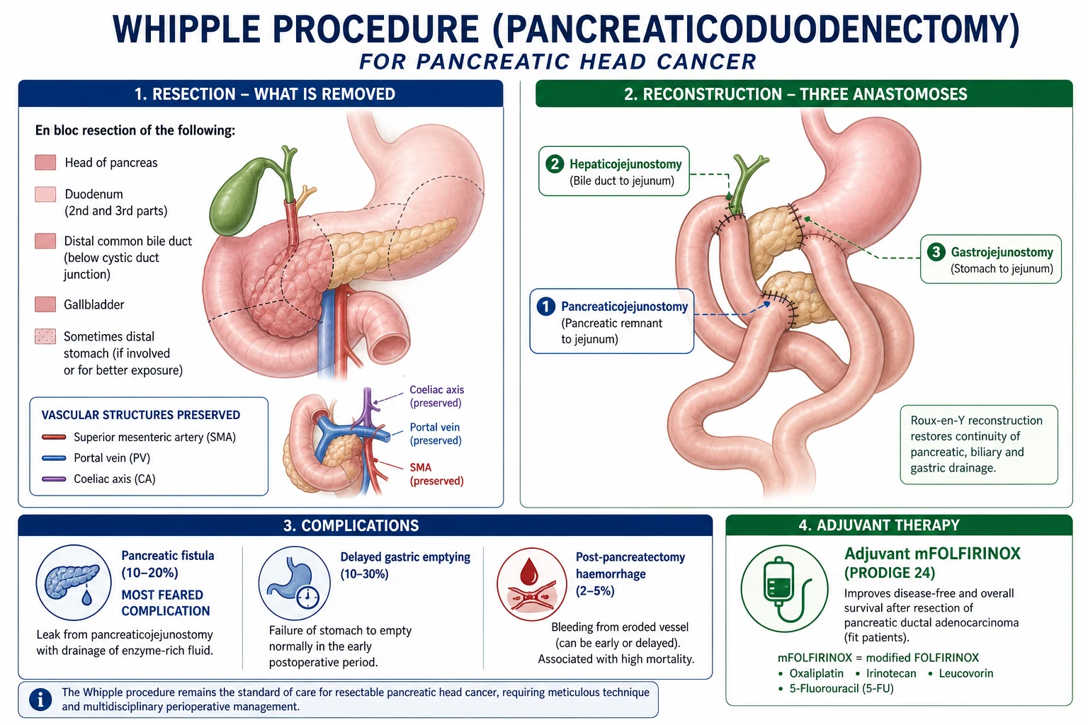
- Surgical excision of the gastrinoma when localised and non-metastatic — offers the only chance of cure. Whipple's procedure for pancreatic head lesions; duodenotomy with resection for duodenal primaries.
- Liver metastasectomy, debulking, and ablative therapies for metastatic disease, combined with long-acting somatostatin analogues (octreotide LAR, lanreotide) for symptomatic control and tumour stabilisation.
- Surveillance: annual fasting gastrin, chromogranin A, imaging, and gastric pH to titrate PPI dose. [1]
NSAID-induced ulcers — prevention and management
NSAIDs remain the second commonest cause of PUD worldwide and the leading cause of ulcer bleeding in older adults. Damage is mediated by both topical mucosal injury and systemic loss of COX-1–derived prostaglandins (PGE2, PGI2) that maintain mucus, bicarbonate, and mucosal blood flow.[1]
Risk stratification (CV risk–balanced):
- High GI risk — age over 60, previous peptic ulcer, H. pylori co-infection, concurrent corticosteroid, anticoagulant (warfarin, DOAC) or antiplatelet (aspirin, clopidogrel) therapy, or high-dose/multiple NSAIDs.
- High CV risk — established coronary, cerebrovascular, or peripheral arterial disease; diabetes with end-organ damage; chronic kidney disease.
- The H. pylori + NSAID interaction is synergistic (≈3 to 4× risk of bleeding); eradication before starting chronic NSAID therapy is recommended where feasible. [1]
Prevention strategy:
- Use the lowest effective NSAID dose, for the shortest duration.
- Co-prescribe a PPI (omeprazole 20 mg OD) in any patient with GI risk factors.
- Substitute a COX-2 selective inhibitor (celecoxib, etoricoxib) in high GI risk — these agents spare COX-1 and the gastric prostaglandin pathway, reducing the risk of ulceration by ~50%; however, COX-2 inhibition removes vascular PGI2 and increases cardiovascular risk, so they are contraindicated after CABG and used cautiously in any high-CV patient.
- In patients requiring both an NSAID and low-dose aspirin, the lowest-dose celecoxib plus PPI or a non-selective NSAID plus PPI may be considered — never combine two NSAIDs.
- H. pylori eradication before starting long-term NSAIDs in endemic populations.[1]
Management of an established NSAID ulcer:
- Stop the NSAID wherever possible. If NSAID continuation is essential (e.g., refractory arthropathy), switch to a COX-2 inhibitor and add a PPI; resolution of ulceration is the goal.
- PPI for 8 weeks (omeprazole 20 mg OD) is the standard healing regimen, and prophylactic doses continued thereafter.
- Eradicate H. pylori if positive — improves healing and reduces rebleeding independently of NSAID withdrawal.
- Reassess antithrombotic therapy with cardiology before stopping aspirin or clopidogrel in secondary-prevention patients — restart PPI cover and resume antiplatelet as soon as safely possible. [1]
Refractory peptic ulcer — diagnostic algorithm
An ulcer that has failed to heal after 8 to 12 weeks of optimised therapy, or recurs within 6 months, is refractory and must be investigated deliberately rather than re-treated empirically. Common aetiologies are listed below in approximate order of frequency.[1][1]
Causes to exclude:
- Ongoing NSAID or aspirin use (including over-the-counter preparations, topical diclofenac, low-dose cardioprotective aspirin) — history taking must be specific.
- Persistent H. pylori infection — false-negative initial test, antibiotic resistance, or non-compliance with the eradication course; retest with urea breath test or stool antigen and consider culture after the second failure.
- Gastric adenocarcinoma or lymphoma — every non-healing gastric ulcer needs repeat biopsy; duodenal ulcers are almost never malignant but must also be re-biopsied if atypical.
- Zollinger-Ellison syndrome — multiple, distal, or recurrent ulcers; check fasting gastrin and secretin test as outlined above.
- Crohn's disease, tuberculosis, cytomegalovirus (CMV) or Helicobacter heilmannii infection — suggested by atypical ulcer morphology, immunocompromise, or systemic features; biopsy with special stains and PCR.
- Idiopathic hypersecretory state — rare; basal acid output over 15 mmol/h in the absence of gastrinoma or H. pylori.
- Non-adherence to PPI, smoking, ongoing psychological stress, or concomitant medications (bisphosphonates, SSRIs that potentiate bleeding). [1]
Stepwise work-up:
- Re-take history focused on NSAID use (prescribed, OTC, topical), compliance, smoking, family history of MEN1.
- Repeat OGD with multiple biopsies from ulcer edge and base + CLO test / histology + CMV / TB immunohistochemistry if atypical.
- Fasting serum gastrin and gastric pH — to exclude ZES (off PPI for 1 to 2 weeks where safe).
- H. pylori retest (urea breath / stool antigen); consider endoscopy with culture if two prior eradications have failed.
- Cross-sectional imaging (CT pancreas protocol) + somatostatin receptor imaging if ZES suspected.
- Treat the underlying cause: PPI high-dose, H. pylori rescue regimen, surgical resection for gastrinoma, chemotherapy/radiotherapy for malignancy, immunomodulation for Crohn's. [1]
Complications & Pitfalls
Complications of PUD (four major — surgical):[1]
- Perforation — 2 to 10% of ulcers; acute peritonitis; emergency surgery.
- Haemorrhage — 15 to 20%; the most common cause of upper GI bleeding; managed endoscopically.
- Gastric outlet obstruction — 2%; from scarring/oedema of chronic DU or prepyloric GU.
- Penetration (not perforation) — posterior DU erodes into the pancreas (causing pancreatitis) or the liver; causes intractable pain radiating to the back.
Complications of ulcer surgery (historical, but exam-yield):
- Dumping syndrome — early (15 to 30 min after eating: vasomotor — dizziness, sweating, palpitations, nausea, from rapid gastric emptying causing fluid shift) and late (1 to 3 h: hypoglycaemia from exaggerated insulin response). Managed with small, frequent, low-carbohydrate meals; octreotide for severe cases.
- Post-vagotomy diarrhoea — up to 20% after truncal vagotomy; bile-salt malabsorption.
- Afferent loop syndrome — after Billroth II; obstruction of the afferent limb causing bilious vomiting that relieves pain.
- Alkaline reflux gastritis — bile reflux into the stomach remnant; causes pain, nausea, bilious vomiting.
- Marginal (anastomotic) ulcer — ulcer at the gastrojejunal anastomosis; may perforate or bleed.
- Anaemia — iron deficiency (reduced acid impairs iron absorption), B12 deficiency (loss of intrinsic factor after gastrectomy).
- Osteoporosis — reduced calcium absorption after gastrectomy.
- Stump carcinoma — gastric cancer developing in the gastric remnant years after partial gastrectomy (alkaline reflux and bacterial overgrowth are carcinogenic). [1]
Classic pitfalls:
- Not biopsying a gastric ulcer — missing gastric carcinoma (5 to 10% of GUs are cancer).
- Not confirming H. pylori eradication — recurrent ulceration.
- Not doing a repeat OGD to confirm gastric ulcer healing — missing non-healing malignancy.
- Not testing for H. pylori before starting long-term PPI — missing curable disease.
- Assuming a perforated ulcer is appendicitis — epigastric pain with free gas vs RLQ pain without gas.
- Forgetting Zollinger-Ellison in patients with multiple, recurrent, or atypical ulcers. [1]
Prognosis & Disposition
Uncomplicated PUD — excellent prognosis with H. pylori eradication + PPI. Healing rate over 90% with appropriate therapy. Recurrence is under 10% after successful H. pylori eradication (vs 50 to 60% recurrence without eradication).[1]
Complicated PUD — prognosis depends on the complication:
- Perforation: mortality 5 to 10% (higher in elderly, delayed presentation, comorbidity).
- Haemorrhage: mortality 5 to 10%; rebleeding risk 10 to 20% (higher if visible vessel or clot on OGD).
- GOO: good with endoscopic dilatation or surgery; may recur. [1]
Follow-up: confirm H. pylori eradication (urea breath test at 4 weeks). Repeat OGD at 8 to 12 weeks for gastric ulcers to confirm healing and exclude malignancy. Duodenal ulcers do not require follow-up OGD (not malignant). [1]
Special Populations
- NSAID users — co-prescribe PPI with NSAIDs for high-risk patients (age over 60, prior ulcer, corticosteroids, anticoagulants/antiplatelets, high-dose/long-term NSAID). Test-and-treat H. pylori before long-term NSAID when practical. Prefer lowest effective NSAID dose; consider COX-2 selective agent plus PPI in very high GI risk if CV risk acceptable. Never restart an NSAID after complicated ulcer without documented gastroprotection plan and indication review.[1][3]
- Elderly — higher perforation and bleed mortality; NSAID/aspirin ulcers predominate; pain may be muted; lower threshold for OGD and admission; careful PPI long-term risk–benefit (fracture/C. diff signals debated — still use when indicated after bleed/perforation).
- Anticoagulated / dual antiplatelet patients — early gastroenterology + cardiology input after bleed; do not casually stop stents’ essential antiplatelets without plan; reverse life-threatening coagulopathy per protocol; early OGD after resus.[3]
- Pregnancy — PUD uncommon (progesterone reduces acid). Prefer antacids, H2-blockers, and PPIs with the best pregnancy safety data (omeprazole/pantoprazole commonly used). Avoid elective surgery; operate for free perforation/uncontrolled bleed. H. pylori eradication timing individualised (prefer postpartum if mild symptoms).
- ICU / burns / head injury — stress-related mucosal disease: prophylaxis for mechanical ventilation over 48 h, coagulopathy, shock, major burns (Curling ulcer), intracranial injury (Cushing ulcer). Prefer IV/PO PPI (e.g. pantoprazole 40 mg IV daily) or H2-blocker per unit protocol; early enteral nutrition protects mucosa.[1]
- Children — duodenal disease more than gastric; H. pylori family clustering; weight-based PPI and antibiotic dosing; exclude other causes of abdominal pain; paediatric gastroenterology for refractory disease.
- CKD / transplant / immunosuppressed — higher bleed risk on antiplatelets/steroids; drug interactions with eradication regimens; lower threshold to scope anaemia or melaena.
Worked metabolic vignette — GOO alkalosis (exam favourite)
A patient with weeks of non-bilious vomiting of old food has Na 130, K 2.6, Cl 80, HCO3 38, urine paradoxically acidic. Mechanism: loss of HCl → hypochloraemic metabolic alkalosis; volume depletion + secondary hyperaldosteronism → renal Na retention with K and H secretion (paradoxical aciduria) until chloride is restored. Treatment: large-bore NG decompression, IV 0.9% sodium chloride with potassium chloride replacement, PPI, then OGD (biopsy any GU/stricture) and definitive dilatation or surgery once optimised. [1]
Evidence, Guidelines & Regional Differences
Maastricht VI Consensus (2022):[1] recommends:
- 14-day triple therapy (PPI + clarithromycin + amoxicillin) as first-line where clarithromycin resistance is under 15%.
- Bismuth quadruple therapy where clarithromycin resistance is over 15% (which includes much of India).
- Confirmation of eradication at least 4 weeks after completing therapy.
- Culture and sensitivity testing after two failed eradication attempts.
Regional differences:
- India — very high H. pylori prevalence (over 50% in adults); high clarithromycin resistance (over 20%) — bismuth quadruple therapy may be preferred; concurrent amoxicillin resistance rising; PUD is common and often presents with complications (perforation, bleeding) due to delayed medical care.
- West — declining H. pylori prevalence (under 30%); NSAID-induced ulcers proportionally more common; PPI prophylaxis widely used. [1]
Historical note: The shift from surgery to medical management of PUD is one of the great triumphs of modern gastroenterology. Before H. pylori eradication, PUD was a chronic, recurrent condition treated with major surgery (vagotomy, antrectomy, gastrectomy). Today, uncomplicated PUD is cured by eradication therapy in the majority of patients.[2]
Perforated peptic ulcer — the clinical picture
BOARD
rigid abdomen on palpation
severe pain, like being stabbed
free gas under diaphragm on erect CXR
Graham omental patch (omentopexy)
H. pylori eradication post-op
Exam Pearls
- DU: pain relieved by food, nocturnal, weight gain. GU: pain worsened by food, weight loss. H. pylori 90% (DU) and 60 to 80% (GU).[1]
- Gastric ulcers are ALWAYS biopsied (5 to 10% are cancer). Duodenal ulcers are almost never malignant.[1]
- H. pylori triple therapy: PPI + clarithromycin + amoxicillin for 14 days. Confirm eradication with urea breath test at 4 weeks.[1]
- Perforated DU: sudden severe epigastric pain, rigid abdomen, free gas under diaphragm on erect CXR. Graham omental patch repair.[1]
- Posterior DU penetrates the pancreas (back pain) or erodes the gastroduodenal artery (massive GI bleed).[1]
- Gastric outlet obstruction: non-bilious vomiting of old food, succussion splash, hypokalaemic hypochloraemic metabolic alkalosis.[1]
- Curling's ulcer = burns. Cushing's ulcer = head injury. Zollinger-Ellison = gastrinoma, multiple ulcers, high fasting gastrin.[1]
- Dumping syndrome: early (15 to 30 min, vasomotor) and late (1 to 3 h, hypoglycaemia). After gastrectomy/vagotomy.[2]
- Billroth I = gastroduodenostomy (after distal gastrectomy). Billroth II = gastrojejunostomy (Roux-en-Y).[2]
- NSAIDs cause ulcers by inhibiting COX-1 (reducing protective prostaglandins). Co-prescribe PPI. COX-2 inhibitors have lower GI risk.[1]
Operative and endoscopic detail (exam detail)
Anatomy examiners love:
- Anterior DU → free perforation into peritoneum → free gas under diaphragm.
- Posterior DU → penetration into pancreas (back pain) or erosion of gastroduodenal artery (GDA) → massive upper GI bleed.
- Lesser-curve GU → risk of malignancy higher than DU; always biopsy.
- Prepyloric / pyloric channel ulcers → scarring → gastric outlet obstruction (GOO). [1]
Bleeding ulcer — endoscopic haemostasis:
- Resuscitate first (see management); restrictive transfusion target Hb about 70–80 g/L in stable patients without ischaemic heart disease.[3]
- OGD for diagnosis and therapy; high-risk stigmata (active spurting/oozing, non-bleeding visible vessel, adherent clot) need therapy.
- Dual modality: adrenaline injection (1:10,000, aliquots) plus thermal contact or mechanical clips — monotherapy with adrenaline alone is inferior.
- After successful endoscopic haemostasis: IV PPI bolus 80 mg then 8 mg/h for 72 h (pantoprazole or omeprazole equivalent).[4]
- Failed endoscopic control → IR embolisation (GDA) or surgery (under-running of bleeding vessel ± ulcer excision / gastrectomy in selected cases).
Perforated ulcer — Graham (omental) patch:
- ABC, NBM, NG tube, IV fluids, IV antibiotics (e.g. co-amoxiclav 1.2 g TDS + metronidazole 500 mg TDS, or piperacillin-tazobactam 4.5 g TDS), IV PPI 80 mg bolus.
- Laparoscopic or open washout; identify perforation (usually anterior DU).
- Graham patch: tongue of omentum secured over perforation with interrupted sutures (classical technique places sutures then ties over omentum).
- Thorough lavage; drains selective; post-op PPI + H. pylori testing/eradication.
- Biopsy gastric ulcers even when perforated if safe; consider malignancy pathway for atypical GU perforations. [1]
GOO from chronic DU scarring:
- Resuscitate, correct hypokalaemic hypochloraemic metabolic alkalosis (paradoxical aciduria teaching point).
- NG decompression, IV PPI, H. pylori eradication.
- Endoscopic dilatation for selected benign strictures; definitive surgery historically truncal vagotomy + gastrojejunostomy or antrectomy variants — now individualised; exclude malignant GOO. [1]
Historical acid-reducing operations (still examined):
- Truncal vagotomy + drainage (pyloroplasty/GJ).
- Highly selective (parietal cell) vagotomy — denervates acid-secreting corpus/fundus, preserves antral innervation (less dumping).
- Billroth I / II and Roux-en-Y reconstructions after partial gastrectomy — know dumping, bile reflux, B12 deficiency, stump carcinoma risk after Billroth II. [1]
Forrest classification of bleeding ulcers (reproduce)
| Forrest | Stigmata | Rebleed risk (order of magnitude) | Action |
|---|---|---|---|
| Ia | Spurting | Highest | Dual endoscopic therapy |
| Ib | Oozing | High | Dual therapy |
| IIa | Non-bleeding visible vessel | High | Dual therapy |
| IIb | Adherent clot | Moderate–high | Consider clot removal + treat base |
| IIc | Flat pigmented spot | Low | Medical |
| III | Clean base | Lowest | Medical / early discharge pathway if stable |
Worked clinical stems (answer these without another book)
Stem A — Perforation. 45 M sudden epigastric pain, board-like rigidity, free gas under right hemidiaphragm.
Perforated peptic ulcer. ABC, IV access, fluids, NG, co-amoxiclav + metronidazole (or local), IV PPI, urgent surgery for Graham patch, post-op H. pylori eradication. [1]
Stem B — Bleeding posterior DU. Melaena, HR 110, BP 95/60, Hb 78, on ibuprofen.
Ulcer bleed / GDA territory. Two large-bore IV, crossmatch, restrictive transfusion, IV PPI 80 mg then 8 mg/h, urgent OGD dual therapy; stop NSAID; eradicate H. pylori. [1]
Stem C — GOO. Recurrent vomiting of old food, succussion splash, metabolic alkalosis.
Pyloric stenosis from chronic DU (or malignancy). Correct electrolytes, NG, PPI, OGD with biopsy, dilatation or surgery. [1]
Stem D — Non-healing GU. GU still present at 12 weeks on PPI, biopsies "chronic gastritis" only once.
Re-biopsy extensively; exclude cancer, H. pylori, surreptitious NSAID, hypersecretory states; do not label "benign" without healing + adequate histology. [1]
Stem E — ZES suspicion. Multiple DU beyond D1, diarrhoea, recurrent despite PPI.
Measure fasting gastrin (off PPI if safe / with caution), gastric pH, seek gastrinoma (MEN1); high-dose PPI; oncology/endocrine pathway. [1]
Stem F — H. pylori after antibiotics. Completed triple therapy, still dyspeptic.
Urea breath test ≥4 weeks after antibiotics and ≥2 weeks off PPI; if positive → bismuth quadruple or susceptibility-guided therapy. [1]
OSCE / short-case performance script
- ABCDE for acute abdomen or GI bleed; DRE for melaena.
- Focused history: NSAIDs, steroids, anticoagulants, alcohol, prior ulcer, H. pylori treatment.
- State Glasgow-Blatchford (pre-endoscopy intervention risk) vs Rockall (post-endoscopy mortality risk) roles.
- Verbalise exact PPI infusion and triple therapy doses.
- Consent themes: perforation repair, conversion, rebleed, need for IR/surgery if endoscopy fails.
- Safety-net: return if melaena, syncope, severe pain; GU healing check OGD. [1]
Extended viva bank (model outlines)
- DU vs GU pain patterns and malignancy risk.
- Why biopsy every GU?
- GDA anatomy and posterior DU bleed.
- Lau PPI infusion evidence after endoscopic therapy.[4]
- Triple vs bismuth quadruple and clarithromycin resistance (India).
- Graham patch technique and when to consider definitive ulcer surgery.
- Dumping — early vs late.
- H. pylori tests — which for diagnosis vs test of cure.
- Stress ulcers (Curling/Cushing) in burns and head injury.
- MEN1 / gastrinoma work-up outline.
Common exam traps (fail patterns)
- Single-agent adrenaline for high-risk bleed.
- Liberal transfusion without target.
- Testing H. pylori for cure while still on PPI.
- Calling free gas "medical" and delaying theatre.
- Forgetting NSAID cessation and gastroprotection plan.
- Missing alkalosis correction in GOO. [1]
Self-check coverage map
| Examiner dimension | Covered? |
|---|---|
| Definition/classification (Johnson GU, DU/GU) | Yes |
| Epidemiology & risk (HP, NSAID) | Yes |
| Pathophysiology (urease, COX, defence) | Yes |
| Presentation + atypical | Yes |
| Differentials with discriminators | Yes |
| Bedside / scores (Blatchford, Rockall, Forrest) | Yes |
| Investigations | Yes |
| Resuscitation with doses | Yes |
| Medical eradication regimens | Yes |
| Surgery/endoscopy for complications | Yes |
| ZES, refractory ulcer | Yes |
| Special populations | Yes |
| Evidence & regional (resistance) | Yes |
| Exam pearls | Yes |
References
- [1]Kempenich JW, Sirinek KR Acid Peptic Disease Surg Clin North Am, 2018.PMID 30243454
- [2]Zittel TT, Jehle EC, Becker HD Surgical management of peptic ulcer disease today--indication, technique and outcome Langenbecks Arch Surg, 2000.PMID 10796046
- [3]Laine L, Barkun AN, Saltzman JR, et al. ACG Clinical Guideline: Upper Gastrointestinal and Ulcer Bleeding Am J Gastroenterol, 2021.PMID 33929377
- [4]Lau JY, Sung J, Hill C, et al. Effect of intravenous omeprazole on recurrent bleeding after endoscopic treatment of bleeding peptic ulcers N Engl J Med, 2000.PMID 10922420