General Surgery · General Surgery
Perianal Abscess and Anal Fistula
Also known as Perianal abscess · Anal fistula · Fistula-in-ano · Perianal sepsis · Cryptoglandular abscess
Perianal abscess and anal fistula (fistula-in-ano) are two stages of a single cryptoglandular disease process. Obstruction of an anal gland duct at the dentate line produces an acute pus collection (abscess) which, on drainage, may leave a permanent epithelialised tract (fistula) in 30 to 50 percent of patients. Abscess presents with severe throbbing perianal pain, fever, and a tender fluctuant mass; fistula presents with recurrent discharge and recurrent abscesses. Goodsall's rule predicts the internal opening. Park's classification grades fistulas by sphincter involvement. Abscess is treated by incision and drainage; low fistula by fistulotomy; high or complex fistula by a loose seton, LIFT, or advancement flap to preserve the sphincter. Crohn's perianal fistula demands anti-TNF and sphincter-conserving surgery.
On this page & tools
Your progress
Saved locally on this device.
Exam tags
Red flags
Overview & Definition
Perianal abscess and anal fistula (fistula-in-ano) are not two diseases but two stages of a single cryptoglandular infection.[1][1]
- Perianal abscess — the acute stage: a localised collection of pus in the perianal or perirectal spaces, produced by obstruction and then infection of an anal gland at the dentate line.
- Anal fistula — the chronic stage: a permanent abnormal tract lined by granulation and epithelium, running between an internal opening at the anal canal (almost always at the dentate line) and an external opening on the perianal skin. It forms when an abscess drains — spontaneously or at surgery — but the connecting tract fails to heal because both openings are continuously re-epithelialised. [1]
The two conditions sit on a continuum: approximately 30 to 50 percent of perianal abscesses go on to develop a fistula after drainage, which is why every abscess patient is warned of this at follow-up.[1] The economic and quality-of-life burden is large — fistula disease is one of the commonest reasons for repeat anorectal surgery — yet the principle that governs every decision is simple and worth memorising now: eliminate the septic source while preserving the sphincter. Everything that follows in this topic is a working-out of that single principle across anatomy, classification, imaging, and technique.
Applied Surgical Anatomy
A confident answer to any perianal question begins with the anatomy, because every classification, every surgical decision, and every incontinence risk reduces to the relationship of the tract to the sphincter complex and the spaces around it.[1]
The anal canal is about 4 cm long. Two definitions coexist and the distinction is examinable: [1]
- The surgical (anatomical) anal canal runs from the anorectal ring to the anal verge — this is the length relevant to the operating surgeon.
- The embryological (histological) anal canal runs from the dentate line to the anal verge, defined by the change from columnar (endoderm) to squamous (ectoderm) epithelium. [1]
The dentate (pectinate) line lies roughly 2 cm proximal to the anal verge and is the most important landmark in the canal. At this line a ring of 6 to 12 anal glands (the crypts of Morgagni) open upwards into small pockets called anal sinuses (crypts). The glands themselves dive into the submucosa and penetrate the internal anal sphincter; some extend into the intersphincteric plane. This is the structural weakness the cryptoglandular theory exploits — debris can obstruct a duct, and the gland lying within or between the sphincters is then primed to suppurate.[1]
The sphincter complex has two concentric components: [1]
- Internal anal sphincter (IAS) — involuntary, smooth muscle, a thickened continuation of the circular layer of the rectum, in continuous tonic contraction (it generates about 70 to 85 percent of resting anal pressure).
- External anal sphincter (EAS) — voluntary, striated muscle, divided into subcutaneous, superficial, and deep parts; it generates squeeze pressure and is the muscle that, if divided, produces incontinence. [1]
Between the two lies the intersphincteric plane — a surgical motorway used deliberately by the surgeon (in fistulotomy and in LIFT) because dissecting here disturbs no functional muscle. Posteriorly and circumferentially, the puborectalis (part of levator ani) forms a U-shaped sling that pulls the anorectal junction forward, creating the anorectal angle. The palpable upper border of this sling is the anorectal ring — a critical landmark: a fistulotomy that opens a tract above the ring divides puborectalis and causes catastrophic incontinence, whereas division below it is generally safe.[1]
The perirectal spaces determine where pus can spread and therefore which abscess forms: [1]
Perianal space
commonest abscess site
- Subcutaneous, around anal verge
- Direct drainage of obstructed gland downward
- **Easily palpable, tender, fluctuant**
- Drained under local or GA
Ischioanal (ischiorectal) space
lateral spread
- Wedge-shaped, lateral to sphincter, below levator
- Large potential space — abscess may be huge
- Bilateral communication = **horseshoe**
- Needs GA drainage
Intersphincteric space
between IAS and EAS
- Where the gland lies and sepsis begins
- Deep, palpable only on DRE as induration
- May extend up or down
- Often drained via the anal canal
Supralevator space
above levator — beware
- Above puborectalis, around rectum
- From upward spread OR pelvic source
- **Imaging mandatory** before drainage
- Wrong route of drainage can create an extrasphincteric fistula
A submucosal abscess sits beneath the anal mucosa, above the dentate line, and is rare; it may be missed externally and is found at proctoscopy. The practical point: the same initial event (an obstructed gland) can present in five different spaces depending on the direction of spread, and the space dictates the symptoms, the operative approach, and the risk. [1]
Classification
Perianal abscess — by anatomical location
| Location | Frequency | Features |
|---|---|---|
| Perianal (subcutaneous) | Commonest, about 60% | Superficial, exquisitely painful, easily palpable and fluctuant |
| Ischiorectal | about 20% | Deep, may grow large before detection; bilateral forms a horseshoe |
| Intersphincteric | about 5% | Between IAS and EAS; tender on DRE, little external swelling |
| Supralevator | about 2.5% | Above levator; may originate intra-abdominally (diverticulitis, Crohn's) |
| Submucosal | Rare | Beneath anal-rectal mucosa; found at proctoscopy |
Anal fistula — Park's classification (by relationship to the sphincter)
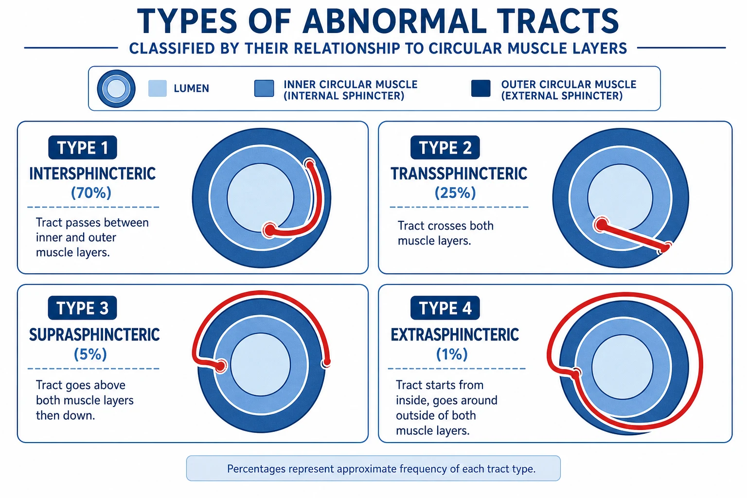
Park's classification, published in 1976, remains the global standard because it is built around the muscle the surgeon must protect.[1]
| Type | Frequency | Course of the tract |
|---|---|---|
| Intersphincteric | 70% (commonest) | Passes through the internal sphincter and ends in the perianal skin through the intersphincteric plane |
| Transsphincteric | 25% | Crosses both internal and external sphincter into the ischiorectal fossa |
| Suprasphincteric | 5% | Ascends above puborectalis, crosses levator, then descends to skin |
| Extrasphincteric | under 1% | Runs from rectum (above the sphincters) directly to skin; usually a pelvic source — Crohn's, malignancy, or iatrogenic |
A separate, imaging-based scheme — the St James's University Hospital MRI classification — grades fistulas into five types from simple intersphincteric (Grade 1) up to horseshoe and extrasphincteric disease (Grade 5), and is what the radiologist will write on the MRI report. Clinicians pair Park's classification (the surgeon's operative map) with the St James's grade (the radiologist's roadmap).[2][3]
A fistula is called simple when it is intersphincteric or low transsphincteric, involves less than 30 percent of the external sphincter, and has a single tract in an otherwise normal anus. It is complex when it involves more than 30 percent of the external sphincter, has a high internal opening, is recurrent or horseshoe, coexists with Crohn's disease, has failed previous surgery, or sits in a previously irradiated or incontinent patient.[2] This simple-versus-complex split is the single most useful bedside discriminator because it dictates the entire operation.
Epidemiology & Risk Factors
Perianal abscess and fistula are common surgical conditions — among the commonest reasons for an emergency surgical admission with an anorectal complaint. The incidence runs at about 15 to 20 per 100,000 population per year, with a lifetime prevalence of about 1 in 10,000. The peak age is 30 to 50 years, and there is a striking male predominance of 2 to 4:1 (attributed to the androgenic effect on anal gland secretion).[1]
The risk factors cluster into those that predispose to obstruction and those that impair healing or immunity: [1]
- Crohn's disease — the single most important systemic association; perianal fistula can be the presenting feature of Crohn's, and perianal disease occurs in up to a third of Crohn's patients over their lifetime. Always think of Crohn's when fistulas are multiple, recurrent, complex, or accompanied by diarrhoea.
- Diabetes mellitus — higher risk of severe, necrotising infection and Fournier's gangrene; undiagnosed diabetes can present as a perianal abscess.
- Immunosuppression — HIV/AIDS, chemotherapy, long-term corticosteroids, post-transplant — atypical and aggressive infection, poor healing.
- Other inflammatory bowel disease — ulcerative colitis less commonly than Crohn's.
- Prior anorectal surgery or trauma — including obstetric injury and habitual anal intercourse.
- Tuberculosis in endemic regions — causes a thin, watery, "tubercular fistula".
- Malignancy — rarely, a chronic non-healing fistula harbours an anal or rectal cancer (fistula-cancer).
- Smoking — strongly associated with Crohn's perianal disease and with poorer healing.
- Chronic diarrhoea or constipation — mechanically promotes gland duct obstruction. [1]
Pathophysiology
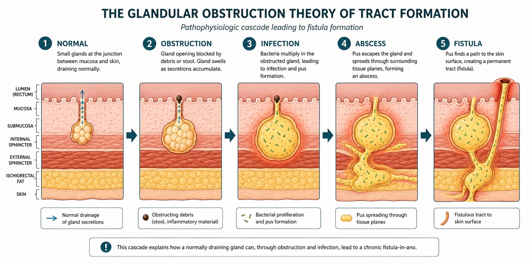
The cryptoglandular theory is the accepted pathogenesis of the great majority (over 90 percent) of cryptoglandular fistulas.[1] Walking the cascade step by step is how examiners want it described:
- The anal canal carries 6 to 12 anal glands at the dentate line, opening into anal crypts and extending into the internal sphincter or the intersphincteric plane.
- Obstruction of a gland duct — by inspissated stool, oedema, foreign debris, or sometimes a faecolith — causes stasis.
- Trapped secretion becomes a culture medium; bacterial overgrowth (typically mixed aerobic and anaerobic flora — E. coli, Bacteroides, Streptococcus, Staphylococcus, and enterococci) generates pus.
- The obstructed gland distends with pus into a small intersphincteric abscess — the point of origin of every cryptoglandular abscess.
- Pus then spreads along the path of least resistance:
- Downward and subcutaneously = perianal abscess (commonest).
- Laterally across the external sphincter into the ischiorectal fossa = ischiorectal abscess.
- Upward above the levator = supralevator abscess.
- Circumferentially across both fossae via the deep postanal space = horseshoe abscess.
- If the abscess ruptures spontaneously or is drained, a communicating tract may persist between the original internal opening at the dentate line and the skin. Both openings are continuously re-epithelialised; the tract lining becomes granulation tissue and then epithelium, which is why the tract cannot close spontaneously. This persistent epithelialised tract is an anal fistula.[1]
The proportion that completes this acute-to-chronic transition is the figure examiners love: 30 to 50 percent of abscesses develop a fistula after drainage.[1]
Goodsall's rule
Goodsall's rule is a bedside rule that predicts the location of the internal opening from the position of the external opening — invaluable at EUA when you cannot easily see where the tract goes.[1]
Imagine a transverse line drawn across the anus: [1]
- An external opening anterior to the line has a short, straight (radial) tract running directly to the nearest anal crypt at the dentate line.
- An external opening posterior to the line has a curved tract that opens at the posterior midline (6 o'clock) crypt. [1]
Exception: an anterior external opening that lies more than 3 cm from the anal verge usually behaves like a posterior one — its tract curves to the posterior midline — because it originates from a posterior midline gland. [1]
Goodsall's rule is accurate in about 80 percent of cases overall, but is less reliable in recurrent fistulas and in Crohn's disease, where imaging and EUA take over. [1]
Perianal sepsis — the numbers an examiner wants
Clinical Presentation
Perianal abscess (acute)
The abscess presents as an acute, severe, perianal problem, and the tempo matters because it drives the urgency of drainage.[1]
- Severe, constant, throbbing perianal pain that is worse on sitting, walking, and defecation and that keeps the patient awake — the pain of pus under pressure.
- Swelling, redness, and a lump in the perianal region.
- Fever, malaise, and sometimes rigors — systemic signs of infection.
- Urinary retention may be the presenting complaint, produced by intense pelvic-floor spasm and pain. [1]
Examination findings: [1]
- A tender, fluctuant, erythematous swelling with surrounding induration and cellulitis.
- Regional inguinal lymphadenopathy may be palpable.
- Digital rectal examination (DRE) is often too painful to complete; when possible it may reveal an internal component, induration, or a high abscess.
- Systemic signs of sepsis: fever, tachycardia, and in severe cases hypotension. [1]
A deep abscess (ischiorectal, intersphincteric, supralevator) can be deceptively hidden — the patient is in severe pain but the perineum looks nearly normal, and only DRE or imaging reveals the collection. This "pain out of proportion to the external findings" pattern is a key teaching point and a classic reason for delayed diagnosis. [1]
Anal fistula (chronic)
The fistula presents as a chronic, relapsing perianal discharge.[1][1]
- Persistent or intermittent perianal discharge — purulent, bloody, or mucopurulent — that soils underwear and requires a pad.
- Recurrent perianal abscesses: the tract blocks, an abscess reforms, it discharges, the swelling settles, and the cycle repeats. A history of "the same lump that bursts and comes back" is almost diagnostic.
- Perianal itching (pruritus ani) and skin excoriation from chronic discharge.
- Pain is usually milder than with an abscess and may worsen only with defecation; severe pain suggests abscess recurrence. [1]
Examination findings: [1]
- An external opening — a small punctum or papule on the perianal skin, often with surrounding skin discolouration and a bead of visible discharge.
- A palpable subcutaneous cord running from the external opening toward the anus, best felt in superficial fistulas.
- DRE may detect the internal opening as a pit or indurated nodule at the dentate line, and assesses sphincter tone (crucial for surgical planning).
- Apply Goodsall's rule at the bedside to predict the internal opening before theatre. [1]
Low fistula
intersphincteric, ~70%
- Tract below the anorectal ring
- **Fistulotomy** (lay open) is safe and curative
- Involves little or no external sphincter
- Recurrence under 10%, incontinence negligible
High or complex fistula
transphincteric / suprasphincteric
- Tract involves a significant portion of EAS
- **Loose seton, LIFT, or advancement flap**
- Never divide sphincter blindly
- Incontinence risk is real — image first
Differential Diagnosis
Every perianal lump or chronic sinus is not a cryptoglandular abscess or fistula. The differentials and their distinguishing features:[1]
| Condition | Key distinguishing features |
|---|---|
| Pilonidal abscess | In the natal cleft (sacrococcygeal area), not perianal; contains hair; no connection to the anal canal |
| Hidradenitis suppurativa | Multiple chronic abscesses and sinuses in axillae, groin, perineum; not connected to anal canal; apocrine-gland distribution |
| Crohn's perianal disease | Multiple or complex fistulas, large fleshy skin tags ("elephant ears"), deep anal fissures, strictures; associated GI symptoms; biopsy shows non-caseating granulomas |
| Fournier's gangrene | Necrotising fasciitis of the perineum; rapidly spreading erythema, crepitus, skin necrosis, severe systemic toxicity — a surgical emergency |
| Infected sebaceous cyst | Superficial discrete fluctuant lump with a central punctum; not connected to the anal canal |
| Tubercular fistula (endemic areas) | Underlying pulmonary TB; thin, watery discharge; multiple tracts; histology shows caseating granulomas |
| Anal / rectal cancer | Hard, irregular, often non-tender mass or non-healing ulcer; biopsy is mandatory for any atypical or non-healing lesion |
| Rectovaginal / anovaginal fistula | Passage of flatus or stool per vagina; obstetric, surgical, or Crohn's aetiology |
| Perianal hidradenoma / rare tumours | Slow-growing solid nodule; diagnosis on histology |
Clinical & Bedside Assessment
Perianal abscess
- Inspect the perianal area for swelling, erythema, fluctuance, and the extent of cellulitis.
- Palpate for fluctuance and inguinal nodes.
- DRE — often too painful; if feasible, look for internal extension or a deep collection.
- Screen for sepsis: vital signs, signs of systemic infection.
- Check the blood glucose: an undiagnosed diabetic may present with perianal sepsis.
- Assess for red flags of necrotising infection: rapidly spreading erythema, crepitus, skin necrosis, bruising, or pain out of proportion to the findings. [1]
Anal fistula
- Inspect the perianal skin for the external opening — a punctum with surrounding skin change — and note its exact clock position and distance from the verge.
- Apply Goodsall's rule to classify the opening as anterior or posterior to the transverse anal line and predict the internal opening.
- Palpate the tract: run a finger along the skin from the external opening toward the anus; a subcutaneous cord may be palpable.
- DRE to feel the internal opening at the dentate line, assess sphincter tone, and gauge how much sphincter is involved — the single most important factor in choosing the operation.
- Proctoscopy may directly visualise the internal opening and exclude haemorrhoids or a tumour. [1]
For complex or recurrent fistulas, examination under anaesthesia (EUA) is the gold standard — combining inspection, DRE, anoscopy, gentle probing of the tract, and dye injection (methylene blue or hydrogen peroxide) to find and confirm the internal opening.[2]
Investigations
Perianal abscess
The diagnosis is clinical — a perianal abscess is drained, not imaged.[1]
- Blood glucose to exclude diabetes.
- Full blood count for leukocytosis.
- Blood cultures if the patient is systemically septic.
- Swab of pus for culture and sensitivity at the time of drainage.
- Imaging (CT or MRI) only for complex, recurrent, deep, or supralevator abscesses, suspected necrotising infection (look for gas in soft tissues), or an immunocompromised host where the clinical picture is unreliable. [1]
Anal fistula
- MRI of the pelvis / perineum (with an endoanal or surface coil) is the gold standard for the complex or recurrent fistula. It maps the primary tract, its relationship to the sphincter, secondary extensions, horseshoeing, and any associated collection — all information the surgeon needs before a second operation. Essential before any surgery on a complex fistula.[2][3]
- Endoanal ultrasound (EAUS) is a quicker, cheaper alternative, less detailed than MRI but useful in theatre; hydrogen peroxide injected into the tract enhances definition.
- Examination under anaesthesia (EUA) with probing and dye injection remains the definitive clinical assessment and is often both diagnostic and therapeutic.
- Fistulography — injecting contrast through the external opening under fluoroscopy — is largely obsolete but may be used when MRI is unavailable.
- Colonoscopy and ileoscopy if Crohn's disease is suspected (multiple or complex fistulas, diarrhoea, weight loss, young age) to look for skip lesions and obtain biopsies.
- Biopsy of any atypical tract at EUA is mandatory to exclude Crohn's, tuberculosis, or malignancy.[3]
Management — Resuscitation
The resuscitation bundle is reserved for the patient with a large abscess and systemic sepsis or suspected necrotising infection.[1][1]
- IV access and fluid resuscitation.
- IV analgesia (opioid).
- Broad-spectrum IV antibiotics covering Gram-negative and anaerobic organisms — for example co-amoxiclav 1.2 g IV every 8 hours, or ceftriaxone 1 to 2 g IV once daily plus metronidazole 500 mg IV every 8 hours. Antibiotics alone are not sufficient — the abscess must be drained, and antibiotics do not cure a collection.
- Urgent surgical assessment for incision and drainage under general or regional anaesthesia.
- Diabetic patients: strict glucose control with an insulin sliding scale, broader antibiotic cover, and a lower threshold for imaging and ICU support. [1]
Suspected Fournier's gangrene (necrotising fasciitis)
This is a surgical emergency.[1]
- Immediate IV resuscitation and broad-spectrum triple-antibiotic cover for polymicrobial infection (a Gram-negative agent, an anaerobe cover, and often a penicillin or carbapenem).
- Urgent, aggressive surgical debridement — wide removal of all necrotic skin, subcutaneous tissue, and fascia until healthy bleeding tissue is reached. Multiple returns to theatre are the rule, not the exception. A defunctioning colostomy and, rarely, orchiectomy may be required.
- Mortality is 20 to 40 percent, higher with delay. The Laboratory Risk Indicator for Necrotising Fasciitis (LRINEC) score can support — but never delay — the clinical decision to operate. [1]
Management — Definitive & Stepwise
Perianal abscess
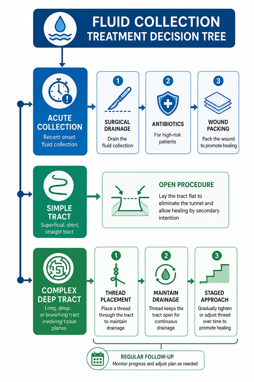
Incision and drainage (I&D) is the definitive treatment; the operation is more important than any antibiotic.[1][1]
- Under general or regional anaesthesia (local infiltration only for a very superficial abscess).
- A radial or cruciate incision over the most fluctuant point, close to the anus to keep any subsequent fistula short.
- Evacuate all pus and break loculations with a finger or artery forceps.
- Send pus for culture and sensitivity.
- Curette the cavity wall to remove necrotic debris.
- For a superficial cavity, loose packing with saline-soaked gauze or an alginate dressing — not too tight, so drainage is maintained. For a deep or large cavity, a mushroom (Malecot) catheter left in the cavity for 2 to 3 weeks is preferred because it keeps the drainage track open and reduces recurrence.[1]
- Do NOT primarily close the wound — primary closure risks recurrence and seeding.
- Antibiotics are not routinely required for an uncomplicated abscess after adequate drainage. Add them when there is cellulitis extending beyond the abscess, systemic sepsis, diabetes, immunosuppression, a prosthetic valve or high-risk cardiac lesion, or recurrent disease.[1]
- Follow-up at 2 to 4 weeks to check healing and warn about fistula development (30 to 50 percent).
Antibiotics for perianal sepsis (when indicated)
Anal fistula
The governing principle: eliminate the tract and the internal opening while preserving the sphincter.[1][2][1] The choice of procedure is dictated by how much external sphincter the tract involves.
Fistulotomy (lay-open) — for a low (simple) fistula that involves little or no external sphincter (intersphincteric or low transsphincteric tract, below the anorectal ring, under 30 percent of EAS).[1]
- A probe is passed from the external to the internal opening.
- The overlying tissue is divided along the entire length of the tract, laying it open and converting a tunnel into a gutter; a small amount of internal sphincter in the floor is accepted.
- The wound heals by secondary intention over 2 to 6 weeks with regular sitz baths and dressings.
- Success over 90 percent, recurrence under 10 percent. Incontinence is minimal for a truly low fistula and is proportional to the amount of muscle divided. [1]
Loose (draining) seton — the cornerstone of managing a high or complex fistula.[1][3]
- A non-absorbable suture or silicone vessel loop is passed through the tract from the external to the internal opening and tied loosely.
- The seton maintains drainage, prevents abscess recurrence, and induces gradual peritract fibrosis — it deliberately does not divide the sphincter.
- It is left in place for weeks to months, often as the first stage of a staged procedure or as definitive long-term drainage (especially in Crohn's). [1]
Cutting seton — the seton is progressively tightened over weeks to months, slowly dividing the sphincter while the tract fibroses behind it. It risks incontinence and is falling out of favour; modern practice prefers a loose seton followed by a sphincter-sparing definitive procedure. [1]
LIFT procedure (ligation of the intersphincteric fistula tract) — for transsphincteric fistulas.[4]
- The tract is identified in the intersphincteric plane through a small perianal incision, ligated and divided at both ends, and the distal tract is curetted.
- Sphincter-sparing — no functional muscle is divided.
- Success rate about 70 to 80 percent, with minimal continence disturbance — now a first-line sphincter-sparing option for transsphincteric disease. [1]
Endorectal advancement flap — for complex or high fistulas. [1]
- A broad-based mucosal-muscular flap is raised from the rectum, the internal opening is closed, and the flap is advanced down to cover it, eliminating the source of sepsis while leaving the sphincter intact.
- Success 60 to 80 percent; can be repeated. [1]
Sphincter-sparing and minimally invasive options — reserve for complex or recurrent disease where sphincter preservation is paramount:[2][3]
- Fibrin glue and anal fistula plug (collagen) — biologic closure of the tract; low morbidity but variable and often disappointing durability (success 15 to 60 percent).
- VAAFT (video-assisted anal fistula treatment) — the tract is visualised with a fistuloscope, the internal opening is identified, the tract is diathermied, and the opening is closed. Sphincter-sparing.
- In the UK, NICE interventional procedures have assessed several of these (e.g. fibrin glue for simple fistulas) as options with appropriate consent. [1]
Crohn's perianal fistula
Crohn's perianal disease demands a multidisciplinary, sphincter-conserving strategy — the surgeon controls sepsis, the gastroenterologist treats the underlying inflammation.[3]
- Avoid sphincter division — the risk of non-healing and disabling incontinence is high in inflamed, immunosuppressed tissue.
- Control sepsis first: examination under anaesthesia, drainage of abscesses, and loose seton placement.
- Medical therapy with anti-TNF:
- Infliximab 5 mg/kg IV at weeks 0, 2, and 6, then 5 mg/kg every 8 weeks for maintenance — the ACCENT II trial showed that maintenance infliximab keeps perianal fistulas closed in roughly twice as many patients as placebo.[5][6]
- Adalimumab 160 mg subcutaneously at week 0, 80 mg at week 2, then 40 mg every other week — an alternative, useful in patients who lose response to or are intolerant of infliximab.
- Antibiotics (metronidazole 400 mg three times daily, ciprofloxacin 500 mg twice daily) for secondary infection and as an adjunct.
- Treat active luminal disease with nutritional support, immunomodulators (azathioprine, methotrexate), and anti-TNF.
- For severe, refractory perianal Crohn's, a defunctioning stoma (loop ileostomy) or, ultimately, proctocolectomy may be required.
Choosing the fistula operation — a stepwise logic
Assess how much external sphincter the tract involves (DRE, EUA, MRI).
Simple / low fistula (under 30% EAS, below anorectal ring) → **fistulotomy (lay open)**.
Transsphincteric with more muscle → **LIFT** (sphincter-sparing).
High transsphincteric / suprasphincteric → **loose seton** to control sepsis first.
After sepsis control → **advancement flap** or staged procedure; reserve cutting seton for selected cases.
Complex / recurrent / Crohn's → **MRI + EUA**, loose seton, biologics, no sphincter division.
Specific Subtypes & Scenarios
- Horseshoe abscess and fistula: sepsis involving both ischiorectal fossae through the deep postanal space, with a posterior midline internal opening. Management is the Hanley procedure — drainage of both sides plus posterior midline unroofing (counter-incision) of the deep postanal space, often with setons. Failure to recognise a horseshoe is a classic cause of recurrence.[1]
- Supralevator abscess: arises either from upward extension of a cryptoglandular abscess or from an intra-abdominal source (diverticulitis, appendicitis, Crohn's, pelvic sepsis). Imaging is mandatory to identify the source, because the route of drainage must match the origin — draining a supralevator abscess of pelvic origin through the sphincter creates an iatrogenic extrasphincteric fistula.[1]
- Recurrent fistula after surgery: reassess with MRI and EUA; actively seek an underlying cause — Crohn's, tuberculosis, HIV, or malignancy — and biopsy the tract.[3]
- Tubercular fistula (endemic regions): thin watery discharge, multiple tracts, underlying pulmonary TB, and caseating granulomas on histology. Anti-tubercular therapy (ATT — rifampicin, isoniazid, pyrazinamide, ethambutol) comes first; surgery only for non-healing disease after adequate ATT.
- HIV-related perianal disease: higher risk of atypical infections and malignancy (anal intraepithelial neoplasia). Manage conservatively, biopsy every atypical lesion, and use CD4-guided therapy.
- Fistula-associated cancer: chronic, long-standing fistulas (especially Crohn's) rarely harbour squamous cell carcinoma or mucinous adenocarcinoma — a non-healing tract must be biopsied.[3]
Complications & Pitfalls
Of the abscess
- Recurrence (10 to 15 percent), usually from inadequate drainage or undiagnosed extension.
- Fistula formation (30 to 50 percent).
- Fournier's gangrene — necrotising fasciitis; a surgical emergency; highest risk in diabetics and the immunocompromised.
- Sepsis and septicaemia, especially with deep abscesses.
- Urinary retention from severe pain. [1]
Of fistula surgery
- Recurrence (5 to 10 percent after fistulotomy; higher for complex fistulas and Crohn's).
- Incontinence — the most feared complication. Risk is proportional to the amount of external sphincter divided and is lowest for intersphincteric fistulas and highest for high transsphincteric ones. Always assess sphincter involvement before dividing any muscle. Even loose setons can cause minor soiling; cutting setons and fistulotomy carry the greatest risk.
- Anal stenosis from excessive scarring.
- Delayed diagnosis of an underlying cause (Crohn's, TB, malignancy).[3]
Classic pitfalls
- Treating an abscess with antibiotics alone without incision and drainage — pus must be evacuated.
- Dividing too much sphincter in a high fistula without first assessing the tract — causing avoidable incontinence.
- Missing a horseshoe component and draining only one side — recurrence is then inevitable.
- Not suspecting Crohn's disease in a patient with multiple or recurrent fistulas — colonoscopy is required.
- Missing Fournier's gangrene — perianal sepsis with skin necrosis, crepitus, or systemic toxicity is a surgical emergency.
- Not biopsying an atypical or non-healing fistula — missing malignancy or TB.
- Draining a supralevator abscess of pelvic origin trans-sphincterically and creating an extrasphincteric fistula. [1]
Prognosis & Disposition
Perianal abscess does well after incision and drainage; recurrence is 10 to 15 percent and a fistula develops in 30 to 50 percent. Most patients are managed as a day case or short stay and warned at discharge about the symptoms of recurrence.[1]
Anal fistula prognosis tracks complexity. Low fistulas: fistulotomy success over 90 percent, recurrence under 10 percent, minimal incontinence. High or complex fistulas: higher recurrence and incontinence rates; often need staged procedures and long follow-up. Crohn's fistulas: chronic and recurrent; the realistic aim is control of sepsis and preservation of continence, not cure.[2]
Disposition: simple abscesses and low fistulas are discharged the same day with sitz-bath and review instructions. Complex fistulas, post-Fournier's patients, and those with sepsis or comorbidity need inpatient care, imaging, and a planned return to theatre. [1]
Special Populations
- Diabetics: higher risk of severe and necrotising infection (Fournier's gangrene). Enforce strict glucose control with an insulin sliding scale, use broader antibiotic cover, and keep a low threshold for imaging, debridement, and ICU admission.[1]
- Immunocompromised (HIV, chemotherapy, transplant, steroids): atypical and aggressive organisms, poor wound healing. Favour conservative surgery, broad antibiotic/antifungal cover, biopsy of all lesions, and early multidisciplinary input.
- Crohn's disease: perianal disease is common and may be the presenting feature. Avoid sphincter division, control sepsis with setons, and treat with anti-TNF. Involve gastroenterology early.[3]
- Pregnancy: a perianal abscess is drained — drainage is safe in pregnancy. Defer fistulotomy to the postpartum period if possible to limit sphincter manipulation; if a fistula is symptomatic, a loose seton is the safe interim option.[1]
- Children: perianal abscess and fistula in boys (often infants) are usually simple and cryptoglandular; fistulotomy is generally well tolerated, but always consider underlying causes in atypical or recurrent paediatric disease.
Evidence, Guidelines & Regional Differences
German S3 Guidelines (Ommer et al., 2017):[1] recommend that every abscess be drained surgically, with antibiotics reserved for systemic infection or high-risk patients; fistulotomy for low fistulas; seton or LIFT for high fistulas; MRI for all complex or recurrent fistulas; and anti-TNF plus seton drainage for Crohn's disease.
ASCRS Guidelines (Vogel et al., 2016; Gaertner et al., 2022):[2][3] mirror these recommendations and emphasise sphincter preservation in high fistulas, the role of LIFT and advancement flaps as sphincter-sparing options, and anti-TNF therapy (infliximab, adalimumab) as the evidence-based medical treatment for Crohn's perianal fistula, supported by the ACCENT II trial.[6]
ACCENT II — Sands et al., NEJM 2004
PMID 14985485
Randomised, double-blind, maintenance trial in 306 patients with draining perianal or abdominal fistulas
Key finding
Maintenance infliximab kept fistulas closed in roughly twice as many patients as placebo (about 36% vs 19% at week 54) and lengthened time to loss of response.
Practice change
Established infliximab as the evidence-based medical therapy for fistulising Crohn's disease and underpins current anti-TNF dosing.
Present et al., NEJM 1999 — infliximab induction RCT
PMID 10228190
Double-blind, placebo-controlled trial in 94 patients with draining perianal Crohn's fistulas (infliximab 5, 10, 20 mg/kg vs placebo at weeks 0, 2, 6)
Key finding
About two-thirds of infliximab-treated patients had a 50% or greater reduction in draining fistulas versus about one-quarter on placebo; the 5 mg/kg dose was best tolerated.
Practice change
First RCT to prove anti-TNF efficacy for perianal Crohn's fistulas; the basis of the 5 mg/kg induction regimen.
Australia / New Zealand: colorectal-society practice closely tracks the ASCRS and German guidance — fistulotomy for low fistulas, MRI for complex/recurrent disease, and LIFT and advancement flaps as first-line sphincter-sparing procedures. Anti-TNF is standard for Crohn's perianal fistula.
India / resource-limited settings: the disease is common, and tuberculosis and filariasis are rare but real causes of atypical fistulas. MRI may be unavailable, so EUA with probing and dye injection remains the practical standard. ICMR guidelines recommend co-amoxiclav or ceftriaxone plus metronidazole for perianal sepsis.[1]
Prevention
There is no specific prophylaxis for cryptoglandular disease, but several measures reduce recurrence and severity: [1]
- Prompt and complete drainage of the first abscess, with breakdown of all loculations — the single biggest preventable cause of recurrence.
- Warning the patient about the symptoms of fistula formation and recurrence, so that early review happens.
- Aggressive control of diabetes and immunosuppression in affected patients to prevent necrotising infection.
- Smoking cessation, which improves Crohn's perianal outcomes.
- Multidisciplinary management of Crohn's disease (anti-TNF, nutrition, immunomodulators) to reduce perianal complications.
- Treatment of tuberculosis where it underlies the fistula, before any surgery. [1]
Exam Pearls
- Cryptoglandular theory — obstruction of an anal gland at the dentate line causes an abscess, then a fistula in 30 to 50 percent.[1]
- Goodsall's rule — anterior external opening = straight radial tract; posterior = curves to 6 o'clock posterior midline; exception: anterior openings over 3 cm from the verge may curve.[1]
- Park's classification — intersphincteric (70%), transsphincteric (25%), suprasphincteric (5%), extrasphincteric (under 1%).[1]
- Abscess — always incision and drainage; antibiotics only for cellulitis, sepsis, diabetes, immunosuppression, prosthetic valves.[1]
- Low fistula = fistulotomy (lay open); high fistula = loose seton (avoid sphincter division = avoid incontinence).[1]
- LIFT = ligation of intersphincteric fistula tract; sphincter-sparing; success 70 to 80 percent.[4]
- Crohn's fistula — anti-TNF (infliximab 5 mg/kg weeks 0/2/6 then q8w; adalimumab 160/80/40 q2w) plus seton; never divide the sphincter.[3][6]
- 30 to 50 percent of abscesses develop a fistula after drainage.[1]
- Fournier's gangrene = necrotising fasciitis of the perineum; diabetics at highest risk; emergency debridement; mortality 20 to 40 percent.[1]
- Horseshoe abscess — both ischiorectal fossae, posterior midline internal opening; drain both sides (Hanley procedure).[1]
- Anorectal ring — formed by puborectalis; a fistulotomy above it causes catastrophic incontinence.
- Supralevator abscess — image before draining; transsphincteric drainage of a pelvic-source abscess creates an extrasphincteric fistula.[1]
Goodsall's rule simplified
ABC
external opening anterior to the transverse line = straight radial tract
external opening posterior = curves to 6 o'clock posterior midline
anterior opening over 3 cm from the verge = exception, may curve
Park's fistula types — from commonest to rarest
ITES
70% — the commonest, between IAS and EAS
25% — crosses both sphincters into the ischiorectal fossa
under 1% — rectum direct to skin; pelvic source
5% — above puborectalis, crosses levator, to skin
Quick self-test: a 38-year-old man has a recurrent perianal discharge. The external opening lies at 3 o'clock, 1 cm from the verge. Where is the internal opening, and what is the operation?
By Goodsall's rule, an anterior external opening within 3 cm of the verge has a straight radial tract to the nearest anal crypt at about 3 o'clock on the dentate line. If EUA confirms a low intersphincteric tract, the operation is fistulotomy (lay-open); if it crosses significant external sphincter, use a loose seton and plan a LIFT or advancement flap.
Exam application bank (NEET-PG / INICET)
One-line answer
Perianal abscess and anal fistula (fistula-in-ano) are two stages of a single cryptoglandular disease process. Obstruction of an anal gland duct at the dentate line produces an acute pus collection (abscess) which, on drainage, may leave a permanent epithelialised tract (fistula) in 30 to 50 percent of patients. Abscess presents with severe throbbing perianal pain, fever, and a tender fluctuant mass; fistula presents with recurrent discharge and recurrent abscesses. Goodsall's rule predicts the internal opening. Park's classification grades fistulas by sphincter involvement. Abscess is treated by incision and drainage; low fistula by fistulotomy; high or complex fistula by a loose seton, LIFT, or advancement flap to preserve the sphincter. Crohn's perianal fistula demands anti-TNF and sphincter-conserving surgery.
Worked stems (answer without another resource)
Stem 1 — Classic presentation. Map symptoms to mechanism; name the first investigation and first treatment step with dose/route if drug therapy is standard. [1]
Stem 2 — Unstable / complicated. List red flags that force immediate resuscitation, theatre, ICU, antidote, or reperfusion — and what you do in the first 15 minutes. [1]
Stem 3 — Atypical group. Elderly, pregnancy, child, or immunocompromised: how presentation and thresholds change. [1]
Stem 4 — Differential trap. Name the three closest mimics and one discriminator for each. [1]
Stem 5 — Disposition. Who goes home with safety-netting, who is admitted, who needs HDU/ICU/theatre, and what follow-up is mandatory. [1]
Rapid viva checklist
- Definition + classification
- Pathophysiology chain
- Bedside signs / criteria
- Score with exact components (if any)
- Emergency bundle
- Definitive therapy with doses
- Complications of disease and of treatment
- Special populations
- Guideline/trial name if classic
- Three exam traps
Coverage self-check
If you cannot answer any stem above from this page alone, re-read the matching section — the page is intended to be self-sufficient for final-prof and NEET-PG/INICET questions on Perianal Abscess and Anal Fistula.
References
- [1]Ommer A, Herold A, Berg E, et al. German S3 guidelines: anal abscess and fistula (second revised version) Langenbecks Arch Surg, 2017.PMID 28251361
- [2]Vogel JD, Johnson EK, Morris AM, et al. Clinical Practice Guideline for the Management of Anorectal Abscess, Fistula-in-Ano, and Rectovaginal Fistula Dis Colon Rectum, 2016.PMID 27824697
- [3]Gaertner WB, Hagerman GF, Feingold DL, et al. The American Society of Colon and Rectal Surgeons Clinical Practice Guidelines for the Management of Anorectal Abscess, Fistula-in-Ano, and Rectovaginal Fistula Dis Colon Rectum, 2022.PMID 35732009
- [4]Rojanasakul A, Pattanaarun J, Sahakitrungruang C, Tantiphlachiva K. Total anal sphincter saving technique for fistula-in-ano; the ligation of intersphincteric fistula tract J Med Assoc Thai, 2007.PMID 17427539
- [5]Present DH, Rutgeerts P, Targan S, et al. Infliximab for the treatment of fistulas in patients with Crohn's disease N Engl J Med, 1999.PMID 10228190
- [6]Sands BE, Anderson FH, Bernstein CN, et al. Infliximab maintenance therapy for fistulizing Crohn's disease N Engl J Med, 2004.PMID 14985485