General Surgery · General Surgery
Perioperative VTE Prophylaxis
Also known as Venous thromboembolism prophylaxis · DVT prophylaxis · Thromboprophylaxis · PE prevention
Venous thromboembolism (VTE) = deep vein thrombosis (DVT) + pulmonary embolism (PE). It is the leading preventable cause of hospital death. Surgical patients carry a ten- to twenty-fold increased risk. Prevention rests on Virchow's triad (stasis, hypercoagulability, endothelial injury), risk stratification (Caprini for surgical, Padua for medical patients) and a stepwise ladder: early mobilisation, mechanical methods (TEDS, IPC) and pharmacological prophylaxis (enoxaparin 40 mg SC OD, fondaparinux 2.5 mg SC OD, rivaroxaban 10 mg PO OD). High-risk patients get both. Extended prophylaxis for twenty-eight days after major orthopaedic or cancer surgery. Massive PE is an emergency: oxygen, haemodynamic support, systemic thrombolysis (alteplase).
On this page & tools
Your progress
Saved locally on this device.
Exam tags
Red flags
Overview & Definition
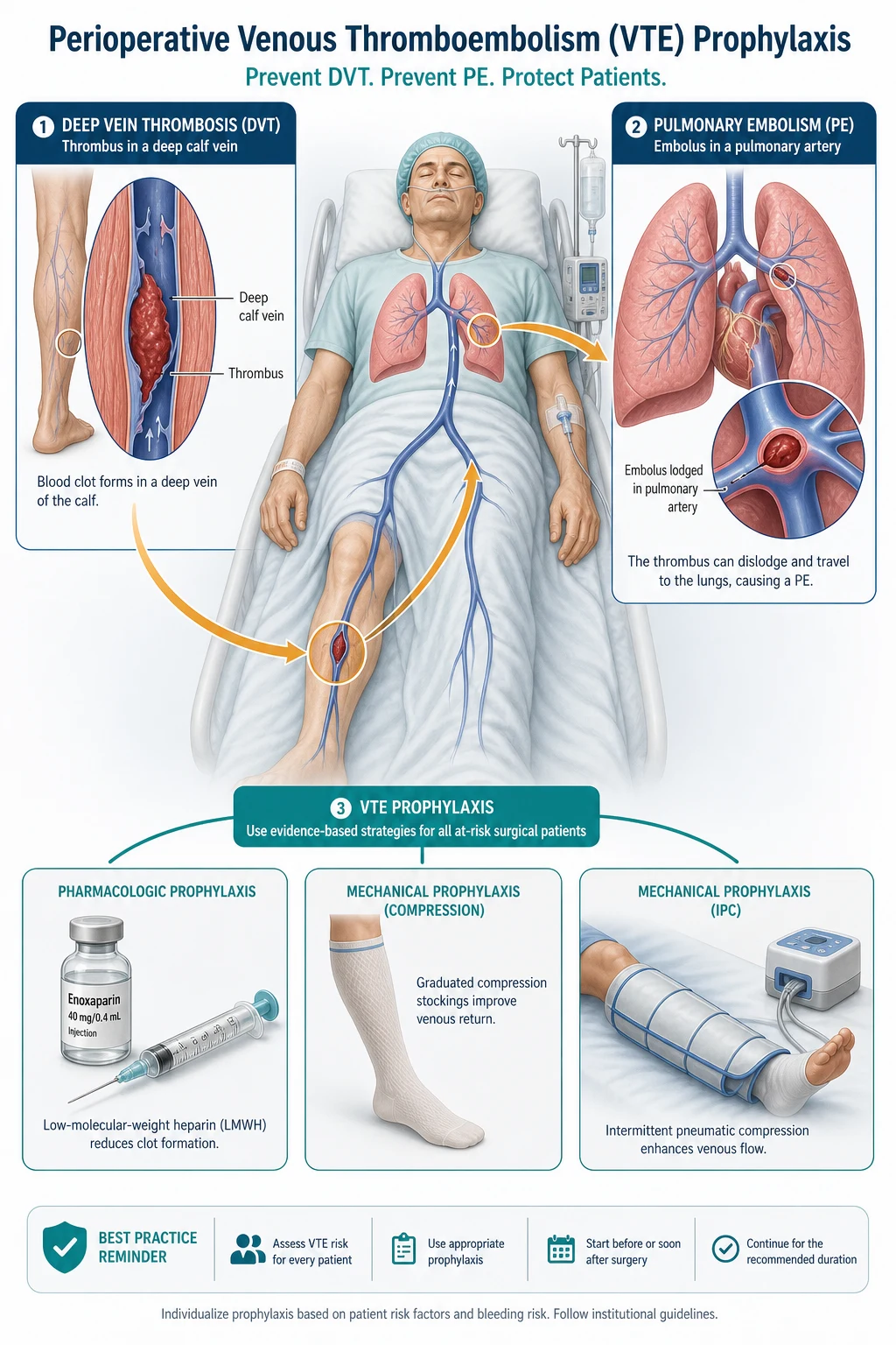
Venous thromboembolism (VTE) is the unified clinical term for deep vein thrombosis (DVT) and its embolic complication, pulmonary embolism (PE). The two entities share the same pathophysiology, risk factors and prevention strategy, and are therefore considered a single disease process.[1]
In the surgical context, perioperative VTE prophylaxis is the systematic, evidence-based application of risk stratification, mechanical methods, pharmacological anticoagulation and early mobilisation to prevent clot formation in patients undergoing operation. It is not optional — VTE is the single most common preventable cause of in-hospital death, and a fatal PE is estimated to occur in roughly 0.2 to 1 percent of unprotected major-surgery patients. The landmark multinational ENDORSE cross-sectional audit of more than sixty-eight thousand hospitalised patients found that roughly forty-one percent of surgical patients were at high VTE risk, yet a substantial proportion did not receive guideline-concordant prophylaxis.[1]
The conceptual shift over the last three decades has been from treatment of established VTE to prevention. A pulmonary embolus that never forms cannot kill a patient, and prophylaxis reduces VTE events by fifty to seventy percent. The challenge at the bedside is to balance thrombosis prevention against bleeding risk — the central tension of this entire topic. [1]
- ACCP (American College of Chest Physicians, 9th edition, 2012) and ASH (American Society of Hematology, 2018) use the term "thromboprophylaxis" and stratify surgical patients into low, moderate, high and very high risk.
- NICE CG92 (UK) mandates a documented VTE and bleeding assessment within fourteen hours of admission for every adult, with mechanical or pharmacological options chosen accordingly.
- NICE/UK practice favours graduated compression stockings (TEDS) plus LMWH; in the UK the term "anti-embolism stockings" is used in place of "TEDS".
- Indian (ICMR / ICMR-aligned hospital) and most South Asian practice follows ACCP principles, with enoxaparin 40 mg SC OD the workhorse agent.
Classification
Perioperative VTE prevention is classified by the type of intervention and by the patient's risk stratum. [1]
Pharmacological
anticoagulation
- Low-molecular-weight heparin (LMWH): enoxaparin 40 mg SC OD — the standard
- Dalteparin 5000 IU SC OD — alternative LMWH
- Unfractionated heparin (UFH) 5000 IU SC every 8-12 h — renal failure, HIT history
- Fondaparinux 2.5 mg SC OD — synthetic pentasaccharide factor Xa inhibitor
- Direct oral anticoagulants (DOACs): rivaroxaban 10 mg, apixaban 2.5 mg BD, dabigatran 220 mg — mainly orthopaedic
- Vitamin K antagonist (warfarin) — rarely used for primary prophylaxis
Mechanical
physical methods
- Graduated compression stockings (TEDS / anti-embolism stockings)
- Intermittent pneumatic compression (IPC) — calf or thigh sleeves
- Venous foot pump (impulse) — for immobilised trauma
- Early and frequent mobilisation — the simplest and most underused
- Inferior vena cava (IVC) filter — only when anticoagulation is contraindicated and DVT is present
Risk strata
Caprini score
- Very low (Caprini 0): early ambulation alone
- Low (Caprini 1-2): mechanical (IPC)
- Moderate (Caprini 3-4): LMWH/LDUH OR IPC
- High/very high (Caprini ≥5): pharmacological + mechanical (dual)
VTE prophylaxis duration by scenario
DVT itself is anatomically classified into distal (calf) DVT — below the popliteal vein, lower embolic potential but may propagate — and proximal DVT — popliteal, femoral, iliac veins, the source of most clinically important PEs. PE is classified by haemodynamic consequence into submassive PE (right ventricular strain but stable blood pressure) and massive PE (systolic BP below ninety mmHg or drop by forty mmHg for over fifteen minutes — thrombolysis candidate). [1]
Epidemiology & Risk Factors
VTE is genuinely a disease of hospitalisation: roughly fifty to seventy percent of all symptomatic DVT and PE events occur in patients who are, or have recently been, in hospital, and ten to thirty percent of unprotected major-surgery patients develop DVT on screening imaging.[1][1]
Baseline (untreated) DVT incidence without prophylaxis, by procedure: [1]
| Surgical setting | Distal DVT (screening) | Proximal DVT | Fatal PE |
|---|---|---|---|
| Major general/abdominal surgery | 15 to 30 percent | 2 to 4 percent | 0.2 to 0.9 percent |
| Hip replacement (THR) | 40 to 60 percent | 4 to 10 percent | 1 to 5 percent |
| Knee replacement (TKR) | 40 to 60 percent | 8 to 20 percent | 1 to 3 percent |
| Hip fracture repair | 40 to 50 percent | 10 to 20 percent | 4 to 10 percent |
| Major trauma / spinal cord injury | 40 to 60 percent | 10 to 20 percent | up to 5 percent |
| Neurosurgery (craniotomy) | 25 to 35 percent | 3 to 5 percent | 1 to 3 percent |
The ENDORSE study (sixty-eight thousand patients across thirty-two countries) demonstrated that 41.5 percent of surgical and 41.5 percent of medical inpatients met criteria for high VTE risk, confirming that thromboprophylaxis is a global, cross-specialty responsibility rather than an orthopaedic or cancer-only concern.[1]
Patient-related risk factors (each adds points to the Caprini score): [1]
- Age — risk rises steeply after forty and again after sixty; age over seventy-five carries the highest single-patient weight.
- Previous VTE — the strongest single risk factor (relative risk roughly eight-fold).
- Active malignancy — and cancer chemotherapy; venous compression, procoagulant tumour products, and postoperative immobility combine.
- Obesity (BMI above thirty) and the metabolic syndrome.
- Pregnancy and the puerperium; combined oral contraceptive pill, hormone replacement therapy, tamoxifen.
- Inherited thrombophilia — Factor V Leiden, prothrombin G20210A mutation, antithrombin/protein C/protein S deficiency, antiphospholipid syndrome.
- Acute medical illness — heart failure (NYHA III-IV within one month), COPD exacerbation, pneumonia, sepsis, acute stroke with paralysis, inflammatory bowel disease.
- Varicose veins, central venous catheter, nephrotic syndrome, myeloproliferative disorders. [1]
Surgical/acute risk factors: general anaesthesia longer than forty-five minutes, pelvic/lower-limb surgery, immobilising plaster cast, bed rest over seventy-two hours, and the surgical stress response itself (see Pathophysiology). [1]
Pathophysiology
All VTE arises from Virchow's triad, described by Rudolf Virchow in 1856, and every surgical patient violates all three limbs simultaneously — the molecular basis of the ten- to twenty-fold excess risk.[1]
Virchow's triad
SHE
immobility, anaesthesia, bed rest, venous obstruction, paralysis
surgical stress, malignancy, pregnancy, OCP, thrombophilia
surgical trauma, venous cannulation, previous DVT, indwelling catheter
1. Venous stasis. General and neuraxial anaesthesia abolish the calf-muscle pump; intra-operative venodilation slows flow; perioperative immobilisation, pain, opioids and catheters keep the patient supine. Venous blood pools in the valve pockets of the deep calf veins — the classic site of clot initiation. Reduced shear stress also down-regulates endothelial thrombomodulin, tipping the balance toward coagulation. [1]
2. Hypercoagulability (the surgical stress response). Surgery activates the extrinsic (tissue-factor) coagulation pathway: damaged tissues release tissue factor, which complexes with factor VIIa to activate factor X and generate thrombin (factor IIa). Thrombin converts fibrinogen to fibrin, activates platelets, and is itself a potent procoagulant through positive feedback (activation of factors V and VIII). Perioperatively there is also a rise in fibrinogen, factor VIII and von Willebrand factor, platelet activation and aggregation, and attenuation of fibrinolysis (raised plasminogen activator inhibitor-1). Malignancy compounds this through tumour mucins and tissue factor; pregnancy through oestrogen-driven increases in clotting factors and acquired resistance to activated protein C. [1]
3. Endothelial injury. Direct surgical trauma disrupts the venous endothelium, exposing subendothelial collagen and tissue factor. Venous cannulation, intravenous catheters, orthopaedic manipulation of the leg, and previous DVT (which damages valves) all denude the protective endothelial monolayer. The normal endothelium is antithrombotic — it expresses thrombomodulin, heparan sulphate, tissue-factor pathway inhibitor and releases prostacyclin and nitric oxide. Injury flips this phenotype to a prothrombotic one. [1]
Cascade of events: thrombus typically begins in the valve sinuses of the calf deep veins as a platelet-fibrin nidus. It may lyse spontaneously, remain confined, or propagate proximally into the popliteal, femoral and iliac veins. A proximal clot (especially iliofemoral) is the one that detaches and embolises to the pulmonary arterial tree, producing PE. Roughly half of proximal DVTs embolise; calf-vein clots embolise far less often but cannot be safely ignored because up to twenty percent propagate proximally. [1]
Clinical Presentation
Most perioperative DVTs are clinically silent — they declare themselves only when they embolise. This silence is precisely why prophylaxis, not detection, is the goal. [1]
Deep vein thrombosis (classic features):
- Calf or thigh pain, swelling, warmth and erythema of the affected limb.
- Calf tenderness on deep palpation and a tense, woody feel.
- Disproportionate limb swelling: a calf circumference more than three centimetres greater than the contralateral leg is significant.
- Superficial venous dilation and visible collateral veins.
- Homans' sign (calf pain on forced dorsiflexion of the foot) — historical and unreliable, no longer recommended; it is painful and can dislodge clot.
- Pratt sign (pain on squeezing the calf) — likewise unreliable. [1]
Atypical presentations to expect in surgical patients:
- The elderly may show only unexplained tachycardia, confusion, or a fall.
- The diabetic or neuropathic patient may have a painless swollen leg.
- Postpartum and pregnant patients — left leg predominance from iliac artery compression of the left iliac vein (May-Thurner anatomy).
- Massive iliofemoral DVT can present as phlegmasia alba dolens (white, painful, swollen leg) progressing to phlegmasia cerulea dolens (blue, tense limb with impending venous gangrene) — a vascular emergency. [1]
Pulmonary embolism:
- Dyspnoea (sudden onset) — the most common symptom, present in over eighty percent.
- Pleuritic chest pain — from pulmonary infarction, in roughly half.
- Haemoptysis, cough, syncope.
- Tachypnoea and tachycardia — the most sensitive bedside signs.
- Hypoxia (low PaO2), raised JVP, right ventricular heave, loud pulmonary component of S2.
- Massive PE: sudden cardiovascular collapse, electromechanical dissociation, cardiac arrest, sudden death.
- Atypical PE: isolated new-onset atrial fibrillation, unexplained fever, or panic/anxiety in the postoperative patient. [1]
Differential Diagnosis
A swollen leg or postoperative dyspnoea is not VTE until proven, but several mimics must be actively distinguished: [1]
| Mimic | Distinguishing features |
|---|---|
| Cellulitis | Unilateral warmth, erythema with clear demarcation, fever, leucocytosis, entry wound; responds to antibiotics; D-dimer low if measured |
| Ruptured Baker (popliteal) cyst | Sudden calf pain with knee effusion; cyst seen on ultrasound; bruising may track into the ankle (crescent sign); history of osteoarthritis |
| Calf muscle tear / gastrocnemius rupture | Sudden exertional calf pain after a "pop"; ultrasound shows intramuscular haematoma, patent deep veins |
| Superficial thrombophlebitis | Tender, palpable, firm cord in a varicose superficial vein; no deep-system involvement on duplex; low embolic risk unless it crosses into the deep system at the saphenofemoral junction |
| Heart failure (bilateral oedema) | Bilateral, dependent, pitting oedema; raised JVP, basal crackles, echocardiogram; responds to diuretics |
| Lymphoedema | Non-pitting, chronic, dorsum-of-foot squaring, Stemmer sign positive; no acute tenderness |
| PE mimics (dyspnoea) | Pneumonia (fever, consolidation, productive cough), pneumothorax (sudden pleuritic pain, reduced breath sounds), ACS (ECG changes, troponin), costochondritis, anxiety |
Clinical & Bedside Assessment
Every adult admitted to hospital must have a documented VTE risk assessment and a parallel bleeding risk assessment, ideally within fourteen hours of admission and again before and after surgery. Two validated scores dominate practice. [1]
Caprini Risk Assessment Model (surgical patients)
The Caprini score is the standard surgical VTE risk tool, summing weighted risk factors to a total that maps to a prophylaxis tier.[9][1]
1 point each: age forty-one to sixty; minor planned surgery (under forty-five minutes); BMI above twenty-five; swollen legs; varicose veins; pregnancy or postpartum; history of unexplained or recurrent miscarriage; oral contraceptives or HRT; sepsis within one month; serious lung disease (including pneumonia) within one month; abnormal pulmonary function (COPD); acute myocardial infarction; congestive cardiac failure within one month; bed rest; inflammatory bowel disease; medical patient currently at bed rest. [1]
2 points each: age sixty-one to seventy-four; arthroscopic surgery; major open surgery lasting over forty-five minutes; laparoscopic surgery over forty-five minutes; malignancy (present or previous); confined to bed over seventy-two hours; immobilising plaster cast; central venous access line. [1]
3 points each: age seventy-five or older; personal or family history of VTE in one parent or sibling; Factor V Leiden; other congenital thrombophilia (prothrombin mutation, antithrombin/protein C/S deficiency); lupus anticoagulant, anticardiolipin or anti-beta-2-glycoprotein antibodies; elevated homocysteine; heparin-induced thrombocytopenia (HIT). [1]
5 points each: stroke within one month; elective major lower-extremity arthroplasty (THR/TKR); hip, pelvis or leg fracture within one month; acute spinal cord injury or paralysis within one month; multiple trauma within one month. [1]
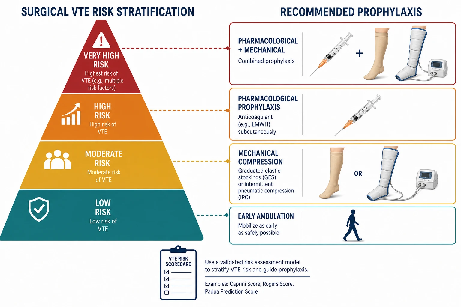
Risk strata and recommended action: [1]
| Caprini total | Risk category | Approx. DVT risk (no prophylaxis) | Recommended prophylaxis |
|---|---|---|---|
| 0 | Very low | less than 0.5 percent | Early ambulation alone |
| 1 to 2 | Low | 1 to 2 percent | Mechanical — IPC preferred |
| 3 to 4 | Moderate | 2 to 4 percent | LMWH, LDUH or IPC |
| 5 or more | High / very high | 4 to 20 percent | Pharmacological + mechanical (dual) |
Padua Prediction Score (medical patients)
For non-surgical (medical) inpatients, the Padua Prediction Score is the validated tool.[8]
- 3 points each: active cancer (local/regional recurrence, metastatic, or chemo/radiotherapy within six months); previous VTE (excluding superficial thrombophlebitis); reduced mobility (bed rest at least three days); known thrombophilic condition.
- 2 points: recent (within one month) trauma and/or surgery.
- 1 point each: elderly age (seventy or over); heart and/or respiratory failure; acute myocardial infarction or ischaemic stroke within one month; acute infection and/or rheumatologic disorder; obesity (BMI thirty or more); ongoing hormonal treatment. [1]
A Padua score of four or more defines high risk (ninety-day VTE incidence about eleven percent without prophylaxis); below four is low risk (about 0.3 percent).[8]
IMPROVE and bleeding risk
The IMPROVE score is an alternative medical-patient model; a score of two or more indicates high risk. Equally important is the bleeding risk assessment: active gastroduodenal ulcer, bleeding within three months before admission, platelets below fifty, age over eighty-five (or seventy-five for ACCP), hepatic/renal failure, ICU admission, and concurrent anticoagulant/antiplatelet therapy all raise bleeding risk. When bleeding risk is high, prefer mechanical prophylaxis alone until the bleeding risk falls. [1]
Investigations
Investigations are for suspected established VTE (prophylaxis itself requires no routine monitoring for LMWH). They are guided by clinical pre-test probability. [1]
Wells score for DVT (pre-test probability):
- Active cancer (+1); paralysis/paresis/plaster cast of lower extremity (+1); bedridden at least three days or major surgery within twelve weeks (+1); localised tenderness along the deep venous system (+1); entire leg swollen (+1); calf swelling at least three centimetres more than the other leg (+1); pitting oedema confined to the symptomatic leg (+1); collateral superficial non-varicose veins (+1); previously documented DVT (+1); alternative diagnosis at least as likely (minus two).
- Two-level outcome: score of two or less = "DVT unlikely" (about five percent have DVT); score of three or more = "DVT likely" (about twenty to fifty percent). [1]
Wells score for PE (simplified two-tier): clinical signs/symptoms of DVT (+1); PE more likely than alternative (+1); heart rate above one hundred (+1); immobilisation at least three days or surgery within four weeks (+1); previous DVT/PE (+1); haemoptysis (+1); malignancy (+1). Score of one or less = "PE unlikely"; score of two or more = "PE likely". [1]
Diagnostic tests:
- D-dimer — a fibrin degradation product. Highly sensitive, poorly specific (raised in malignancy, infection, pregnancy, postoperatively, and after trauma). A negative D-dimer in a low-probability patient effectively excludes DVT or PE; a raised value mandates imaging. D-dimer is unhelpful in postoperative patients (predictably elevated) and is therefore sparingly used.
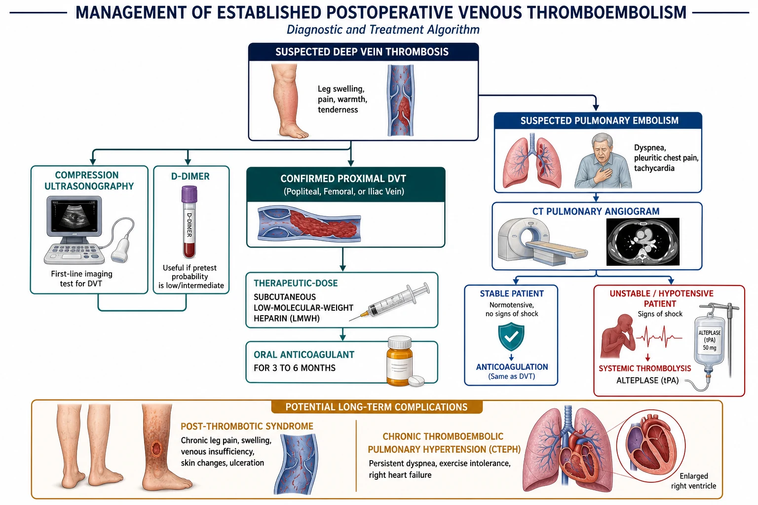
- Compression ultrasonography (venous duplex) — the first-line and gold-standard imaging for DVT. Non-compressibility of a vein under probe pressure is diagnostic; absent or phasic flow and lack of colour augmentation support the diagnosis. Sensitivity for proximal DVT exceeds ninety-five percent; it is less reliable for isolated calf DVT (serial scanning at one week if negative and suspicion persists).
- CT pulmonary angiography (CTPA) — the first-line imaging for PE. Filling defect(s) in the pulmonary arterial tree are diagnostic; it also identifies alternative diagnoses (pneumonia, aortic dissection). It is the test of choice in haemodynamically stable patients.
- Ventilation-perfusion (V/Q) scan — used when CTPA is contraindicated (severe renal failure, contrast allergy, pregnancy), but interpretation requires a chest X-ray and is only definitive when the scan is normal or shows high-probability mismatched defects.
- Echocardiography, ECG and troponin/BNP — in suspected submassive or massive PE show right ventricular strain (right heart dilation, McConnell sign, raised troponin/BNP, right axis deviation, S1Q3T3 pattern — a non-specific and late sign).
- Lower-limb venous ultrasound — performed in suspected PE if CTPA is unavailable, as proximal DVT confirms the need for treatment. [1]
Management — Resuscitation
Established massive PE is a perioperative emergency. The resuscitation bundle is time-critical: [1]
- Call for help — arrest team, ICU, anaesthetics, and haematology.
- Airway and breathing — high-flow oxygen (fifteen litres via non-rebreather) targeting saturations above ninety-four percent; consider non-invasive ventilation if respiratory failure.
- Circulation — large-bore intravenous access; cautious fluid bolus (crystalloid 250 to 500 mL) because excessive fluid worsens right-heart failure; vasopressors/inotropes (noradrenaline, dobutamine) for shock; avoid excessive fluid resuscitation.
- Empirical anticoagulation — therapeutic-dose unfractionated heparin IV bolus (eighty units/kg) then infusion (eighteen units/kg/h) if bleeding risk allows; UFH is preferred because it is rapidly reversible, fast-acting and easily monitored (APTT) in the perioperative window.
- Thrombolysis — for massive PE with hypotension/shock: alteplase (rt-PA) 50 mg IV over five minutes (or one hundred mg over two hours per older regimens). In peri-arrest, give thrombolysis empirically without confirmatory CTPA — the risk of missing a massive PE exceeds the bleeding risk.
- Surgical embolectomy or catheter-directed thrombolysis — if thrombolysis is contraindicated (recent major surgery, active bleeding, intracranial lesion) or fails.
- Reversal of prophylactic anticoagulation if life-threatening bleeding occurs — protamine sulphate (1 mg per 1 mg of enoxaparin, within eight to twelve hours, partial reversal) for LMWH; protamine fully reverses UFH. [1]
For the unstable postoperative patient with suspected PE, the decision between empirical thrombolysis and CTPA balances the freshness of the surgical wound against the immediacy of death — involve the surgical team and senior clinician immediately. [1]
Management — Definitive & Stepwise Prophylaxis
The definitive strategy is risk-stratified prophylaxis, applied to every patient and reviewed daily. [1]
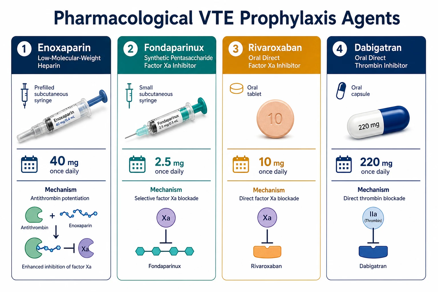
Pharmacological prophylaxis dosing
| Agent | Class | Prophylactic dose | Route | Timing | Notes |
|---|---|---|---|---|---|
| Enoxaparin | LMWH | 40 mg once daily (high-risk orthopaedic: 30 mg twice daily) | SC | Start 12 h pre-op or 12 h post-op | Standard agent; reduce to 20 mg OD if CrCl below 30 mL/min |
| Dalteparin | LMWH | 5000 IU once daily (orthopaedic: 5000 IU then 5000-7500 IU daily) | SC | Evening before, then daily | Once-daily; oncology extended-dose |
| Tinzaparin | LMWH | 75 IU/kg or 4500 IU once daily | SC | Daily | Less renal accumulation than enoxaparin |
| Unfractionated heparin (UFH) | Heparin | 5000 IU every 8-12 h | SC | Two hours pre-op, then every 8 h | Preferred in severe renal failure and in HIT history (no — avoid if HIT); rapidly reversible |
| Fondaparinux | Pentasaccharide, factor Xa | 2.5 mg once daily | SC | 6-8 h post-op | Non-inferior to LMWH; contraindicated if CrCl below 30 mL/min; no HIT risk |
| Rivaroxaban | Direct Xa inhibitor (DOAC) | 10 mg once daily | PO | 6-10 h post-op | Orthopaedic only (RECORD); no routine monitoring; avoid in severe hepatic/renal impairment |
| Apixaban | Direct Xa inhibitor (DOAC) | 2.5 mg twice daily | PO | 12-24 h post-op | Orthopaedic (ADVANCE); twice-daily dosing |
| Dabigatran | Direct thrombin inhibitor (DOAC) | 220 mg once daily (two 110 mg capsules, half-dose start) | PO | 1-4 h post-op (half dose), then full | Orthopaedic (RE-NOVATE); dyspepsia, GI side-effects |
| Warfarin | Vitamin K antagonist | Variable, INR-guided (target 2-3) | PO | — | Rarely for primary prophylaxis; needs bridging and INR monitoring |
Mechanical prophylaxis
| Method | Mechanism | Indication | Cautions |
|---|---|---|---|
| Graduated compression stockings (TEDS) | Reduce venous stasis; improve venous return | Low/moderate risk; adjunct to LMWH | Do not use in severe peripheral arterial disease (ankle pressure below eighty mmHg), severe leg oedema, deformity, acute superficial thrombophlebitis; measure and fit correctly (thigh-length preferred) |
| Intermittent pneumatic compression (IPC) | Cyclical inflation empties deep veins; boosts endogenous fibrinolysis | Moderate risk; when pharmacological contraindicated | Remove to ambulate; check skin integrity; contraindicated in acute DVT (rare relative) |
| Venous foot pump | Mimics the plantar venous plexus pump | Immobilised trauma, intra-operative | Skin checks |
| Early mobilisation | Restores calf-muscle pump | All patients, every shift | The cheapest, safest, most underused measure |
| IVC filter | Traps emboli | Confirmed proximal DVT + absolute contraindication to anticoagulation; recurrent PE despite anticoagulation | Temporary (retrievable) preferred; not a substitute for prophylaxis; long-term risk of filter thrombosis and post-thrombotic syndrome |
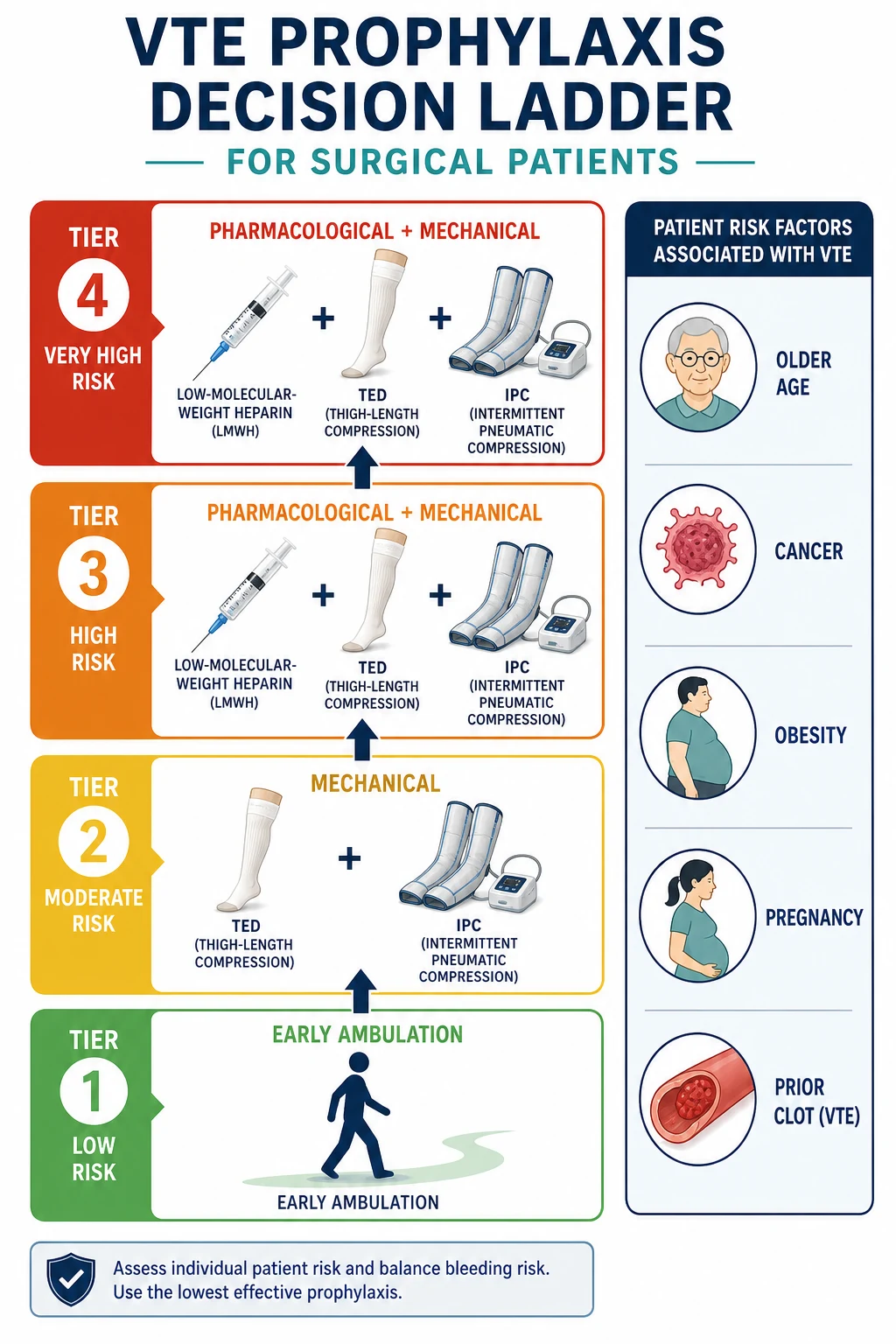
Stepwise prophylaxis ladder
- Very low risk (Caprini 0): early and frequent ambulation. No pharmacological or mechanical prophylaxis routinely required.
- Low risk (Caprini 1-2): mechanical prophylaxis — IPC is preferred over stockings alone.
- Moderate risk (Caprini 3-4): LMWH, low-dose UFH or IPC (choose one; add the other if bleeding risk is low and benefit desired).
- High/very high risk (Caprini at least 5): combined pharmacological + mechanical (dual therapy) — the additive benefit is well established.
- Start timing: LMWH/fondaparinux within twelve hours of surgery (commonly 6-12 h postoperatively, or evening before for elective surgery per local protocol). IPC applied before induction of anaesthesia and continued until the patient is fully mobile. Neuraxial caution: allow at least twelve hours after a prophylactic LMWH dose before spinal/epidural needle insertion, and at least four hours after catheter removal before the next dose, to avoid spinal haematoma. [1]
Treatment of established VTE (when prophylaxis has failed)
- Acute anticoagulation — therapeutic-dose LMWH (enoxaparin 1 mg/kg twice daily or 1.5 mg/kg once daily) or UFH infusion, overlapping with an oral agent. DOACs (rivaroxaban 15 mg twice daily for three weeks then 20 mg once daily; apixaban 10 mg twice daily for a week then 5 mg twice daily) are now first-line for many patients as they need no parenteral lead-in.
- Duration — at least three months for provoked (surgical) proximal DVT/PE; three to six months typically. Extended therapy (indefinite) for unprovoked VTE, recurrent VTE, cancer-associated thrombosis (six months then ongoing while cancer active).
- Thrombolysis — for massive PE (above) and, selectively, limb-threatening iliofemoral DVT (phlegmasia) by catheter-directed route.
- Compression stockings for symptom relief and post-thrombotic syndrome prevention. [1]
Specific Subtypes & Scenarios
Major orthopaedic surgery (highest elective risk). THR, TKR and hip-fracture repair carry the highest VTE risk of any elective procedure. Extended prophylaxis for twenty-eight to thirty-five days is standard, supported by the RECORD (rivaroxaban), ADVANCE (apixaban), RE-NOVATE (dabigatran) and RE-MODEL (dabigatran knee) programmes.[3][4] DOACs (rivaroxaban 10 mg OD, apixaban 2.5 mg BD, dabigatran 220 mg OD) and LMWH are all guideline-acceptable; the choice balances cost, monitoring and bleeding. Hip-fracture patients are elderly with multiple comorbidities — fondaparinux or LMWH is preferred over early DOAC.
Cancer surgery (ENOXACAN II). Patients undergoing curative abdominal/pelvic cancer surgery require extended-duration enoxaparin for twenty-eight days postoperatively, which reduced venographic DVT from twenty-one to twelve percent in the landmark trial without excess bleeding.[2] Hospitalised medical patients with active cancer also warrant prophylaxis throughout admission.
Neurosurgery (craniotomy and spinal surgery). Intracranial and spinal surgery carries high VTE risk but also high bleeding consequence — the classic dilemma. Mechanical prophylaxis (IPC, with or without TEDS) is started intra-operatively; pharmacological prophylaxis (LMWH) is typically deferred for forty-eight to seventy-two hours postoperatively once haemostasis is secure, then added. Post-haemorrhagic traumatic brain injury follows a similar delayed-start strategy. [1]
Major trauma and spinal cord injury. Among the highest VTE-risk groups. Early mechanical prophylaxis is applied immediately, and LMWH commenced once major bleeding is excluded (typically within thirty-six to seventy-two hours). IVC filters are reserved for patients with confirmed proximal DVT in whom anticoagulation is absolutely contraindicated. [1]
Bariatric surgery. Extreme obesity alters drug distribution; standard LMWH doses may be subtherapeutic and some centres dose-adjust (e.g. enoxaparin 40 mg twice daily or higher-weight dosing). High risk from immobility, OSA and metabolic syndrome — combined mechanical and pharmacological prophylaxis is routine. [1]
Major general/abdominal and urological surgery. LMWH (enoxaparin 40 mg SC OD) plus IPC for moderate-to-high risk; five to seven days or until full ambulation. Laparoscopic procedures over forty-five minutes still warrant risk assessment — many fall into the moderate category. [1]
Medical patients (medical VTE prophylaxis). Acutely ill medical inpatients with heart failure, severe respiratory disease, sepsis or immobility benefit from prophylactic LMWH or fondaparinux for the duration of impaired mobility, supported by MEDENOX (enoxaparin), PREVENT (dalteparin) and ARTEMIS (fondaparinux).[6][7] Critically ill ICU patients receive prophylaxis unless actively bleeding.
Vascular surgery. Tailor to the procedure: high-risk aortic or major reconstructions get LMWH plus IPC; lower-extremity bypass requires individualised balancing of graft patency and bleeding. [1]
Complications & Pitfalls
Thrombotic complications of inadequate prophylaxis:
- Fatal PE — the catastrophic, largely preventable outcome.
- Post-thrombotic syndrome (PTS) — chronic venous hypertension after DVT from valve destruction and persistent obstruction. Affects twenty to fifty percent of DVT patients: leg heaviness, swelling, pain, hyperpigmentation, lipodermatosclerosis and venous ulcers. Reduced by early adequate anticoagulation; role of compression stockings is debated.
- Chronic thromboembolic pulmonary hypertension (CTEPH) — organised residual PE remodels pulmonary arteries; presents months to years later with exertional dyspnoea and right heart failure; treated by pulmonary endarterectomy.
- Recurrent VTE — risk of recurrence after a first event is up to thirty percent at ten years. [1]
Bleeding complications of over-prophylaxis:
- Wound haematoma (the most common, and a particular concern after neurosurgery and urological procedures).
- Gastrointestinal, retroperitoneal, and intracranial bleeding.
- The bleeding versus thrombosis balance is the central clinical judgement — there is no risk-free option. [1]
Heparin-induced thrombocytopenia (HIT) — the paradoxical prothrombotic complication of heparin. An IgG antibody against platelet factor 4 (PF4)-heparin complexes causes platelet activation and aggregation. Suspect a platelet fall of fifty percent or below one hundred and fifty thousand between days five and fourteen of heparin exposure (or sooner if prior exposure). The 4Ts score (Thrombocytopenia, Timing, Thrombosis, oTher cause) estimates pre-test probability; confirm with an anti-PF4 ELISA or serotonin-release assay. Management: stop ALL heparin (LMWH, UFH, line flushes) and substitute a non-heparin anticoagulant (argatroban, danaparoid, fondaparinux, or a DOAC). Fondaparinux is attractive for prophylaxis in patients with a HIT history because it has essentially no cross-reactivity. [1]
Key pitfalls:
- Omitting the risk assessment — the most common reason prophylaxis is missed.
- Timing errors with neuraxial anaesthesia — spinal haematoma is devastating; respect the twelve-hour (prophylactic LMWH) and twenty-four-hour (therapeutic) windows.
- Renal dosing neglected — enoxaparin and fondaparinux accumulate in renal failure; check creatinine clearance.
- Confusing prophylactic and therapeutic LMWH doses — 40 mg OD is prophylactic; 1 mg/kg twice daily is therapeutic.
- Inadequate duration in orthopaedic/cancer surgery — stopping at discharge loses the benefit of extended prophylaxis.
- Bleeding risk ignored — applying pharmacological prophylaxis to a patient with active bleeding or severe thrombocytopenia. [1]
Prognosis & Disposition
With appropriate risk-stratified prophylaxis, VTE events fall by fifty to seventy percent and fatal PE becomes rare. Without it, the untreated event rates quoted in the Epidemiology table apply. After a confirmed DVT or PE, anticoagulation continues for at least three months (provoked) to lifelong (unprovoked, recurrent, or cancer-associated). [1]
Disposition: pharmacological prophylaxis continues until the patient is fully ambulating and acute illness has resolved (medical patients) or for twenty-eight days (major orthopaedic and cancer surgery). Patients discharged on DOACs or LMWH need education on injection technique, bleeding precautions, and warning signs (dyspnoea, chest pain, leg swelling, bleeding). Early follow-up bridges the inpatient-to-community transition, and mechanical prophylaxis continues at home if mobility remains impaired. [1]
Mortality of massive PE untreated is around thirty percent; with prompt recognition, anticoagulation and thrombolysis it falls below ten percent. Recurrent VTE risk is highest in the first month after an event. [1]
Special Populations
Pregnancy and the postpartum period. Pregnancy is a hypercoagulable, pro-stasis state and VTE remains a leading cause of maternal death. LMWH is the agent of choice throughout pregnancy and lactation — it does not cross the placenta. Warfarin is teratogenic (first trimester) and causes fetal bleeding (third trimester), and DOACs are contraindicated in pregnancy and breastfeeding. Caesarean section, especially emergency, and additional risk factors (previous VTE, thrombophilia, obesity) warrant LMWH prophylaxis for at least six weeks postpartum. Neuraxial labour analgesia requires the same LMWH-timing precautions as surgical anaesthesia. [1]
Renal impairment. LMWHs are renally cleared. For creatinine clearance below thirty mL/min, reduce enoxaparin to 20 mg SC OD, switch to UFH (5000 IU SC every 8-12 h), or use tinzaparin (less renally cleared). Fondaparinux is contraindicated below thirty mL/min. UFH is fully reversible with protamine, an advantage in perioperative renal failure. [1]
Elderly. Higher baseline VTE risk, more comorbidity, more polypharmacy, and higher bleeding risk — reassess risk daily and favour IPC plus cautious pharmacological dosing. Avoid DOACs in severe renal impairment common in this group. [1]
The already-anticoagulated patient. For patients on long-term warfarin or a DOAC, manage perioperative anticoagulation by bridging: stop the DOAC two to five days pre-op (drug- and renal-dependent), stop warfarin five days pre-op, bridge with therapeutic-dose LMWH (last dose twenty-four hours pre-op) if high thromboembolic risk (mechanical mitral valve, recent VTE, AF with prior stroke), and resume post-operatively when haemostasis is secure. [1]
Children. Rarely need surgical prophylaxis except after major trauma, adolescent OCP use with surgery, or central venous lines. Dose LMWH weight-based (enoxaparin 0.5 mg/kg twice daily, anti-Xa monitored). [1]
Severe liver disease and thrombocytopenia. Already coagulopathic — bleeding risk usually dominates; mechanical prophylaxis is often the safer default. [1]
Evidence, Guidelines & Regional Differences
Landmark VTE prophylaxis trials
Guidelines:
- ACCP (American College of Chest Physicians) 9th edition (2012) — the international reference for risk-stratified prophylaxis; recommends LMWH over UFH for most surgical patients and dual therapy for high risk.
- ASH (American Society of Hematology, 2018) — broadly concordant with ACCP.
- NICE Clinical Guideline CG92 (UK) — mandates universal VTE and bleeding risk assessment on admission, favours anti-embolism stockings plus LMWH, and sets the twenty-eight-day extended prophylaxis benchmark.
- ESVS (European Society for Vascular Surgery) and ESC —- provide cardiovascular and venous-disease-specific guidance.
- Indian practice — largely ACCP-aligned; enoxaparin 40 mg SC OD is the workhorse, with fondaparinux as a useful alternative given its once-daily, HIT-free profile and lower cost. [1]
Controversies. (i) The net benefit of extended DOAC prophylaxis in medical patients (MAGELLAN showed efficacy but at the cost of bleeding; MARINER was neutral overall). (ii) Whether asymptomatic distal DVTs detected on surveillance ultrasound warrant treatment. (iii) The optimal agent in orthopaedics — DOACs are convenient and effective but costlier and lack reversal agents for all (idarucizumab reverses dabigatran; andexanet reverses apixaban/rivaroxaban). (iv) The role of DOACs immediately post-neurosurgery remains limited by bleeding concerns. [1]
- UK (NICE): anti-embolism stockings (thigh-length, measured) plus LMWH; universal admission assessment; strong emphasis on twenty-eight-day orthopaedic and cancer prophylaxis.
- USA (ACCP/ASH): Caprini-driven; IPC preferred for low risk; DOACs widely used in orthopaedics; fondaparinux accepted.
- India / South Asia: ACCP principles applied; enoxaparin 40 mg SC OD dominant; fondaparinux valued for cost, once-daily dosing and no HIT risk; DOAC use growing in private tertiary centres.
- Europe: ACCP/ESVS; dabigatran and rivaroxaban well established in orthopaedics after the European-led RECORD and RE-NOVATE programmes.
Exam Pearls
- Virchow's triad: STASIS, HYPERCOAGULABILITY, ENDOTHELIAL INJURY — and surgery violates all three.[1]
- VTE is the leading PREVENTABLE cause of hospital death.
- Caprini score = surgical patients; Padua score = medical patients. A Padua score of four or more is high risk; a Caprini score of five or more warrants dual therapy.[8][9]
- LMWH (enoxaparin 40 mg SC OD) is the standard pharmacological prophylaxis. Therapeutic enoxaparin is 1 mg/kg twice daily (or 1.5 mg/kg once daily) — do not confuse the two.
- Fondaparinux 2.5 mg SC OD — synthetic factor Xa inhibitor, no HIT risk, contraindicated if CrCl below thirty.
- DOAC orthopaedic doses: rivaroxaban 10 mg OD, apixaban 2.5 mg twice daily, dabigatran 220 mg OD — all for twenty-eight days after hip/knee surgery.[3][4]
- Extended twenty-eight-day prophylaxis after major orthopaedic or cancer surgery (ENOXACAN II for cancer).[2]
- Mechanical: TEDS + IPC + early mobilisation. Both modalities (pharmacological + mechanical) for high risk.
- Neuraxial rule: twelve hours after prophylactic LMWH before spinal/epidural; twenty-four hours after therapeutic; four hours after catheter removal before next dose.
- HIT: platelet fall of fifty percent or below one hundred and fifty, days five to fourteen of heparin — stop ALL heparin, give argatroban/danaparoid/fondaparinux; fondaparinux has no cross-reactivity.
- Renal impairment (CrCl below thirty): reduce enoxaparin to 20 mg OD or switch to UFH; fondaparinux contraindicated.
- Pregnancy: LMWH (does not cross placenta); warfarin teratogenic, DOACs contraindicated.
- Massive PE (unstable): oxygen, fluids/inotropes, alteplase 50 mg IV over five minutes; empirical if peri-arrest.
- Post-thrombotic syndrome: chronic venous insufficiency in twenty to fifty percent after DVT — heavy leg, oedema, ulcers.
- Investigations: DVT = compression ultrasound; PE = CTPA (V/Q if contraindicated/pregnant); D-dimer only useful if low probability and negative.
- Wells DVT: score of two or less = unlikely; three or more = likely.
- IVC filter: only when anticoagulation contraindicated with confirmed proximal DVT — not a substitute for prophylaxis.
- ENDORSE taught us that ~42 percent of inpatients are high-risk and many are under-prophylaxed.[1]
Exam application bank (NEET-PG / INICET)
One-line answer
Venous thromboembolism (VTE) = deep vein thrombosis (DVT) + pulmonary embolism (PE). It is the leading preventable cause of hospital death. Surgical patients carry a ten- to twenty-fold increased risk. Prevention rests on Virchow's triad (stasis, hypercoagulability, endothelial injury), risk stratification (Caprini for surgical, Padua for medical patients) and a stepwise ladder: early mobilisation, mechanical methods (TEDS, IPC) and pharmacological prophylaxis (enoxaparin 40 mg SC OD, fondaparinux 2.5 mg SC OD, rivaroxaban 10 mg PO OD). High-risk patients get both. Extended prophylaxis for twenty-eight days after major orthopaedic or cancer surgery. Massive PE is an emergency: oxygen, haemodynamic support, systemic thrombolysis (alteplase). [1]
Worked stems (answer without another resource)
Stem 1 — Classic presentation. Map symptoms to mechanism; name the first investigation and first treatment step with dose/route if drug therapy is standard. [1]
Stem 2 — Unstable / complicated. List red flags that force immediate resuscitation, theatre, ICU, antidote, or reperfusion — and what you do in the first 15 minutes. [1]
Stem 3 — Atypical group. Elderly, pregnancy, child, or immunocompromised: how presentation and thresholds change. [1]
Stem 4 — Differential trap. Name the three closest mimics and one discriminator for each. [1]
Stem 5 — Disposition. Who goes home with safety-netting, who is admitted, who needs HDU/ICU/theatre, and what follow-up is mandatory. [1]
Rapid viva checklist
- Definition + classification
- Pathophysiology chain
- Bedside signs / criteria
- Score with exact components (if any)
- Emergency bundle
- Definitive therapy with doses
- Complications of disease and of treatment
- Special populations
- Guideline/trial name if classic
- Three exam traps
Coverage self-check
If you cannot answer any stem above from this page alone, re-read the matching section — the page is intended to be self-sufficient for final-prof and NEET-PG/INICET questions on Perioperative VTE Prophylaxis.
References
- [1]Cohen AT, Tapson VF, Bergmann JF, et al. (ENDORSE). Venous thromboembolism risk and prophylaxis in the acute hospital care setting (ENDORSE study): a multinational cross-sectional study Lancet, 2008.PMID 18242412
- [2]Bergqvist D, Agnelli G, Cohen AT, et al. (ENOXACAN II). Duration of prophylaxis against venous thromboembolism with enoxaparin after surgery for cancer N Engl J Med, 2002.PMID 11919306
- [3]Lassen MR, Raskob GE, Gallus A, et al. (ADVANCE-3). Apixaban versus enoxaparin for thromboprophylaxis after hip replacement N Engl J Med, 2010.PMID 21175312
- [4]Eriksson BI, Dahl OE, Rosencher N, et al. (RE-NOVATE). Dabigatran etexilate versus enoxaparin for prevention of venous thromboembolism after total hip replacement: a randomised, double-blind, non-inferiority trial Lancet, 2007.PMID 17869635
- [5]Cohen AT, Spiro TE, Buller HR, et al. (MAGELLAN). Rivaroxaban for thromboprophylaxis in acutely ill medical patients N Engl J Med, 2013.PMID 23388003
- [6]Leizorovicz A, Cohen AT, Turpie AG, et al. (PREVENT). Randomized, placebo-controlled trial of dalteparin for the prevention of venous thromboembolism in acutely ill medical patients Circulation, 2004.PMID 15289368
- [7]Cohen AT, Davidson BL, Gallus AS, et al. (ARTEMIS). Efficacy and safety of fondaparinux for the prevention of venous thromboembolism in older acute medical patients: randomised placebo controlled trial BMJ, 2006.PMID 16439370
- [8]Barbar S, Noventa F, Rossetto V, et al. A risk assessment model for the identification of hospitalized medical patients at risk for venous thromboembolism: the Padua Prediction Score J Thromb Haemost, 2010.PMID 20738765
- [9]Cronin M, Dengler N, Krauss ES, et al. Completion of the Updated Caprini Risk Assessment Model (2013 Version) Clin Appl Thromb Hemost, 2019.PMID 30939900