General Surgery · General Surgery
Peripheral Arterial Disease
Also known as PAD · Peripheral vascular disease · Intermittent claudication · Lower limb ischaemia · Critical limb ischaemia
Peripheral arterial disease (PAD) is atherosclerotic narrowing of the lower limb arteries. Presents on a spectrum: asymptomatic PAD (ABPI under 0.9, no symptoms), intermittent claudication (calf pain on walking, relieved by rest), chronic limb-threatening ischaemia (CLTI) with rest pain (severe nocturnal foot pain relieved by dependency) and tissue loss (ulceration, gangrene), and acute limb ischaemia (ALI) with the 6 Ps. Smoking is the dominant modifiable risk factor; diabetes multiplies risk 2-4 fold. ABPI under 0.9 confirms PAD; toe-brachial index replaces ABPI when medial Mönckeberg calcinosis makes ankle vessels non-compressible (diabetes, CKD). Fontaine classification: I (asymptomatic), II (claudication), III (rest pain), IV (gangrene). TASC II grades aortoiliac and femoropopliteal lesions A-D, guiding endovascular vs surgical revascularisation. WIfI (Wound-Ischaemia-foot Infection) risk-stratifies CLTI amputation risk. Management ladder: stop smoking + supervised exercise + antiplatelet + statin (best medical therapy, BMT, for all), cilostazol for symptomatic claudication, low-dose rivaroxaban + aspirin (COMPASS regimen) for symptomatic PAD, angioplasty ± stent for focal TASC A-B disease, bypass with autologous vein for extensive TASC C-D disease, fasciotomy after revascularisation for reperfusion compartment syndrome, and amputation for unsalvageable limb. Acute limb ischaemia: emergency IV unfractionated heparin, Fogarty catheter embolectomy for embolic, catheter-directed thrombolysis for thrombotic. Time window 4-6 hours before irreversible muscle necrosis.
On this page & tools
Your progress
Saved locally on this device.
Exam tags
Red flags
Overview & Definition
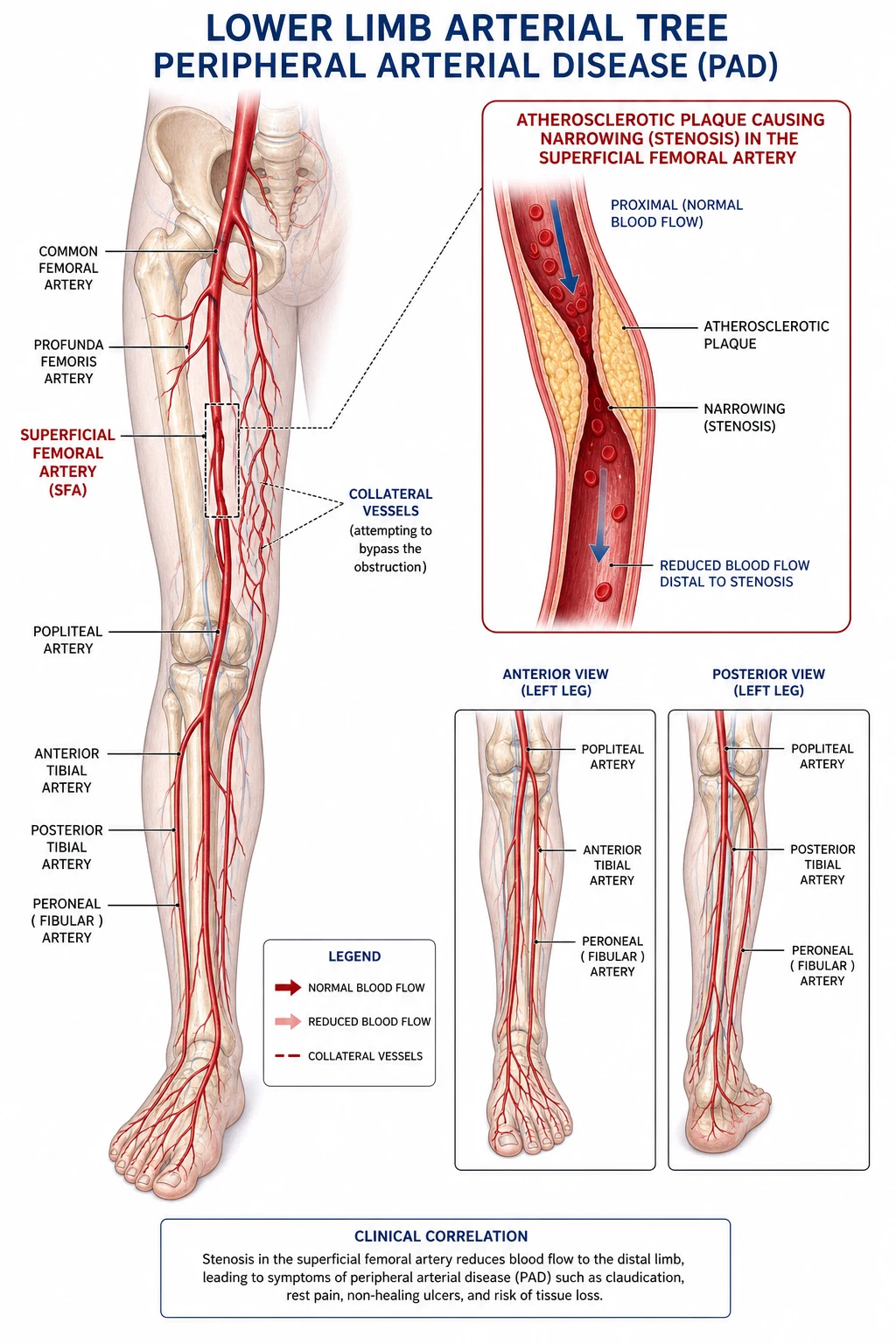
Peripheral arterial disease (PAD) is a chronic atherosclerotic condition affecting the arteries of the lower limbs (and — far less commonly — the upper limbs, mesenteric, renal, or carotid arteries). It is part of the same systemic atherosclerotic process that causes coronary artery disease and cerebrovascular disease — patients with PAD have a 2 to 4 times higher risk of myocardial infarction (MI) and stroke, and most of them die from a cardiovascular event rather than from the leg itself.[1][3]
PAD exists on a clinical spectrum from asymptomatic PAD (ABPI under 0.9 with no reported symptoms — found in roughly half of all cases on screening) through intermittent claudication (reproducible muscle pain on walking, relieved by rest) to chronic limb-threatening ischaemia (CLTI) — the modern term that replaces "critical limb ischaemia" and bundles Fontaine III (rest pain) and Fontaine IV (tissue loss, ulceration, gangrene). A fourth entity — acute limb ischaemia (ALI) — describes a sudden arterial occlusion (embolism or in-situ thrombosis) that threatens the limb within hours and is a vascular surgical emergency. Severity worsens as stenosis progresses from moderate narrowing through critical (over 70% lumen area reduction) stenosis to total occlusion.[1][2]
Key concepts the examiner rewards:
- Polyvascular disease — any PAD patient has atherosclerosis elsewhere. Most have coronary or carotid disease; PAD is a coronary risk equivalent on every global risk calculator.
- The asymptomatic majority — over half of community-dwelling PAD cases are asymptomatic. They still carry the same cardiovascular risk and deserve the same BMT as claudicants.
- Stages of chronic disease are not linear — patients may remain at Fontaine II for decades, or progress rapidly to CLTI after a plaque rupture event; progression depends on risk-factor control rather than on a single stenosis.[3]
Classification
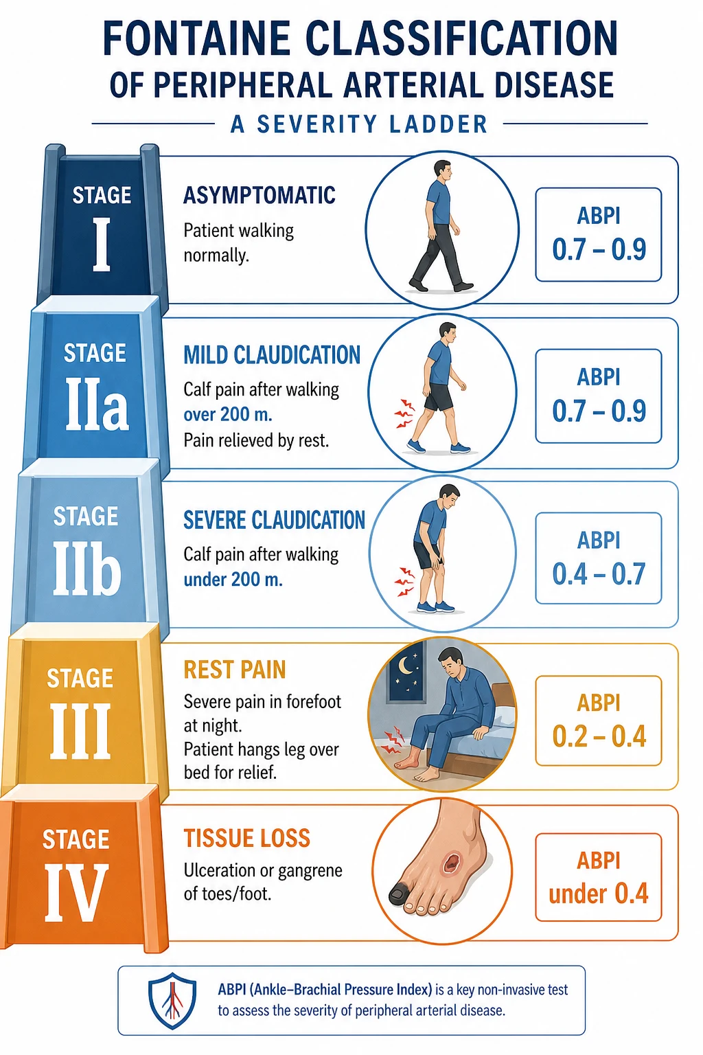
Fontaine classification (the standard clinical staging):[1]
| Stage | Description | Typical ABPI | Clinical priority |
|---|---|---|---|
| I | Asymptomatic (detected on ABPI screening) | 0.7 to 0.9 | Risk-factor modification, no intervention |
| IIa | Mild claudication (pain after walking over 200 m) | 0.5 to 0.7 | Supervised exercise + BMT |
| IIb | Severe claudication (pain after walking under 200 m) | 0.4 to 0.5 | BMT; consider revascularisation if refractory |
| III | Rest pain (severe pain in foot at night, relieved by dependency) | 0.3 to 0.4 | Urgent vascular referral; revascularisation |
| IV | Tissue loss (ulceration, gangrene) | under 0.3 | Critical limb ischaemia; urgent revascularisation or amputation |
Clinical staging pearls (the exam framework):
- Stages I and II = chronic limb-threatening ischaemia absent — these are not limb-threatening; treatment is conservative (smoking cessation, supervised exercise, antiplatelet, statin).
- Stages III and IV = chronic limb-threatening ischaemia (CLTI) — these are limb-threatening and require urgent vascular assessment and revascularisation where feasible. The Global Vascular Guidelines (GVG 2019) now prefer the term CLTI over the older "critical limb ischaemia" because it encompasses the full spectrum of ischaemia severity, tissue loss, and infection (the WIFI staging: Wound, Ischaemia, foot Infection).[1][2]
- Fontaine III = rest pain without ulceration. Fontaine IV = ulceration or gangrene. Together, III + IV = CLTI.
- Leriche syndrome is a specific Fontaine IIb/III pattern caused by aortoiliac occlusion (terminal aortic occlusion). The classic triad: (1) buttock claudication, (2) erectile dysfunction (impotence from compromised internal iliac perfusion), and (3) absent femoral pulses bilaterally. Patients may also develop thigh claudication and trophic skin changes in both legs. On examination, listen for an aortic bruit; confirm with CTA or MRA of the abdominal aorta and iliacs. Treatment: aortobifemoral bypass (gold standard for extensive disease) or aortoiliac angioplasty + stent (for focal iliac stenoses). Smoking cessation and best medical therapy are mandatory in all cases.[1]
Rutherford categories (alternative, used in research and revascularisation trials): the original Rutherford 0 to 6 scale maps to Fontaine but adds haemodynamic and anatomical granularity, used widely in trial entry criteria and endovascular reporting.[1]
| Rutherford | Clinical | ABPI | Equivalent Fontaine |
|---|---|---|---|
| 0 | Asymptomatic | over 0.9 | — (or I) |
| 1 | Mild claudication | 0.7 to 0.9 | I / IIa |
| 2 | Moderate claudication | 0.5 to 0.7 | IIb |
| 3 | Severe claudication | 0.4 to 0.5 | IIb |
| 4 | Rest pain | 0.3 to 0.4 | III |
| 5 | Minor tissue loss (ulcer, focal gangrene) | 0.3 to 0.4 | IV |
| 6 | Major tissue loss (extensive gangrene above transmetatarsal) | under 0.3 | IV |
TASC II lesion classification (the revascularisation roadmap): the Inter-Society Consensus document (TASC II, 2007) grades lesions in the aortoiliac and femoropopliteal segments as A, B, C, or D based on anatomical complexity. A and B lesions favour endovascular first; C and D lesions favour open surgical reconstruction; modern practice shifts favour endovascular-first for most femoropopliteal disease up to 25 cm in experienced centres.[1]
TASC II — aortoiliac (AI) segment:[1]
| Type | Description | Default strategy |
|---|---|---|
| A | Unilateral or bilateral short (under 3 cm) common iliac stenosis; unilateral short external iliac stenosis | Endovascular |
| B | Short (under 3 cm) infrarenal aortic stenosis; unilateral CIA occlusion; short (under 3 cm) external iliac stenosis with heavy calcification | Endovascular |
| C | Bilateral CIA occlusions; bilateral external iliac stenoses (3-10 cm, not extending into CFA); unilateral EIA stenosis extending into CFA; unilateral EIA occlusion with severe contralateral disease | Endovascular-first often, surgical for fit patients with extensive disease |
| D | Infrarenal aortic occlusion; aortoiliac occlusion (Leriche); diffuse disease involving the aorta and both iliac arteries requiring open repair; bilateral EIA occlusions | Surgical (aortobifemoral bypass) |
TASC II — femoropopliteal (FP) segment:[1]
| Type | Description | Default strategy |
|---|---|---|
| A | Single stenosis under 10 cm; single occlusion under 5 cm | Endovascular |
| B | Multiple stenoses each under 5 cm ("stuttering"); single stenosis or occlusion 5-15 cm not involving infrageniculate popliteal; single or multiple lesions in the absence of continuous tibial vessels | Endovascular |
| C | Single stenosis or occlusion over 15 cm; multiple stenoses or occlusions each 5-15 cm with or without heavy calcification | Surgical or endovascular (endovascular increasingly favoured) |
| D | Chronic total occlusion (CTO) of CFA or SFA over 20 cm; chronic occlusion of popliteal artery and proximal trifurcation vessels | Surgical (bypass) if vein conduit available |
WIfI staging for Chronic Limb-Threatening Ischaemia (CLTI): the Society for Vascular Surgery WIfI system stratifies amputation risk (very low / low / medium / high) and guides revascularisation urgency by grading three axes independently:[5]
| Grade | W (Wound) | I (Ischaemia) | fI (foot Infection) |
|---|---|---|---|
| 0 | No ulcer; no gangrene | ABPI over 0.8; toe pressure over 60 mmHg; TcPO₂ over 60 mmHg | Uninfected |
| 1 | Small shallow ulcer (no bone / tendon); toes gangrenous | ABPI 0.6-0.8; toe pressure 40-60 mmHg; TcPO₂ 40-60 mmHg | Mild (erysipelas-like) |
| 2 | Deeper ulcer (bone, joint, tendon exposure); widespread gangrene | ABPI 0.4-0.6; toe pressure under 40 mmHg; TcPO₂ under 40 mmHg | Moderate (deep abscess, osteomyelitis) |
| 3 | Extensive deep ulcer; extensive gangrene | ABPI under 0.4; toe pressure under 20 mmHg; TcPO₂ under 20 mmHg | Severe (systemic sepsis) |
For instance, a patient with W2 I3 fI0 (deep forefoot ulcer, severe ischaemia, no infection) is high risk of major amputation without urgent revascularisation. WIfI underpins the Global Vascular Guidelines (GVG 2019) risk stratification and the choice of revascularisation strategy by Infrainguinal Disease (GLASS) stage — the GLASS grading mirrors TASC but is applied within a "patient x lesion x operator" decision framework.[5][1]
By clinical syndrome:
- Intermittent claudication (IC) — reproducible calf/thigh/buttock pain on walking, relieved within minutes by rest. Not limb-threatening.
- Critical limb ischaemia (CLI) — rest pain for over 2 weeks, non-healing ulcer, or gangrene. Limb-threatening. Urgent revascularisation needed.
- Acute limb ischaemia (ALI) — sudden onset (hours). The 6 Ps. Surgical emergency. [1]
Fontaine & ABPI — numbers examiners test
Epidemiology & Risk Factors
PAD is one of the three major atherosclerotic vascular diseases (alongside coronary and cerebrovascular disease) and is among the most underdiagnosed. The landmark global burden study by Fowkes and colleagues (Lancet 2013) pooled 34 community-based studies (112,027 participants) using the ABPI under 0.9 definition and estimated that approximately 202 million people worldwide were living with PAD in 2010, an increase of 23.5% from the 2000 estimate of 164 million; the increase was substantially steeper in low- and middle-income countries (28.7%) than in high-income countries (13.1%), reflecting the epidemiological transition of atherosclerosis to the developing world.[4] The Global Burden of Disease 2019 update estimates that 236 million adults aged 25 and over were affected by 2015, and the age-standardised prevalence has continued to rise.[3][4]
Pooled prevalence by age bracket (synthesised from Fowkes 2013 and Criqui 2021):[4][3]
| Age band | Crude prevalence (ABPI under 0.9) |
|---|---|
| 40 to 49 years | ~3 to 5% |
| 50 to 59 years | ~6 to 10% |
| 60 to 69 years | ~10 to 15% |
| Over 70 years | 15 to 25% |
Symptom distribution: across the full PAD population, roughly 50% are asymptomatic (ABPI detected on screening), 30% have atypical leg symptoms not recognised as claudication, 15 to 20% have classical intermittent claudication, and 5 to 10% have CLTI. Mortality and cardiovascular event rates climb sharply across this gradient — even asymptomatic PAD carries a 2- to 4-fold cardiovascular mortality risk.[3]
Risk factors — quantitative breakdown
| Risk factor | Effect estimate | Notes |
|---|---|---|
| Smoking (current) | OR ~2.5–4.5 for PAD; OR ~2–3 for claudication | Dose-related (pack-years); strongest modifiable risk factor; risk declines substantially within 5 to 10 years of cessation but never returns to baseline |
| Diabetes mellitus | OR ~2.0–4.0 | Highest predictor of amputation; each 1% rise in HbA1c increases PAD risk ~25 to 30% |
| Age (per decade over 50) | ~1.5 to 2.0 fold per decade | Reflects cumulative exposure to other risk factors plus age-related vascular remodelling |
| Hypertension | OR ~1.5–2.5 | Adds independently to PAD risk; tightly linked to stroke and MI risk in PAD |
| Dyslipidaemia (LDL over 130 mg/dL) | OR ~1.5–2.0 | Apolipoprotein B and lipoprotein(a) increasingly recognised as strong predictors |
| Chronic kidney disease (eGFR under 60) | OR ~2–4 | Strongest non-traditional risk factor; accelerates medial calcification |
| Hyperhomocysteinaemia | OR ~1.5–2.0 per 5 μmol/L rise | Modifiable with B-vitamins in some studies but no clear mortality benefit (HOPE-2, SEARCH) |
| C-reactive protein (hsCRP) | Marker of inflammatory burden | Reflects plaque activity and predicts progression |
| Family history of premature ASCVD | OR ~1.5 | Particularly if first-degree relative with PAD or AAA before 60 |
| Race / ethnicity | Black and South Asian populations ~2 fold higher age-adjusted prevalence | Reflects higher diabetes and hypertension burden |
| Male sex | Modestly higher prevalence OR ~1.2–1.5 | Women are more likely to be underdiagnosed |
Smoking — the single most effective intervention: smokers develop PAD a decade earlier than non-smokers, have more aggressive multi-level disease, and have a 2- to 4-fold higher amputation rate. Smoking cessation is the most effective intervention to halt progression — benefits begin within weeks (improved endothelial function, lowered fibrinogen) and accumulate over 5 to 10 years. Offer behavioural support + nicotine replacement therapy (NRT) ± varenicline or bupropion to every patient.[1][3]
Diabetes specifics: people with diabetes have a 2- to 4-fold higher PAD prevalence, more distal (infrapopliteal) disease, medial Mönckeberg calcinosis that renders the ABPI unreliable (over 1.3), and an 8- to 15-fold higher amputation rate than non-diabetics. Because neuropathy masks the classical claudication pain, diabetics often present first with a non-healing ulcer (the "diabetic foot" — see Specific Subtypes).[3]
Chronic kidney disease: both a cause and a complication of PAD. eGFR under 60 mL/min/1.73 m² is associated with a 2- to 4-fold higher PAD prevalence; dialysis patients have 10 to 30% prevalence. CKD drives medial arterial calcinosis (Mönckeberg sclerosis), accelerates atherosclerosis, and limits contrast-based imaging. The combination of PAD + CKD + diabetes is the highest-risk triad for major adverse cardiovascular events (MACE) and major adverse limb events (MALE).[3]
Hyperhomocysteinaemia: elevated plasma homocysteine (over 15 μmol/L) confers an OR of ~1.5 to 2.0 for PAD, independent of other risk factors. Pathophysiology is uncertain — possibly endothelial injury, oxidative stress, and promotion of LDL oxidation. Treating with B-vitamins (folic acid, B6, B12) lowers homocysteine but does not improve cardiovascular or limb outcomes in large randomised trials (HOPE-2, SEARCH), so screening is not routinely recommended.[3]
Screening — who deserves an ABPI?
The ACC/AHA 2024 guideline recommends a one-time ABPI for anyone with lower extremity symptoms suggestive of PAD, non-healing lower extremity wounds, age 65 and over, age 50 to 64 with risk factors (smoking, diabetes, dyslipidaemia, hypertension, family history), or known atherosclerotic disease in another bed (coronary, carotid, renal, mesenteric).[1]
PAD at a glance — the numbers examiners test
Pathophysiology
Atherosclerosis — the molecular cascade
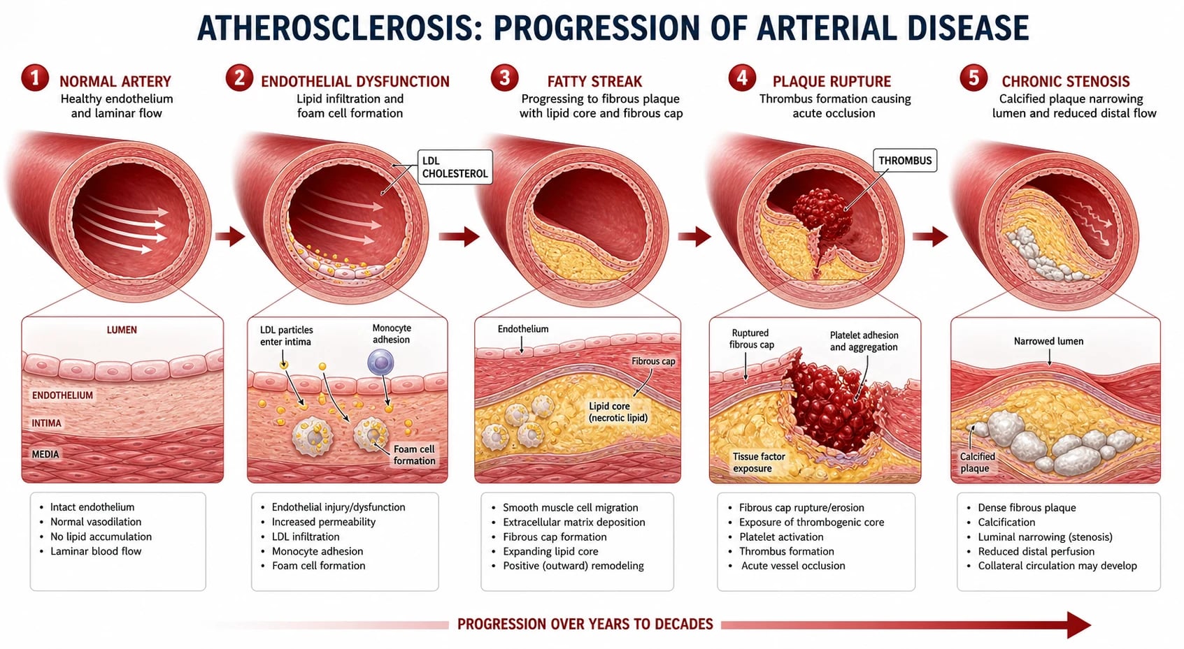
PAD is overwhelmingly caused by atherosclerosis, a chronic inflammatory response-to-injury of the arterial wall, and shares the same pathogenesis as coronary and carotid atherosclerosis. Six overlapping stages generate the mature obstructing plaque.[3]
- Endothelial dysfunction — inciting noxious stimuli (smoking toxins, hyperglycaemia, hypertension shear stress, dyslipidaemia, oxidative stress, homocysteine, inflammation) reduce nitric oxide (NO) bioavailability and increase endothelial permeability. LDL particles infiltrate the subendothelial intima.
- LDL retention and oxidation — subendothelial LDL is oxidised by reactive oxygen species; oxidised LDL is chemotactic for monocytes and toxic to endothelial cells.
- Monocyte recruitment and foam-cell formation — monocytes adhere to dysfunctional endothelium (via VCAM-1 / ICAM-1), transmigrate into the intima, and differentiate into macrophages that engulf oxidised LDL via scavenger receptors (SR-A, CD36) to become lipid-laden foam cells. Foam cells aggregate as the fatty streak — the earliest visible lesion.
- Fibrous plaque formation — smooth muscle cells migrate from the media, proliferate, and secrete collagen to form a fibrous cap over a lipid necrotic core of dead foam cells, cholesterol crystals, and cellular debris. The plaque is now haemodynamically significant when the lumen is reduced by over 70% of cross-sectional area.
- Plaque calcification and remodelling — chronic plaques undergo dystrophic calcification (medial in Mönckeberg; intimal in mature atheroma) and positive outward remodelling that preserves the lumen until late in the disease.
- Plaque rupture, erosion, or fissuring exposes the thrombogenic lipid core and collagen to flowing blood → platelet aggregation → acute thrombus formation → acute limb ischaemia. Vulnerable plaques have a thin fibrous cap, large lipid core, dense macrophage infiltrate, and microcalcification.[3]
The haemodynamic rule — why a critical stenosis matters
Resting muscle uses a small fraction of arterial inflow (under 20%), so even a sizeable stenosis may be asymptomatic at rest. During exercise, oxygen demand rises 3 to 5 fold and the diseased artery cannot recruit additional flow. The Hagen-Poiseuille relationship describes laminar flow through a tube: [1]
Q = (π × ΔP × r⁴) / (8 × η × L)
where Q is flow, ΔP is the pressure gradient, r is the lumen radius, η is blood viscosity, and L is vessel length. Flow is proportional to the fourth power of the radius — a 50% reduction in radius halves flow by 6.25-fold (1/0.5⁴) if all else were equal (in vivo, distensibility and turbulence attenuate this). Once stenosis reaches ~70% cross-sectional area reduction, distal perfusion pressure falls at rest and pain emerges on exertion (claudication); multi-level occlusion collaterals fail and even resting perfusion is inadequate (rest pain and tissue loss).[3]
[1]Inflammatory and prothrombotic milieu
Atherosclerosis is sustained by chronic innate and adaptive immunity — macrophages, T-cells, B-cells, dendritic cells — releasing pro-inflammatory cytokines (IL-1β, IL-6, TNF-α), matrix metalloproteinases (MMP-2, MMP-9) that thin the fibrous cap, and pro-thrombotic tissue factor. High-sensitivity CRP (hsCRP) is the most clinically useful biomarker; elevated hsCRP independently predicts cardiovascular events and PAD progression and is the rationale for anti-inflammatory therapies such as colchicine in coronary disease (the CANTOS and LoDoCo2 trials — not yet licensed in PAD).[3]
Collateral circulation and compensation
When an artery narrows slowly, pre-existing anastomotic arterioles enlarge and new collaterals sprout under VEGF and PDGF signalling. Well-developed collaterals can maintain perfusion at near-baseline levels despite occlusion of a major vessel; this is why some patients have normal or borderline ABPI despite significant aortoiliac disease. Collateral capacity is the main reason resting ABPI is insensitive, and why the exercise ABPI is required when symptoms are atypical or resting ABPI is borderline.[1]
Vascular calcification — intimal vs medial
Intimal calcification is the focal calcification inside atherosclerotic plaque; it disrupts the fibrous cap and is part of atherosclerotic disease. Medial (Mönckeberg) calcification is diffuse, concentric calcification of the tunica media, common in diabetes, CKD, and elderly patients. It does not narrow the lumen directly but makes arteries non-compressible (rigid pipes), falsely elevating ABPI to over 1.3. In a diabetic or uraemic patient, a high ABPI is never reassuring — use toe-brachial index (TBI) instead.[3]
Plaque biology — from lipid theory to plaque stability
Classical teaching emphasises plaque size and stenosis. Modern vascular biology emphasises plaque vulnerability: a large eccentric plaque with a thin fibrous cap, lipid-rich necrotic core, dense macrophage infiltrate, and spotty neovascularisation from vasa vasorum is prone to rupture and thrombosis even when only mildly stenotic. This explains why some acute limb ischaemia events arise from stenoses that looked moderate on duplex — and why aggressive lipid-lowering (high-intensity statin, target LDL under 1.8 mmol/L) stabilises plaque independent of regression. [1]
Acute-on-chronic events
When a chronic plaque ruptures, the resulting thrombus either:
- spontaneously lyses (subclinical, often picked up only on surveillance imaging),
- extends and occludes the vessel (acute-on-chronic limb ischaemia), or
- embolises distally (atheroembolism — blue toe syndrome, cholesterol embolisation with livedo reticularis and eosinophilia, acute kidney injury).[2]
PAD at a glance — the numbers examiners test
Clinical Presentation
PAD presents across a clinical severity spectrum; the same pathology produces widely different presentations depending on collaterals, comorbidity, and individual perception. [1]
Intermittent claudication (the classic, non-limb-threatening presentation)
Claudication is ischaemic muscle pain brought on by walking and relieved by rest within minutes.[1][1]
- Calf pain (most common — superficial femoral / popliteal disease) comes on after a reproducible walking distance (the patient's individual "claudication distance") and is relieved by standing still within 2 to 5 minutes.
- Buttock pain during walking → suspect aortoiliac disease; when combined with erectile dysfunction and absent femoral pulses it is Leriche syndrome.
- Thigh pain → external iliac or common femoral disease.
- Foot pain (instep pain) → infrapopliteal / tibial disease — common in diabetics and in Buerger disease.
- Nocturnal cramps (calf) are a frequent early symptom and overlap with simple cramp, but in PAD recur with exertion.
- Bilateral disease may limit examination of the worse leg unless each is assessed individually.
Critical limb ischaemia / Chronic limb-threatening ischaemia (CLTI)
CLTI is limb at risk without timely revascularisation.[1][5]
- Rest pain — severe, burning pain in the forefoot or toes (occasionally the digits alone) that wakes the patient at night (nocturnal hypotension reduces perfusion further). Relieved within minutes by hanging the leg over the side of the bed or sleeping in a chair because gravity augments arterial flow and reduces oedema-associated capillary compression.
- Non-healing ulcers — typically at the distal points of the toes, lateral malleoli, or pressure points; described as "punched-out" (clearly demarcated, full-thickness, with a pale grey-yellow necrotic base and minimal granulation tissue); deep and painful, contrasting with venous ulcers (shallow, painless, around the gaiter area) and neuropathic ulcers (painless, hyperkeratotic rim, on plantar surface over pressure points).
- Gangrene — dry (black, mummified, well-demarcated, dry) or wet (green-black, malodorous, with surrounding cellulitis). Dry gangrene converts to wet gangrene if infected; wet gangrene is a surgical urgency because it can progress to fulminant sepsis.
- Tissue loss the size of a dime on the great toe in a diabetic may conceal extensive deep tissue infection tracking along tendon sheaths to the plantar space; always probe an ulcer and assess the depth with a sterile probe, and check plain X-ray of the foot for osteomyelitis or gas.
Acute limb ischaemia (ALI) — the surgical emergency
ALI is an arterial occlusion of minutes to hours that threatens the limb within the 4 to 6-hour ischaemic window before irreversible muscle necrosis (and the 24-hour window beyond which systemic reperfusion injury, hyperkalaemia, acidosis, and myoglobinuric renal failure become life-threatening).[2]
The classic presentation is the 6 Ps (memorise in this order):[2]
| P | Sign | Mechanism / clinical interpretation |
|---|---|---|
| Pain | Sudden, severe, distal, unrelieved by analgesia | Earliest and most sensitive sign |
| Pallor | Waxy white skin, dead-white nail beds | Loss of arterial inflow |
| Pulselessness | Absence of distal pulses (compare to contralateral limb) | Diagnostic; the level of absent pulse localises occlusion |
| Paraesthesia | Reduced sensation, "pins and needles" in the toes or foot | Sensory nerves are the most ischaemia-sensitive axons |
| Paralysis | Inability to move the foot or toes (the "motor" sign) | Motor weakness = impending muscle necrosis; most useful sign of threatened limb |
| Perishing cold | Cold limb even at warm ambient temperature | End-stage of in-flow loss |
Two clinical classes by aetiology:[1][2]
- Embolic ALI — sudden onset, often a previously well limb; no preceding claudication; common source = atrial fibrillation (left atrial appendage thrombus), then post-MI mural thrombus, valvular disease, atrial myxoma, paradoxical embolism via PFO, or proximal aneurysm.
- Thrombotic ALI — sudden onset on a background of claudication; occludes on a pre-existing plaque that had been compensated by collaterals that now fail. Often a more distal occlusion (popliteal or tibial) — but more extensive, because the patent proximal vessel hardens the plaque.
Atypical presentations — the examiner's traps
The exam tests atypical presentations routinely because they are the ones missed in clinic.
- Diabetic patients lose protective sensation (peripheral neuropathy); they may present with a painless foot ulcer rather than claudication. Always suspect ischaemia in any diabetic foot lesion, even when pain is absent.
- Elderly patients attribute leg pain to "arthritis" or "old age" — direct ABPI screening in everyone over 65 (or over 50 with risk factors) is high-yield.[1]
- Women have similar prevalence to men but report fewer classic claudication symptoms; they are more likely to present with walking impairment rather than reported pain, and are underdiagnosed.[3]
- CKD patients have dense medial calcification; the ABPI is unreliable (falsely elevated), and the diagnosis rests on toe-brachial index (TBI), transcutaneous oximetry, and waveform analysis.
- Asymptomatic PAD — found on screening; these patients rarely have an audible femoral or pedal bruit, and self-report atypical exertional leg fatigue. They still share the cardiovascular risk of symptomatic PAD and deserve BMT.
Examination findings — the focused lower-limb vascular exam
- Absent or weak peripheral pulses (femoral, popliteal, posterior tibial, dorsalis pedis). Compare side to side; an asymmetric pulse deficit is more informative than absolute grading in a noisy system. Pedis/tibial pulses are absent in 5 to 10% of healthy people (anatomical variant), so an isolated missing pedal pulse without symptoms is non-diagnostic.
- Cool skin, pallor on elevation (Buerger test), dependent rubor (redness when the leg hangs down) in severe disease.
- Loss of hair on the dorsum of the foot and lower leg, shiny skin, thickened brittle nails (trophic changes from chronic ischaemia).
- Bruits over the femoral or abdominal aorta indicate turbulent flow through a stenosis.
- Capillary refill time > 3 seconds suggests reduced arterial inflow.
- Ulcer morphology (see Specific Subtypes for the full comparison).
- Neurological exam — to differentiate diabetic neuropathy from spinal stenosis and to look for ALI-precipitated sensorimotor loss.
- 6 Ps for ALI; always assess viability by distinction between motor and sensory loss.[2]
Intermittent claudication (PAD)
vascular
- Calf pain on walking, relieved by standing still
- Reproducible walking distance
- Absent pulses
- ABPI under 0.9
Neurogenic claudication
spinal stenosis
- Pain relieved by sitting or leaning forward
- Neurological symptoms (numbness, weakness)
- Normal pulses and ABPI
- MRI shows spinal stenosis
Venous claudication
post-DVT
- Bursting pain, leg swelling
- History of DVT
- Normal ABPI
- Venous duplex shows obstruction
Differential Diagnosis
The lower-limb pain syndromes can mimic each other; each of the following must be considered before naming the lesion claudication. [1]
| Condition | Mechanism | Key distinguishing features |
|---|---|---|
| Neurogenic claudication (lumbar spinal stenosis) | Compression of cauda equina roots in the narrowed spinal canal | Pain relieved by sitting or leaning forward ("shopping-trolley sign"), not just standing; neurological symptoms (numbness, paresthesia, weakness, reduced reflexes); normal ABPI; MRI lumbar spine shows stenosis |
| Venous claudication | Outflow obstruction from iliofemoral post-thrombotic disease | Bursting thigh/calf pain on walking, worse on standing; leg swelling, varicose veins, history of DVT; normal ABPI; venous duplex shows obstruction; pain relieved by leg elevation |
| Deep vein thrombosis (DVT) | Acute deep venous thrombus | Unilateral swelling, warmth, calf tenderness (not exertion-related); pleuritic chest pain if pulmonary embolism; Wells score + D-dimer + venous ultrasound |
| Baker cyst rupture | Popliteal cyst decompresses into calf | Sudden calf pain and bruising behind knee; history of knee osteoarthritis; ultrasound shows fluid collection; ABPI normal |
| Chronic exertional compartment syndrome | Raised compartment pressure during exercise | Pain in a specific muscular compartment after running/jumping, slow resolution; compartment pressure measurement is diagnostic; ABPI normal |
| Popliteal artery entrapment syndrome | Anatomic compression of popliteal artery by gastrocnemius | Young athlete (typically male, 20-40) with calf claudication on exertion; pulses disappear on active plantar flexion or passive dorsiflexion; MRI/MRA confirms; ABPI may be normal at rest |
| Cystic adventitial disease | Mucinous cyst in the popliteal artery wall | Intermittent calf claudication in young, athletic, non-smoking men; ABPI falls with knee flexion (Ishi's sign); MRI shows cyst |
| Endofibrosis of the external iliac artery | Intimal hyperplasia in high-performance cyclists and runners | Exercise-induced thigh pain in an athlete; normal resting ABPI but positive exercise ABPI; CTA/MRA shows subtle stenosis |
| Musculoskeletal (osteoarthritis, muscle strain, hernia) | Joint or muscle pathology | Pain on specific movements, not walking distance-dependent; normal ABPI; X-ray changes; localised tenderness; pain worsens with stairs or specific directions but not always with walking |
| Aortoiliac aneurysm / horseshoe kidney | Pelvic mass compressing iliac vessels | Back/abdominal pain; pulsatile abdominal mass; CTA confirms |
| Raynaud phenomenon | Vasospastic arterial occlusion | Cold- or emotion-triggered colour changes (white → blue → red); affects fingers; normal ABPI unless underlying CTD (scleroderma, SLE) |
| Vasculitis (Takayasu, giant cell arteritis, ANCA-associated) | Inflammatory arterial wall pathology | Young female (Takayasu); constitutional symptoms; elevated inflammatory markers; multiple pulse deficits; imaging shows long smooth stenoses |
Intermittent claudication (PAD)
vascular
- Calf pain on walking, relieved by standing still
- Reproducible walking distance
- Absent pulses
- ABPI under 0.9
Neurogenic claudication
spinal stenosis
- Pain relieved by sitting or leaning forward
- Neurological symptoms (numbness, weakness)
- Normal pulses and ABPI
- MRI shows spinal stenosis
Venous claudication
post-DVT
- Bursting pain, leg swelling
- History of DVT
- Normal ABPI
- Venous duplex shows obstruction
Clinical & Bedside Assessment
Focused lower-limb vascular history
A targeted history must precede examination. The eight questions that make a diagnosis are: (1) Symptoms — pain at rest, on walking, or both? (2) Distance — reproducible claudication distance? (3) Relief pattern — standing, sitting, leaning forward, or dependency? (4) Site — calf, thigh, buttock, foot? (5) Onset — sudden (ALI) vs insidious (chronic)? (6) Risk factors — smoking, diabetes, BP, lipids, CKD, family history? (7) Cardiovascular comorbidity — known CAD, prior MI, stroke, AAA? (8) Functional impact and social role — does it prevent work, leisure, or self-care?[1]
Pulse examination — the bedside roadmap
Palpate femoral, popliteal, posterior tibial, and dorsalis pedis arteries in a standard order, comparing side to side. The clinical convention is to grade as 0 absent, 1 weak/thready, 2 normal, 3 bounding. The classically taught pulse grading used the 0–4 scale (0 absent, 1 thready, 2 normal, 3 increased, 4 bounding) — both are accepted.[1]
| Absent pulse (with normal proximal pulse) | Likely lesion |
|---|---|
| Femoral pulse absent bilaterally | Aortoiliac occlusion (Leriche); consider aortic dissection extension |
| Femoral pulse absent unilaterally | Ipsilateral iliac occlusion or common femoral occlusion |
| Femoral present, popliteal absent | Superficial femoral artery (SFA) occlusion |
| Popliteal present, pedal pulses absent | Popliteal-tibial occlusive disease (Buerger, infrapopliteal atherosclerosis, embolic shower) |
| Pedal pulse absent alone | Often a benign anatomical variant — confirm with ABPI before declaring disease |
Anatomical caveat: in 5 to 10% of healthy adults the dorsalis pedis is congenitally absent or replaced by a lateral perforator; an isolated missing pedal pulse without symptoms should not be over-interpreted.[1]
Buerger's angle and the elevation-dependency test
- Elevation pallor: patient supine, both legs elevated to 45 degrees for 1 minute. Pallor of the plantar surface within 10 to 15 seconds (or under 30 seconds) indicates severe ischaemia; the angle at which pallor first appears is the Buerger angle — under 20 degrees = severe ischaemia, over 40 degrees is normal.
- Dependency rubor: with the patient then sitting up, the foot becomes deep red within 30 to 60 seconds (dependent rubor) because of reactive vasodilation in the chronically ischaemic microcirculation. Together these are the bedside hallmarks of severe PAD/CLTI.[1]
Six Ps of acute limb ischaemia — recap of the bedside hallmark
Pain, Pallor, Pulselessness, Paraesthesia, Paralysis, Perishing cold. Motor loss (paralysis) is the most useful sign of threatened limb — movement of toes and foot within seconds can distinguish salvageable from non-salvageable ischaemia. Sensory loss (paraesthesia) precedes motor loss as ischaemia progresses.[2]
Ankle-Brachial Pressure Index (ABPI) — the cornerstone bedside test
Measure the highest ankle systolic pressure (posterior tibial or dorsalis pedis) using a continuous-wave 5 MHz handheld Doppler probe and a 10 to 12 cm sphygmomanometer cuff at the ankle. Divide by the higher of the two brachial systolic pressures.[1]
- Normal: 0.9 to 1.3.
- ABPI over 1.3 suggests medial calcinosis (non-compressible arteries, common in diabetes/CKD — falsely elevated; use toe pressures instead).[3]
- ABPI under 0.9 = PAD. Under 0.5 = severe. Under 0.3 = CLTI.[1]
ABPI interpretation — band by band
| ABPI | Interpretation | Action |
|---|---|---|
| Over 1.40 | Non-compressible (medial calcinosis); do not interpret as "supernormal" | Measure toe-brachial index (TBI) or toe pressures; rule out falsely elevated reading |
| 1.00 to 1.40 | Normal | Reassure; no PAD; if symptomatic, consider alternative diagnosis |
| 0.91 to 0.99 | Borderline (equivocal) | Repeat after exercise; if drops under 0.9, confirms PAD |
| 0.71 to 0.90 | Mild PAD | Best medical therapy; no urgent intervention |
| 0.41 to 0.70 | Moderate PAD | Best medical therapy; consider revascularisation if lifestyle-limiting |
| 0.00 to 0.40 | Severe PAD / CLTI | Urgent vascular referral; revascularisation or amputation |
How to perform ABPI (standard technique)
- Patient lies supine for 5 to 10 minutes before measurement (volume shifts affect the reading).
- Place a sphygmomanometer cuff just above the ankle.
- Use a 5 MHz handheld Doppler probe over the dorsalis pedis and posterior tibial arteries in turn; record the systolic pressure at which the Doppler signal returns as the cuff is deflated.
- Repeat for the other ankle, then both brachial arteries (right and left).
- ABPI = highest ankle pressure (per side) ÷ highest brachial pressure.
- A fall in ankle pressure of more than 20 mmHg or a drop in ABPI of more than 0.15 after treadmill exercise (standard Bruce protocol or 3.2 km/h at 10% grade for 5 minutes) confirms haemodynamically significant PAD in patients with borderline resting ABPI.[1]
Exercise ABPI (treadmill test) — when to use
Use when resting ABPI is borderline (0.91 to 0.99) or symptoms are atypical. A patient who can complete 5 minutes of standard treadmill exercise without ABPI dropping rules out significant PAD with high negative predictive value. Contraindications: rest pain, recent MI, unstable angina, severe aortic stenosis — do not exercise these patients.[1]
Toe-brachial index (TBI) — when to use
In diabetes, chronic kidney disease, or any ABPI over 1.3 (medial calcinosis renders ankle pressures unreliable). A small toe cuff and photoplethysmography record digital pressures.
- TBI over 0.75 = normal
- TBI 0.4 to 0.75 = mild-moderate PAD
- TBI under 0.4 = severe PAD / CLTI; under 0.25 predicts failure of wound healing.[1][2]
Common ABPI pitfalls (the exam favourites)
- Falsely elevated ABPI (over 1.3) in diabetes / CKD from medial Mönckeberg calcification — never trust a high reading in a diabetic; use toe pressures.
- Falsely normal ABPI in a young athlete with endofibrosis of the iliac artery (rare — typically cyclists); use exercise ABPI.
- Single-vessel leg — if only one tibial artery is patent, ABPI may still be borderline even with severe proximal disease; combine with waveform analysis.
- Resting ABPI may be normal in single-level aortoiliac disease if collaterals are excellent — exercise ABPI required.
- Do not measure ABPI on a leg with recent revascularisation, open ulcer, or below-knee cast — wait until the skin is intact.
- Bilateral brachial pressures must be measured and the higher used — subclavian stenosis can depress one arm reading by 20+ mmHg. [1]
Resting ABPI
first-line screening
- Single supine measurement
- Sensitivity ~95% for stenosis > 50%
- Unreliable in diabetes / CKD (medial calcinosis)
- Cheap, bedside, repeatable
Exercise ABPI
borderline / atypical symptoms
- Treadmill 3.2 km/h, 10% grade, 5 min
- Drop > 0.15 confirms haemodynamic PAD
- Reveals single-level iliac disease missed at rest
- Avoid in critical limb ischaemia
Toe-brachial index (TBI)
diabetes / CKD / ABPI > 1.3
- Photoplethysmography on the great toe
- TBI < 0.7 = PAD; < 0.25 = CLTI
- Bypasses medial calcinosis
- Predicts wound-healing potential
Investigations
Investigations are chosen by what the result will change: confirm the diagnosis, locate the lesion(s), quantify severity, and inform revascularisation planning. Most decisions can be made with bedside ABPI + duplex ultrasound; cross-sectional imaging (CTA, MRA, DSA) is reserved for revascularisation planning and disputed diagnoses. [1]
Non-invasive anatomical and functional tests
| Test | What it does | When to use | Cautions / pitfalls |
|---|---|---|---|
| Resting ABPI | Confirms / quantifies PAD | Screening, baseline, follow-up | Unreliable over 1.3 (medial calcinosis) |
| Exercise ABPI | Provokes claudication | Borderline resting ABPI; atypical symptoms | Avoid in CLTI, recent MI, severe aortic stenosis |
| Toe-brachial index (TBI) | Bypasses medial calcinosis | Diabetes, CKD, ABPI > 1.3 | Requires photoplethysmography; cold toes can yield false low readings |
| Segmental limb pressures + pulse volume recordings (PVR/PPG) | Inflates cuffs at thigh, calf, ankle to localise the level of maximal drop | When ABPI is abnormal but the anatomical site is unclear; useful in diabetics | Cuffs are bulky; not as sensitive as duplex |
| Duplex ultrasound (the workhorse of vascular imaging) | B-mode + colour Doppler + spectral analysis at every named vessel from aorta to pedal | First-line for any PAD symptom or ABPI under 0.9 | Operator-dependent; can underestimate heavily calcified or tandem lesions |
| CT angiography (CTA) | Computed tomography of the entire arterial tree with iodinated IV contrast | Pre-operative planning for bypass; identifying TASC C/D lesions; distinguishing high-grade stenosis from occlusion | Iodinated contrast (caution in CKD); heavy calcification can overestimate stenosis |
| MR angiography (MRA) | Contrast-enhanced or non-contrast MRA (e.g. QISS, fresh-blood imaging) of the arterial tree | Alternative to CTA; CKD stage 3b-4 (with newer macrocyclic gadolinium); younger patients (avoids radiation) | Gadolinium (NSF risk in severe CKD); overestimates stenosis; artefact from metal implants |
| Catheter digital subtraction angiography (DSA) | Percutaneous catheter + iodinated contrast; gold standard for anatomical detail | Performed at the time of endovascular intervention; rarely used purely diagnostically | Invasive; bleeding, dissection, distal embolisation, contrast nephropathy; radiation |
| Transcutaneous oxygen pressure (TcPO₂) | Skin-surface electrode measures tissue oxygen delivery; reflects microvascular perfusion | Predicts wound healing (over 40 mmHg = likely; under 20 mmHg = unlikely); picks the amputation level | Requires warm skin and trained operator; insensitive to MAC changes |
| Skin perfusion pressure (SPP) | Laser Doppler measurement of skin perfusion | Pre-amputation assessment; wound healing potential at specific level | Less standardised than TcPO₂ |
| Indocyanine green (ICG) fluorescence angiography | Real-time perfusion imaging during operation | Intra-operative decision-making on completeness of revascularisation | Limited availability |
Bloods — cardiovascular risk and comorbidity screen
- FBC — exclude anaemia (worsens ischaemia); polycythaemia (hyperviscosity); thrombocytosis.
- U&E / eGFR — renal function (drives contrast safety); creatinine may be elevated by cholesterol emboli or rhabdomyolysis.
- Lipid profile — total cholesterol, LDL-C, HDL-C, triglycerides; fasting not essential for total/HDL/LDL.
- HbA1c — diagnose / monitor diabetes; targets under 7% in PAD.
- hsCRP — inflammatory biomarker; high levels predict cardiovascular events and PAD progression.
- Fibrinogen, D-dimer, Lp(a), apolipoprotein B — emerging biomarkers, used selectively.
- ECG — atrial fibrillation (embolic risk for ALI); silent ischaemia; Q-waves from prior MI.
- Echocardiogram — if ALI from cardiac source (left atrial thrombus, valve vegetation, myxoma) suspected. [1]
Imaging interpretation — what to look for in the report
- Stenosis grading on duplex: peak systolic velocity (PSV) ratios across the stenosis — PSV ≥ 2× the proximal segment = ≥ 50% stenosis; ≥ 4× = ≥ 70% (the duplex criteria used in most vascular laboratories). End-diastolic velocity > 40 cm/s in the SFA suggests high-grade stenosis.
- Waveform analysis: a normal arterial waveform is triphasic (sharp upstroke, brief reverse flow, forward diastolic component). As stenosis develops the reverse flow component is lost (biphasic waveform), and then the diastolic component disappears (monophasic waveform with slow upstroke and delayed peak). At rest, a monophasic pedal waveform in a symptomatic patient is equivalent to a positive ABPI.[1]
- CTA reading: identify inflow (aorta, iliac), outflow (CFA, SFA, popliteal), and run-off (tibial, pedal). Comment on quality of the ipsilateral GSV (for bypass planning). Note heavily calcified segments where CTA may overestimate stenosis.
- DSA lesions to report: site (above / below inguinal ligament), length, occlusion vs stenosis, degree of calcification, quality of run-off, presence of collaterals.[1]
Venous assessment — when needed
If revascularisation considers a bypass with autologous vein conduit, perform venous duplex mapping of the great (long) and small (short) saphenous veins (diameter ≥ 2.5 mm, no prior thrombophlebitis, continuous length) and the cephalic/basilic if arm vein conduit is needed (rare). [1]
Management — Resuscitation
Acute limb ischaemia (ALI) is a surgical emergency. The objective is to restore arterial flow before irreversible muscle necrosis (within the 4 to 6 hour ischaemic window), treat reperfusion injury, and prevent thrombus propagation or embolisation. The bench-to-bedside priorities are listed in order; do not delay any step.[2]
| Priority | Action | Detail |
|---|---|---|
| 1 | Immediate IV unfractionated heparin | Bolus 80 IU/kg (typically 5,000-10,000 IU) then infusion 18 IU/kg/h (aPTT 2 to 2.5× control). Prevents clot propagation, propagates endogenous thrombolysis, and protects the contralateral limb from a new embolism. |
| 2 | Adequate IV opioid analgesia | Morphine 5 to 10 mg IV titrated (or equivalent). Pain is severe and protracted; analgesia helps the bed-side exam by reducing sympathetic vasoconstriction. |
| 3 | Urgent vascular surgical referral | Within 30 minutes of presentation for any threatened limb; activate the on-call vascular team. |
| 4 | Position limb dependently | Head-down, limb below heart level to improve gravity-assisted perfusion. Avoid elevation (worsens ischaemia). Avoid compression — no pillow under the heel, no crepe bandage. |
| 5 | Confirm viability by the TASC II / Rutherford categories: Class I viable (no sensory loss, no muscle weakness, audible arterial and venous Doppler) → no immediate threat; Class IIa marginally threatened — salvageable (minimal sensory loss, mild weakness, often inaudible arterial Doppler, audible venous Doppler) → urgent treatment; Class IIb immediately threatened (sensory loss beyond toes with rest pain, paralysis of forefoot muscles, no Doppler signal) → immediate embolectomy or thrombolysis; Class III irreversible (profound anaesthesia, paralysis/contracture, inaudible arterial AND venous Doppler, marbling) → primary amputation. | |
| 6 | Embolectomy (Fogarty catheter) under local/general anaesthesia for an embolic clot of recent onset | Pass the embolectomy catheter distally through an arteriotomy (typically common femoral), inflate the balloon, and withdraw; do this repeatedly until brisk in-flow and back-bleeding. Distal run-off completion angiogram. Consider on-table thrombolysis if there is residual distal thrombus. |
| 7 | Catheter-directed thrombolysis (CDT) for thrombotic ALI on a pre-existing plaque | Infuse alteplase (rtPA) 0.5 to 1.0 mg/h through a multi-sidehole catheter placed across the thrombus, often combined with mechanical thrombectomy (e.g. AngioJet, Indigo). Monitor fibrinogen and bleeding. Avoid CDT if there is intracranial bleeding risk, recent surgery, active GI bleed, or the limb is already irreversibly damaged — too slow for a Rutherford IIb limb. |
| 8 | Hybrid procedures in the operating theatre | Combine open surgical exposure of a vessel with catheter-based treatment of the occlusion (e.g. femoral endarterectomy + patch with intra-operative iliac angioplasty/stent). |
| 9 | Fasciotomy (two-incision, four-compartment) if the limb has been ischaemic > 4-6 hours or there is suspected reperfusion compartment syndrome | Prophylactic four-compartment fasciotomy of the leg via medial and lateral incisions removes the need to recognise the compartment syndrome after revascularisation. Do not close primarily; use a vacuum-assisted closure or shoelace technique. |
| 10 | Primary major amputation for Rutherford class III (irreversible) ischaemia | Above-knee (BKA or AKA) chosen for unreconstructable disease, sepsis, or frailty; below-knee only if there is a viable knee joint for prosthetic mobility. |
| 11 | Reperfusion syndrome management when revascularising a long-ischaemic limb | Washout of lactate, potassium, myoglobin; risk of cardiac arrhythmia, acute kidney injury, and DIC. Give IV fluids (1 mL/kg/h balanced crystalloid, ensuring urine output over 100 mL/h), correct hyperkalaemia, consider urinary alkalinisation with sodium bicarbonate (controversial), monitor CK and renal function; renal replacement therapy may be needed. |
Management — Definitive & Stepwise
All patients with PAD (regardless of stage) receive: best medical therapy (BMT).[1][2]
Best Medical Therapy (BMT)
Every PAD patient — Fontaine I through IV, asymptomatic or CLTI — receives the same best medical therapy (BMT) backbone. BMT slows disease progression, reduces cardiovascular mortality, and improves symptoms. The seven components are:[1][2][3]
- Smoking cessation — the single most important intervention. Halt progression and reduces amputation risk. Offer behavioural support + pharmacotherapy (varenicline 1 mg BD after titration, nicotine replacement, bupropion, cytisine).
- Antiplatelet therapy — aspirin 75 mg OD or clopidogrel 75 mg OD (if aspirin-intolerant). Reduces MACE by approximately 25%.[1]
- Statin — atorvastatin 40 to 80 mg OD (high-intensity). Reduces MACE and slows plaque progression. Target LDL under 1.8 mmol/L.[1]
- Blood pressure control — target under 130/80 mmHg. ACE inhibitor or ARB preferred (additional vascular benefit).
- Diabetes control — HbA1c target under 53 mmol/mol (7%); GLP-1 RAs are first-line in PAD with diabetes.
- Supervised exercise programme — 30 to 45 minutes walking, 3 times/week, 12 weeks. Improves claudication distance by 100-200%. First-line for claudication.[1]
- Cilostazol — a phosphodiesterase-3 inhibitor; improves claudication symptoms in ~50%. Contraindicated in heart failure.
Best Medical Therapy — drug-by-drug detail
Smoking cessation — the single most effective intervention.[1][3]
- Each patient is offered behavioural support (counselling, brief intervention) combined with pharmacotherapy: nicotine replacement therapy (patch + fast-acting lozenge/gum), varenicline (partial nicotinic acetylcholine receptor agonist; first-line), bupropion, or cytisine where licensed.
- The risk of disease progression and amputation falls sharply within 1 year of cessation and reaches near-never-smoker levels after 5 to 10 years for MI but never returns to baseline for PAD-specific endpoints.
- A pragmatic "5As" — Ask, Advise, Assess, Assist, Arrange follow-up — should be performed at every clinic visit; relapse is the rule, not the exception.[1]
Antiplatelet therapy — the three options:[1][2]
| Drug | Dose | Evidence base | Notes |
|---|---|---|---|
| Aspirin | 75 mg OD orally (range 75-100 mg) | Reduces MI, stroke, vascular death by ~25% (CAPRIE, ATT) | First-line for most patients; give with PPI if GI bleed risk; no benefit of higher doses |
| Clopidogrel | 75 mg OD orally | Non-inferior to aspirin; CAPRIE showed marginal benefit in PAD subgroup | First-line if aspirin-intolerant or after revascularisation |
| Aspirin + low-dose rivaroxaban | Aspirin 75 to 100 mg OD + rivaroxaban 2.5 mg BD | COMPASS trial: 24% reduction in MACE (MI, stroke, CV death) and 35% reduction in major adverse limb events (MALE) including amputation | Recommended by ESC 2024 and ACC/AHA 2024 for symptomatic PAD (Fontaine IIb-IV) without high bleeding risk |
The COMPASS regimen in practice:[2] for symptomatic PAD (claudication limiting lifestyle, prior revascularisation, or amputation), add rivaroxaban 2.5 mg BD to aspirin 75 to 100 mg OD. Exclusions: active peptic ulcer, recent stroke or TIA within 1 year, concurrent dual antiplatelet therapy, eGFR under 15 mL/min/1.73 m², indication for therapeutic anticoagulation (e.g. atrial fibrillation), high bleeding risk. Monitor for bleeding; discontinue if clinically significant.
Dual antiplatelet therapy (DAPT — aspirin + clopidogrel): generally not recommended long-term in PAD outside of post-intervention windows. After endovascular stenting, DAPT for 1 to 6 months is common (varies by location: bare-metal stent 1 month; drug-coated or below-knee longer). After bypass surgery, aspirin alone is preferred; adding clopidogrel does not improve graft patency (the CASPAR trial in below-knee bypasses was negative). [1]
Statin — the dose matters:[1] atorvastatin 40 to 80 mg OD (high-intensity) is the standard for symptomatic PAD. Target LDL cholesterol under 1.8 mmol/L (under 70 mg/dL); under 1.4 mmol/L if very high risk (recent MI, diabetes with target-organ damage). High-intensity statins reduce MACE in PAD by ~25% and slow plaque progression. Statin-related muscle symptoms necessitate dose reduction or alternative (rosuvastatin, pravastatin, ezetimibe, PCSK9 inhibitor).
Antihypertensive: target under 130/80 mmHg. ACE inhibitor (ramipril 10 mg OD) or ARB are preferred — the HOPE trial showed ramipril reduced MI, stroke, and CV death in a large PAD subgroup. ACE inhibitors also slow renal progression in PAD + CKD. Beta-blockers are not contraindicated in PAD (the myth of "peripheral steal" is unproven); beta-blockers are appropriate when there is concurrent CAD or HF. [1]
Diabetes: HbA1c under 53 mmol/mol (under 7%) is the standard target; GLP-1 receptor agonists (liraglutide, semaglutide) are first-line in PAD with diabetes because they reduce MACE independently of glucose control. SGLT2 inhibitors (empagliflozin, dapagliflozin, canagliflozin) reduce HF and renal endpoints but have not shown clear PAD-specific benefit; they are used for the standard indications.[1]
Cilostazol — the symptomatic therapy: 100 mg BD orally (start at 50 mg BD if intolerant). Contraindicated in heart failure of any severity (black-box warning — PDE-3 inhibition has caused sudden death). Useful in lifestyle-limiting claudication after BMT has been optimised; improves maximal walking distance by 30 to 50% but does not reduce MACE. Reversible on stopping; side-effects (headache, flushing, palpitations, diarrhoea) lead to discontinuation in up to 15% of patients.[1]
Other pharmacotherapy for claudication: the ESC 2024 guideline adds naftidrofuryl oxalate 100 mg TDS as a second-line option (not licensed in all markets); pentoxifylline 400 mg TDS is now considered largely ineffective; cilostazol is the only widely used drug with a meaningful symptomatic effect. [1]
Diet, weight, and lifestyle: Mediterranean diet (or low-fat, low-salt diet if hypertensive), restrict alcohol, DASH diet if hypertensive, BMI under 25 kg/m² target where feasible. Annual influenza vaccination reduces cardiovascular events in PAD. Wear appropriate footwear (closed, soft, no seams), inspect feet daily (especially diabetic). [1]
[1]Supervised Exercise Programme (SEP) — the first-line therapy for claudication
Why supervised exercise beats walking advice alone: a structured programme improves pain-free walking distance by 100 to 200% and maximum walking distance by 50 to 100%, comparable to endovascular intervention for claudication (in the CLEVER trial, supervised exercise outperformed iliac stenting for claudication at 6 months). The mechanism is not just improved collateral flow — it includes improved endothelial function, muscle metabolism (mitochondrial biogenesis, improved oxygen extraction), pain modulation, and walking economy.[1]
Standard protocol (TASC II / ACC-AHA):[1]
- Frequency: 3 sessions per week.
- Duration: 30 to 45 minutes per session (excluding warm-up and cool-down).
- Total length: minimum 12 weeks (most UK programmes run 12 to 24 weeks).
- Mode: intermittent treadmill walking — walk to near-maximal claudication pain, rest until pain subsides, repeat. Cycle or arm ergometry may be substituted where treadmill is unavailable.
- Intensity: prescribed at a workload that brings on claudication within 3 to 5 minutes; rest; repeat.
- Setting: hospital-based or community-based supervised sessions (home-based "exercise prescriptions" are second-best; the supervision matters).
Contraindications: Fontaine III (rest pain), Fontaine IV (tissue loss), uncontrolled hypertension, unstable angina, recent MI, severe aortic stenosis, or any condition that prevents safe treadmill exercise. [1]
Outcome measures to track: pain-free walking distance (PWD), maximum walking distance (MWD), ABPI change, quality-of-life scores (SF-36, Walking Impairment Questionnaire). [1]
Interventional treatment (for those who fail BMT or have CLTI)
Indications for revascularisation
- Chronic limb-threatening ischaemia (CLTI) — Fontaine III or IV. Absolute indication. Limb is at risk of major amputation within weeks to months without restoration of in-line flow.[1][2]
- Lifestyle-limiting claudication (Fontaine IIb) that fails best medical therapy — persistent symptoms after 3 to 6 months of supervised exercise, antiplatelet, statin, and smoking cessation preventing work, self-care, or essential activities.[1]
- Disabling claudication in a young, otherwise healthy patient with a clearly identifiable focal lesion — early intervention may be appropriate.
- Rapidly progressive symptoms (claudication distance falling over weeks despite BMT).
- Claudication that threatens livelihood (e.g. a postman, a surgeon, a sales representative who must walk long distances).
NOT indicated for: asymptomatic PAD (Fontaine I), mild claudication that responds to BMT, or frail elderly patients in whom intervention would not improve quality of life. [1]
The Heart Team / Vascular Team decision
Choice of endovascular-first vs surgical bypass is a multidisciplinary decision involving a vascular surgeon, interventional radiologist, vascular medicine specialist, anaesthetist, and the patient. The Global Vascular Guidelines (2019) recommend a structured GLASS (Global Anatomic Staging System) decision underpinned by the WIfI clinical stage.[5][1] Patient factors (age, fitness, life expectancy, vein conduit availability, comorbidities, frailty) and lesion factors (TASC grade, WIfI ischaemia grade, calcification, run-off, length) sit alongside operator expertise in the choice.
Decision flow: which intervention for which patient
Endovascular (angioplasty ± stent)
The first-line intervention for most focal lesions. Performed under local anaesthesia with sedation (conscious sedation) via common femoral, brachial, radial, or popliteal access under duplex or angiographic guidance.[1]
| Procedure | Best for | Technical notes | Patency |
|---|---|---|---|
| Plain balloon angioplasty (POBA) | Short stenoses < 10 cm; tibial vessels for wound healing | Low-cost, no metal left behind | 50 to 70% patency at 1 year for femoropopliteal; 80%+ aortoiliac at 5 years |
| Bare-metal stent (BMS) | Focal SFA/popliteal stenoses, recoil after POBA, dissection | Stent fractures at the adductor hiatus — flexor site | 60 to 75% at 1 year for SFA |
| Drug-coated balloon (DCB) angioplasty | SFA/popliteal disease under 25 cm | Paclitaxel-coated balloon — the PACIFIC, IN.PACT SFA, LEVANT 2 trials showed superior 1 to 2 year patency versus POBA. A 2018 meta-analysis raised a mortality signal that has since been substantially allayed. | 70 to 85% at 1 year for femoropopliteal |
| Drug-eluting stent (DES) | SFA/popliteal disease, especially in-stent restenosis | Polymer-loaded sirolimus everolimus eluting stents | 75 to 85% at 1 year |
| Bioabsorbable scaffold | A niche for short SFA lesions in young patients | Scaffold degrades over 2-3 years; limited long-term data | Limited |
| Covered stent (Viabahn/Passager) | Long SFA occlusions, aneurysm exclusion | ePTFE with self-expanding nitinol; requires 6 mm vessel | 60 to 80% at 1 year for long lesions |
| Infrapopliteal angioplasty | CLTI with tissue loss and tibial disease | Drug-coated or sirolimus-coated balloons; angiosome-guided revascularisation (foot is supplied by anterior tibial, posterior tibial, or peroneal "angiosomes") | Limb-salvage 70 to 80% at 1 year, lower patency |
| Aortoiliac angioplasty + stent | TASC A-C iliac lesions; focal aortic stenosis | "Kissing balloon/stent" for aortic bifurcation lesions | 80 to 90% at 5 years, 70 to 80% at 10 years |
| Intravascular lithotripsy (IVL, Shockwave) | Heavily calcified iliac or femoropopliteal lesions | Pulsatile sonic pressure fractures intimal calcium | 75 to 85% at 1 year |
| Rotational atherectomy (Rotarex, Jetstream) | Acute or subacute thrombus, in-stent restenosis | Mechanical debulking; reduces thrombus burden | 70% patency at 6 to 12 months |
| Special — pedal access / pedal-plantar loop technique | Tibial/pedal occlusion with poor proximal access | Retrograde tibial or plantar puncture under US guidance | 90% technical success in experienced hands |
ANGIO
Surgical bypass
For extensive, multi-level disease not suitable for angioplasty, or failed endovascular — autologous vein grafts consistently outperform prosthetic grafts in below-knee targets.[1]
| Bypass | Indication | Conduit | Patency |
|---|---|---|---|
| Aortobifemoral bypass | TASC D aortoiliac occlusion / Leriche | Dacron (Y) graft | 85 to 90% at 5 years |
| Aortoiliac endarterectomy | Focal aortic bifurcation disease | Direct endarterectomy + patch | 80 to 90% at 5 years |
| Femorofemoral crossover | Unilateral iliac occlusion in a high-risk patient | Dacron | 60 to 75% at 5 years |
| Axillobifemoral bypass | Aortic occlusion in a poor surgical candidate | Dacron, axillary to both femorals | 50 to 70% at 5 years (lower patency than aortobifemoral) but acceptable for high-risk |
| Femoropopliteal bypass above knee | TASC C-D SFA occlusion, normal popliteal run-off | Reversed autologous saphenous vein (preferred) or PTFE if no vein | 70 to 80% at 5 years (vein), 50 to 60% (PTFE) |
| Femoropopliteal bypass below knee | Popliteal disease, infrageniculate target | In-situ or reversed GSV; PTFE acceptable only above knee | 65 to 75% at 5 years (vein) |
| Femorodistal (tibial/pedal) bypass | CLTI with tissue loss; only tibial/pedal run-off | Only autologous vein (GSV, arm vein, composite) | 50 to 65% at 5 years |
| Obturator bypass | Infected groin with intact external iliac | Dacron via obturator foramen | 60% at 5 years (limited indication) |
| Common femoral endarterectomy + patch | Focal CFA disease (avoid external compression of a stent) | Bovine pericardium or Dacron patch; eversion endarterectomy option | 85%+ at 5 years |
Landmark revascularisation trials
BASIL trial (Adam et al., Lancet 2005; J Vasc Surg 2010)
CLEVER (Murphy et al., Circulation 2012; PMID 22090168)
BEST-CLI trial (Farber et al., NEJM 2022)
IN.PACT SFA / DCB meta-analyses (Katsanos et al.)
Critical limb ischaemia (CLTI) — definitive bundle
- Urgent revascularisation (endovascular or bypass) within days to salvage the limb; the GLASS stage and WIfI stage drive operative urgency.
- Wound care: debridement of necrotic tissue, appropriate dressings (modern hydrocolloid, hydrofibre, or biological skin substitutes), offloading (total-contact casting for plantar ulcers); do not underestimate the role of a wound-care specialist nurse.
- Antibiotics for infection (infected ulcers, wet gangrene) — empirically cover Staphylococcus aureus, Streptococcus, Enterobacteriaceae, and anaerobes pending culture; avoid fluoroquinolone monotherapy for diabetic foot osteomyelitis (limited bone penetration, high resistance rates).
- Glycaemic optimisation peri-operatively (peri-operative target 7.8 to 10 mmol/L for inpatients — avoid hypo- and hyperglycaemia).
- Pressure offloading with appropriate footwear; total contact cast (TCC) for non-infected plantar neuropathic ulcers.
- Major amputation (below or above knee) for non-reconstructible disease, irreversible ischaemia, uncontrolled sepsis, or intractable pain. The aim is a stump that will heal and allow prosthetic mobilisation.[1]
Revascularisation — the indications at a glance
Specific Subtypes & Scenarios
Leriche syndrome (aortoiliac occlusive disease)
The classic triad of buttock claudication + erectile dysfunction + absent femoral pulses bilaterally, reflecting occlusion of the aortoiliac segment at or just below the renal arteries (so-called coronary orifice syndrome in some texts). Present in middle-aged men (women present later with buttock claudication alone).[1]
- Onset: usually 50 to 60 years; smokers, diabetics.
- Findings: absent femoral pulses bilaterally; bilateral lower-limb claudication (buttock + thigh ± calf); erectile dysfunction (from compromised internal iliac flow); absent hip flexion-induced claudication if severe; aortic or femoral bruit; CTA/MRA confirms.
- Treatment: aortobifemoral bypass (gold standard for extensive disease, 85-90% patency at 5 years); aortoiliac angioplasty + stent for focal disease; aggressive BMT. Younger patients with focal disease are reasonable candidates for endovascular-first.
Buerger disease (thromboangiitis obliterans)
A non-atherosclerotic segmental inflammatory thrombosis of small and medium-sized arteries and veins, almost exclusively in young male smokers (typically under 45). Autoimmune mechanism with hypersensitivity to tobacco constituents; HLA associations.
- Presentation: severe distal ischaemia — instep foot claudication, digital ischaemia, ulceration, gangrene, migratory superficial thrombophlebitis, Raynaud phenomenon, upper limb involvement in ~50%.
- Diagnostic criteria (Shionoya 1983; Mills 2009): age under 45, smoking history, infrapopliteal arterial occlusions (angiography shows characteristic "corkscrew" collaterals — the Martorell sign), no atherosclerotic risk factors except smoking, exclusion of autoimmune disease, hypercoagulable state, and proximal source of emboli.
- Treatment: complete smoking cessation is the only intervention proven to halt progression and avoid amputation. Vasodilators, sympathectomy, prostaglandin analogues, therapeutic angiogenesis (e.g. intramuscular VEGF gene therapy in trials) and bosentan (a dual endothelin-receptor antagonist) have limited or no evidence. Bypass is rarely feasible (distal disease). Amputation may eventually be required in 30 to 50% of those who continue smoking.[1]
Diabetic foot (the most-examined clinical syndrome in PAD)
A combination of three inter-locked pathologies: neuropathy (loss of protective sensation from distal symmetric polyneuropathy), ischaemia (atherosclerotic PAD), and infection (often polymicrobial). Diabetic foot is the commonest cause of non-traumatic lower-limb amputation globally.[1]
- Wagner classification (1 = superficial ulcer, 2 = deep ulcer to tendon/capsule, 3 = deep with osteomyelitis/abscess, 4 = forefoot gangrene, 5 = whole-foot gangrene).
- IWGDF / University of Texas classification incorporates depth, infection, and ischaemia together (analogous to WIfI): grade A0 to D3.
- Diabetic foot attack: any new hot, swollen, discharging foot in a person with diabetes regardless of ABPI — warrants emergency assessment by a multidisciplinary foot team (MDFT).
- Imaging: X-ray foot for gas, osteomyelitis (cortical erosion); MRI for soft-tissue extent and osteomyelitis; duplex and CTA to plan revascularisation if limb ischaemic.
- Wound care + antibiotics + offloading + revascularisation are the four pillars. Ischaemic component = treat as CLTI (revascularisation within days). Neuropathic forefoot ulcer without ischaemia = offloading and conservative care.
- Empirical antibiotics: cover Gram-positives (S. aureus including MRSA where prevalent) and anaerobes; piperacillin-tazobactam ± vancomycin empirically; tailor to culture; avoid PICC line fluoroquinolone monotherapy (poor bone penetration for osteomyelitis).
Acute limb ischaemia (ALI)
Already covered in detail under Management — Resuscitation. [1]
Embolic (cardiac source, typically AF, MI mural thrombus, valve disease, atrial myxoma, paradoxical embolism via PFO, or proximal aneurysm) — sudden onset on previously well limb; embolus often at common femoral bifurcation. Treatment: Fogarty embolectomy via femoral approach.[2]
Thrombotic (in-situ on plaque or graft) — sudden on a background of claudication or known stenosis; occludes on a previously compensated vessel; often involves the superficial femoral or popliteal level. Treatment: catheter-directed thrombolysis (faster-acting lytic like alteplase), mechanical thrombectomy, or surgical thromboendarterectomy + patch. [1]
Popliteal artery aneurysm
Beyond atherosclerotic PAD, consider in any older man with a popliteal pulsatile mass behind the knee — half of popliteal aneurysms are bilateral; a third are associated with an abdominal aortic aneurysm (AAA).
- Complicated by distal embolisation (athero-thrombus shower, "blue toe syndrome") or acute thrombosis.
- Diagnosis: duplex ultrasound; CTA if intervention considered.
- Treatment: when above 2 cm or symptomatic, repair by open posterior approach with interposition graft or exclusion + bypass (medial approach); endovascular stent-grafts (Viabahn) are alternatives in high-risk patients. [1]
Popliteal artery entrapment syndrome (PAES)
Anatomic compression of the popliteal artery — typically by the medial head of gastrocnemius or, in functional PAES, with hypertrophy. Young athletic patients (runners, cyclists) with calf claudication on strenuous activity; pulses disappear on active plantar flexion or passive dorsiflexion (provocation tests); ABPI may be normal at rest. CTA/MRA in the stress position. Treatment: muscle release, occasionally bypass in established disease. [1]
Cystic adventitial disease
Rare; a gelatinous cyst in the adventitia of the popliteal artery narrows the lumen dynamically. Young non-smoking males; intermittent claudication; ABPI falls with knee flexion (Ishi's sign); MRI shows the cyst. Treatment: aspiration or surgical excision; vessel resection with interposition graft if extensive. [1]
Endofibrosis of the external iliac artery
Intimal hyperplasia in high-volume endurance athletes (cyclists, triathletes, marathoners). Exercise-induced thigh or buttock pain; resting ABPI is normal; exercise ABPI is positive. CTA/MRA shows a subtle stenosis of the external iliac. Treatment: activity modification, endoluminal release, vessel release or vein patch angioplasty. [1]
Atheroembolism ("blue toe syndrome", "purple toe syndrome", "trash foot")
Lacunar atheromatous debris showering distally — usually following anticoagulation, thrombolysis, angiography, vascular surgery, or aortic manipulation. Mottled, livedo reticularis or "blue" toes with palpable pedal pulses (because the emboli are small). Lab: eosinophilia, elevated ESR/CRP, decreased complement, rising creatinine. Distinguish from acute large-vessel occlusion. Treatment: stop anticoagulation where possible, start statin, treat the proximal source; prostacyclin analogue iloprost may help. Renal disease progression is the feared outcome.[1]
Vasculitis affecting peripheral arteries
- Takayasu arteritis: young Asian female, large-vessel stenoses (aorta, subclavian, carotid, renovisceral, iliac). Pulseless arm "pulseless disease".
- Giant cell arteritis: elderly female, head and upper-limb claudication, jaw claudication, high ESR/CRP; treat with prednisolone 40-60 mg/day before biopsy.
- IgG4-related aortitis / periaortitis: medial fibrosis occluding aortoiliac segments.
- Buerger disease (above): inflammatory thrombosis of small/medium vessels.[1]
Complications & Pitfalls
Complications of PAD itself
- Progression to CLTI — about 25% of claudicants progress over 5 years to rest pain, ulceration, or gangrene; another 5 to 10% undergo revascularisation; 1 to 2% require major amputation.[3]
- Major adverse cardiovascular events (MACE) — the leading cause of death. 5-year mortality of 15 to 30% (and ~50% at 10 years), mostly from MI, stroke, and vascular death. PAD is a CHD risk equivalent on every global risk calculator.[3]
- Major adverse limb events (MALE) — acute limb ischaemia, urgent revascularisation, or major amputation; the COMPASS trial showed aspirin + rivaroxaban 2.5 mg BD halved MALE in symptomatic PAD.
- Infection of ischaemic tissue — wet gangrene, cellulitis, deep foot abscess, and osteomyelitis (especially in diabetic foot); may progress to fulminant sepsis.
- Atheroembolism ("blue toe", "purple toe", "trash foot") — showers from a proximal lesion (often after aortic or vascular manipulation); mottled or livedo reticularis on the toes with palpable pedal pulses; lab: eosinophilia, low complement, rising creatinine.
- Acute-on-chronic presentations — a chronic plaque ruptures and occludes a previously compensated vessel; the limb presents acutely despite prior chronic symptoms.
- Depression — the symptomatic burden, repeated hospital visits, and fear of amputation cause major psychological morbidity; up to 30% of CLTI patients have depression.
Complications of intervention
| Complication | Mechanism | Mitigation |
|---|---|---|
| Graft occlusion / thrombosis (10 to 20% within 2 years for femoropopliteal bypass) | Intimal hyperplasia at anastomoses, progression of disease, hypercoagulability | Vein graft surveillance (duplex at 1, 3, 6, 12 months then annually); treat high-grade stenosis before occlusion |
| Graft infection (1 to 3% for prosthetic grafts; ~0.5% for vein grafts) | Contamination during surgery; haematogenous seeding from remote source | Aseptic technique; antibiotics at induction and 24 h peri-op; cover any subsequent dental/UTI procedure in prosthetic grafts |
| Contrast nephropathy | Iodinated contrast in CKD | Pre-hydration; avoid nephrotoxic drugs (NSAIDs, gentamicin); consider CO2 angiography or MRA in advanced CKD |
| Access-site haematoma / pseudoaneurysm | Inadequate compression of the puncture site | Manual compression; US-guided compression; thrombin injection for pseudoaneurysm |
| Distal embolisation ("trash foot") | Plaque disruption during endovascular manipulation | Filter protection; careful wire and catheter manipulation |
| Arterial dissection | Wire perforation / subintimal dissection | Stent or surgery when flow-limiting |
| Reperfusion injury / compartment syndrome | Sudden restoration of flow to a long-ischaemic limb raises compartment pressure and releases free radicals | Prophylactic 4-compartment fasciotomy if ischaemia > 4-6 h; IVF; bicarbonate for rhabdomyolysis |
| Endoleak (covered stents / stent-grafts) | Incomplete sealing of aneurysm or graft | CTA surveillance; type-specific management |
| In-stent restenosis | Neointimal hyperplasia at 6-24 months | Surveillance duplex; re-intervention for high-grade restenosis (DCB) |
| Sexual dysfunction / retrograde ejaculation | Disruption of peri-aortic sympathetic plexus in aortobifemoral bypass | Pre-operative counselling; nerve-sparing technique |
Surveillance after revascularisation
- Vein bypass: duplex ultrasound at 1, 3, 6, 12 months then annually; peak systolic velocity ratio ≥ 2 or monophasic waveforms trigger revision.
- Endovascular: clinical review + duplex at 1, 3, 6, 12 months then annually.
- Risk-factor control (statin, antiplatelet, smoking cessation, BP control, glucose control) is for life.
- Patient-reported outcomes: walking distance, quality of life (SF-36, VascuQol, WIQ) annually.
- Lifelong antiplatelet; for symptomatic PAD without high bleed risk, aspirin 75 mg OD + rivaroxaban 2.5 mg BD (COMPASS). [1]
Classic pitfalls — the exam favourites
- Not measuring ABPI in patients with atypical leg pain — PAD is underdiagnosed.
- Not treating PAD as systemic disease — prescribing antiplatelet and statin only as an afterthought.
- Not offering smoking cessation — the most effective intervention; every visit.
- Missing acute limb ischaemia — a cold, pulseless, painful limb is an emergency; give IV heparin first.
- Falsely reassuring ABPI in diabetes — medial calcinosis can make ABPI normal even with severe disease. Use toe pressures.
- Operating without imaging — duplex + CTA before any intervention; define inflow, run-off, and conduit.
- Choosing a non-vein graft for femorodistal bypass when a usable GSV was available; the BEST-CLI trial confirmed vein superiority.
- Closing a fasciotomy site primarily — postpone closure (VAC or shoelace) until the swelling resolves.
- Failing to involve the multidisciplinary foot team for any diabetic foot lesion (diabetologist, vascular surgeon, orthotist, podiatrist, wound-care nurse).
- Treating only the leg — the leading cause of death in PAD is not the leg; treat the patient as the BMT numbers above. [1]
Prognosis & Disposition
Intermittent claudication:[3]
- 75% of claudicants remain stable or improve with BMT over 5 years.
- 25% progress — of these, 5 to 10% require intervention, 1 to 2% need amputation.
- 5-year mortality: 15 to 30% — mostly from cardiovascular events (MI, stroke). PAD is a coronary equivalent.
Critical limb ischaemia:
- Without revascularisation: 1-year amputation rate approximately 40%, mortality approximately 25%.
- With successful revascularisation: limb salvage rate 70 to 80% at 1 year.
- 5-year survival: 40 to 50% (worse than many cancers). [1]
Acute limb ischaemia:
- Limb salvage depends on time to revascularisation. Under 6 hours = good prognosis. Over 12 hours = high risk of amputation and death.
- Mortality 10 to 15% (underlying cardiovascular disease). [1]
Special Populations
Diabetic patients
Diabetes accelerates atherosclerosis and shifts the disease into distal, infrapopliteal arteries, where bypass is harder and the procedural success lower. Diabetic patients also develop:
- Medial Mönckeberg calcinosis → ABPI over 1.3 (falsely elevated); use toe-brachial index (TBI over 0.75 normal) or TcPO₂.
- Distal symmetric polyneuropathy → painless ulceration; loss of protective sensation.
- Microvascular disease → impaired wound healing.
- Increased risk of infection → wet gangrene, sepsis, osteomyelitis; amputation rate 8 to 15 fold higher than non-diabetics.
- Higher procedural complication rates including contrast nephropathy (commonly co-exists with CKD), in-stent restenosis, and graft infection.[1]
Practical approach: annual ABPI (or TBI) screening over age 50; foot inspection at every visit; multidisciplinary foot team for any new lesion; aggressive BMT (HbA1c under 7%; GLP-1 RA first-line; SGLT2 inhibitor if HF or CKD); avoid embolectomy/thrombolysis in extensive infrapopliteal disease (distal target impossible). [1]
Chronic kidney disease (CKD)
- CKD accelerates atherosclerosis (eGFR under 60 carries OR ~2-4 for PAD prevalence; dialysis patients 10-30% PAD prevalence).
- Medial calcification is the rule, not the exception: rely on TBPI or toe pressure, not ABPI.
- Contrast nephropathy is a real concern; mitigate with IV pre-hydration, minimise contrast volume (CO₂ angiography or non-contrast MRA), avoid nephrotoxic drugs.
- Renal disease carries ~10-fold higher MACE/MALE than the general PAD population; revascularisation can still be life- and limb-saving.
- Renal dose considerations: rivaroxaban 2.5 mg BD is approved down to eGFR 15 mL/min/1.73 m² (COMPASS exclusion); the statin dose is unchanged; cilostazol dose needs no change. [1]
Elderly
- Symptomatic presentation is often delayed; the atypical leg fatigue or walking impairment dominates over classic claudication.
- Multimorbidity (CAD, HF, prior stroke) increases surgical risk.
- Endovascular-first for most lesions in patients without a long life expectancy.
- Frailty assessment (Clinical Frailty Scale) should inform decisions about revascularisation vs primary amputation.
- Quality of life and mobility trump longevity goals in frail patients.
Pregnancy
- PAD is uncommon in pregnancy, but the relevant vascular conditions are pregnancy-related venous thromboembolism and, rarely, spontaneous arterial dissection (carotid, renal, iliac).
- Vasoactive drugs such as statins, cilostazol, and ACE inhibitors are contraindicated; beta-blockers and labetalol are reasonable.
- Anticoagulation with low-molecular-weight heparin (enoxaparin 1 mg/kg BD) is preferred over warfarin in pregnancy. [1]
Young patients (under 45) with PAD
- Buerger disease (tobacco use; distal disease; Raynaud; superficial thrombophlebitis) — first.
- Popliteal artery entrapment, cystic adventitial disease, endofibrosis — consider in athletes.
- Vasculitis (Takayasu, Behçet, SLE, anti-phospholipid syndrome) — uncommon but recognisable.
- Premature atherosclerosis — investigate for familial hypercholesterolaemia (LDL over 5 mmol/L with family history; tendon xanthomas), Lp(a) (over 50 mg/dL is a marker of premature atherosclerosis), or homocysteinaemia. [1]
Immunosuppressed / transplant recipients
- Cyclosporin and sirolimus can cause or aggravate atherosclerosis; post-transplant PAD progresses rapidly.
- Renal transplant candidates require careful ABPI ± TBI pre-transplant.
Anticoagulated patients
- PAD + AF: avoid COMPASS (use therapeutic anticoagulation); add low-dose aspirin is NOT recommended (bleeding outweighs benefit).
- PAD + prosthetic heart valve: full-dose DOAC or warfarin (per valve indication) + DAPT after stenting for the required period; transition back to monotherapy at end of course.
- PAD + cancer: aspirin + low-dose rivaroxaban (advanced data on VTE prophylaxis in cancer) is an accepted option in CLTI patients with cancer.
Evidence, Guidelines & Regional Differences
Landmark trials and what they changed
| Trial / study | Population | Intervention vs comparator | Year | Headline finding |
|---|---|---|---|---|
| Fowkes Lancet 2013 (PMID 23915883) | 34 studies, 112,027 participants | Systematic review of community PAD prevalence | 2013 | 202 million people living with PAD in 2010; 23.5% rise from 2000 |
| CAPRIE (Steering Committee, Lancet 1996) | 19,185 patients (ischaemic stroke, MI, PAD) | Clopidogrel 75 mg OD vs aspirin 325 mg OD | 1996 | 8.7% relative-risk reduction for MI, stroke, vascular death; PAD subgroup benefited most |
| HOPE (Yusuf, NEJM 2000) | High-risk patients including a PAD subgroup | Ramipril 10 mg OD vs placebo | 2000 | 22% reduction in MI, stroke, CV death; PAD subgroup benefited similarly |
| CAPRIE / HOPE post-hoc PAD subgroup analyses | PAD subgroup | Aspirin + ramipril + statin | various | Confirmed combination therapy benefit |
| Heart Protection Study (Lancet 2002) | 20,536 high-risk patients | Simvastatin 40 mg OD vs placebo | 2002 | First definitive statin RCT with PAD subgroup benefit (~25% MACE reduction) |
| CLEVER (Murphy, Circulation 2012) | Aortoiliac claudication | SET vs primary stenting vs OMC | 2012 | SET outperformed stenting for 6-month walking performance |
| COMPASS (Anand, NEJM 2017; PAD subset Lancet 2018) | 27,395 stable atherosclerotic vascular disease (PAD subgroup 7,479) | Rivaroxaban 2.5 mg BD + aspirin vs aspirin alone | 2017 | 24% reduction in MACE; 35% reduction in MALE including major amputation; established low-dose rivaroxaban regimen for symptomatic PAD |
| BASIL (Adam, Lancet 2005; 2010) | 452 CLTI | Open vein bypass vs balloon angioplasty | 2005 | Vein bypass superior for overall survival beyond 2 years in patients with usable GSV |
| VOYAGER-PAD (Bonaca, NEJM 2020) | 6,564 symptomatic PAD after revascularisation | Rivaroxaban 2.5 mg BD + aspirin vs aspirin alone | 2020 | 17% reduction in MALE/MACE; supported extended low-dose rivaroxaban |
| BEST-CLI (Farber, NEJM 2022) | 1,830 CLTI | Open vein bypass vs endovascular | 2022 | Vein bypass superior to endovascular for MALE and amputation in the vein-cohort |
| Katsanos DCB meta-analysis | Femoropopliteal RCTs | Drug-coated balloon vs POBA | 2018 | Improved patency; transient mortality signal largely allayed in 2019 update |
| PROPEL (Ra, NEJM 2017) | CLTI (failed prior revascularisation) | JVS-100 gene therapy vs placebo | 2017 | No benefit; gene therapy remains experimental |
Current guidelines — what each recommends
ACC/AHA 2024 (Gornik et al.)[1]
- Screen at-risk populations with ABPI: over 65, over 50 with risk factors, diabetics over 50, known ASCVD elsewhere.
- BMT for all PAD patients (antiplatelet, statin, smoking cessation, exercise, BP control).
- SET first-line for claudication.
- Revascularisation for CLTI or refractory lifestyle-limiting claudication.
- Cilostazol for symptomatic claudication (not in heart failure).
- Low-dose rivaroxaban + aspirin for symptomatic PAD without high bleed risk.
- Multidisciplinary vascular team for complex decisions.
ESC 2024 (Mazzolai et al.)[2]
- Coordination with cardiovascular risk-reduction programme for cardiovascular prevention.
- Asymptomatic PAD: BMT like symptomatic PAD; screen at-risk for polyvascular disease.
- Claudication: SET first; cilostazol / naftidrofuryl second-line.
- CLTI: endovascular or surgical revascularisation based on GLASS staging.
- Lifelong follow-up and surveillance after revascularisation.
- Wound-healing centre integration for diabetic foot and CLTI.
- Foot care programme for diabetic foot.
TASC II (2007)[1]
- Anatomical lesion classification (A-D) — for aortoiliac and femoropopliteal — that historically guided endovascular vs surgical decision-making. The current trend is endovascular-first for most lesions up to TASC C in experienced centres; surgical bypass reserved for TASC D or extensive multilevel disease with suitable conduit.
WIfI / Global Vascular Guidelines (2019)[5][1]
- WIfI staging integrates wound, ischaemia, and foot infection for CLTI.
- GLASS grades the anatomical disease pattern to inform revascularisation strategy.
- Patient x lesion x operator decision framework.
Regional and guideline deltas
[1] [1] [1] [1]Evidence gaps and controversies
- When to add COMPASS in asymptomatic PAD — VOYAGER-PAD and the asymptomatic PAD subgroup of COMPASS leave a residual judgement call.
- Ticagrelor vs clopidogrel in PAD — the EUCLID trial was neutral; ticagrelor is approved for CAD but not specifically for PAD.
- Vorapaxar (PAR-1 antagonist) — TRA 2°P-PAD subgroup benefit on MALE but bleeding offset.
- Rivaroxaban 2.5 mg BD in CKD stage 4-5 — registry data suggest benefit but COMPASS excluded eGFR under 15.
- Stem cell and gene therapy for "no-option" CLTI — small trials; no definitive indication.
- Annual ABPI screening — cost-effectiveness data favour targeted screening rather than universal. [1]
Exam Pearls
The 12 facts that decide any PAD answer
- ABPI under 0.9 = PAD. Under 0.5 = severe. Under 0.3 = CLTI. Over 1.3 = medial Mönckeberg calcinosis — use toe pressures (TBI under 0.7 = PAD).[1]
- Fontaine: I (asymptomatic), IIa (claudication over 200 m), IIb (claudication under 200 m), III (rest pain), IV (tissue loss/gangrene). Rutherford 0-6 aligns: 4 (rest pain), 5 (minor tissue loss), 6 (major tissue loss).[1][1]
- TASC II (2007) A-B-C-D grading of aortoiliac and femoropopliteal lesions. A and B = endovascular-first; D = surgical bypass (aortobifemoral for aortoiliac; femorodistal with vein for femoropopliteal).[1]
- WIfI (Wound, Ischaemia, foot Infection) — risk-stratifies CLTI amputation risk (very low to high); the Global Vascular Guidelines 2019 combine WIfI with the GLASS anatomical staging.[5]
- Smoking = #1 modifiable risk factor. Smokers develop PAD a decade earlier. Stop smoking = the only intervention that truly halts disease.[1]
- Diabetes = 2-4× risk; distal disease; medial calcinosis → use toe pressures; amputation risk 8-15× higher.[3]
- Claudication = relieved by STANDING still in 2-5 minutes. Neurogenic = relieved by SITTING / leaning forward. Rest pain (CLTI) = relieved by HANGING LEG DOWN.[1][1]
- Acute limb ischaemia: 6 Ps — Pain, Pallor, Pulselessness, Paraesthesia, Paralysis, Perishing. Motor loss = most useful for viability. Emergency: IV heparin 80 IU/kg bolus + 18 IU/kg/h then Fogarty embolectomy (embolic) or catheter-directed thrombolysis (thrombotic). Time window 4-6 h.[2]
- PAD = CHD/MI risk equivalent. 5-year mortality 15-30% (mostly from MI/stroke); the leading cause of death is not the leg.[3]
- BMT for all: antiplatelet (aspirin 75 mg OD, or clopidogrel if intolerant; consider COMPASS if symptomatic and not high bleed risk), high-intensity statin (atorvastatin 40-80 mg OD), smoking cessation, BP control (below 130/80 with ACE-I), diabetes (HbA1c below 7%, GLP-1 RA first-line), and supervised exercise.[1]
- Angioplasty/stent for focal, short (under 10 cm) lesions; bypass with autologous vein for extensive TASC D or failed endovascular. CLTI = urgent revascularisation. Amputation for non-reconstructible / irreversible / sepsis.[1]
- Leriche syndrome = buttock claudication + erectile dysfunction + absent femoral pulses = aortoiliac occlusion → aortobifemoral bypass (or focal iliac stent).[1]
- Buerger disease: young male smoker, distal arterial occlusion, superficial thrombophlebitis, Raynaud; stop smoking = the only effective treatment.[1]
- Diabetic foot: neuropathy + ischaemia + infection. Medial calcinosis → ABPI unreliable → use toe pressures. Always check foot X-ray for osteomyelitis (gas, cortical erosion). Total-contact cast for non-infected plantar neuropathic ulcer.[1]
- Reperfusion compartment syndrome: after revascularisation of a long-ischaemic limb; 4-compartment fasciotomy prevents Volkmann contracture, AKI from rhabdomyolysis, and death.[1]
Mnemonic: the 6 Ps of acute limb ischaemia
Pain, Pallor, Pulselessness, Paraesthesia, Paralysis, Perishing cold — in the order in which they progress. Motor loss (Paralysis) = imminent muscle necrosis.[2]
Mnemonic: COMPASS — when to use low-dose rivaroxaban
Compelling symptomatic PAD (claudication + lifestyle impact, prior revascularisation, or amputation) Once aspirin OD + rivaroxaban BD (2.5 mg) Multidisciplinary vascular team + patient consent Prevent MALE and MACE (24% MACE, 35% MALE reduction) Avoid in: high bleed risk, eGFR below 15, AF (use therapeutic DOAC instead), recent stroke/TIA Same dose down to eGFR 15 Survey for bleeding symptoms monthly for the first 6 months [1]
Mnemonic: 5A smoking cessation
Ask every patient at every visit\nAdvise clearly: "the most important thing you can do for your leg (and your heart) is to stop smoking"\nAssess readiness to set a quit date\nAssist with pharmacotherapy (NRT, varenicline) and behavioural support\nArrange follow-up in 1-2 weeks [1]
High-yield comparison: ALI embolic vs thrombotic
Embolic ALI
cardiac or proximal source
- Sudden onset, previously well limb
- No prior claudication
- Often at common femoral bifurcation
- Fogarty embolectomy is treatment of choice
Thrombotic ALI
on pre-existing plaque
- Sudden on a background of claudication / known PAD
- Occludes a previously compensated vessel
- Often more distal (SFA, popliteal)
- Catheter-directed thrombolysis ± stenting or surgical bypass
Venous gangrene
look-alike
- DVT progressing to phlegmasia cerulea dolens / alba dolens
- Painless in advanced ischaemia (goldberg)
- Urgent anticoagulation + thrombolysis/thrombectomy ± fasciotomy
- Sometimes proceed to amputation
High-yield comparison: claudication pattern vs level of disease
Calf claudication
femoropopliteal
- Most common
- SFA / popliteal occlusion
- Angioplasty / femoropopliteal bypass
Buttock + thigh claudication
aortoiliac
- Leriche if associated with impotence + absent femoral pulses
- Aortobifemoral or iliac stent
Foot / instep claudication
infrapopliteal / Buerger
- Distal tibial vessels
- Diabetic foot syndrome or Buerger disease
- Tibial angioplasty / pedal bypass; limited options
Borderline / pitfall ABPI results
ABPI > 1.3
medial Mönckeberg
- Diabetes / CKD / elderly
- Never reassuring
- Use toe-brachial index (TBI < 0.7 = PAD)
ABPI 0.91 - 0.99
borderline
- Repeat after treadmill exercise
- A drop > 0.15 confirms PAD
- May reveal single-level iliac disease missed at rest
Normal ABPI + typical symptoms
consider alternative
- Spinal stenosis / venous claudication
- Imaging of spine / venous duplex
- Rest NL but exercise ABPI may be positive
Frequently-misremembered facts (and the corrections)
- Cilostazol is NOT for acute relief — it takes 4 to 12 weeks for symptomatic improvement; the only fast-acting oral vasodilator in PAD is naftidrofuryl (modest at best) and PAD acute presentations go via IV heparin, not vasodilators.
- Beta-blockers are not contraindicated in PAD — the old "steal" fear is unproven; they are appropriate for coexisting CAD or HF.
- The ABPI is NOT enough in diabetes — toe pressures are required.
- Rest pain being relieved by HANGING the leg down is a high-yield exam pearl — it is the gravity-assisted flow test that distinguishes CLTI from musculoskeletal pain.
- Sudden ischaemia = ALI = VIABILITY determined by Doppler, not by arteriogram — absent arterial + absent venous Doppler = irreversible, requires primary amputation.
- Buerger disease is a clinical diagnosis — biopsy or angiography not strictly required; cessation of smoking is the ONLY treatment that works.
- Fasciotomy is prophylactic in severe ALI — don't wait for compartment syndrome to develop.
- Claudication distance reproduces on treadmill, but a diabetic may not feel pain — rely on ABPI / TBI / TcPO₂. [1]
Examination stems the exam loves
- "A 60-year-old smoker gets buttock pain walking 100 m and erectile dysfunction; femoral pulses absent; ABPI 0.45 on each side." → Leriche syndrome; aortobifemoral or iliac stent.
- "A 70-year-old diabetic with ABPI 1.5 cannot walk to the bus stop without rest; his toe pressure is 35 mmHg." → ABPI falsely elevated in medial calcinosis; diabetic foot risk; TBI confirms PAD; staged management.
- "A 58-year-old with AF wakes up with a cold, painful right leg with absent femoral pulse; sensation to mid-foot is reduced." → Embolic ALI; viability assessment: motor function + Doppler; IV UFH + Fogarty embolectomy.
- "A 30-year-old heavy smoker with painful toes but normal pulses elsewhere." → Buerger disease; smoking cessation.
- "A 65-year-old with rest pain at night, ABPI 0.35, ulcer over the big toe." → CLTI (Fontaine IV / WIfI high); urgent revascularisation + multidisciplinary foot care.
- "A patient is 6 hours post-femoro-popliteal bypass with a tense swollen leg and painful passive flexion of toes." → Reperfusion compartment syndrome; 4-compartment fasciotomy.
- "A 58-year-old has claudication at 50 m after 6 months of BMT and SET. What next?" → Endovascular-first for focal disease; bypass if TASC D / extensive. [1]
Key pharmacotherapy doses for viva (the table the examiner will probe)
[1]Exam application bank (NEET-PG / INICET)
One-line answer
Peripheral arterial disease (PAD) is atherosclerotic narrowing of the lower limb arteries. Presents on a spectrum: asymptomatic PAD (ABPI under 0.9, no symptoms), intermittent claudication (calf pain on walking, relieved by rest), chronic limb-threatening ischaemia (CLTI) with rest pain (severe nocturnal foot pain relieved by dependency) and tissue loss (ulceration, gangrene), and acute limb ischaemia (ALI) with the 6 Ps. Smoking is the dominant modifiable risk factor; diabetes multiplies risk 2-4 fold. ABPI under 0.9 confirms PAD; toe-brachial index replaces ABPI when medial Mönckeberg calcinosis makes ankle vessels non-compressible (diabetes, CKD). Fontaine classification: I (asymptomatic), II (claudication), III (rest pain), IV (gangrene). TASC II grades aortoiliac and femoropopliteal lesions A-D, guiding endovascular vs surgical revascularisation. WIfI (Wound-Ischaemia-foot Infection
Worked stems (answer without another resource)
Stem 1 — Classic presentation. Map symptoms to mechanism; name the first investigation and first treatment step with dose/route if drug therapy is standard. [1]
Stem 2 — Unstable / complicated. List red flags that force immediate resuscitation, theatre, ICU, antidote, or reperfusion — and what you do in the first 15 minutes. [1]
Stem 3 — Atypical group. Elderly, pregnancy, child, or immunocompromised: how presentation and thresholds change. [1]
Stem 4 — Differential trap. Name the three closest mimics and one discriminator for each. [1]
Stem 5 — Disposition. Who goes home with safety-netting, who is admitted, who needs HDU/ICU/theatre, and what follow-up is mandatory. [1]
Rapid viva checklist
- Definition + classification
- Pathophysiology chain
- Bedside signs / criteria
- Score with exact components (if any)
- Emergency bundle
- Definitive therapy with doses
- Complications of disease and of treatment
- Special populations
- Guideline/trial name if classic
- Three exam traps
Coverage self-check
If you cannot answer any stem above from this page alone, re-read the matching section — the page is intended to be self-sufficient for final-prof and NEET-PG/INICET questions on Peripheral Arterial Disease.
References
- [1]Gornik HL, Aronow HD, Goodney PP, et al. 2024 ACC/AHA/AACVPR/APMA/ABC/SCAI/SVM/SVN/SVS/SIR/VESS Guideline for the Management of Lower Extremity Peripheral Artery Disease: A Report of the American College of Cardiology/American Heart Association Joint Committee on Clinical Practice Guidelines Circulation, 2024.PMID 38743805
- [2]Mazzolai L, Teixido-Tura G, Lanzi S, et al. 2024 ESC Guidelines for the management of peripheral arterial and aortic diseases Eur Heart J, 2024.PMID 39210722
- [3]Criqui MH, Matsushita K, Aboyans V, et al. Lower Extremity Peripheral Artery Disease: Contemporary Epidemiology, Management Gaps, and Future Directions: A Scientific Statement From the American Heart Association Circulation, 2021.PMID 34315230
- [4]Fowkes FGR, Rudan D, Rudan I, et al. Comparison of global estimates of prevalence and risk factors for peripheral artery disease in 2000 and 2010: a systematic review and analysis Lancet, 2013.PMID 23915883
- [5]Mills JL Sr, Conte MS, Armstrong DG, et al. The Society for Vascular Surgery Lower Extremity Threatened Limb Classification System: risk stratification based on wound, ischemia, and foot infection (WIfI) J Vasc Surg, 2014.PMID 24126108