General Surgery · General Surgery
Splenic Injury and Splenectomy
Also known as Splenic rupture · Splenic trauma · Splenectomy · Post-splenectomy sepsis · OPSI
Splenic injury is the most common intra-abdominal organ injury in blunt trauma (25% of cases). The AAST grading system (Grade I–V, revised 2018) guides management. Non-operative management (NOM) is successful in over 90% of haemodynamically stable patients. Splenectomy is reserved for haemodynamic instability, peritonitis, or failed NOM. The critical post-operative concern is overwhelming post-splenectomy infection (OPSI) — a 1–5% lifetime risk of fulminant sepsis from encapsulated organisms (S. pneumoniae, H. influenzae, N. meningitidis) with up to 50–70% mortality. Prevention requires vaccination (pneumococcal, meningococcal, Hib), lifelong antibiotic prophylaxis, and patient education.
On this page & tools
Your progress
Saved locally on this device.
Exam tags
Red flags
Overview
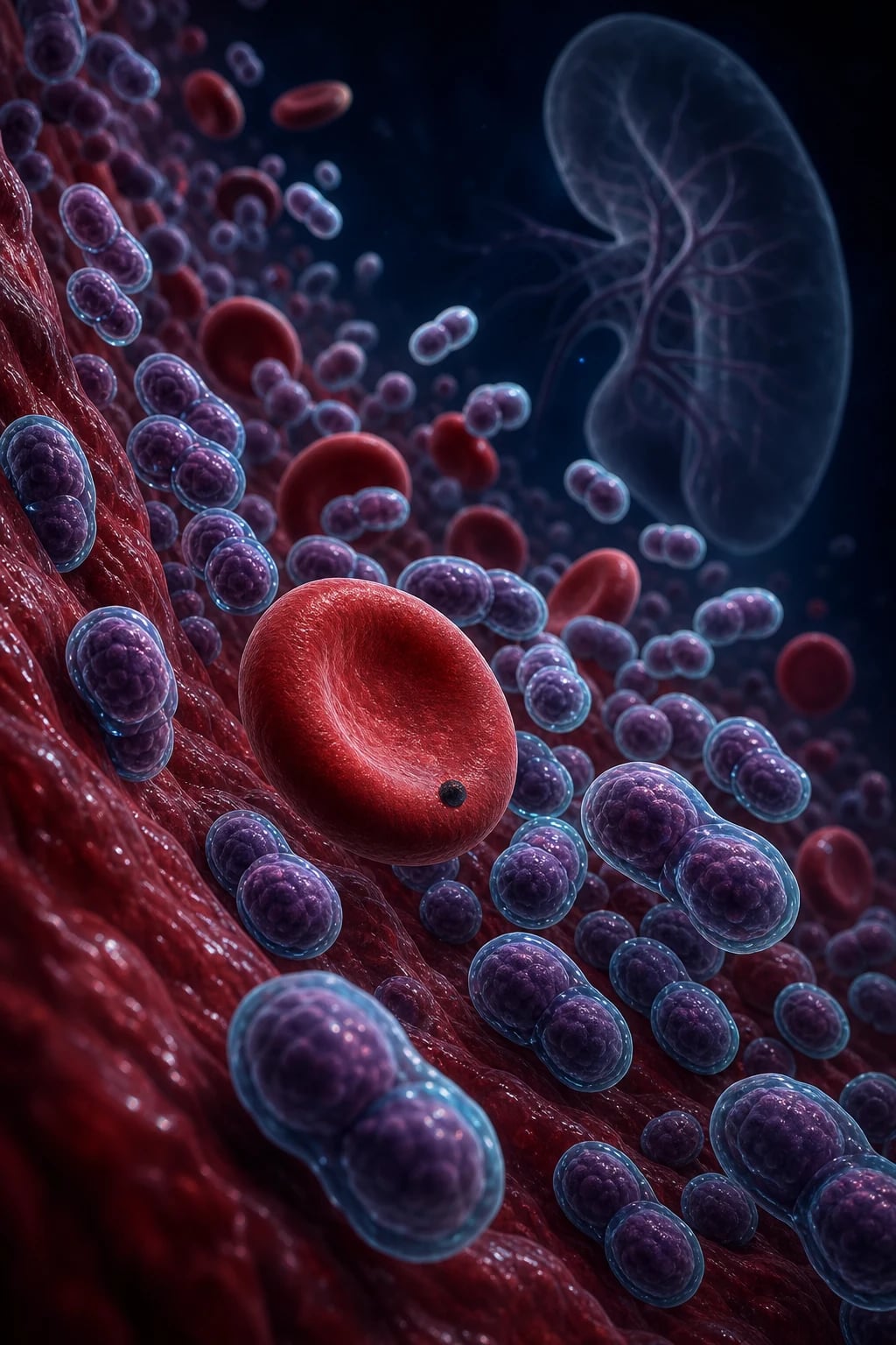
Splenic injury encompasses any traumatic damage to the spleen — from a minor subcapsular haematoma to complete shattering of the parenchyma. It is the most commonly injured intra-abdominal organ in blunt trauma, accounting for approximately 25% of all blunt abdominal injuries.[8] The evolution from routine splenectomy toward non-operative management (NOM) in over 90% of stable patients represents one of the great paradigm shifts in modern trauma surgery.[9] Where the 1970s trauma laparotomy almost invariably ended in splenectomy, the contemporary standard mandates spleen-preserving strategies whenever the patient's physiology permits, because the immunological cost of asplenia is lifelong and potentially lethal.
The spleen performs three critical immunological functions that splenectomy abolishes: filtration of opsonised encapsulated bacteria and senescent/abnormal erythrocytes from the blood, production of opsonins (tuftsin and properdin) that potentiate neutrophil phagocytosis, and antibody synthesis (particularly early IgM responses to novel antigens). Removal of these defences creates a permanent vulnerability to overwhelming post-splenectomy infection (OPSI) — a fulminant, rapidly fatal septicaemia caused by encapsulated organisms that the asplenic host cannot clear.[5]
[1]Anatomy
Surface Anatomy and Topography
The spleen sits in the left upper quadrant beneath ribs 9-11, protected by the diaphragm, the lower left rib cage, and surrounding viscera. It is shaped like a coffee bean, with a diaphragmatic surface (convex, smooth, related to ribs 9-11 and the costodiaphragmatic recess of the left pleura) and a visceral surface (concave, related to the stomach anteriorly, the left kidney posteriorly, the splenic flexure of colon inferiorly, and the pancreatic tail at the hilum). The long axis lies along the course of the tenth rib. [1]
The hilum lies on the visceral surface along the central long axis and transmits the splenic artery, splenic vein, lymphatics, and autonomic nerves within the splenorenal ligament. The spleen is entirely intraperitoneal except for its bare area at the hilum; this peritonealisation is what allows a massive haemoperitoneum to develop after rupture. [1]
Ligamentous Attachments (Surgical Relevance)
The spleen is suspended by four peritoneal ligaments that must be sequentially divided during mobilisation. Each carries specific vascular or visceral contents that dictate the order and danger of division: [1]
- Gastrosplenic ligament (anterior) — derived from the dorsal mesogastrium. Contains the short gastric vessels (4-5 arteries and veins running along the upper part) and the left gastroepiploic vessels along the lower part. Avulsed by traction on the stomach during fundoplication or passed behind to deliver the spleen. Vascular content: short gastric + left gastroepiploic vessels.
- Splenorenal (lienorenal) ligament (posterior) — also from dorsal mesogastrium. Contains the splenic artery and vein, the tail of the pancreas, and lymphatics. This is the critical vascular pedicle. Division here during splenectomy must protect the pancreatic tail. Vascular/visceral content: splenic artery, splenic vein, pancreatic tail.
- Splenophrenic ligament (superior) — avascular peritoneal reflection between the upper pole and the diaphragm. Usually divided first to begin mobilisation. Avascular.
- Splenocolic ligament (inferior) — between the lower pole and the splenic flexure of the colon. May contain the inferior polar vessels or branches of the left gastroepiploic in up to 25%. Traction here during left colectomy avulses the lower pole capsule — a classic iatrogenic injury. Variable vascular content. [1]
LIGAMENTS
Segmental (Vascular) Anatomy — Couinaud-like Concept
Although the spleen does not have true Couinaud segments like the liver, it has a functional segmental anatomy based on the arterial branching pattern that is the anatomical basis of partial splenectomy. The splenic artery divides at the hilum into 2-3 principal lobar (segmental) arteries, each supplying a discrete avascular plane between segments. Two classic branching patterns are described: [1]
- Magistral pattern (~30%) — the artery enters the hilum as a single trunk and branches late, within 3-4 cm of the hilum. The trunk is short and central. Easier to ligate en masse at the hilum.
- Distributive pattern (~70%) — the artery branches early and extensively (5-6 cm or more proximal to the hilum), sending long, separate segmental branches across a wide hilum. Individual branch ligation is required; mass ligation risks injuring the pancreatic tail. [1]
Each segmental artery is end-arterial with no significant intraparenchymal collaterals, so ligation of one branch produces a sharp line of demarcation on the splenic surface — the surgeon then transects along this ischaemic line. Two polar arteries are surgically important: the superior polar artery (arising separately from the splenic artery in approximately 65% of people) and the inferior polar artery (from the left gastroepiploic or distal splenic artery). Selective ligation of a polar artery devascularises only that pole, enabling an anatomical partial splenectomy. [1]
Blood Supply and Venous Drainage
Arterial:
- Splenic artery — the largest branch of the coeliac trunk (after the common hepatic and left gastric). It follows a characteristically tortuous, caterpillar-like course along the superior border of the pancreas, giving off dorsal pancreatic, pancreatic magna, caudal pancreatic, and short gastric branches before reaching the hilum.
- Short gastric arteries (4-5) — arise from the distal splenic artery and supply the gastric fundus; must be individually ligated during splenectomy.
- Left gastroepiploic artery — branch of the splenic artery running along the greater curvature; contributes to the lower pole supply via the splenocolic/gastrosplenic ligaments. [1]
Venous:
- Splenic vein — forms at the hilum from 5-6 tributaries, runs posterior to and grooves the superior border of the pancreas, receives the inferior mesenteric vein (IMV) in approximately 40%, then joins the superior mesenteric vein (SMV) behind the pancreatic neck to form the portal vein. Splenic vein thrombosis is a recognised post-splenectomy complication (incidence up to 7.5%), and isolated splenic vein thrombosis causes left-sided (sinistral) portal hypertension with gastric varices. [1]
Tail of Pancreas — the Hidden Risk
Clinical pearl: The tail of the pancreas lies within 1 cm of the splenic hilum in 73% of people and directly touches the spleen in 30%. Pancreatic tail injury (with subsequent fistula, abscess, or pancreatitis) is a recognised complication of splenectomy — the incidence of post-splenectomy pancreatic fistula is 3-8%. During splenectomy the pancreatic tail must be gently swept medially off the hilum, mass ligation avoided in favour of individual vessel ligation, and a closed-suction drain placed whenever there is any concern about pancreatic trauma or electrocautery proximity.[10]
Epidemiology
MECHANISM
Blunt mechanisms (over 90% of cases) include motor vehicle collisions (driver-side impact, pedestrian strikes), falls from height, bicycle handlebar compression, direct blows (assault, sports), and blast injury. Penetrating mechanisms (stab, gunshot) less commonly injure the spleen in isolation — they typically require laparotomy regardless of organ injured. The male predominance reflects trauma demographics. Concomitant injuries are the rule rather than the exception: left lower rib fractures (ribs 9-11) coexist in up to 20%, and associated left kidney, pancreatic tail, stomach, and left hepatic lobe injuries are common — these must be actively excluded on CT. [1]
AAST Grading System
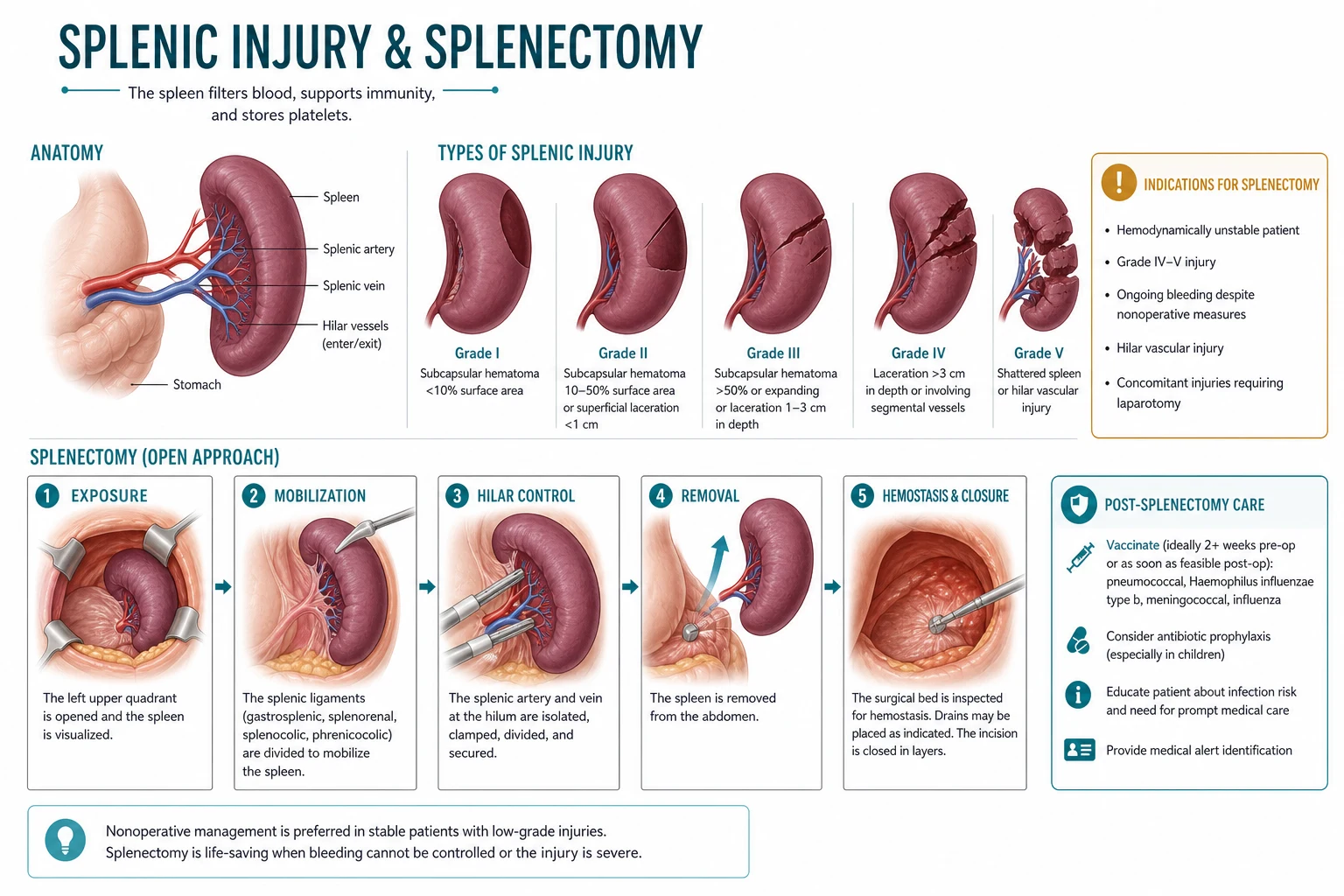
The American Association for the Surgery of Trauma (AAST) splenic injury scale (first published 1989, revised 1994 and again in 2018) provides a standardised grading system that correlates with both injury severity and management decisions.[1][2] The 2018 revision refined the definitions of Grade IV, distinguished vascular from parenchymal injuries more rigorously, and emphasised that the grade is determined by imaging (CT) or operative findings, with CT now the primary modality.
| Grade | Injury Type | Description | Typical Management |
|---|---|---|---|
| I | Subcapsular haematoma | under 10% surface area | NOM |
| I | Capsular tear | under 1 cm parenchymal depth | NOM |
| II | Subcapsular haematoma | 10-50% surface area | NOM |
| II | Intraparenchymal haematoma | under 5 cm | NOM |
| II | Laceration | 1-3 cm depth, not involving trabecular vessels | NOM |
| III | Subcapsular haematoma | over 50% or expanding/ruptured | NOM + angioembolisation |
| III | Intraparenchymal haematoma | over 5 cm or expanding | NOM + angioembolisation |
| III | Laceration | over 3 cm or involving trabecular vessels | NOM + angioembolisation |
| IV | Laceration | Segmental or hilar vessel injury devascularising over 25% | NOM if stable, surgery if unstable |
| V | Laceration | Shattered spleen | Surgery (usually splenectomy) |
| V | Vascular | Hilar injury devascularising entire spleen | Surgery (splenectomy) |
CT Findings and Vascular Injuries (2018 Update)
The 2018 update formalised the role of vascular injury on CT as a modifier of management. A contrast blush (active extravasation of contrast), a pseudoaneurysm, or an arteriovenous fistula identifies patients at high risk of ongoing bleeding who benefit from angioembolisation even when their parenchymal grade is modest. Typical CT descriptors per grade: [1]
- Grade I — thin subcapsular crescent of blood under 10% of the splenic convexity; superficial capsular irregularity. Minimal free fluid.
- Grade II — moderate subcapsular or intraparenchymal haematoma (10-50% surface area or under 5 cm); shallow laceration not reaching the hilum. Trace perihepatic/pelvic free fluid.
- Grade III — large/expanding subcapsular or intraparenchymal haematoma (over 50% surface or over 5 cm); laceration extending into the hilum involving trabecular vessels. Moderate haemoperitoneum (Morrison's pouch, pelvis).
- Grade IV — laceration involving segmental or hilar vessels with devascularisation of over 25% of the parenchyma. Often a contrast blush at the hilum or within a segmental territory.
- Grade V — shattered spleen (multiple fragments with complete parenchymal disruption) or hilar vascular injury devascularising the entire organ. Massive haemoperitoneum. [1]
In many Indian and global centres, AAST Grade IV-V injuries in haemodynamically stable patients are managed with angiographic embolisation rather than surgery, preserving splenic immune function. The key determinant is haemodynamic stability, not grade alone. However, a shattered spleen (Grade V parenchymal) almost always requires splenectomy even if the patient transiently responds to resuscitation.
Clinical Presentation
Symptoms
- Left upper quadrant pain — most common presenting symptom; may radiate to the left flank
- Left shoulder tip pain (Kehr's sign) — referred pain from diaphragmatic irritation by intraperitoneal blood irritating the phrenic nerve roots C3-C5; the shoulder tip shares C4 dermatomal innervation with the diaphragmatic peritoneum
- Diffuse abdominal pain — with peritonism if hollow viscus injury coexists
- Signs of haemorrhagic shock — dizziness, syncope, thirst, confusion, oliguria (progressive as blood loss exceeds 30% of circulating volume) [1]
Signs
Specific eponymous signs:
- Kehr's sign — referred left shoulder tip pain (diaphragmatic irritation by blood). Worsened by Trendelenburg position or deep inspiration. Present in approximately 50% of splenic ruptures.[8]
- Balance's sign — fixed dullness in the left flank (coagulated perisplenic blood) with shifting dullness elsewhere. Suggests perisplenic clot that has not dispersed.
- Ballance's sign — fixed dullness to percussion in the left upper quadrant that does not shift with position changes (due to a contained perisplenic haematoma).
- Saegesser's sign — tenderness on palpation of the left sternocleidomastoid muscle (phrenic nerve root irritation at C3-C4).
Investigations
Immediate (ATLS Protocol)
All trauma patients follow the ATLS primary survey (ABCDE). Splenic injury is identified during C (circulation) assessment. Two large-bore peripheral cannulae, bloods (group and crossmatch minimum 4 units), and activation of the massive transfusion protocol in the unstable patient are immediate priorities. [1]
Imaging
| Investigation | Role | Sensitivity |
|---|---|---|
| FAST scan | Bedside, rapid, identifies free fluid in LUQ/Morrison's pouch/Pouch of Douglas | 60-90% for significant haemoperitoneum |
| CT with IV contrast | Gold standard for stable patients; grades injury, detects contrast extravasation (blush), pseudoaneurysm, AV fistula, associated injuries | Near 100% for parenchymal injury |
| Angiography | Diagnostic + therapeutic (embolisation for active bleeding/pseudoaneurysm) | Therapeutic in 15-30% of Grade III-V |
| CEUS (contrast-enhanced ultrasound) | Detects active bleeding in stable patients; useful for follow-up and in pregnancy/ionising radiation concerns | Comparable to CT for parenchymal injury |
Blood Tests
- FBC — serial haemoglobin monitoring (initial Hb may be normal in acute haemorrhage as equilibration takes hours; a falling trend is more significant than any single value)
- Crossmatch — minimum 4 units; activate massive transfusion protocol if unstable (target ratio 1:1:1 PRBC:FFP:platelets)
- Coagulation — PT/INR, APTT — correct any coagulopathy (the trauma-induced coagulopathy of severe shock)
- Serum amylase/lipase — assess for concurrent pancreatic injury; a rising trend over 24-48 hours suggests pancreatic duct injury
- ABG/VBG — lactate and base deficit as markers of shock severity and resuscitation adequacy (normalisation predicts survival)
- Group and screen — if transfusion anticipated but not immediately required [1]
Non-Operative Management (NOM)
NOM has become the standard of care for haemodynamically stable patients with splenic injury, regardless of grade. Success rates exceed 90% in carefully selected patients and reach 95%+ in children.[9] The philosophy reflects the recognition that the spleen is not expendable — its immune function is irreplaceable, and the morbidity of a splenectomy (lifelong OPSI risk) is far greater than the risk of failed observation in a well-selected, monitored patient.
Criteria for NOM
- Haemodynamically stable (SBP over 90 mmHg after 2L crystalloid resuscitation, with stable or improving lactate)
- No peritonitis on abdominal examination
- No other intra-abdominal injury requiring laparotomy (e.g., hollow viscus perforation, severe liver injury requiring surgery)
- No evidence of active contrast extravasation on CT (if blush present — add angioembolisation, do not abandon NOM)
- Transfusion requirement within physiological parameters (typically under 2 units in 24 hours; exceeding this is a criterion for failure) [1]
NOM Protocol — Bedrest, Monitoring, and Repeat Imaging
NOM is active management, not passive neglect. The protocol is graded by injury severity: [1]
- Setting: ICU admission for Grade III-V injuries for 24-48 hours; a monitored ward bed is acceptable for Grade I-II in a stable patient.
- Haemodynamic monitoring: continuous HR, BP, SpO2; hourly urine output (target over 0.5 mL/kg/h).
- Serial abdominal examinations every 2-4 hours to detect developing peritonitis (a new sign of peritonism mandates reassessment and often laparotomy).
- Serial FBC every 6 hours for 24 hours, then every 12 hours until stable. A fall in haemoglobin of over 2 g/dL, or a continuing downward trend after resuscitation, suggests ongoing bleeding and triggers reassessment.
- Bed rest for 24 hours; mobilise once stable and Hb trend flat.
- Repeat CT: not routine in recovering patients. Indicated at 48-72 hours for Grade III-V injuries, at any time for clinical deterioration (dropping Hb, new pain, fever, haemodynamic instability), and before discharge in higher-grade injuries to exclude a developing pseudoaneurysm.
- Avoid antiplatelet agents and anticoagulants during the acute observation period; DVT prophylaxis with mechanical devices initially, transitioning to chemical prophylaxis once bleeding is controlled (typically 48 hours). [1]
Failure of NOM and Activity Restriction
Failure of NOM (5-10% of adults) is defined by: haemodynamic deterioration despite resuscitation; ongoing transfusion requirement (over 2 units in 24 hours, or any transfusion in children where the threshold is stricter); new peritonitis; or CT evidence of a developing pseudoaneurysm/AV fistula on follow-up imaging. Failure most commonly occurs in the first 48-72 hours but delayed failures up to 2 weeks are described. [1]
Activity restriction timeline after discharge:
- No strenuous activity or heavy lifting for 2 weeks (Grade I-II), 4-6 weeks (Grade III-IV), and up to 3 months (Grade V managed non-operatively).
- No contact sports for at least 6 weeks to 3 months depending on grade, and until CT-confirmed healing.
- Patient education on the symptoms of delayed splenic rupture (new abdominal pain, syncope, pallor) — re-present immediately. [1]
Angioembolisation
Angioembolisation is the procedural adjunct that has allowed NOM to extend into high-grade injuries. By selectively occluding the bleeding vessel while preserving splenic parenchyma and collateral inflow, it converts a high-failure subgroup into successful NOM.[4]
Indications:
- Active contrast extravasation (blush) on CT in a stable or transient-responding patient
- Pseudoaneurysm formation (a delayed-rupture time bomb)
- Arteriovenous fistula
- AAST Grade IV-V in stable patients (selected centres, especially if blush present)
- Moderate splenic injury with signs of ongoing bleeding but haemodynamic stability [1]
Technique — proximal vs distal:
- Arterial access via common femoral artery (Seldinger, 5-6 French sheath).
- Selective catheterisation of the coeliac axis then the splenic artery with a Cobra or Simmons catheter; superselective catheterisation of the injured segmental branch with a microcatheter.
- Proximal (main splenic artery) embolisation — coils (platinum or stainless steel) deployed in the main splenic artery proximal to the pancreatic magna branch. This reduces pulse pressure to the spleen while preserving flow via collateral inflow from the short gastric and left gastroepiploic arteries, so the spleen is not infarcted. Preferred for multiple/poorly localised injuries or hilar injuries.
- Distal (superselective) embolisation — gelfoam (a temporary agent, recanalises in ~2 weeks) or microcoils deployed in the specific segmental/polar branch supplying the injury. Preferred for a single, focal, accessible blush or pseudoaneurysm. Preserves maximal parenchyma.
- Some centres combine both: proximal coil + distal gelfoam for high-grade injuries.
- Success rate: 85-95%, with failure defined by need for splenectomy despite embolisation. [1]
Complications of angioembolisation:
- Post-embolisation syndrome (~30%): fever, left upper quadrant pain, leucocytosis, ileus — self-limiting over 1-3 days.
- Splenic infarction (5-15%): more common after distal gelfoam; usually clinically silent if under 50%.
- Splenic abscess (1-5%): requires antibiotics ± percutaneous/surgical drainage.
- Pseudoaneurysm recurrence or new pseudoaneurysm (~5%): rationale for repeat CT before discharge in high-grade injuries.
- Access-site complications (haematoma, dissection, pseudoaneurysm). [1]
Operative Management
Indications for Surgery
- Haemodynamic instability despite resuscitation (absolute indication)
- Peritonitis on abdominal examination (suggests hollow viscus injury or transperitoneal contamination)
- Failed NOM (ongoing transfusion requirement, haemodynamic deterioration, new pseudoaneurysm after angioembolisation)
- Associated hollow viscus injury requiring laparotomy
- Other intra-abdominal injuries mandating exploration (e.g., severe liver injury, major vascular injury) [1]
Operative Procedures
The choice between spleen-preserving techniques (splenorrhaphy, partial splenectomy) and total splenectomy depends on the grade of injury, the patient's physiological state, and the presence of other injuries. In the damage-control setting (coagulopathy, hypothermia, acidosis — the lethal triad), total splenectomy is performed rapidly to control bleeding; splenic salvage is a luxury of the stable patient. [1]
Splenorrhaphy (Spleen-Preserving Repair)
For Grade I-II injuries when the spleen must be mobilised during surgery for other reasons, or for selected Grade III injuries in a stable patient. The principle is to achieve haemostasis while preserving the organ. Techniques, applied in escalating fashion: [1]
- Topical haemostatic agents — Surgicel (oxidised cellulose), Floseal (gelatin-thrombin matrix), Gelfoam, fibrin glue (Tisseel). Useful for surface oozing and small capsular tears.
- Argon beam coagulation — a non-contact electrocautery that coagulates the splenic surface with a stream of ionised argon gas; effective for diffuse capsular bleeding with minimal thermal depth.
- Capsular suture repair — 2-0 or 3-0 absorbable suture (Vicryl/PDS) on a tapered needle, placed with an omental or Surgicel buttress to prevent the suture cutting through the friable parenchyma. Horizontal mattress sutures for deeper lacerations.
- Mesh splenorrhaphy — an absorbable mesh (Vicryl/polyglactin) "splenic wrap" fashioned into a bag; the mobilised spleen is encased and the mesh tightened to provide tamponade. Useful for Grade II-III with multiple lacerations; time-consuming and now largely supplanted by angioembolisation. [1]
Partial (Subtotal) Splenectomy
For polar injuries (upper or lower pole devascularisation, Grade III-IV) where the damaged segment can be sacrificed and the remainder preserved. The technique exploits the avascular intersegmental plane: [1]
- Mobilise the spleen to the midline (divide splenophrenic, splenocolic ligaments; reflect the gastrosplenic and splenorenal ligaments).
- Identify and selectively ligate the segmental/polar artery supplying the injured segment (e.g., superior polar artery for an upper pole injury).
- Observe the line of demarcation on the splenic surface (becomes ischaemic/dusky within minutes).
- Transect along the demarcation line using a scalpel or electrocautery, compressing the parenchyma between the fingers or with a stapler.
- Oversew the cut edge with a continuous 2-0 absorbable suture with an omental buttress; supplement with argon beam and haemostatic agents.
- Preserve at least 25% of the splenic parenchyma — this is the minimum required to maintain adequate immune function (opsonin production and encapsulated organism clearance). Verify viability of the remnant (pink, bleeding edge). [1]
Total Splenectomy
For Grade IV-V injuries, hilar vascular injury with total devascularisation, the damage-control setting, or failed splenic salvage. The open procedure is the reference standard; laparoscopic splenectomy is reserved for elective indications (ITP, hereditary spherocytosis, small to moderate spleens). [1]
SPLENECTOMY STEPS
Critical intra-operative points:
- Ligate the splenic artery before the vein where possible — reduces splenic engorgement and back-bleeding, and allows the spleen to "auto-transfuse" ~300 mL of stored blood into the circulation before removal.
- Avoid mass ligation of the hilum — risks injury to the pancreatic tail and post-operative fistula. Individual vessel ligation (suture ligatures or vascular stapler) is safer.
- Search for accessory spleens before closure — failure to remove them in elective haematological splenectomy (e.g., ITP) leads to recurrence; in trauma, retained accessory splenic tissue mitigates but does not reliably prevent OPSI. [1]
Laparoscopic Splenectomy (Elective)
Reserved for elective indications with normal or modestly enlarged spleens (under ~20 cm). Patient in right lateral decubitus, ports in the left subcostal region. Ligaments divided with harmonic scalpel/energy device; hilum secured with an endoscopic vascular stapler. Advantages: less pain, shorter stay. Contraindicated in trauma (no tactile assessment, inability to pack, time pressure) and massive splenomegaly. [1]
Post-Splenectomy Management
OPSI — Overwhelming Post-Splenectomy Infection
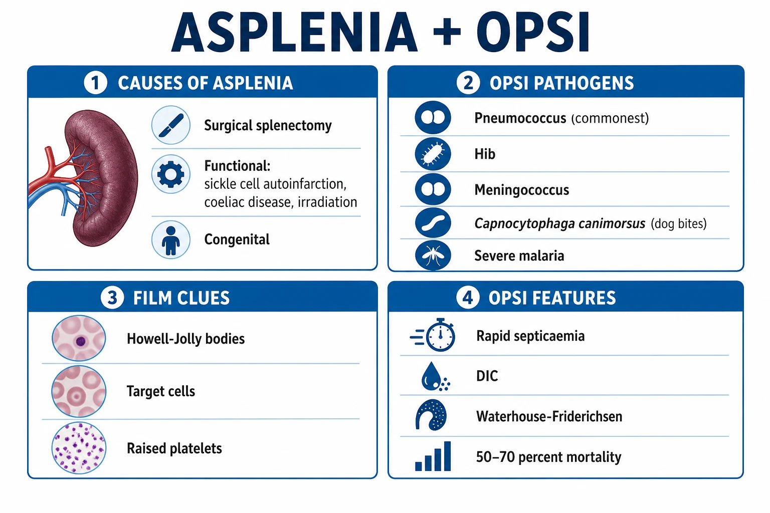
OPSI is the most feared complication of splenectomy. It is a medical emergency with mortality of 50-70% despite optimal intensive care treatment.[5] The clinical hallmark is a fulminant course: a previously well patient develops a flu-like prodrome that within hours evolves into septicaemic shock, disseminated intravascular coagulation (DIC), purpura fulminans, and multi-organ failure.
Causative organisms (encapsulated bacteria and others):
- Streptococcus pneumoniae — 50-90% of cases; the dominant pathogen[6]
- Haemophilus influenzae type b — declining due to widespread Hib vaccination; still a threat in unvaccinated adults
- Neisseria meningitidis — meningococcaemia with Waterhouse-Friderichsen syndrome
- Capnocytophaga canimorsus — dog bite-associated sepsis; frequently fulminant in asplenic patients
- Salmonella species (especially in sickle cell disease with autoinfarction)
- Escherichia coli, Klebsiella, group B streptococcus — less common
- Intracellular parasites: Babesia microti (babesiosis, tick-borne) and Plasmodium falciparum (severe malaria) — the asplenic host cannot clear intraerythrocytic parasites
Pathophysiology: The spleen performs three critical functions for clearing encapsulated organisms: (1) production of opsonins (tuftsin — a tetrapeptide that potentiates neutrophil phagocytosis, and properdin — an alternative complement pathway component), (2) mechanical phagocytosis and filtration of blood-borne bacteria by splenic macrophages, and (3) early IgM antibody production to novel polysaccharide antigens. Without the spleen, encapsulated organisms — which resist non-specific complement lysis and require opsonisation for clearance — proliferate unchecked in the bloodstream. The result is fulminant septicaemia, DIC, Waterhouse-Friderichsen syndrome (bilateral adrenal haemorrhage from DIC), purpura fulminans, and death within 24-48 hours.[5]
Clinical presentation of OPSI:
- Prodromal phase (4-12 hours): mild fever, rigors, myalgia, headache, nausea, vomiting — non-specific and easily mistaken for a viral illness.
- Fulminant phase: high fever, septicaemic shock with hypotension refractory to fluids, DIC with purpura fulminans (extensive cutaneous and visceral thrombosis), adrenal haemorrhage (Waterhouse-Friderichsen), meningitis, and rapid progression to multi-organ failure and death.
- The entire course from first symptom to death may be under 24 hours. [1]
Exam application bank (NEET-PG / INICET)
One-line answer
Splenic injury is the most common intra-abdominal organ injury in blunt trauma (25% of cases). The AAST grading system (Grade I–V, revised 2018) guides management. Non-operative management (NOM) is successful in over 90% of haemodynamically stable patients. Splenectomy is reserved for haemodynamic instability, peritonitis, or failed NOM. The critical post-operative concern is overwhelming post-splenectomy infection (OPSI) — a 1–5% lifetime risk of fulminant sepsis from encapsulated organisms (S. pneumoniae, H. influenzae, N. meningitidis) with up to 50–70% mortality. Prevention requires vaccination (pneumococcal, meningococcal, Hib), lifelong antibiotic prophylaxis, and patient education.
Worked stems (answer without another resource)
Stem 1 — Classic presentation. Map symptoms to mechanism; name the first investigation and first treatment step with dose/route if drug therapy is standard. [1]
Stem 2 — Unstable / complicated. List red flags that force immediate resuscitation, theatre, ICU, antidote, or reperfusion — and what you do in the first 15 minutes. [1]
Stem 3 — Atypical group. Elderly, pregnancy, child, or immunocompromised: how presentation and thresholds change. [1]
Stem 4 — Differential trap. Name the three closest mimics and one discriminator for each. [1]
Stem 5 — Disposition. Who goes home with safety-netting, who is admitted, who needs HDU/ICU/theatre, and what follow-up is mandatory. [1]
Rapid viva checklist
- Definition + classification
- Pathophysiology chain
- Bedside signs / criteria
- Score with exact components (if any)
- Emergency bundle
- Definitive therapy with doses
- Complications of disease and of treatment
- Special populations
- Guideline/trial name if classic
- Three exam traps
Coverage self-check
If you cannot answer any stem above from this page alone, re-read the matching section — the page is intended to be self-sufficient for final-prof and NEET-PG/INICET questions on Splenic Injury and Splenectomy.
[1]Vaccination Protocol
Vaccination is the cornerstone of OPSI prevention. The principle is to prime the immune system with T-cell-dependent conjugate vaccines first, then broaden coverage with polysaccharide vaccines. Ideally administered at least 14 days before elective splenectomy (to capture the peak antibody response while the spleen can still filter and process antigen) or at least 14 days after emergency splenectomy (deferred to allow immune recovery and to avoid the immunosuppressive effect of acute trauma/surgery and transfusion).[7]
| Vaccine | Type | Dose | Schedule |
|---|---|---|---|
| PCV13 (Prevenar 13) | Pneumococcal conjugate | 0.5 mL IM | 1 dose |
| PPSV23 (Pneumovax 23) | Pneumococcal polysaccharide | 0.5 mL IM/SC | 1 dose, 8 weeks after PCV13; booster at 5 years |
| MenACWY | Meningococcal conjugate (quadrivalent) | 0.5 mL IM | 1 dose; booster every 5 years |
| MenB (Bexsero, Trumenba) | Meningococcal group B | 0.5 mL IM | 2 doses (Bexsero 4 weeks apart; Trumenba 0, 6 months) |
| Hib (Haemophilus influenzae type b) | Conjugate | 0.5 mL IM | 1 dose (if not previously vaccinated) |
| Influenza | Inactivated | Annual | Every year |
Order of administration and exact intervals:
- PCV13 first — the conjugate primes a T-cell-dependent response, generating immunological memory and a stronger, more durable antibody response.
- PPSV23 at least 8 weeks later (minimum 8 weeks; some guidelines allow up to 1 year) — broader serotype coverage (23 serotypes vs PCV13's 13) but T-cell-independent and shorter-lived.
- MenACWY + MenB + Hib can be co-administered (different sites) at the first visit or sequentially.
- PPSV23 booster at 5 years (one repeat dose; a third dose is not routinely given).
- MenACWY booster every 5 years for life.
- Annual influenza vaccine — reduces the risk of secondary bacterial pneumonia.
- Timing of emergency splenectomy: if possible vaccinate at least 14 days post-operatively, ideally before discharge; if the patient is discharged sooner, vaccinate at the 2-week follow-up visit. [1]
- UK: Asplenic patients carry a medical alert card and standby antibiotics. NHS provides free pneumococcal, meningococcal, Hib, and annual influenza vaccines. British Committee for Standards in Haematology (BCSH) guidelines recommend lifelong antibiotic prophylaxis.[12]
- India: PCV13 was introduced into the national immunisation schedule in 2021. Asplenic adults should additionally receive PPSV23, MenACWY, MenB, and Hib. Annual influenza vaccine recommended. Access to conjugate vaccines can be limited in rural settings — reinforce standby antibiotics and education.[7]
- US: ACIP guidelines recommend PCV15 or PCV20 followed by PPSV23 for asplenic adults (PCV20 simplifies the schedule — single dose, no PPSV23 needed). MenACWY booster every 5 years; MenB series for high-risk, with a booster 1 year after completion then every 2-3 years if risk persists.
Antibiotic Prophylaxis
WHO/UK guidelines: Lifelong antibiotic prophylaxis for all asplenic patients, with particular emphasis on:
- Children under age 16 (highest risk)
- Adults over age 50
- Patients with impaired immune function (malignancy, immunosuppression)
- The first 2 years post-splenectomy (the highest-risk period, when OPSI incidence peaks)
- Patients with an inadequate vaccination record [1]
Penicillin V (phenoxymethylpenicillin) 250-500 mg twice daily is the traditional regimen; amoxicillin 250 mg once daily is an alternative with better pneumococcal cover and once-daily convenience. In penicillin allergy, erythromycin 250 mg BD or clarithromycin. Conscientious adherence is the challenge — standby (rescue) antibiotics carried at all times are the safety net for breakthrough fever regardless of whether prophylaxis is being taken. [1]
Patient Education
Every asplenic patient MUST receive — documented and reinforced at each visit: [1]
- Medical alert card/bracelet — clearly identifying asplenic status ("SPLEEN REMOVED — RISK OF SEVERE INFECTION")
- Standby antibiotics — oral amoxicillin 1g (or cefalexin/clarithromycin if penicillin-allergic) for self-administration at the first sign of significant fever, with instruction to still seek emergency medical care
- Education on warning signs — fever, rigors, lethargy, myalgia, confusion, purpura = seek emergency care immediately, not the next GP appointment
- Dog bite awareness — Capnocytophaga canimorsus risk; seek antibiotics (amoxicillin-clavulanate) for any dog bite, even trivial
- Malaria prophylaxis — essential and rigorous for travel to endemic areas (asplenic patients develop severe cerebral malaria with high mortality); combine chemoprophylaxis with meticulous mosquito avoidance
- Tick bite awareness — babesiosis risk in endemic areas (Northeastern USA)
- Annual influenza vaccination and maintenance of the booster schedule (PPSV23 at 5 years, MenACWY every 5 years)
- A written management plan shared with the GP, with escalation triggers [1]
Complications
Early (within 30 days)
| Complication | Incidence | Management |
|---|---|---|
| Rebleeding | 2-5% | Return to theatre; splenectomy if NOM/splenorrhaphy attempted |
| Pancreatic fistula | 3-8% | Conservative (drain, octreotide, nutritional support); ERCP + stent if persistent |
| Gastric fistula | Rare (under 1%) | Conservative vs surgical repair |
| Atelectasis (left lower lobe) | 20-30% | Chest physiotherapy, incentive spirometry |
| Subphrenic abscess | 3-5% | Antibiotics + percutaneous drainage |
| Portal/splenic vein thrombosis | 5-7.5% | Anticoagulation 3-6 months; screen with Doppler USS (consider early post-op screening in high-risk) |
| Wound infection | 3-5% | Antibiotics, wound care |
| Post-embolisation syndrome | ~30% (post-angio) | Supportive — antipyretics, analgesia; self-limiting |
| Post-splenectomy thrombocytosis | Common | Aspirin if platelets over 1000 x 10^9/L to reduce thrombosis risk |
Late (after 30 days)
| Complication | Incidence | Notes |
|---|---|---|
| OPSI | 1-5% lifetime | Highest in first 2 years; children at highest risk |
| Thrombocytosis (persistent) | Less common | Usually transient; thrombosis risk; aspirin if platelets over 1000 x 10^9/L |
| Atherosclerotic events | Modestly increased | Long-term cardiovascular risk; possible role of platelet activation |
| Pulmonary hypertension | Rare but recognised | Screen at long-term follow-up; mechanism unclear |
| Ischaemic heart disease | Modestly increased | Meta-analyses show ~1.5-fold increased risk |
Portal/splenic vein thrombosis deserves specific attention. After splenectomy the splenic vein stump thromboses in up to 7.5% of patients (and over 50% have asymptomatic thrombus on routine screening). Extension into the portal vein and superior mesenteric vein causes acute portal hypertension, bowel congestion, and mesenteric ischaemia. Risk factors include massive splenomegaly, myeloproliferative disease, and post-operative thrombocytosis. Many centres now perform routine Doppler ultrasound on day 5-7 after splenectomy for large spleens and treat with therapeutic anticoagulation (3-6 months) when detected. [1]
Special Situations
Paediatric Splenic Injury
Children have even higher NOM success rates (95%+) than adults due to greater splenic tissue elasticity, a thicker capsule relative to organ size, and more robust compensatory mechanisms. Splenectomy is avoided at all costs in children because of the dramatically higher OPSI risk — mortality from OPSI in children splenectomised under age 5 may reach 50% before modern vaccination. Key differences and the APSA (American Pediatric Surgical Association) guidelines: [1]
- NOM is preferred for almost all grades, including most Grade IV-V injuries in stable children.
- Angioembolisation is used less frequently than in adults (children's vessels are smaller and access more technically demanding).
- Blood transfusion threshold is lower — transfuse to a haemoglobin of 7 g/dL rather than the adult 8-9 g/dL; accept more transfusion to avoid surgery.
- Splenorrhaphy and partial splenectomy are preferred over total splenectomy if surgery is unavoidable.
- ICU observation: Grade IV-V for 24 hours; Grade I-III on a ward.
- Bed rest: typically 24 hours, then ambulation; discharge when tolerating diet and stable.
- APSA activity restriction (post-discharge): Grade I-II — 3 weeks; Grade III — 4 weeks; Grade IV-V — 5 weeks before return to full activity/contact sports. This is graded by injury severity, not a blanket restriction.
- Lifelong antibiotic prophylaxis is mandatory in children (at least until age 16, often lifelong per UK guidance), with the full conjugate-then-polysaccharide vaccine schedule. [1]
Accessory Spleens (Splenunculi)
Accessory spleens are present in 10-15% of the general population and in up to 30% of patients with haematological disease (hereditary spherocytosis, ITP, thalassaemia). They are small nodules of normal splenic tissue formed from failure of fusion of the splenic anlage in the dorsal mesogastrium. [1]
Locations (in order of frequency):
- Splenic hilum — 54%
- Splenorenal/gastrosplenic ligaments
- Tail of pancreas (pancreatic accessory spleens)
- Greater omentum
- Mesentery of small bowel
- Lesser sac
- Left ovary / broad ligament (pelvis)
- Rarely: testis (along the line of embryological descent), liver, gallbladder, stomach wall [1]
Clinical significance:
- In elective splenectomy for haematological disease (especially ITP), failure to identify and remove all accessory spleens is a leading cause of recurrence — accessory tissue hypertrophies and recapitulates the hypersplenic state. Meticulous search and removal is mandatory.
- After traumatic splenectomy, accessory spleens may provide partial immune function but are insufficient to reliably prevent OPSI — full vaccination and prophylaxis are still required.
- Splenosis (see below) is the acquired counterpart — autotransplanted splenic implants after trauma — and should not be confused with congenital accessory spleens. [1]
Splenosis
Splenosis is the autotransplantation of splenic tissue onto peritoneal surfaces following traumatic splenic rupture. Fragments of splenic pulp implant and vascularise on the peritoneum, omentum, mesentery, and even the pleura or pericardium if the diaphragm was breached. Incidence after splenic trauma is 25-65% on imaging at follow-up. Implants are usually multiple, small (mm to several cm), and asymptomatic. [1]
Clinical significance:
- Implants can grow over decades and may mimic other pathology (peritoneal nodules, masses) — diagnosed by Tc-99m heat-damaged red cell scan (specific for splenic tissue).
- They provide partial immune function but inadequate to prevent OPSI — patients still require vaccination and prophylaxis.
- Surgical removal is only indicated for symptomatic implants (pain, obstruction, torsion). [1]
Iatrogenic Splenic Injury
Iatrogenic injury is increasingly recognised as a cause of "medical" splenectomy. It occurs during:
- Left colectomy — traction on the splenic flexure avulses the splenocolic ligament and lower pole capsule (the classic mechanism)
- Hiatal hernia repair / Nissen fundoplication — traction on the stomach avulses short gastric vessels and the gastrosplenic ligament
- Left nephrectomy / adrenalectomy — direct retraction injury
- Distal pancreatectomy — the spleen is en bloc or retracted; concurrent splenectomy is common
- Upper abdominal vascular surgery (coeliac, splenic artery aneurysm)
- Diagnostic procedures — colonoscopy (rare), percutaneous procedures, gastric banding [1]
Management: Intraoperative recognition is critical. Small capsular tears can be managed with topical haemostatics, argon beam, or splenorrhaphy (spleen-preserving). If splenectomy is required for iatrogenic injury, vaccinate before discharge (ideally at least 14 days post-operatively), document the asplenic state prominently, and provide the full patient education package. Iatrogenic splenectomy is a recognised source of medicolegal liability — meticulous operative technique and prompt recognition are essential. [1]
Spontaneous (Pathological) Splenic Rupture
Pathological (abnormal) spleens are vulnerable to rupture with minimal or no trauma — sometimes termed "spontaneous" though a trivial trigger (cough, strain, palpation) is often identifiable in retrospect. Causes: [1]
- Infectious mononucleosis (EBV) — the most common cause of spontaneous rupture in young adults; the spleen is enlarged, hyperaemic, and the capsule infiltrated by lymphoid hyperplasia. Peak rupture risk in weeks 2-3 of illness.
- Malaria (falciparum, vivax) — acute splenomegaly
- Haematological malignancy — acute leukaemia, lymphoma (especially after cytoreduction)
- Haemolytic anaemia — hereditary spherocytosis, sickle cell disease (autoinfarcted spleen in adults; acute sequestration crisis in children), thalassaemia
- Splenic abscess / infarction with capsular inflammation
- Connective tissue disease — systemic lupus, rheumatoid (Felty syndrome)
- Pregnancy — rare [1]
Advice in infectious mononucleosis: Avoid contact sports and strenuous activity for at least 3-4 weeks (some guidelines recommend until splenomegaly has resolved clinically and on imaging, which may take 6-8 weeks). Risk of rupture is highest in weeks 2-3 of illness. Any patient with EBV who develops acute abdominal pain and signs of shock has a splenic rupture until proven otherwise. [1]
Delayed Splenic Rupture
The classic "delayed rupture" (occurring 48 hours to weeks after injury) was historically attributed to the two-hit model: an initial subcapsular haematoma that ruptures the capsule as it expands. Modern understanding attributes most "delayed" events to pseudoaneurysm formation — a partially injured segmental artery that progressively enlarges and ruptures into the peritoneum days to weeks later. This understanding underpins the repeat CT before discharge for high-grade injuries and the activity restriction timeline. Presentation is re-attendance with new abdominal pain, syncope, and shock in a patient with a recent (often seemingly minor) splenic injury. Management is urgent angioembolisation or surgery depending on stability. [1]
Elective Splenectomy Indications
Beyond trauma, splenectomy is performed for:
- Hereditary spherocytosis — moderate-severe chronic haemolysis; curative (removes the site of destruction of the abnormal spherocytes)
- Immune thrombocytopenic purpura (ITP) — refractory to steroids, rituximab, and TPO-receptor agonists; response rate ~80% but recurrence from accessory spleens is common
- Thalassaemia major — massive splenomegaly with hypersplenism and transfusion requirement
- Splenic abscess — rare; percutaneous drainage first-line, splenectomy for multifocal or failed drainage
- Wandering spleen — ptotic spleen with a long mobile mesentery prone to torsion; splenopexy preferred in children, splenectomy if infarcted
- Splenic artery aneurysm — selective indication; interventional radiology first-line
- Staging laparotomy in selected lymphomas (historical; now rare with modern imaging)
- Sickle cell disease with acute splenic sequestration crisis (children) or autosplenectomy complications [1]
For all elective splenectomies, vaccinate at least 14 days pre-operatively — this is one of the few opportunities to truly optimise immune protection before the spleen is removed. [1]
Key Exam Points
OPSI
High-yield MCQ topics:
- AAST grading — Grade IV-V typically require surgery in unstable patients; 2018 update emphasised CT vascular injuries (blush, pseudoaneurysm).
- Kehr's sign — referred left shoulder tip pain from diaphragmatic irritation by intraperitoneal blood (phrenic nerve C3-C5).
- NOM criteria — stable, no peritonitis, no other injury requiring surgery, under 2 units transfusion.
- OPSI organisms — S. pneumoniae (most common, 50-90%), H. influenzae, N. meningitidis, Capnocytophaga canimorsus, Babesia microti.
- Vaccination schedule — PCV13 first, then PPSV23 at 8 weeks; booster PPSV23 at 5 years; MenACWY every 5 years; annual influenza.
- Antibiotic prophylaxis — penicillin V 250-500 mg BD (or amoxicillin 250 mg OD); lifelong in children under 16.
- Angioembolisation — indications (contrast blush, pseudoaneurysm, Grade III-V stable); proximal coil vs distal gelfoam; success 85-95%.
- Pancreatic tail injury risk — within 1 cm of hilum in 73%; always inspect, drain if concerned; fistula 3-8%.
- Partial splenectomy — preserve at least 25% for immune function; ligate segmental artery, transect at line of demarcation.
- Accessory spleens — 10-15% incidence; hilum most common (54%); remove all in elective haematological splenectomy.
- Spontaneous rupture — EBV mononucleosis is the classic cause; avoid contact sports 3-4 weeks.
- Paediatric — 95%+ NOM; APSA activity restriction graded by grade (3-5 weeks). [1]
Viva trigger: "A 25-year-old male presents after an MVC with LUQ pain, hypotension, and left shoulder pain. FAST is positive. What is your approach?"
- Immediate laparotomy (unstable + positive FAST = no CT)
- ATLS protocol: ABCDE, two large-bore cannulae, crossmatch 4 units, activate massive transfusion protocol
- Intra-operative: midline incision, evacuate blood, pack all four quadrants, systematically remove packs from least to most likely bleeding area
- If splenic injury Grade IV-V: rapid mobilisation, ligate splenic artery then vein, individually ligate short gastrics, inspect pancreatic tail, search for accessory spleens, place drain if pancreatic concern
- Post-op: vaccinate at 14 days (PCV13, PPSV23 at 8 weeks, MenACWY, MenB, Hib, influenza), antibiotic prophylaxis, patient education, medical alert card, standby antibiotics
- Discharge advice: lifelong OPSI vigilance, dog bite and malaria awareness [1]
Viva trigger 2: "A 45-year-old woman undergoes splenectomy for ITP. What pre-operative preparation is essential?"
- Vaccinate at least 14 days BEFORE elective splenectomy (PCV13, then PPSV23 at 8 weeks — so plan ahead; MenACWY, MenB, Hib, influenza)
- Standby antibiotics and medical alert card organised before discharge
- Counsel on lifelong OPSI risk and penicillin prophylaxis
- Meticulous search for and removal of accessory spleens (up to 30% in haematological disease) to prevent recurrence [1]
References
- [1]Moore EE, et al. Comparison of the characteristics and features of pressure garments used in the management of burn scars Burns, 1998.PMID 9688198
- [2]Kozar RA, et al. International guidelines for groin hernia management Hernia, 2018.PMID 29330835
- [3]Bessoud B, et al. Long-lived memory T lymphocyte responses against SARS coronavirus nucleocapsid protein in SARS-recovered patients Virology, 2006.PMID 16690096
- [4]Sclafani SJ, et al. Drug exposure in pregnancy and heart defects J Cardiovasc Pharmacol, 2011.PMID 21499119
- [5]Holdsworth RJ, et al. Herd immunity and the varicella vaccine: is it a good thing? Arch Pediatr Adolesc Med, 2001.PMID 11296069
- [6]Waghorn DJ. An outbreak of mycobacterial furunculosis associated with footbaths at a nail salon N Engl J Med, 2002.PMID 11986410
- [7]Rubin LG, et al. Concordance for clonal hematopoiesis is limited in elderly twins Blood, 2020.PMID 31697828
- [8]Peitzman AB, et al. Fluoride excretion in children after sevoflurane anaesthesia Br J Anaesth, 2002.PMID 12393764
- [9]Stassen NA, et al. Phosphoinositide lipid second messengers: new paradigms for transepithelial signal transduction Pflugers Arch, 2005.PMID 15614575
- [10]Cocanour CS, et al. Specialized practice reduces inpatient mortality, length of stay, and cost in the care of colorectal patients Dis Colon Rectum, 2011.PMID 21654243
- [11]Diamond CK, et al. Predictive clinical model of tumor response after chemoradiation in rectal cancer Oncotarget, 2017.PMID 28938543
- [12]Davies JM, et al. Profiling analysis of differential gene expression between hematogenous and peritoneal metastatic sublines of human pancreatic cancer using a DNA chip J Exp Clin Cancer Res, 2004.PMID 15595644