General Surgery · General Surgery
Surgical Site Infection
Also known as SSI · Postoperative wound infection · Wound infection
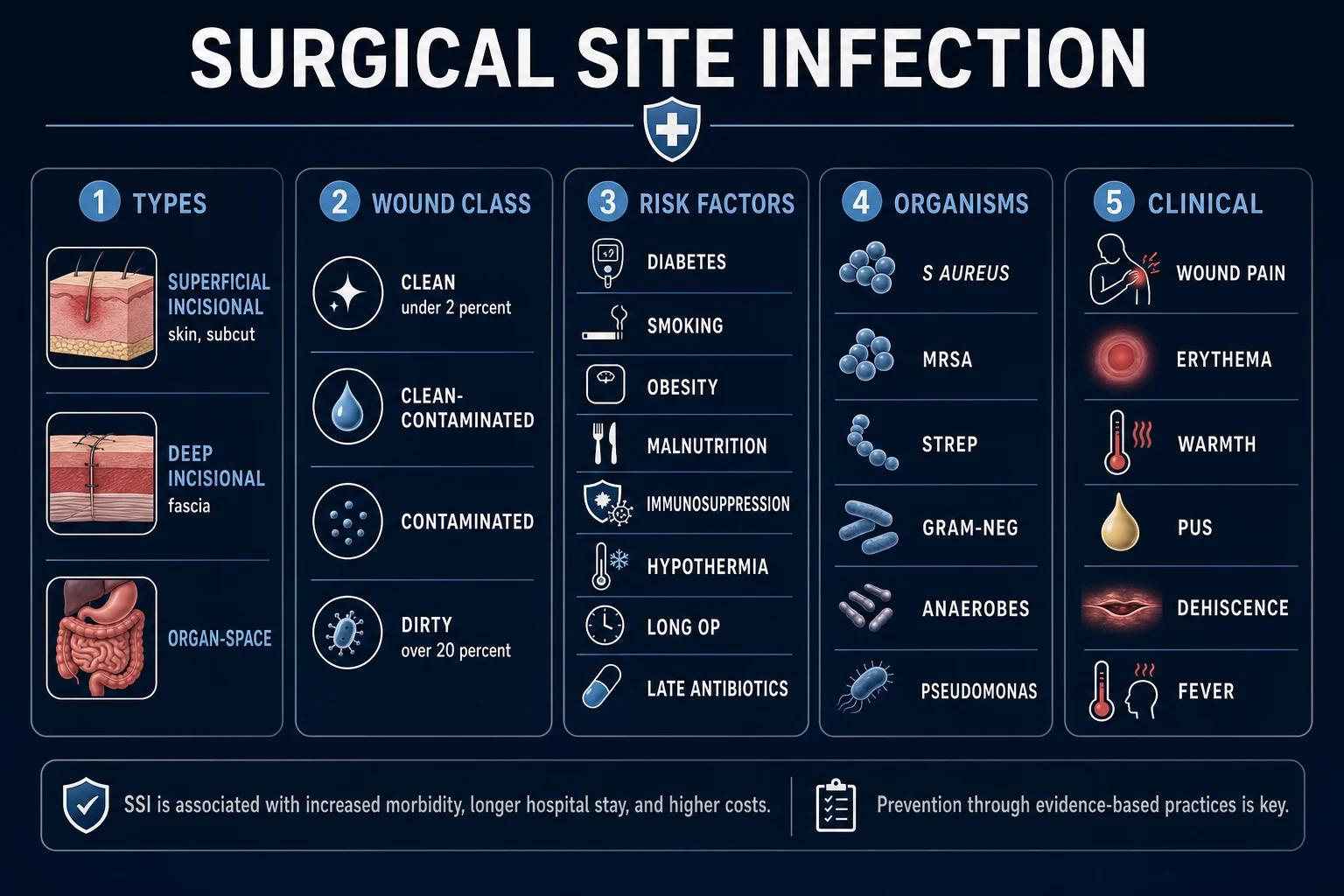
SSI = infection at the surgical site within 30 days (or 90 days if implant placed), classified by CDC into superficial incisional (skin/subcut), deep incisional (fascia/muscle) and organ/space. Affects 2 to 5% of procedures, doubles mortality, and adds 7 to 10 days to length of stay. Most common organism in clean surgery: Staphylococcus aureus. Prevention bundle: prophylactic antibiotics within 60 min of incision (re-dose at 4 h or after 1500 mL blood loss), normothermia, glycaemic control, clipping (not shaving), supplemental oxygen, chlorhexidine skin prep, WHO Surgical Safety Checklist.
On this page & tools
Your progress
Saved locally on this device.
Exam tags
Red flags
Overview & Definition
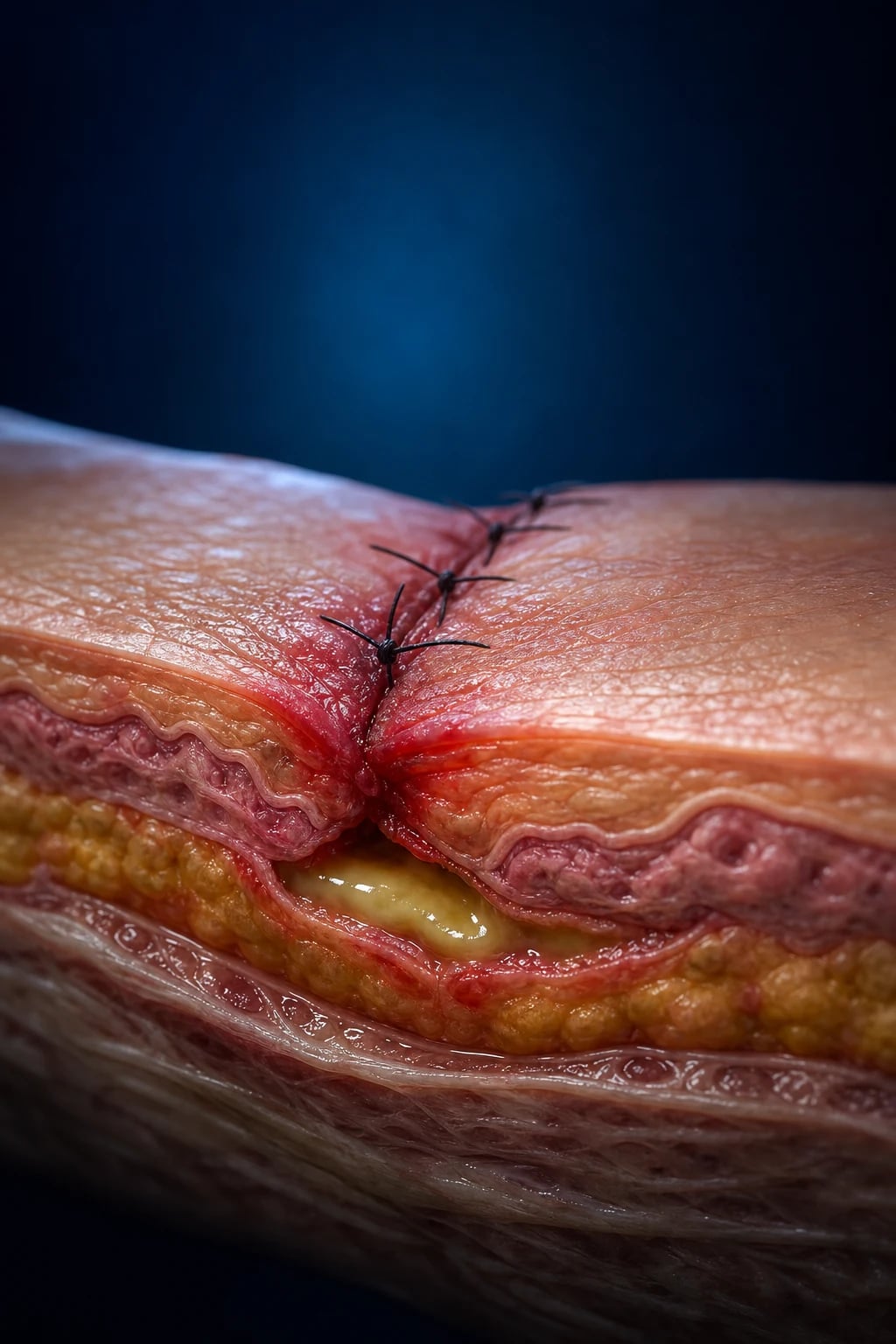
Surgical site infection (SSI) is any infection occurring at or near the surgical incision, or in any deep organ or space manipulated during surgery, that arises within 30 days of the operative procedure — or within 90 days if an implant (prosthetic joint, vascular graft, mesh, cardiac device, breast prosthesis) is left in place.[3][1] It is among the most common healthcare-associated infections (HAIs) in surgical patients and is largely preventable through evidence-based perioperative care.
The current CDC definition dates to Horan et al. 1992[3] and was reaffirmed in the CDC 2017 Guideline for the Prevention of Surgical Site Infection by Berríos-Torres et al.[1] A diagnosis of SSI requires at least one of the following: (i) purulent drainage from the incision or a drain placed into the deep space; (ii) an organism isolated from an aseptically obtained sample of fluid or tissue from the incision or deep space; (iii) the surgeon deliberately opens the wound in a patient with fever, pain or tenderness (unless culture-negative); (iv) radiological evidence of a deep abscess or collection; or (v) the surgeon or attending physician's clinical diagnosis of SSI.[3]
SSI is not a single entity but a heterogeneous family of infections distinguished by anatomical depth, microbial source, and surgical context. Mastery requires understanding four interlinked concepts: the CDC depth classification (superficial, deep, organ/space), the traditional wound-contamination class (clean, clean-contaminated, contaminated, dirty), the NNIS/NHSN risk index (a 0 to 3 score), and the microbial flora appropriate to each operative site.[2][3]
Classification
CDC classification by depth
Superficial incisional
skin and subcutaneous tissue only
- Most common SSI type (about 60%)
- Within 30 days of surgery
- Erythema, warmth, pain, **purulent drainage**
- Does not involve fascia or muscle
- Manage: open wound, dressings ± oral antibiotics
Deep incisional
fascia and muscle layer
- Involves deep soft tissue (fascia, muscle)
- Within 30 days (or 90 days if implant) AND one of: purulent drainage, dehiscence, abscess
- Higher fever, more systemic illness
- Requires surgical exploration, debridement, IV antibiotics
Organ/space
any anatomical site opened/manipulated
- Most serious; e.g. peritoneal abscess, mediastinitis, prosthetic joint infection, intra-abdominal collection
- Within 30 days (or 90 days if implant)
- Often requires **drainage ± re-operation**
- Example: anastomotic leak after colorectal surgery
The single most important distinction at the bedside is whether the infection is confined to the body wall (superficial or deep incisional) or involves a deeper cavity or organ (organ/space) — the latter usually requires imaging, drainage, and often re-operation.[1][1]
Traditional wound-contamination class (NRC / SENIC / CDC)
The four-class wound contamination scheme, originally defined by the National Research Council (1964) and refined by CDC, predicts baseline SSI risk and guides the choice of prophylactic antibiotics.[2]
Wound class and SSI risk
NNIS / NHSN risk index
The National Nosocomial Infections Surveillance (NNIS) risk index — now maintained by the NHSN — is a 0 to 3 score that adjusts SSI rates for case-mix and allows fair comparison between surgeons, hospitals and procedures. It assigns one point for each of:[2]
- ASA physical status score greater than 2 (i.e. ASA III, IV or V — mild to severe systemic disease);
- A contaminated (class 3) or dirty/infected (class 4) wound;
- An operation duration exceeding the 75th percentile for that specific procedure (e.g. longer than T hours, where T is published per procedure in NHSN tables). [1]
A patient with NNIS score 3 has up to a ten-fold higher SSI risk than one with score 0. NNIS-adjusted SSI rates are the basis of public reporting in the USA and NHSN surveillance.[2]
Epidemiology & Risk Factors
SSI is the most common nosocomial infection in surgical patients. In the CDC NHSN 2014 multistate point-prevalence survey, HAI prevalence in US acute-care hospitals was approximately 4%, with SSIs contributing a major share in surgical patients.[9] Overall SSI rates in high-income settings range from 1.9 to 5% of inpatient procedures; rates in low- and middle-income countries are two to three times higher.[4]
SSI increases length of stay by 7 to 10 days, doubles mortality (attributable mortality 1 to 3%, much higher for mediastinitis and organ/space infections), and increases readmission risk two- to ten-fold. The economic burden is substantial: a UK study by Coello et al. estimated an additional 6.5 to 11 days of stay and £814 to £10,523 per case in extra cost, while US estimates range from US$10,000 to 25,000 per infection.[18]
Patient-related risk factors
Procedure-related risk factors
- Wound class (clean → dirty, see above).[2]
- Operation duration — risk rises approximately linearly with time.
- Approach — laparoscopic and minimally invasive surgery has lower SSI rates than open surgery.[1]
- Prophylactic antibiotic timing and choice — see Pathophysiology and Prevention.[6]
- Perioperative hypothermia (core temperature under 36C) doubles SSI rates.[10]
- Perioperative hypoxia, hypotension, inadequate resuscitation, blood transfusion.[11]
- Hair removal by shaving (razor creates micro-lacerations) vs clipping.[16]
- Poor glycaemic control — even non-diabetics with stress hyperglycaemia are at risk.[1]
- Foreign material — sutures, drains, mesh, prosthesis all reduce inoculum threshold.
- Surgical technique — rough tissue handling, excessive diathermy, haematoma, dead space, hypoperfusion.
Pathophysiology
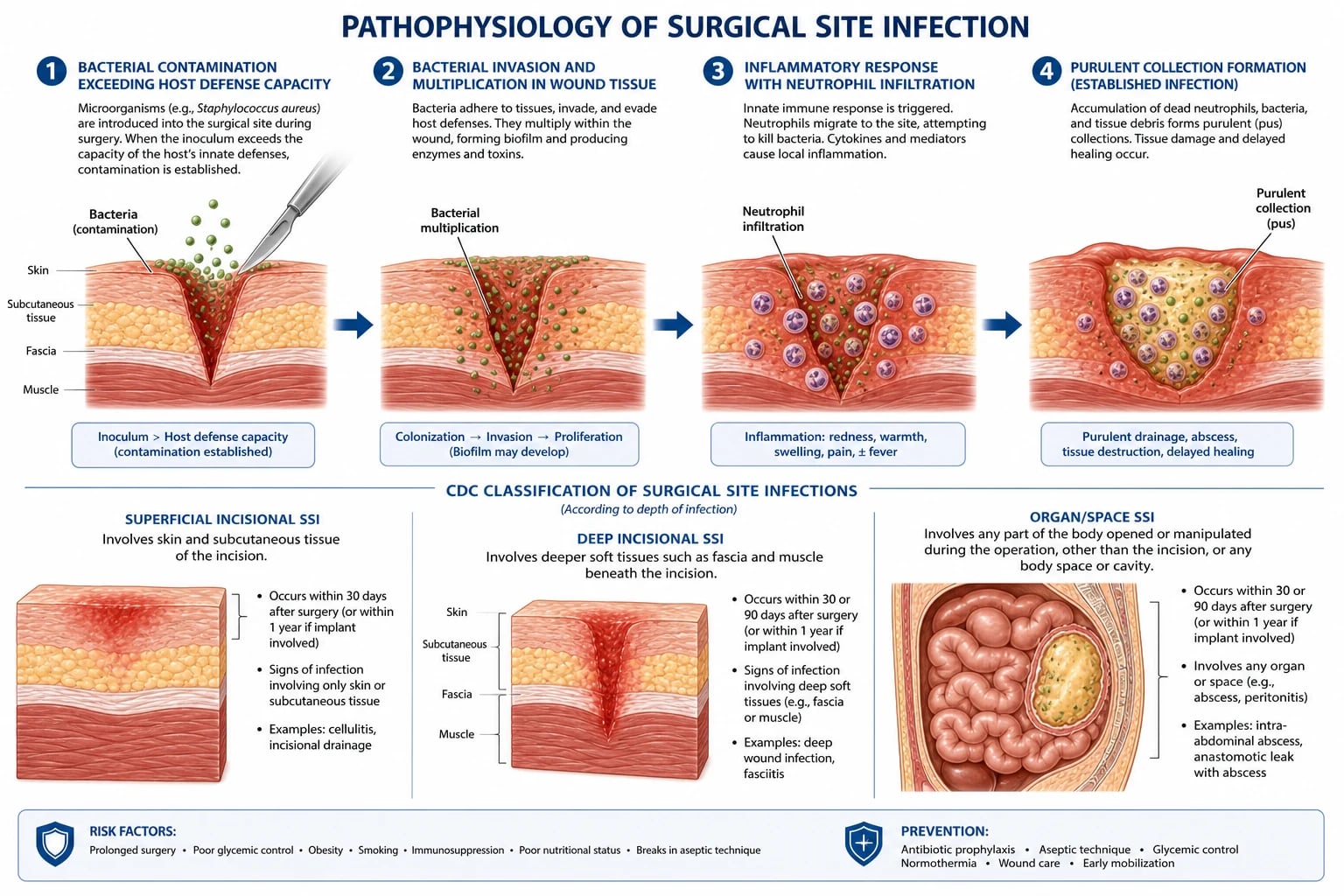
An SSI develops when the bacterial inoculum, virulence and microenvironment at the surgical site overcome the patient's local and systemic host defences. The classical quantitative threshold is more than 10 to the fifth power organisms per gram of tissue — below this, healthy tissue usually contains the contamination; above it, clinical infection is likely. Foreign material (suture, mesh, prosthesis) reduces this threshold by several orders of magnitude, which explains why implant surgery is uniquely vulnerable.[2][1]
Sources of contamination
- Endogenous flora (about 80%) — the patient's own skin, gastrointestinal, genitourinary or respiratory tract. This is the dominant source and explains why site-specific prophylaxis works.[2]
- Skin: Staphylococcus aureus, coagulase-negative staphylococci (CoNS), Corynebacterium.
- Upper GI / small bowel: streptococci, lactobacilli, Enterobacterales.
- Distal small bowel / colon: Enterobacterales (E. coli, Klebsiella, Enterobacter, Proteus), enterococci, anaerobes (Bacteroides fragilis, Clostridium, Peptostreptococcus).
- Vagina: lactobacilli, group B Streptococcus, anaerobes, Enterobacterales.
- Exogenous (surgical team, theatre air, instruments) — typically S. aureus, CoNS, and (in contaminated theatres) Gram-negatives including Pseudomonas.[2]
- Haematogenous spread — distant focus (endocarditis, IV line infection, dental abscess) seeds the operative site; rare but classic for early prosthetic joint infection.
Biofilm and implant infection
Within hours of implantation, bacteria adhere to the surface of sutures, mesh, prosthetic joints, vascular grafts and cardiac devices and secrete an extracellular glycocalyx (biofilm). Within the biofilm, bacteria are metabolically quiescent (persister cells), protected from host neutrophils and up to 1000 times more resistant to antibiotics than planktonic organisms.[1] Biofilm infection therefore often persists despite prolonged antibiotics and frequently requires implant removal for cure. This underlies the high stakes of SSI in orthopaedic, cardiac, vascular and hernia surgery.
The host response and oxygen
Tissue injury triggers the inflammatory cascade: complement activation, fibrinogen conversion to fibrin, platelet aggregation and recruitment of neutrophils and macrophages. Neutrophils kill bacteria by phagocytosis and the respiratory (oxidative) burst, which is critically dependent on tissue oxygen tension. Several landmark studies have demonstrated that subcutaneous oxygen tension correlates inversely with SSI risk: this is the mechanistic basis for perioperative normothermia (which improves tissue perfusion and oxygen delivery), supplemental inspired oxygen, fluid resuscitation, pain control (which reduces catecholamine-driven vasoconstriction), and smoking cessation.[10][11][12]
Why patient comorbidity matters mechanistically
- Diabetes / hyperglycaemia: impairs neutrophil chemotaxis, phagocytosis and oxidative burst; glycation of collagen weakens scars.[1]
- Smoking: nicotine causes vasoconstriction; carbon monoxide reduces oxygen carriage; collagen synthesis falls.
- Malnutrition: deficiency of protein, vitamin C (cross-linking of collagen), vitamin A, zinc, and copper impairs fibroblast function and angiogenesis.
- Obesity: increased tissue pressure reduces capillary flow and oxygen tension; longer operations; under-dosed antibiotics; dead space favours seroma and infection.
- Hypothermia: vasoconstriction and a left-shifted oxyhaemoglobin curve reduce tissue oxygen delivery.[10]
- Corticosteroids / immunosuppression: reduce macrophage and T-cell function and impair collagen deposition.
Clinical Presentation
SSI most commonly declares itself between postoperative days 3 and 10, although organ/space infections (especially prosthetic joint, vascular graft) can present weeks to months later. The cardinal features reflect the depth of infection. [1]
Classic local features (5 cardinal signs of inflammation)
- Rubor — erythema extending more than 1 cm beyond the wound edge; calor (warmth);
- Dolor — pain out of proportion to the expected postoperative course, often the first symptom;
- Tumor — swelling, induration;
- Functio laesa — loss of function (e.g. a limb that cannot bear weight, a wound that gapes). [1]
Purulent drainage
The single most specific feature. Purulent is defined clinically: thick, opaque, yellow, green or brown, sometimes malodorous — distinct from the serous (clear, straw-coloured) transudate of a seroma and the sanguineous (bloody) drainage of a haematoma.[3]
Wound dehiscence
- Superficial dehiscence — separation of skin edges; usually managed with delayed secondary closure or healing by secondary intention.
- Fascial (deep) dehiscence — a surgical emergency; classically presents around postoperative day 5 to 8 with a sudden serosanguineous "pink fluid" discharge ("the wound gave way and the patient felt something pop"); viscera may be visible. Requires emergency return to theatre for resuturing and management of any underlying SSI.[1]
Systemic features
Fever (temperature over 38C), tachycardia, rising CRP (CRP that fails to fall, or rises after day 3, is highly suspicious), leukocytosis. Organ/space SSI may present with sepsis: hypotension, tachypnoea, oliguria, altered mental state. [1]
Organ/space-specific presentations
- Intra-abdominal/pelvic abscess: persistent fever, ileus, localised abdominal or pelvic pain, palpable mass on PR/PV examination, diarrhoea.
- Anastomotic leak: after colorectal surgery, around day 5 to 7 — fever, tachycardia, abdominal pain, ileus, leukocytosis, sometimes faeculent drainage from the wound or drain. CT with water-soluble oral and rectal contrast is diagnostic.[1]
- Mediastinitis (post-sternotomy): fever, wound instability, crepitus over the sternum, purulent drainage, sometimes retrosternal pain.
- Prosthetic joint infection: persistent pain, swelling, reduced range of motion, sinus tract; sometimes only low-grade fever with elevated CRP/ESR.
Atypical presentations (high-risk groups)
[1]Differential Diagnosis
Not every red, swollen or draining wound is an SSI. The complete differential, with distinguishing features, includes: [1]
| Condition | Key distinguishing feature |
|---|---|
| Normal postoperative inflammation | Peaks day 2 to 3, settles thereafter; erythema within 1 cm of wound; no purulence; patient is afebrile with falling CRP |
| Wound haematoma | Early (24 to 72 h); palpable fluctuant collection, surrounding ecchymosis; sterile unless infected; large ones need evacuation |
| Seroma | Clear/straw-coloured sterile transudate; common after mastectomy, axillary or groin dissection; aspiration usually diagnostic; conservative management unless infected |
| Sterile (mechanical) dehiscence | Fascial failure without infection — coughing, obesity, malnutrition, technical suture fault; increased drainage but no purulence; surgeon/attending diagnosis |
| Anastomotic leak | Organ/space SSI after GI surgery; CT with water-soluble contrast; surgical emergency if peritonitis |
| Necrotising fasciitis | Severe pain out of proportion to wound appearance; rapidly progressive dusky skin, bullae, crepitus, systemic toxicity; LRINEC score elevated; surgical emergency |
| Pyoderma gangrenosum (post-surgical) | Rapidly enlarging painful ulcer with undermined violaceous edge; sterile cultures; treated with systemic steroids, not surgery |
| Contact dermatitis (adhesive, dressing) | Pruritic, well-demarcated erythema matching dressing outline; no fever, no purulence; resolves with removal of allergen |
| Cellulitis (without abscess) | Spreading erythema with smooth margins; treat with antibiotics; if rapidly progressive or systemic toxicity consider necrotising infection |
| Suture reaction / stitch abscess | Localised small pustule around a suture; sterile or low-grade S. aureus; remove the suture |
| Drug reaction or viral exanthem | Generalised, unrelated to wound; treats the cause |
A low threshold for considering necrotising fasciitis is essential: missed necrotising infection has a mortality of 30 to 70% and progresses by the hour. The LRINEC score (CRP, WBC, haemoglobin, sodium, creatinine, glucose) helps risk-stratify, but a high clinical suspicion mandates immediate surgical exploration regardless of the score.[1]
Clinical & Bedside Assessment
Inspection and documentation of the wound
Examine the wound under good light, after removing the dressing with sterile technique. Document: [1]
- Erythema: extent in centimetres beyond the wound edge, and whether well-demarcated or spreading.
- Warmth, swelling, induration: measured and described.
- Dehiscence depth: skin only, subcutaneous tissue, fascia, or full thickness with viscera visible.
- Discharge character and volume: serous, sanguineous, serosanguineous, or purulent; malodorous or not; volume per day.
- Surrounding skin: blistering, necrosis, crepitus, lymphangitis.
- Wound edge: undermining (tunnel) depth.
- Drains: site, character of effluent, patency. [1]
A wound probe gently introduced into a sinus or open cavity can establish depth and reveal a hidden collection. A sterile swab should be taken from the deepest aspect (not the surface slough, which is colonised) and sent for Gram stain, culture and sensitivity.[1]
CDC diagnostic criteria
The CDC criteria (Horan 1992, reaffirmed 2017) are the operational definition of SSI.[3][1] A patient meets the criteria if any one of the following is present:
CDC diagnostic criteria for SSI — any ONE is sufficient
Named wound scoring systems
The ASEPSIS wound score (Wilson et al. 1990) is the most widely used research and surveillance instrument, and is examiner-tested: [1]
- A — Additional treatment (antibiotics, debridement, drainage): scored
- S — Serous discharge (days 1 to 7): 0 to 3 per day
- E — Eryththema (days 1 to 7): 0 to 4 per day
- P — Purulent exudate (days 1 to 7): 0 to 10 per day
- S — Separation of deep tissues (days 5 to 14): 0 to 10
- I — Isolation of bacteria
- S — Stay in hospital longer than 14 days [1]
The total (0 to 665) classifies wound healing: 0 to 10 satisfactory; 11 to 20 disturbed; over 20 severe/wound infection.[1]
The Southampton wound scoring system grades from 0 (healing by primary intention) through I to IV (inflammation, haematoma, serous discharge, purulent discharge) to V (pus under skin) and VI (deep cavity with or without tissue breakdown).[1]
Systemic assessment for sepsis
Use the qSOFA (RR over 22, systolic BP under 100 mmHg, altered mental status — 2 of 3 suggests high risk of poor outcome) and full ABCDE if systemic toxicity. Examine drains, examine per rectum/per vaginam for pelvic collections, exclude distant source (lines, lungs, urine).[1]
Investigations
Wound sampling
- Pus or deep tissue (not surface swab) for Gram stain, culture and sensitivity. The surface is colonised and a surface swab may mislead.
- Anaerobic transport if foul-smelling or suspected anaerobic infection (anaerobes die in air).
- MRSA screen (nose, axilla, groin, perineum) on admission for high-risk procedures.[13]
Blood tests
- Full blood count — leukocytosis over 11 × 10 to the ninth per litre, left shift.
- CRP — typically rises from day 1 to a peak around day 3; a CRP that fails to fall or rises after day 3 is highly suspicious for SSI. A normal CRP by day 4 to 5 has a high negative predictive value.
- Procalcitonin — more specific for bacterial sepsis than CRP; supports antibiotic stewardship.
- U&E, LFT, glucose/HbA1c, albumin — baseline, glycaemic and nutritional assessment.
- Lactate — if sepsis suspected (over 2 mmol/L). [1]
Microbiology
- Two sets of blood cultures (peripheral, separate venepunctures, before antibiotics) in any patient with fever over 38.5C, systemic sepsis, or immunocompromise.
- Antibiotic susceptibility results guide de-escalation; request MRSA, ESBL and carbapenemase status as appropriate. [1]
Imaging
- Ultrasound (bedside, point-of-care) — quick, radiation-free; identifies fluid collections in superficial, abdominal wall, and pelvic locations; can guide diagnostic aspiration and drain placement. Particularly useful in children, pregnant women, and the critically ill.
- CT with IV contrast and water-soluble oral and rectal contrast — the gold standard for intra-abdominal and pelvic collections, anastomotic leak, abscess, and gas-forming infection. Avoid barium if perforation or leak is suspected (use water-soluble).
- MRI — preferred for spinal hardware, prosthetic joint, and soft-tissue infection of the extremities; high sensitivity for osteomyelitis.
- Plain radiograph — may show free intra-abdominal gas (perforation), soft-tissue gas (necrotising infection), or sternal instability; limited role.
- Nuclear medicine (white-cell labelled scan, FDG-PET/CT) — used in chronic prosthetic joint infection when conventional imaging is indeterminate. [1]
Special investigations for organ/space infections
- Joint aspirate for cell count, Gram stain and culture — synovial WBC over 3000 per microlitre with neutrophil predominance suggests prosthetic joint infection.
- Multiple deep tissue biopsies (at least 3 to 6) at revision arthroplasty — the modified Birmingham criteria for prosthetic joint infection use synovial WBC, culture and histology.
- Sinogram — contrast injected into a sinus tract may delineate a deep collection or connection to a viscus. [1]
Management — Resuscitation
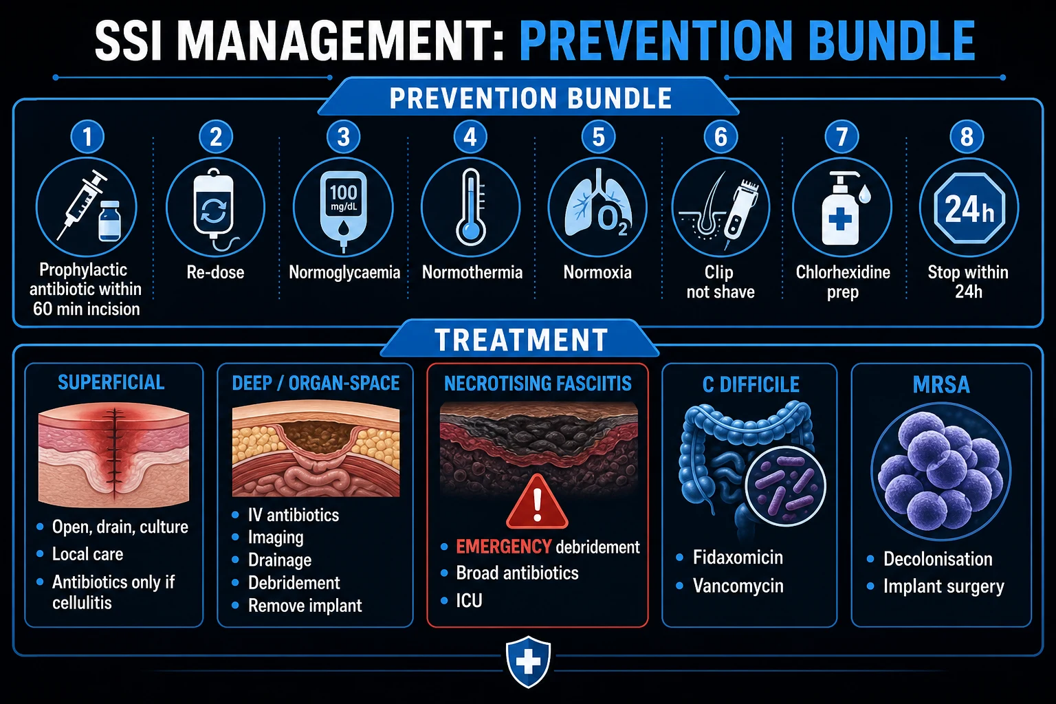
Most SSIs are not immediately life-threatening. However, deep or organ/space SSI presenting with sepsis is a time-critical surgical emergency — Surviving Sepsis Campaign targets apply.[1]
[1]Source control is not a substitute for antibiotics and antibiotics are not a substitute for source control — both are required. Do not delay source control waiting for stability if the patient has necrotising infection, fascial dehiscence with exposed viscera, or an uncontrolled leak.[1]
Management — Definitive & Stepwise
Step 1 — Superficial incisional SSI
- Open the wound: remove the sutures or staples over the infected segment; allow drainage.
- Send pus for Gram stain, culture and sensitivity.
- Irrigate with normal saline; pack lightly with saline-soaked gauze or use a hydrofibre/hydrocolloid dressing (alginates for highly exudative wounds).
- Daily dressing change (or every 48 to 72 h for hydrocolloids); document wound dimensions, exudate and tissue type.
- Targeted antibiotics only if there is surrounding cellulitis or systemic signs:
- Oral flucloxacillin 500 mg four times daily for 5 to 7 days for suspected MSSA;
- Oral doxycycline 100 mg daily or clindamycin 300 mg four times daily if penicillin-allergic;
- Add oral co-amoxiclav 625 mg three times daily or metronidazole if GI/GU source suspected.
- Re-evaluate at 48 hours — if not improving, reconsider diagnosis (deeper infection? resistant organism? necrotising?), escalate to imaging, IV antibiotics, surgical review. [1]
Step 2 — Deep incisional SSI
- Open the entire wound, not just the infected segment.
- Surgical debridement of all necrotic tissue, slough and non-viable fascia; explore for and drain any associated collection.
- Daily surgical dressing change (or vacuum dressing).
- IV antibiotics:
- IV flucloxacillin 1 g every 6 hours for MSSA;
- IV vancomycin 15 to 20 mg/kg every 12 hours (trough 15 to 20) for MRSA or β-lactam allergy;
- Add broad-spectrum cover (piperacillin-tazobactam or ceftriaxone + metronidazole) if polymicrobial/GI source suspected.
- Image to exclude extension to organ/space (CT abdomen/pelvis).
- Duration typically 7 to 14 days; de-escalate once sensitivities available. [1]
Step 3 — Organ/space SSI
- Source control is paramount:
- Percutaneous image-guided drainage (CT or ultrasound) for accessible collections — preferred over surgery where feasible.
- Re-operation for collections not amenable to percutaneous drainage, for anastomotic leak with peritonitis, for retained necrotic tissue, or for failed percutaneous drainage.
- Broad-spectrum IV antibiotics as for the resuscitation bundle; de-escalate to targeted therapy based on cultures.
- Manage the underlying cause — anastomotic leak may need resection and stoma, prosthetic joint infection may need 1-stage or 2-stage revision, mediastinitis may need rewiring and flap cover.
- Duration 7 to 14 days for most organ/space infections; 4 to 6 weeks for osteomyelitis, mediastinitis, and prosthetic joint infection (with infectious diseases input). [1]
Step 4 — Negative-pressure wound therapy (NPWT / VAC)
- Continuous negative pressure of minus 125 mmHg (range 50 to 175).
- Indications: open abdomen, dehisced wounds, large defects after debridement, complex perineal wounds after abdominoperineal resection, diabetic foot ulcers, burns.
- Effects: removes exudate, increases local blood flow, reduces oedema, promotes granulation, contracts the wound edge, reduces frequency of dressing change.
- Prophylactic closed-incision NPWT (ciNPWT) is increasingly used in high-risk closed incisions — obese patients, midline laparotomy, sternotomy in obese diabetics, groin incisions after vascular surgery — to reduce SSI.[4]
Antibiotic duration principles
- Superficial with cellulitis: 5 to 7 days oral.
- Deep: 7 to 14 days IV (transition to oral once afebrile and improving).
- Organ/space with source control: 4 to 7 days after source control in most cases (short courses equivalent to longer for many intra-abdominal infections); longer (4 to 6 weeks IV) for osteomyelitis, mediastinitis, prosthetic joint.
- De-escalate as soon as sensitivities available; engage antimicrobial stewardship. [1]
Escalation triggers
- Failure to improve within 48 to 72 hours of appropriate therapy — reconsider diagnosis, repeat imaging, surgical review.
- Worsening pain, erythema spreading, crepitus, systemic deterioration — suspect necrotising infection or uncontrolled source: emergency surgical review.
- New organ dysfunction — ICU admission. [1]
Specific Subtypes & Scenarios
Orthopaedic prosthetic joint infection
- Organisms: coagulase-negative staphylococci (CoNS) and S. aureus most common; also Streptococcus, Enterococcus, Gram-negatives, anaerobes. Biofilm is the defining feature.[1]
- Diagnosis: clinical (pain, swelling, sinus), serum CRP and ESR (both typically elevated), joint aspiration for cell count and culture (synovial WBC over 3000 per microlitre with neutrophil predominance is suggestive), multiple deep tissue biopsies at revision surgery (3 to 6 samples; modified Birmingham criteria).
- Management:
- Acute (less than 3 weeks post-op): DAIR (Debridement, Antibiotics, Irrigation and Retention of implant) + IV antibiotics (e.g. rifampicin + fluoroquinolone for staphylococci) for 6 weeks.
- Chronic: two-stage exchange (remove implant, antibiotic-loaded cement spacer, IV antibiotics 6 weeks, then reimplantation) — gold standard. One-stage exchange in selected centres.
- Suppressive antibiotics lifelong if implant cannot be removed and patient not fit for surgery.
Colorectal surgery
- Most common organ-space SSI after colorectal surgery is anastomotic leak and pelvic abscess.[1]
- Organisms: enteric Gram-negatives (E. coli, Klebsiella, Enterobacter), enterococci, anaerobes (B. fragilis). Prophylaxis: IV cephalosporin (cefuroxime or ceftriaxone) + metronidazole.
- Prevention: combined mechanical and oral antibiotic bowel preparation (e.g. polyethylene glycol prep + oral neomycin and metronidazole) reduces SSI more than IV antibiotics alone; this is supported by multiple randomised trials and meta-analyses, reversing earlier guidance that bowel prep was unnecessary.[1]
- Anastomotic leak: presents around day 5 to 7; CT with water-soluble contrast; emergency re-operation with diversion (Hartmann's) for generalised peritonitis; percutaneous drainage for localised pelvic abscess.
Caesarean section
- SSI rate 3 to 15%, higher after emergency caesarean and labour.
- Organisms: endogenous vaginal flora — group B Streptococcus, anaerobes, E. coli; S. aureus (including MRSA) from skin.
- Prevention: pre-incision IV cephalosporin (cefazolin 2 g IV — 3 g if body weight over 120 kg) given before skin incision, NOT after cord clamping.[14] A Cochrane meta-analysis confirmed that pre-incision prophylaxis reduces postpartum endometritis and wound infection without neonatal harm.[14] Add azithromycin 500 mg IV for emergency caesarean in labour (Azithro trial).
- Skin preparation: 2% chlorhexidine-alcohol superior to povidone-iodine (Cochrane review).[17]
- Endometritis is the organ-space equivalent: fever, uterine tenderness, foul lochia; treat with IV clindamycin + gentamicin (or co-amoxiclav).
Cardiac surgery (sternotomy and vein-harvest sites)
- Organisms: S. aureus (especially in nasal carriers), CoNS, Gram-negatives.
- Deep SSI = mediastinitis — mortality 10 to 40%.
- Prevention:
- Pre-op screening for S. aureus nasal carriage; carriers decolonised with mupirocin 2% nasal ointment twice daily and chlorhexidine body wash for 5 days.[13][15] The Bode NEJM 2010 trial showed this strategy reduced deep S. aureus SSI by approximately 50% in cardiothoracic and orthopaedic carriers (NNT 8).[13]
- Prophylaxis: IV cefazolin 2 g (vancomycin 1.5 g over 60 min if MRSA risk or β-lactam allergy); re-dose at 4 hours or after 1500 mL blood loss or CPB over 2 hours.
- Prophylactic ciNPWT for high-risk sternotomy (obese, diabetic, bilateral internal mammary harvest).
Vascular graft infection
- Early: S. aureus, Gram-negatives.
- Late: CoNS (indolent, months to years), or graft-enteric erosion (aorto-enteric fistula presenting with herald upper GI bleed).
- Diagnosis: CT angiography; tagged white-cell scan if indeterminate.
- Management: graft excision + extra-anatomic bypass (axillo-bifemoral) or in-situ replacement with rifampicin-soaked or silver-coated Dacron, biological (cadaveric) graft, or autogenous vein. Lifelong suppressive antibiotics if graft retained. [1]
Implant and mesh infections (general principles)
- Biofilm is the central problem: retention of implant usually fails for chronic infection.
- Acute (less than 3 weeks): DAIR + targeted IV antibiotics.
- Chronic: implant removal, antibiotics, delayed reimplantation.
- Mesh infection after hernia repair: try conservative antibiotics; if persistent, mesh removal (often partial). [1]
Complications & Pitfalls
Wound-related
- Superficial dehiscence — heals by secondary intention or delayed primary closure.
- Fascial dehiscence — surgical emergency (see above); may progress to evisceration.
- Incisional hernia — 10 to 30% of patients who develop a midline laparotomy SSI later present with an incisional hernia.
- Hyperthrophic or keloid scarring — disfiguring; more common in dark skin types.
- Chronic sinus / non-healing wound — consider underlying mesh infection, osteomyelitis, fistula, foreign body (retained swab). [1]
Systemic
- Sepsis, septic shock, multiorgan failure — SSI approximately doubles perioperative mortality.
- Secondary pneumonia, UTI, line infection from prolonged ITU stay.
- Venous thromboembolism — prolonged immobility; balance LMWH against wound bleeding. [1]
Antibiotic-related
- Clostridioides difficile colitis — watery diarrhoea, abdominal pain after antibiotic exposure; treat with oral vancomycin or fidaxomicin, stop inciting antibiotic.
- Selection of multidrug-resistant organisms (MRSA, ESBL, VRE, CRE) — antimicrobial stewardship essential.
- Allergic reactions, line infections, ototoxicity/nephrotoxicity (vancomycin, aminoglycosides). [1]
Prognosis and cost
- Increased length of stay (7 to 10 days), readmission (2 to 10-fold higher), disability, lost income, medico-legal consequences.[18]
- Estimated additional cost per SSI: US$10,000 to 25,000 in the USA, £814 to 10,523 in the UK.[18]
Common pitfalls
- Misinterpreting a surface swab — surface colonisation does not diagnose SSI; deep tissue or pus is required.
- Treating only the symptom — opening a wound and giving antibiotics without considering an underlying collection, anastomotic leak, or necrotic tissue will fail.
- Missing necrotising infection — pain out of proportion, crepitus, dusky skin mandate immediate exploration; do not wait for imaging.
- Inadequate source control — leaving necrotic tissue, an un-drained collection, or a leaking anastomosis and hoping antibiotics will work.
- Under-dosing prophylaxis in obesity — cefazolin 2 g is under-dosed for BMI over 40 / weight over 120 kg (use 3 g).[7]
- Wrong antibiotic for the surgical site — clean surgery = cefazolin (S. aureus); colorectal = add metronidazole (anaerobes).
Prognosis & Disposition
- Superficial SSI: most resolve within 2 to 4 weeks with wound opening, dressings and (if needed) oral antibiotics; no long-term sequelae in most cases.
- Deep SSI: 7 to 14 days IV antibiotics; usually 4 to 6 weeks to full wound healing; risk of incisional hernia.
- Organ/space SSI: 7 to 14 days IV antibiotics (longer for osteomyelitis, mediastinitis, prosthetic joint); often requires re-operation; longer recovery.
- Prosthetic joint infection: 6 weeks IV + often implant revision; long-term functional impairment.
- Mediastinitis: mortality 10 to 40%.
- Overall attributable mortality: 1 to 3%, doubled vs uninfected patients. [1]
Discharge criteria
- Afebrile for 24 to 48 hours;
- Falling CRP and improving clinical condition;
- Clean wound on appropriate dressings;
- Tolerates oral intake and oral antibiotics if continued;
- Safe home environment and follow-up arranged (district or surgical nurse for dressing change);
- Patient and family educated about warning signs (spreading erythema, fever, increasing pain). [1]
Follow-up
- Surgical review at 2 to 4 weeks post-discharge;
- Surveillance for incisional hernia at 6 to 12 months;
- Consider MRSA decolonisation on discharge for carriers.[13]
Special Populations
Diabetes mellitus
- HbA1c over 8% (64 mmol/mol) triples SSI risk; perioperative glucose over 11 mmol/L further increases risk.[1]
- Aim for HbA1c under 69 mmol/mol (8.5%) before elective surgery; consider VRIII (variable-rate IV insulin infusion) for poorly controlled diabetics.
- Target blood glucose 6 to 10 mmol/L perioperatively (4 to 12 mmol/L acceptable); avoid both hyper- and hypoglycaemia.
- Use a long-acting basal insulin (e.g. glargine) on the morning of surgery; omit short-acting meal-time insulin when fasting.
Obesity
- BMI over 30 increases SSI risk; over 40 markedly so.
- Tissue hypoxia, longer operations, under-dosed prophylaxis, and difficulty closing dead space.
- Increase antibiotic dose — cefazolin 3 g if weight over 120 kg; vancomycin dose by actual body weight.[7]
- Consider prophylactic ciNPWT for high-risk closed incisions (midline laparotomy, panniculectomy incision).[4]
- Wound closure in layers to obliterate dead space; subcutaneous suction drainage controversial.
Immunosuppressed patients (transplant, chemo, HIV, chronic steroids)
- Atypical and opportunistic organisms — Candida, Aspergillus, mycobacteria (rapidly growing M. abscessus, M. chelonae), Pseudomonas.
- Blunted inflammatory response — subtle signs, low-grade fever only.
- Lower threshold for imaging and biopsy; early involvement of infectious diseases / microbiology.
- Cover broadly empirically (meropenem + vancomycin ± antifungal); narrow once organism identified. [1]
Elderly
- Blunted inflammatory response; atypical presentation (confusion, hypothermia, hypoglycaemia, falls).
- Higher mortality; comorbidity.
- Delirium prevention: optimise pain control, sleep, hydration, mobility; minimise sedatives. [1]
Pregnancy
- Caesarean SSI rate 3 to 15%, higher after emergency caesarean and labour.
- Physiological changes affect drug dosing (increased volume of distribution, increased renal clearance).
- Pre-incision (not cord-clamp) cephalosporin — cefazolin 2 g IV (3 g if over 120 kg) — is safe in pregnancy and lactation and is the standard of care.[14]
- Endometritis is the organ-space equivalent — IV clindamycin + gentamicin.
Paediatrics
- Weight-based dosing — cefazolin 30 mg/kg IV (maximum 2 g); vancomycin 15 mg/kg (maximum 1 g).
- Higher relative fluid requirements; careful fluid balance.
- Use ultrasound preferentially for imaging (radiation avoidance).
- Parents involved in care and consent. [1]
MRSA carriers
- Pre-op screening (nasal swab) for high-risk procedures — cardiac, orthopaedic implant, neurosurgery, prolonged ITU, transfer from another hospital.
- Decolonisation: mupirocin 2% nasal ointment twice daily + chlorhexidine 4% body wash daily for 5 days pre-op.[13][15]
- Prophylaxis: vancomycin 1 to 1.5 g IV over 60 min or teicoplanin 400 mg IV instead of (or in addition to) cefazolin if known MRSA colonisation.[7]
- Universal decolonisation strategies have been shown to reduce MRSA clinical isolates and bacteraemia in ICU (Huang 2013).[19]
Evidence, Guidelines & Regional Differences
Landmark guidelines
- CDC 2017 Guideline for the Prevention of SSI (Berríos-Torres et al., JAMA Surgery 2017)[1] — 9 high-priority recommendations covering parenteral antibiotics, glycaemic control, normothermia, oxygenation, hair removal, and antimicrobial prophylaxis. Replaced the 1999 CDC/HICPAC guideline (Mangram).[2]
- WHO Global Guidelines for SSI Prevention (Allegranzi et al., Lancet Infectious Diseases 2016)[4][5] — 13 pre-operative and 16 intra-/post-operative recommendations; first guideline to specifically address low- and middle-income countries and to make recommendations on glycaemic control, anaesthetic agents, and oxygenation.
- NICE Clinical Guideline CG74 (2008), updated as NG125 (2019) — UK recommendation: prophylactic antibiotics within 60 minutes of incision; do NOT use antibiotic sutures, beads or pouches routinely; clip rather than shave; 80% FiO2 in recovery.
- ASHP / IDSA / SIS / SHEA surgical prophylaxis guideline (Bratzler et al. 2013)[7] — procedure-specific antibiotic recommendations (agent, dose, timing, duration, re-dosing).
Landmark trials
- Classen 1992 NEJM[6] — established the optimal timing of prophylactic antibiotics. SSI rate was 3.8% when antibiotics were given more than 2 hours before incision, 0.6% within 0 to 2 hours (optimal), 1.4% within 3 hours after incision, and 5.9% more than 3 hours after incision. This is the single study underpinning "within 60 minutes" guidance.
- Kurz 1996 NEJM[10] — perioperative normothermia (36 to 37C) reduced SSI after colorectal surgery from 19% to 6%.
- Greif 2000 NEJM[11] — supplemental perioperative oxygen (80% FiO2) reduced SSI after colorectal surgery from 11% to 5%.
- Belda 2005 JAMA[12] — confirmed supplemental oxygen benefit (80% FiO2 for 6 h post-op). Note: the PROXI trial (2009) was neutral, so the benefit remains debated, but WHO and CDC continue to recommend 80% FiO2 while the patient is intubated and in early recovery.
- Bode 2010 NEJM[13] — nasal mupirocin + chlorhexidine body wash in confirmed S. aureus carriers reduced deep S. aureus SSI by about 50% in cardiothoracic and orthopaedic procedures (NNT 8). Confirmed by Cochrane review (Liu 2017).[15]
- Haynes 2009 NEJM[8] — the WHO Surgical Safety Checklist reduced inpatient complications from 11% to 7% and mortality from 1.5% to 0.8% in a global cohort of over 7000 patients.
- Huang 2013 NEJM[19] — universal decolonisation in ICU reduced MRSA clinical isolates and bacteraemia more than targeted screening and isolation.
- Mackeen 2014 Cochrane[14] — pre-incision (vs cord-clamp) antibiotic administration in caesarean section reduced postpartum endometritis and wound infection without neonatal harm.
Cochrane systematic reviews
- Liu 2017[15] — nasal decontamination with mupirocin in S. aureus carriers reduces SSI.
- Tanner 2021[16] — preoperative hair removal: no difference between clipping and no hair removal; shaving is associated with higher SSI than clipping. If hair must be removed, use clippers, not razors.
- Hadiati 2020[17] — skin preparation for caesarean: no clear difference between antiseptic agents, but chlorhexidine-alcohol generally preferred for its rapid, persistent activity.
Controversies
- Optimal oxygen concentration — PROXI trial was neutral; current guidance still recommends 80% FiO2 intraoperatively and in early recovery for patients with normal oxygenation.[4]
- Mechanical bowel preparation — earlier trials suggested mechanical prep alone was useless and possibly harmful; recent meta-analyses and trials (e.g. the elective colorectal surgery literature) show that combined mechanical and oral antibiotic bowel preparation reduces SSI more than IV antibiotics alone.[1]
- Routine vancomycin vs cefazolin prophylaxis — cefazolin remains first-line; vancomycin added for known MRSA carriers, β-lactam allergy, or high local MRSA prevalence; routine dual coverage not recommended due to nephrotoxicity and selection pressure.
- Antibiotic-impregnated suture (triclosan-coated Vicryl Plus) — modest SSI reduction in meta-analyses; WHO suggests use.[4]
- Duration of prophylaxis — single pre-incision dose is as effective as 24-hour coverage for most procedures; prolonged prophylaxis selects resistance and C. difficile.[1]
Regional deltas
India / LMIC: SSI rates in many Indian and sub-Saharan African hospitals are 2 to 3 times higher than in HICs (10 to 15% in some series), driven by overcrowded theatres, limited antibiotic stewardship, irregular antibiotic supply, and inadequate perioperative monitoring.[4] The WHO Global Guidelines 2016 explicitly addressed LMIC contexts.[4][5] The Ayushman Bharat scheme and Indian National Action Plan on Antimicrobial Resistance emphasise SSI surveillance and stewardship. Practical LMIC-relevant measures: clip rather than shave, hand hygiene and aseptic technique (cheap and high-impact), WHO Surgical Safety Checklist, accurate timing of generic cefazolin.
Exam Pearls
Evidence-based SSI prevention bundle — BUNDLE
prophylactic antibiotics within 60 minutes (re-dose at 4 h or after 1500 mL blood loss)
aseptic technique, chlorhexidine-alcohol skin prep, adhesive drape
core temperature 36 to 37C (forced-air warmer)
blood glucose 6 to 10 mmol/L; aim HbA1c under 69 mmol/mol
minimally invasive approach where possible
supplemental 80% FiO2 in recovery
NNIS/NHSN risk index 0 to 3 — ACW
ASA score greater than 2 (+1)
wound class 3 or 4 (contaminated/dirty) (+1)
operation longer than the 75th percentile duration for that procedure (+1)
ASEPSIS wound score components
antibiotics, drainage, debridement
days 1 to 7
days 1 to 7
days 1 to 7
days 5 to 14
culture result
duration of stay
- Definition: SSI within 30 days (or 90 days if implant).
- CDC depth classes: superficial (skin/subcut), deep (fascia/muscle), organ/space (any cavity/joint/mediastinum).
- Wound class risk: clean under 2%, clean-contaminated 3 to 7%, contaminated 10 to 15%, dirty over 27%.
- NNIS/NHSN risk index 0 to 3: ASA over 2 (+1), contaminated/dirty wound (+1), operation longer than 75th percentile (+1).
- Prophylactic antibiotics: within 60 minutes BEFORE incision; re-dose at 4 hours or after 1500 mL blood loss (cefazolin 2 g IV — 3 g if over 120 kg; vancomycin 1.5 g over 60 min if MRSA/β-lactam allergic).
- Most common organism in CLEAN surgery: Staphylococcus aureus (and CoNS).
- Clean-contaminated / contaminated: enteric Gram-negatives (E. coli, Klebsiella) + anaerobes (B. fragilis).
- Classen 1992 NEJM — optimal timing 0 to 60 minutes pre-incision; SSI 0.6% vs 3.8% if too early.[6]
- Bode 2010 NEJM — mupirocin + chlorhexidine decolonisation in S. aureus carriers halves deep SSI in cardiothoracic/orthopaedic surgery.[13]
- Kurz 1996 NEJM — normothermia (36 to 37C) halves SSI after colorectal surgery.[10]
- Greif 2000 NEJM and Belda 2005 JAMA — supplemental 80% FiO2 reduces SSI (debated by PROXI).[11][12]
- Haynes 2009 NEJM — WHO Surgical Safety Checklist reduces complications (11% to 7%) and mortality (1.5% to 0.8%).[8]
- Caesarean: pre-incision (NOT cord-clamp) cefazolin 2 g IV.[14]
- Colorectal: combined mechanical + oral antibiotic bowel prep superior to IV alone.[1]
- MRSA carriers: mupirocin 2% nasal + chlorhexidine body wash for 5 days pre-op; vancomycin/teicoplanin prophylaxis.[13]
- Hair removal: clip not shave (Tanner Cochrane).[16]
- Superficial SSI: open wound, dressings ± oral antibiotics only if cellulitis. Deep SSI: debridement + IV antibiotics. Organ/space SSI: drain + source control + broad-spectrum IV antibiotics.
- NPWT/VAC: minus 125 mmHg for open wounds; prophylactic ciNPWT for high-risk closed incisions.
- Implant infection (biofilm): acute less than 3 weeks — DAIR + IV antibiotics; chronic — implant removal + revision.
Exam application bank (NEET-PG / INICET)
One-line answer
SSI = infection at the surgical site within 30 days (or 90 days if implant placed), classified by CDC into superficial incisional (skin/subcut), deep incisional (fascia/muscle) and organ/space. Affects 2 to 5% of procedures, doubles mortality, and adds 7 to 10 days to length of stay. Most common organism in clean surgery: Staphylococcus aureus. Prevention bundle: prophylactic antibiotics within 60 min of incision (re-dose at 4 h or after 1500 mL blood loss), normothermia, glycaemic control, clipping (not shaving), supplemental oxygen, chlorhexidine skin prep, WHO Surgical Safety Checklist. [1]
Worked stems (answer without another resource)
Stem 1 — Classic presentation. Map symptoms to mechanism; name the first investigation and first treatment step with dose/route if drug therapy is standard. [1]
Stem 2 — Unstable / complicated. List red flags that force immediate resuscitation, theatre, ICU, antidote, or reperfusion — and what you do in the first 15 minutes. [1]
Stem 3 — Atypical group. Elderly, pregnancy, child, or immunocompromised: how presentation and thresholds change. [1]
Stem 4 — Differential trap. Name the three closest mimics and one discriminator for each. [1]
Stem 5 — Disposition. Who goes home with safety-netting, who is admitted, who needs HDU/ICU/theatre, and what follow-up is mandatory. [1]
Rapid viva checklist
- Definition + classification
- Pathophysiology chain
- Bedside signs / criteria
- Score with exact components (if any)
- Emergency bundle
- Definitive therapy with doses
- Complications of disease and of treatment
- Special populations
- Guideline/trial name if classic
- Three exam traps
Coverage self-check
If you cannot answer any stem above from this page alone, re-read the matching section — the page is intended to be self-sufficient for final-prof and NEET-PG/INICET questions on Surgical Site Infection.
References
- [1]Berríos-Torres SI, Umscheid CA, Bratzler DW, et al. Centers for Disease Control and Prevention Guideline for the Prevention of Surgical Site Infection, 2017 JAMA Surg, 2017.PMID 28467526
- [2]Mangram AJ, Horan TC, Pearson ML, et al. Guideline for Prevention of Surgical Site Infection, 1999. Centers for Disease Control and Prevention (CDC) Hospital Infection Control Practices Advisory Committee Am J Infect Control, 1999.PMID 10196487
- [3]Horan TC, Gaynes RP, Martone WJ, et al. CDC definitions of nosocomial surgical site infections, 1992: a modification of CDC definitions of surgical wound infections Infect Control Hosp Epidemiol, 1992.PMID 1334988
- [4]Allegranzi B, Zayed B, Bischoff P, et al. New WHO recommendations on intraoperative and postoperative measures for surgical site infection prevention: an evidence-based global perspective Lancet Infect Dis, 2016.PMID 27816414
- [5]Allegranzi B, Bischoff P, de Jonge S, et al. New WHO recommendations on preoperative measures for surgical site infection prevention: an evidence-based global perspective Lancet Infect Dis, 2016.PMID 27816413
- [6]Classen DC, Evans RS, Pestotnik SL, et al. The timing of prophylactic administration of antibiotics and the risk of surgical-wound infection N Engl J Med, 1992.PMID 1728731
- [7]Bratzler DW, Dellinger EP, Olsen KM, et al. Clinical practice guidelines for antimicrobial prophylaxis in surgery Am J Health Syst Pharm, 2013.PMID 23327981
- [8]Haynes AB, Weiser TG, Berry WR, et al. A surgical safety checklist to reduce morbidity and mortality in a global population N Engl J Med, 2009.PMID 19144931
- [9]Magill SS, Edwards JR, Bamberg W, et al. Multistate point-prevalence survey of health care-associated infections N Engl J Med, 2014.PMID 24670166
- [10]Kurz A, Sessler DI, Lenhardt R. Perioperative normothermia to reduce the incidence of surgical-wound infection and shorten hospitalization. Study of Wound Infection and Temperature Group N Engl J Med, 1996.PMID 8606715
- [11]Greif R, Akca O, Horn EP, et al. Supplemental perioperative oxygen to reduce the incidence of surgical-wound infection N Engl J Med, 2000.PMID 10639541
- [12]Belda FJ, Aguilera L, Garcia de la Asuncion J, et al. Supplemental perioperative oxygen and the risk of surgical wound infection: a randomized controlled trial JAMA, 2005.PMID 16249417
- [13]Bode LG, Kluytmans JA, Wertheim HF, et al. Preventing surgical-site infections in nasal carriers of Staphylococcus aureus N Engl J Med, 2010.PMID 20054045
- [14]Mackeen AD, Packard RE, Ota E, et al. Timing of intravenous prophylactic antibiotics for preventing postpartum infectious morbidity in women undergoing cesarean delivery Cochrane Database Syst Rev, 2014.PMID 25479008
- [15]Liu Z, Norman G, Iheozor-Ejiofor Z, et al. Nasal decontamination for the prevention of surgical site infection in Staphylococcus aureus carriers Cochrane Database Syst Rev, 2017.PMID 28516472
- [16]Tanner J, Norrie P, Melen K. Preoperative hair removal to reduce surgical site infection Cochrane Database Syst Rev, 2021.PMID 34437723
- [17]Hadiati DR, Hakimi M, Nurdiati DS. Skin preparation for preventing infection following caesarean section Cochrane Database Syst Rev, 2020.PMID 32580252
- [18]Coello R, Charlett A, Wilson J, et al. Adverse impact of surgical site infections in English hospitals J Hosp Infect, 2005.PMID 15866006
- [19]Huang SS, Septimus E, Kleinman K, et al. Targeted versus universal decolonization to prevent ICU infection N Engl J Med, 2013.PMID 23718152
- [20]Troeman DPR, Postma DF, van Werkhoven CH, et al. Postoperative Staphylococcus aureus Infections in Patients With and Without Preoperative Colonization JAMA Netw Open, 2023.PMID 37906196