General Surgery · General Surgery
Testicular Torsion
Also known as Torsion of testis · Spermatic cord torsion · Acute scrotum
Testicular torsion is a urological surgical emergency — twisting of the spermatic cord on its longitudinal axis compromises the testicular blood supply, producing ischaemia and, within hours, irreversible infarction. The salvage window is the 6-hour rule: 90 to 100 percent salvage if detorsed within 6 hours, about 50 percent at 6 to 12 hours, and less than 10 percent after 24 hours. Classic presentation: sudden severe unilateral testicular pain, an absent cremasteric reflex, and a high-riding testis lying horizontally. Diagnosis is clinical — never delay surgery for imaging when suspicion is high. Definitive management is immediate scrotal exploration with detorsion, assessment of viability, ipsilateral orchidopexy or orchidectomy, and routine contralateral orchidopexy (the bell-clapper deformity is bilateral in roughly 12 percent anatomically and fixation protects the only remaining testis).
On this page & tools
Your progress
Saved locally on this device.
Exam tags
Red flags
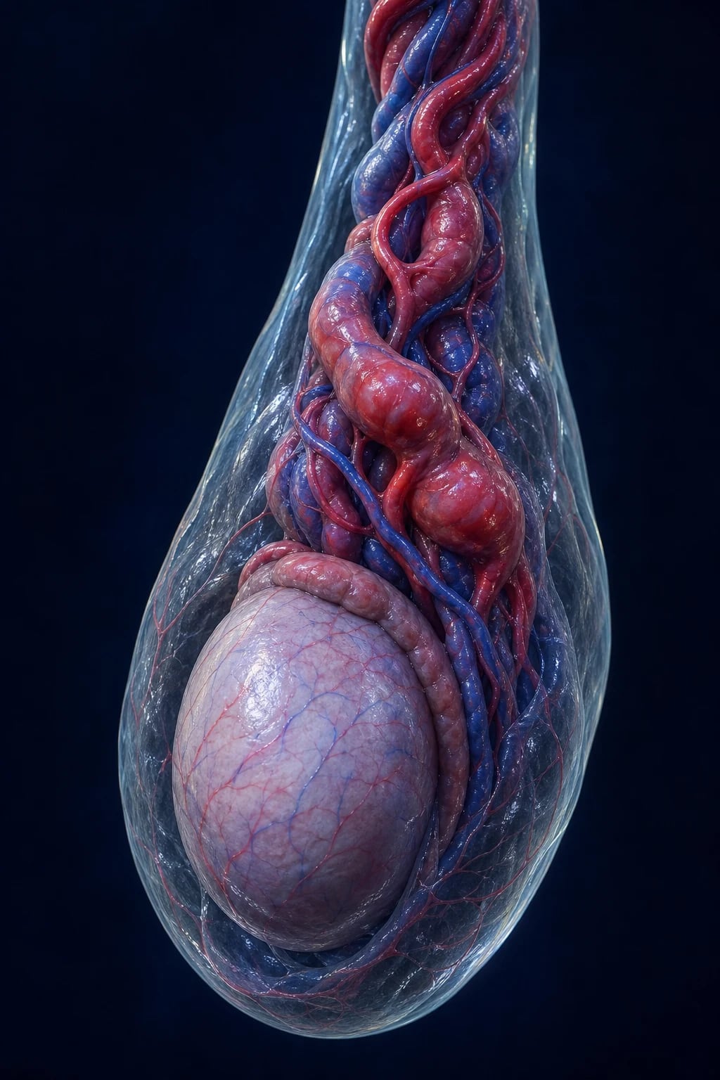
Overview & Definition
Testicular torsion is the twisting of the spermatic cord on its longitudinal axis, which mechanically compromises the testicular blood supply (first the low-pressure venous and lymphatic channels, then the higher-pressure testicular artery) and progresses — unless the cord is untwisted — through ischaemia to infarction and gangrene of the testis. It is one of the few true time-critical urological emergencies: the salvageable window is measured in hours, and the determinant of outcome is the duration of torsion before detorsion.[1]
The doctrine that governs the whole topic can be stated in four words — time is testis. The widely taught salvage gradient is the 6-hour rule: detorsion within 6 hours of onset salvages the testis in roughly 90 to 100 percent of cases; this falls to about 50 percent between 6 and 12 hours, to less than 20 percent between 12 and 24 hours, and to near zero after 24 hours, when orchidectomy is the expected outcome.[1][1] Because of this gradient, any adolescent or young adult male who presents with acute testicular pain has torsion until proven otherwise, and the burden of proof rests on the clinician who would not explore.
Clinically the diagnosis rests on a small set of high-yield findings — sudden severe unilateral scrotal pain, an absent ipsilateral cremasteric reflex, and a high-riding testis with a transverse (horizontal) lie — supported by nausea and vomiting. Imaging (colour Doppler ultrasound) is only an adjunct for the genuinely equivocal presentation and must never delay exploration when the clinical picture is convincing.[1][2]
Classification
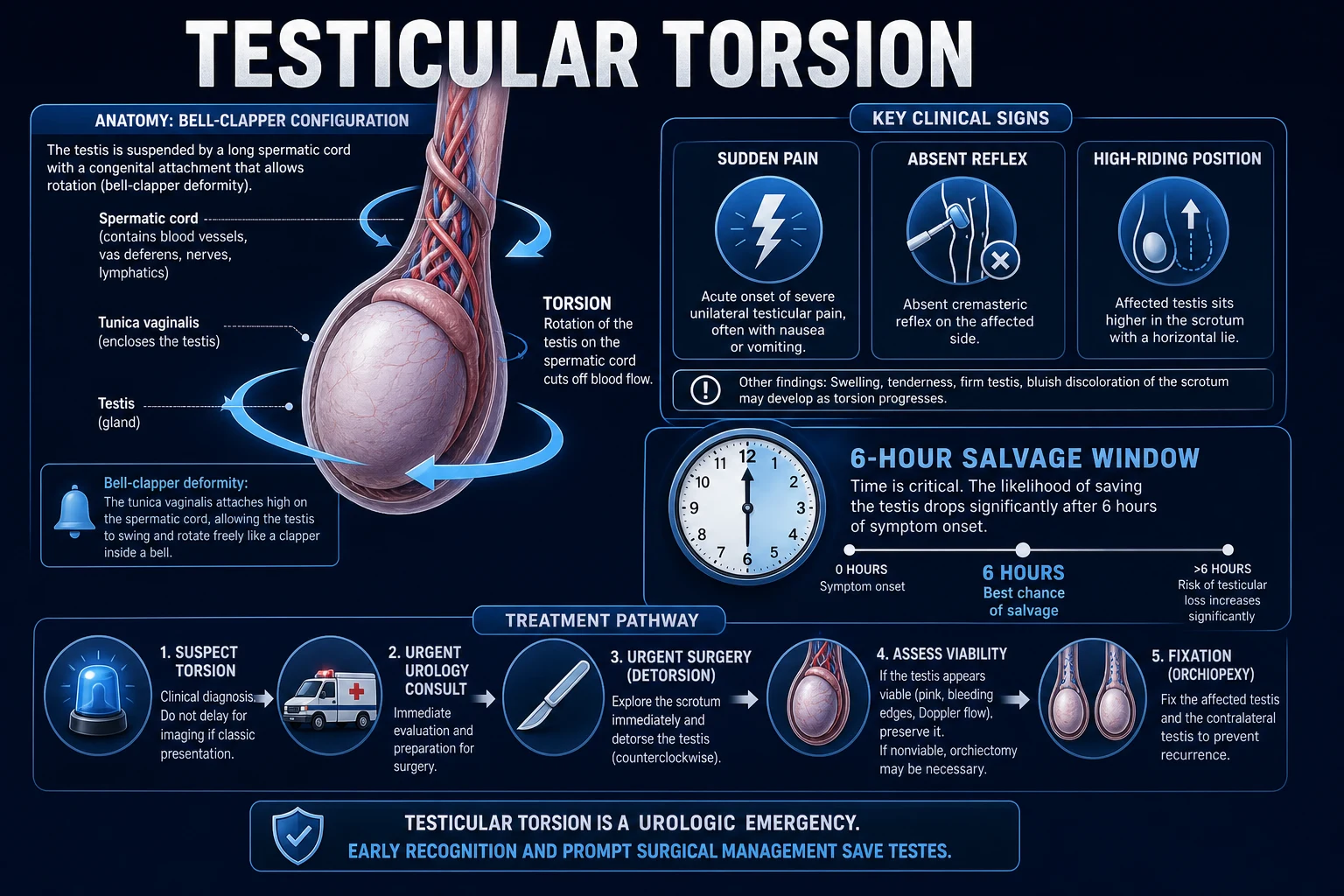
Torsion is best understood by where on the cord the twist occurs and by its temporal behaviour. Three forms are recognised.[1]
Intravaginal torsion
adolescents; the common form
- The twist occurs WITHIN the tunica vaginalis — between the testis and its visceral and parietal layers
- Underlying lesion is the **bell-clapper deformity** (abnormally high investment of the tunica on the cord)
- The testis hangs freely and rotates; typically 360 to 720 degrees
- Peak in **puberty (12 to 18 years)** but occurs at any age; the form most relevant to MBBS exams
Extravaginal torsion
neonates; prenatal/perinatal
- The ENTIRE cord AND its coverings (including tunica vaginalis) twist as a unit, BEFORE the tunica fuses to the scrotal wall
- Almost always **prenatal or perinatal**; the testis is infarcted at birth
- Presents as a **painless, hard, discoloured scrotal mass** in a newborn
- Salvage is usually impossible; the role of surgery is to fix and protect the contralateral testis
Intermittent (subacute) torsion
recurrent self-limiting episodes
- Recurrent acute testicular pain that **spontaneously resolves** as the testis detorses
- Often mislabelled as epididymitis or growing pains
- Carries a high risk of a future **complete, non-resolving torsion**
- Treated by **elective bilateral orchidopexy** — do not wait for a catastrophic episode
A useful additional descriptor at surgery is the degree of rotation. Twists of less than 360 degrees may permit some residual flow and a subacute picture; complete torsion is typically 360 to 720 degrees (one to two full turns), and beyond this venous, then arterial, flow is fully abolished.[1]
Epidemiology & Risk Factors
Testicular torsion has a characteristic bimodal age distribution — a small neonatal peak (extravaginal, mostly perinatal) and a much larger adolescent peak between 12 and 18 years, with most series placing the majority of intravaginal torsion in males aged 12 to 25 years.[1][2]
Testicular torsion — the headline numbers
The single most important anatomical risk factor is the bell-clapper deformity, in which the tunica vaginalis invests the cord too high, leaving the testis unanchored and free to rotate. The deformity is bilateral in roughly 12 percent of anatomical specimens — and because the contralateral testis can therefore torsion at any time, bilateral (contralateral) orchidopexy is performed at every torsion operation even when only one side is symptomatic.[1][1]
Recognised risk factors and precipitants:[1][2]
- Undescended testis (cryptorchidism): torsion of an intra-abdominal or inguinal testis is roughly ten times more common than of a scrotal testis. Any male with an empty hemiscrotum plus acute abdominal or groin pain has torsion of the undescended testis until proven otherwise.
- Puberty and rapid testicular growth: the adolescent growth spurt increases the mass-to-pedicle ratio and is the explanation for the age peak.
- Prior episode of acute scrotal pain that resolved spontaneously (intermittent torsion) — the strongest historical predictor of a future complete torsion.
- Trauma and strenuous exercise: may precipitate torsion in an already-predisposed (bell-clapper) testis, but a careful history usually shows that the pain began either before the trauma or out of proportion to it.
- Cold ambient temperature: provokes cremasteric contraction, the mechanical trigger for rotation; a seasonal predilection for winter months is described.
- Sleep: a substantial minority torsion during sleep, presumably on sudden cremaster contraction.
- Polyorchidism, high investment of gubernaculum, and a long mesorchium are rarer anatomical predispositions.
India and other low-resource settings: late presentation is the dominant problem. Embarrassment about genital symptoms, lack of awareness of the 6-hour window, and reliance on home remedies or traditional healers mean many adolescents present 12 to 24 hours or more after onset, when orchidectomy is already the likely outcome. Cultural reluctance to discuss genital pain — especially with parents — is a major driver of delay. Access to round-the-clock emergency operating capacity may be limited in rural areas, so manual detorsion as a bridge to surgery and rapid referral pathways are particularly important. Public health messaging about testicular self-examination and the urgency of acute scrotal pain is a recognised unmet need.
Pathophysiology
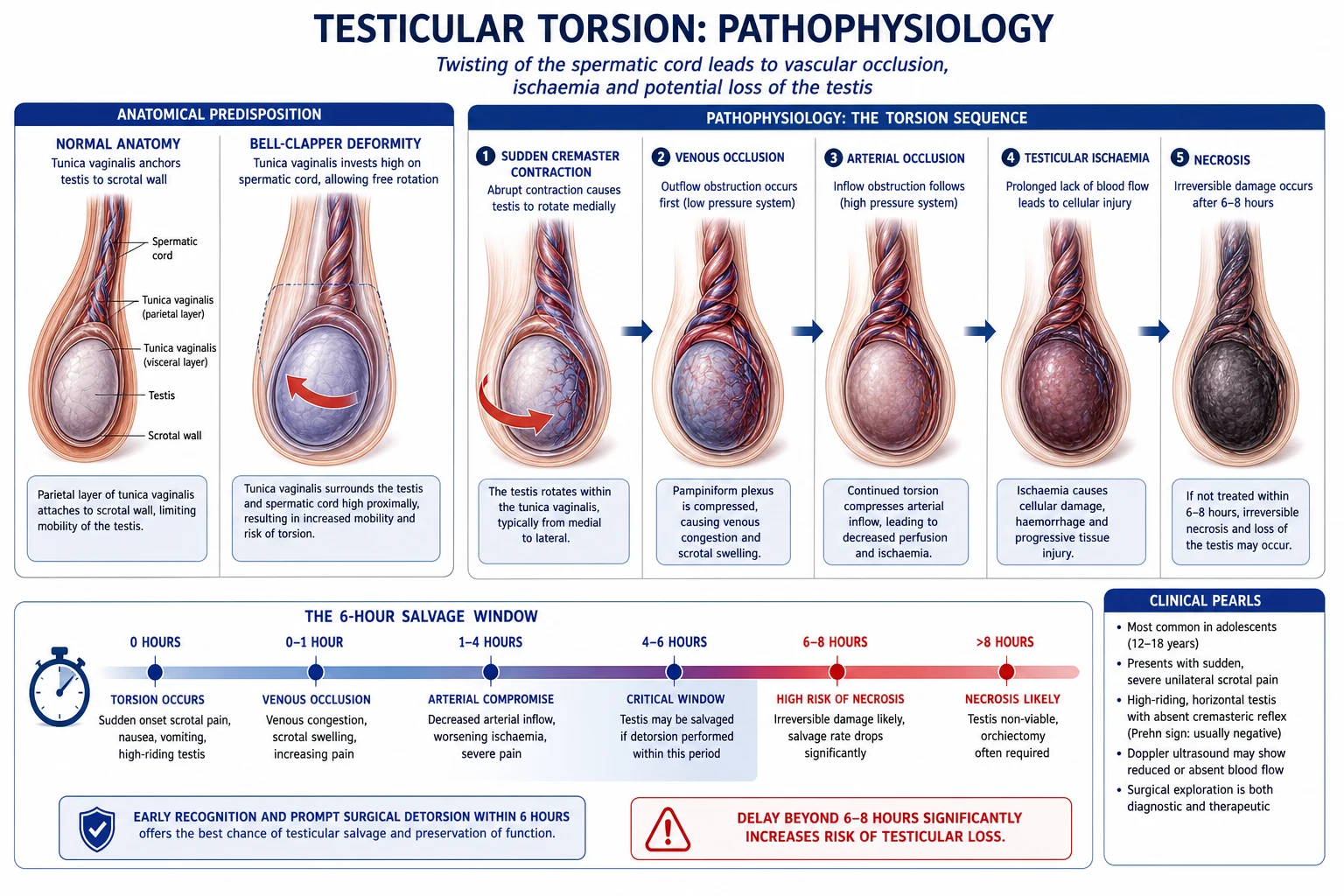
The bell-clapper deformity — the anatomical substrate
In normal anatomy the tunica vaginalis — the peritoneum-derived serous sac — descends with the testis through the inguinal canal and reflects onto the epididymis and posterolateral testis, effectively anchoring the testis to the posterior scrotal wall. In the bell-clapper deformity this reflection is abnormally high: the tunica vaginalis invests the cord itself rather than stopping at the epididymis, so the testis, epididymis and a length of cord lie freely suspended within the tunica — exactly like the clapper inside a bell. With nothing tethering it, the testis can rotate on its vascular pedicle.[1]
This deformity is the substrate for nearly all intravaginal torsion. Because it develops embryologically, it is usually bilateral, which is the rationale for fixing the contralateral testis at the same operation.[1]
The mechanical cascade — why the testis dies
The sequence of events once the cord twists is mechanical and predictable, and it explains the salvage window:[1][2]
- Trigger. A sudden cremasteric contraction (spontaneous, during sleep, after trauma or exercise, or in cold weather) rotates the mobile testis. The torsion is usually medial/inward (about two-thirds of cases); the testis is described as rotating "like closing a book."
- Venous and lymphatic occlusion. The thin-walled, low-pressure pampiniform veins are compressed first. Venous outflow is abolished while arterial inflow continues, producing venous congestion, interstitial oedema, and progressive swelling of the testis within the confined tunica albuginea (a non-compliant fascial envelope).
- Arterial occlusion. Rising intra-tunical pressure and direct mechanical kinking progressively compress the thicker-walled, higher-pressure testicular artery. Once arterial inflow stops, the testis becomes truly ischaemic.
- Infarction. After approximately 6 to 8 hours of complete arterial occlusion, tissue injury becomes irreversible — initially haemorrhagic, then frankly gangrenous. Beyond 24 hours the gland is necrotic and unsalvageable. [1]
Two practical consequences flow from this cascade. First, the degree of torsion matters: a partial twist (less than 360 degrees) may permit trickle flow and a subacute course, whereas 720 degrees abolishes flow within minutes. Second, because venous occlusion precedes arterial occlusion, a testis that has been torsed for only a few hours may look congested and blue but recover completely once detorsed — whereas one torsed for more than 12 hours is typically black, flaccid, and non-viable. The intraoperative decision between orchidopexy and orchidectomy rests on this viability assessment.[1]
Consequences for the contralateral testis — antisperm antibodies
Even when the torsed testis is removed, the contralateral testis is at risk of subsequent subfertility. Breakdown of the blood-testis barrier during ischaemia, and reactive changes after orchidectomy, are associated with the formation of antisperm antibodies and a measurable decline in semen parameters in a subset of patients; the magnitude and clinical impact of this effect remains debated, but it is the rationale for offering baseline and follow-up semen analysis after an episode of torsion.[7]
Clinical Presentation
The classic picture
[1]Associated symptoms
Pain is the headline, but the visceral accompaniments are highly suggestive:[1][1]
- Nausea and vomiting occur in the majority and reflect the visceral afferent pathway of the testis (T10 to L1); their presence with acute scrotal pain substantially raises the likelihood of torsion.
- Lower abdominal or groin pain is common, referred via the genitofemoral and ilioinguinal nerves; a small number of patients present with abdominal pain and minimal scrotal complaints.
- Scrotal swelling, erythema, and a reactive hydrocoele develop over hours as oedema and inflammation progress — these are late signs and their absence early does not exclude torsion.
- Low-grade fever may appear late, particularly if necrosis supervenes; a high fever with gradual onset over days points instead to epididymo-orchitis.
- Urinary symptoms (dysuria, frequency) are typically absent; their presence favours epididymitis, but their absence does not confirm torsion.
Atypical presentations — the traps examiners love
Examiners deliberately probe the atypical presentations because they are the source of missed diagnoses and litigation:[2][8]
- Abdominal or groin pain with an unremarkable scrotum: torsion of an undescended (inguinal or intra-abdominal) testis can present as acute abdomen or a tender groin lump with an empty hemiscrotum. Always examine the scrotum in any male with acute abdominal pain.
- Pain that seems to have settled: an intermittent torsion that has spontaneously detorsed may produce a near-asymptomatic patient by the time of assessment; the history of sudden severe pain that resolved is itself the red flag.
- Trauma: a torsion precipitated by minor trauma may be attributed to the injury; pain disproportionate to the mechanism, or a high-riding testis on the injured side, should prompt exploration.
- Post-pubertal males with "epididymitis": torsion is more likely than epididymitis in males under 25 with acute unilateral pain, and the cremasteric reflex is the discriminator.
- The neonate presents not with pain but with a painless, hard, discoloured (blue-black) scrotal mass, irritability, and reluctance to feed — extravaginal, prenatal torsion.
Differential Diagnosis
The acute scrotum is the umbrella presentation, and the differential is short but the stakes are high. The single most important distinction is torsion versus epididymo-orchitis, because they overlap in age and appearance but demand opposite management.[1][2]
The complete acute scrotum differential, in order of clinical importance:[1][2]
- Testicular torsion — the can't-miss diagnosis; sudden, severe, absent cremasteric reflex, high-riding horizontal testis.
- Epididymitis or epididymo-orchitis — the great mimic; gradual onset over days, fever, pyuria, preserved cremasteric reflex.
- Torsion of the appendix testis (hydatid of Morgagni) — the blue dot sign at the upper pole, milder pain, normal lie.
- Incarcerated or strangulated inguinal hernia — cannot get above the swelling, bowel obstruction signs.
- Traumatic haematocoele or testicular rupture — clear trauma history, bruising, may need exploration.
- Idiopathic scrotal oedema — bilateral, painless, boggy scrotum in a young boy.
- Renal colic — pain radiates to (not from) the scrotum, loin tenderness, haematuria.
- Mumps orchitis — parotitis and viral prodrome in a post-pubertal male.
Each is contrasted in detail below. [1]
| Condition | Onset & tempo | Cremasteric reflex | Prehn sign | Other distinguishing features |
|---|---|---|---|---|
| Testicular torsion | Sudden, severe, hours | Absent | Negative (no relief on elevation) | High-riding horizontal testis; nausea/vomiting; age 12 to 25 |
| Epididymitis / epididymo-orchitis | Gradual, over days | Present | Positive (pain relieved by elevation) | Fever, dysuria, pyuria; tender epididymis first; sexually active or UTI |
| Torsion of appendix testis | Acute, less severe | Present | Variable | Blue dot sign at upper pole; normal testicular lie; prepubertal boys |
| Incarcerated inguinal hernia | Acute groin/scrotal pain | Present | Negative | Cannot get above the swelling; bowel obstruction signs; groin mass |
| Traumatic haematocoele / rupture | After clear trauma | Variable | Negative | Bruising, swelling; history of direct blow; may need exploration |
| Idiopathic scrotal oedema | Insidious | Present | Negative | Bilateral, oedematous but painless scrotum; young boys |
| Renal colic | Acute, colicky | Present | Negative | Pain radiates to (not from) the scrotum; loin tenderness; haematuria |
| Mumps orchitis | Gradual, days | Present | Variable | Parotitis; viral prodrome; post-pubertal males |
Three differentials deserve specific comment. Torsion of the appendix testis (hydatid of Morgagni) classically produces the blue dot sign — a small dark area visible through the scrotal skin at the upper pole representing the infarcted appendage; pain is less severe than in torsion of the testis proper, the testis lies normally, and the cremasteric reflex is preserved.[1] Epididymo-orchitis is the great mimic: gradual onset over days, fever, pyuria, a tender epididymis, and a preserved cremasteric reflex separate it, but in any young male with acute unilateral pain torsion is the default until excluded. Incarcerated hernia is distinguished by the inability to get above the swelling and by signs of bowel obstruction.
Clinical & Bedside Assessment
The diagnosis is made at the bedside. The history alone — sudden severe unilateral pain in a young male — is enough to mandate exploration; physical signs refine but should not over-rule it.[1][1]
Examination technique
A focused scrotal and abdominal examination should take only a few minutes and includes:[1]
- Inspect for asymmetry, swelling, erythema, high-riding testis, horizontal lie, and any external signs of trauma. Compare both sides.
- Palpate gently — assess testicular position, lie, tenderness, and any separate epididymal tenderness or hydrocoele. Try to identify the cord in the scrotum; a thickened, tender, twisted cord ("knot") is pathognomonic when felt.
- Elicit the cremasteric reflex — stroke the inner upper thigh on each side and observe the ipsilateral testis. Asymmetrical or absent reflex on the painful side is the key finding.
- Perform Prehn sign — elevate the scrotum; no relief of pain (negative Prehn sign) supports torsion, while relief (positive Prehn sign) supports epididymitis. Note: Prehn sign is unreliable and must not be used to exclude torsion.
- Examine the abdomen and groin for a hernia, an empty hemiscrotum (undescended testis), and referred tenderness.
The named signs and how reliable they are
Bedside signs — how useful they really are
The cremasteric reflex deserves emphasis. It is mediated by the genitofemoral nerve (L1 to L2) afferently and efferently: stroking the inner thigh contracts cremaster and lifts the testis. Its absence on the painful side is the single most sensitive sign of torsion, but it is not perfect — it can be absent in some normal males, in epididymitis (rarely), and after spinal injury — so a preserved reflex does not exclude torsion. It must be interpreted with the whole picture.[1]
Prehn sign (relief of pain on elevation of the scrotum suggests epididymitis; no relief suggests torsion) is historically taught but unreliable and should never be the basis for a decision to defer exploration. The blue dot sign indicates torsion of the appendix testis, not of the testis itself. The angel sign — an absent cremasteric reflex plus a high-riding testis — is a useful cluster at the bedside.[1]
Investigations
The cardinal rule: diagnosis is clinical
[1]Colour Doppler ultrasound
When the diagnosis is uncertain, colour Doppler ultrasonography is the imaging of choice. The positive finding is absent or markedly reduced intratesticular blood flow on the affected side compared with the contralateral testis, often with a normal or swollen grey-scale testicular parenchyma in early torsion. Reported sensitivity is around 95 percent and specificity around 92 percent in systematic reviews, but this performance is operator-dependent and falls in small or partially-torsed testes, and in prepubertal boys (in whom baseline flow is low).[3]
The crucial caveat is that false negatives occur — partial torsion, spontaneous detorsion at the moment of scanning, and slow collateral flow can all produce an apparently normal Doppler study. A systematic review of ultrasound in the acute scrotum concluded that combined grey-scale (B-mode) and colour Doppler features outperform either alone, with sensitivity approaching 100 percent in well-performed studies, but emphasised that ultrasound findings must always be interpreted in conjunction with the clinical picture, never in isolation.[3]
The TWIST score — a structured clinical risk tool
The Testicular Workup for Ischaemia and Suspected Torsion (TWIST) score is a validated bedside tool that stratifies the probability of torsion from history and examination alone, without imaging. It is a 7-point scale:[5][6]
| TWIST component | Points |
|---|---|
| Hard testis | 2 |
| Testicular swelling | 2 |
| Absent cremasteric reflex | 1 |
| High-riding testis | 1 |
| Nausea / vomiting | 1 |
| Total | 0 to 7 |
The Barbosa risk stratification derived from the score is:[5]
- 0 to 2 — low risk: torsion unlikely; observe, ultrasound if needed.
- 3 to 4 — intermediate risk: ultrasound mandatory, with surgical backup.
- 5 to 7 — high risk: proceed to immediate surgical exploration without imaging.
A systematic review and meta-analysis of nine prospective studies found the Barbosa cut-points had an area under the receiver operating curve of 0.92, with a low-risk sensitivity of 0.98 (effectively ruling torsion out) and a high-risk specificity of 0.97 (effectively ruling it in, justifying immediate exploration). The intermediate group remained genuinely uncertain and required ultrasound.[5] The score is best regarded as a decision-support tool, not a replacement for surgical judgement — and in a high-suspicion clinical setting with an experienced surgeon, exploration remains the default.
Other bedside and laboratory tests
- Urinalysis — to help exclude epididymo-orchitis (pyuria, positive nitrites, bacteriuria). Critically, mild pyuria does not exclude torsion (reactive inflammation around a torsed cord can produce a few white cells); a sterile urine with acute pain favours torsion.
- Full blood count and CRP — a markedly raised white cell count and CRP support infection (epididymitis) but again do not exclude torsion; mild leucocytosis is common in torsion from systemic stress.
- Blood group and save, coagulation, beta-hCG — routine pre-operative bloods once the decision to operate is made; beta-hCG is essential if ectopic pregnancy is in the differential of abdominal pain (where relevant).
- Scrotal exploration is itself the definitive investigation — a "negative" exploration for acute scrotum is acceptable and far safer than a missed torsion. [1]
Imaging pathway: EAU, AUA, EAU/ESPU paediatric, and NICE-aligned pathways all agree that colour Doppler ultrasound is the only recommended imaging modality when imaging is needed, that it must not delay exploration, and that high-resolution ultrasonography and radionuclide scintigraphy are alternatives only in selected centres. MRI is not used in the acute setting. Manual detorsion as a bridge to surgery is endorsed where operating theatre access is delayed.[1]
Management — Resuscitation
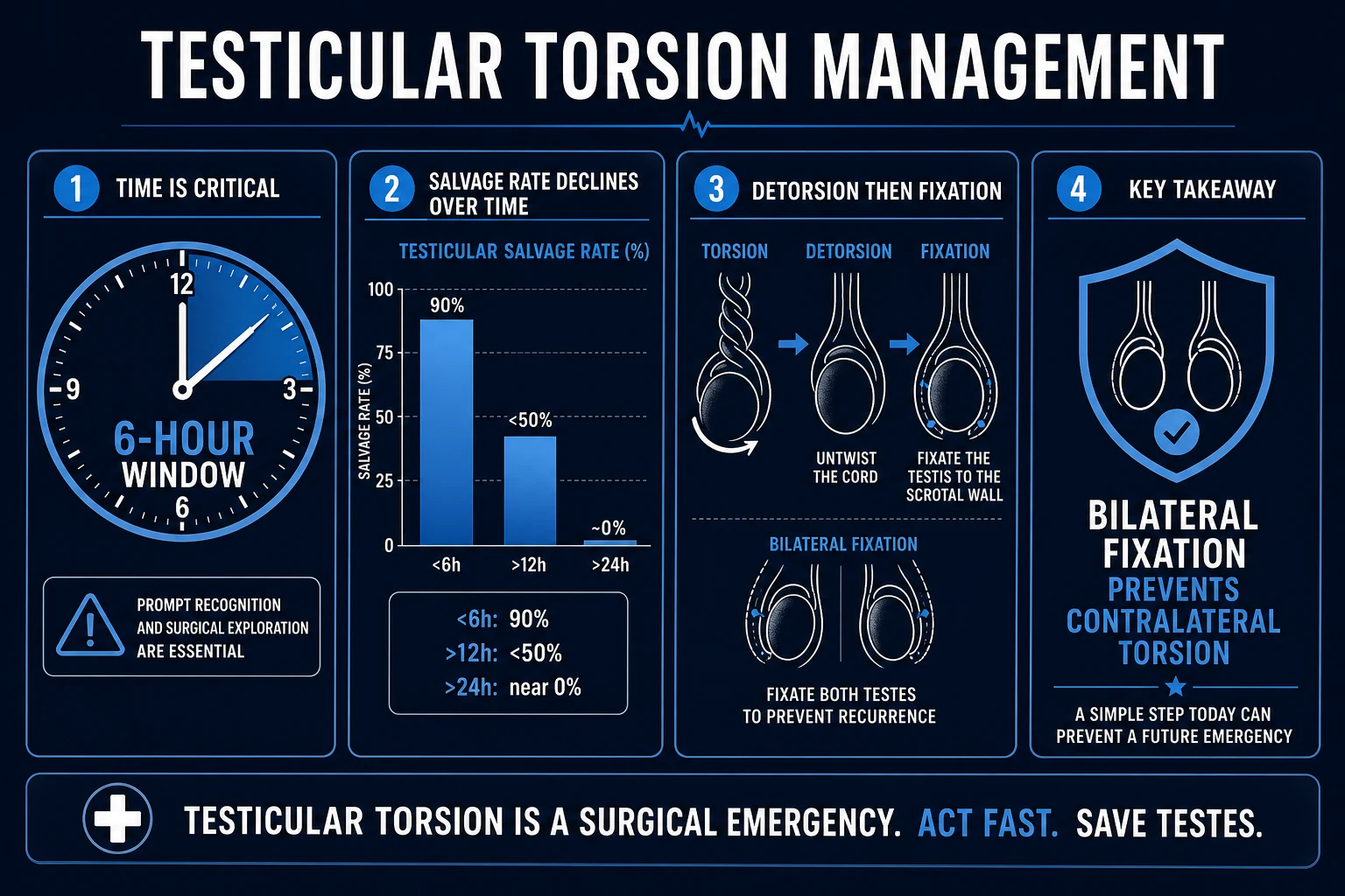
There is no resuscitative delay for torsion: the moment the diagnosis is entertained, the patient is on a path to the operating theatre.[1][2] The resuscitative bundle is small and pragmatic:
- Analgesia: intravenous opioid — morphine 0.1 mg/kg (typical adult 5 to 10 mg) titrated, or fentanyl 1 mcg/kg, with an antiemetic. Adequate analgesia is humane and also facilitates manual detorsion if attempted.
- Antiemetic: ondansetron 4 mg IV (or 0.1 mg/kg in children) for nausea and to support the fasted state.
- Keep nil by mouth from the time of decision; confirm last oral intake to estimate fasting for anaesthesia.
- Consent — for scrotal exploration ± orchidopexy ± orchidectomy, explicitly including the possibility of loss of the testis and the routine fixation of the contralateral side. Document the time-critical nature of the discussion.
- Mark the side with the patient awake and participating; surgical "never-events" include operating on the wrong testis.
- Theatre alert — notify the operating team and anaesthetist immediately; torsion is a Category 1 (immediate) emergency in most theatre prioritisation systems.
- Broad-spectrum antibiotic — cefazolin 1 to 2 g IV (or 25 to 50 mg/kg in children) on induction, particularly if orchidectomy for a necrotic testis is anticipated, to reduce wound infection.
- Single dose of NSAID (e.g. diclofenac 75 mg IM/IV where available) may supplement analgesia but should not replace opioid in severe pain. [1]
The resuscitation question on this topic is short because the answer is: do not resuscitate, operate. Every minute of additional analgesia, IV access, or paperwork that delays the knife is a minute of ischaemia.[1]
Management — Definitive & Stepwise
Immediate surgical exploration — the standard of care
The definitive treatment is immediate scrotal exploration, ideally within 6 hours of symptom onset.[1][1] The operative steps are:[1][1]
- Approach. A transverse hemiscrotal incision (some surgeons prefer a single midline raphe incision to access both sides) through the skin, dartos, and tunica vaginalis; deliver the testis.
- Detorsion. Manually untwist the cord. Because torsion is usually medial (two-thirds of cases), the corrective rotation is lateral/outward — "opening the book." If the direction is unclear, untwist until the testis appears least congested and then confirm by the return of arterial flow. Once detorsed, wrap the testis in warm saline-soaked gauze and observe for return of pink colour and bleeding.
- Assess viability. This is the critical intra-operative judgement:
- A testis that pinks up and bleeds briskly from an incision of the tunica albuginea is viable — proceed to orchidopexy.
- A black, flaccid, non-bleeding testis is non-viable — proceed to orchidectomy.
- If uncertain, incise the tunica albuginea and observe for bright-red bleeding over a few minutes; some surgeons additionally apply warm packs and wait 10 minutes before deciding. There is no single, universally agreed viability threshold — a "save at all costs in the young" approach is reasonable because the downside of leaving a marginally viable testis (atrophy) is less severe than the irreversible loss of a potentially recoverable one.
- Orchidopexy (if viable). Fix the testis to the scrotal wall with non-absorbable sutures (classically 3-0 or 4-0 polypropylene/Ethibond, three to four points) placed through the tunica albuginea and the dartos layer. Some surgeons additionally create a dartos pouch to prevent recurrent rotation.
- Orchidectomy (if non-viable). Divide and ligate the cord at the external ring — the vas deferens and vessels separately — and send the specimen for histology. A testicular prosthesis (silicone) can be inserted at a later elective sitting, or at the same operation by some protocols; offer and document the discussion.
- Contralateral orchidopexy. Always fix the opposite testis at the same operation, using the same non-absorbable technique, because the bell-clapper deformity is bilateral and the risk of metachronous torsion of the only remaining testis is unacceptable. This is one of the few absolutely non-negotiable steps in urological surgery. [1]
Manual detorsion — the bridge to surgery
When operating theatre access is delayed (rural setting, list backlog, prolonged transfer), manual detorsion at the bedside or in the emergency department can restore flow as a temporising measure. The technique:[1][2]
- Provide intravenous analgesia and procedural sedation as needed.
- Grasp the testis gently and rotate it medial to lateral / outward — "opening the book" — because torsion is usually medial. One to two full rotations may be required.
- Success is signalled by an abrupt relief of pain and a softer, lower-riding testis; if pain worsens, the torsion was lateral (one in three cases) and the direction should be reversed.
- Success rates of 25 to 80 percent are reported; even after successful manual detorsion, the testis must still be explored and fixed in the same admission, because retorsion is common and partial detorsion can leave compromised flow.
Manual detorsion direction
OPENBOOK
rotate the testis outward/laterally, like opening a book
sudden relief of pain signals successful detorsion
still proceed to surgical exploration and orchidopexy
detorsion is a bridge, not a cure
the bell-clapper deformity is bilateral
torsion is lateral in about a third — reverse direction if pain worsens
give IV opioid ± sedation before attempting detorsion
confirm restored flow; arrange urgent fixation
Neonatal torsion — special management
In perinatal/neonatal extravaginal torsion the testis is almost always non-salvageable at birth (it torsed in utero and is necrotic). The objectives of surgery are therefore different: confirm the diagnosis, remove the necrotic testis, and explore and fix the contralateral testis to protect the only remaining gonad. Whether the contralateral testis should always be explored and fixed in neonatal torsion is debated — some units advocate urgent exploration within 24 hours to fix the contralateral side and exclude asynchronous torsion, while others adopt a more conservative approach with close observation — but the prevailing modern practice favours early contralateral scrotal exploration and orchidopexy.[1][2]
Intermittent torsion — elective fixation
Any patient with a convincing history of recurrent episodes of acute testicular pain that resolved spontaneously should be offered elective bilateral orchidopexy, regardless of a normal Doppler at the time of clinic review. The natural history of untreated intermittent torsion is progression to a complete, non-detorsing episode with loss of the testis.[1][1]
Specific Subtypes & Scenarios
Intravaginal torsion (adolescents) — the archetypal case
This is the form that dominates exams and clinical practice. The substrate is the bilateral bell-clapper deformity, the trigger is a sudden cremasteric contraction, and the presentation is the classic triad. Management is immediate exploration, detorsion, viability-guided orchidopexy or orchidectomy, and contralateral fixation. Outcomes track the time-to-detorsion almost linearly.[1][1]
Extravaginal (neonatal) torsion
The whole cord and its coverings twist before the tunica vaginalis has fused to the scrotal wall. Presentation is a painless, hard, discoloured scrotal mass in a neonate (often noted at birth). The testis is typically already infarcted; the operation is for diagnosis, removal, and contralateral protection. Bilateral prenatal torsion is catastrophic (anorchia) and rare.[1]
Torsion of an undescended testis
An undescended (inguinal or intra-abdominal) testis has a roughly tenfold higher torsion risk than a scrotal one. The presentation is acute abdominal or groin pain with an empty scrotum and a tender lump in the groin. This is a frequently missed diagnosis in the emergency department — always palpate the scrotum in any male with acute abdominal pain. Management is urgent exploration (inguinal approach for an inguinal testis), detorsion, and orchidopexy to the scrotum if viable, or orchidectomy if not.[1]
Torsion of testicular appendages
The appendix testis (hydatid of Morgagni) and the appendix epididymis are Müllerian and Wolffian remnants respectively that can themselves torsion, especially in prepubertal boys. The picture is less severe than testicular torsion: a more gradual onset, a normal testicular lie, a preserved cremasteric reflex, and the blue dot sign at the upper pole. Management is conservative (rest, scrotal support, NSAIDs such as ibuprofen 5 to 10 mg/kg orally) once testicular torsion has been confidently excluded; the infarcted appendage atrophies over one to two weeks.[1][8]
Post-orchidopexy ("recurrent") torsion
Torsion after a previous orchidopexy is rare but possible if absorbable sutures were used or fixation was inadequate — this is why non-absorbable sutures are mandatory. Suspect it in any patient with acute scrotal pain and a prior fixation, and manage as for primary torsion.[1]
Complications & Pitfalls
Complications of the disease
- Testicular necrosis and atrophy — the direct consequence of delayed detorsion. A salvaged testis may still atrophy over months if viability was marginal; atrophy rates of 5 to 40 percent are reported even after "successful" salvage, depending on ischaemia time.[1][4]
- Infertility and subfertility — diminished semen parameters (reduced count, motility, and morphology) occur in a subset after unilateral torsion, related to duration of ischaemia and possibly to antisperm antibody formation after blood-testis barrier breakdown. The effect is measurable but variable; bilateral torsion or loss of both testes causes infertility/azoospermia.[7]
- Infection — retention of a necrotic testis leads to scrotal abscess and systemic sepsis; this is why non-viable tissue must be removed.
- Psychological and cosmetic impact — loss of a testis in a young man carries significant body-image and psychological consequences; testicular prosthesis should be offered and discussed sensitively.
Complications and risks of surgery
- Wound infection and haematoma — the most common surgical morbidity; minimise with meticulous haemostasis and a single peri-operative dose of cefazolin.
- Recurrent torsion — if fixation is inadequate or absorbable sutures are used; prevented by non-absorbable sutures and (optionally) a dartos pouch.
- Testicular atrophy — after salvage of a marginally viable testis; counsel patients pre-operatively.
- Chronic testicular pain — a small fraction report persistent discomfort after orchidopexy.
- Injury to the epididymis or vas — a technical risk during fixation. [1]
Pitfalls — the avoidable errors
[1]Prognosis & Disposition
The dominant determinant of outcome is the duration of symptoms before detorsion. The widely taught salvage gradient is:[1][1][4]
| Time from onset to detorsion | Approximate salvage rate |
|---|---|
| Less than 6 hours | 90 to 100 percent |
| 6 to 12 hours | ~50 percent |
| 12 to 24 hours | less than 20 percent |
| More than 24 hours | near 0 percent (orchidectomy) |
A 5-year series of 114 paediatric patients reported an overall salvage rate of 55 percent; on multivariate analysis, the only independent predictor of salvage was a symptom duration of less than 6 hours (odds ratio 22.5) — inter-hospital transfer, time of day, insurance status, and ethnicity did not independently affect outcome, underscoring that duration is king.[4]
Disposition
- After orchidopexy: discharge within 24 hours with scrotal support, oral analgesia, and advice to avoid strenuous activity for 4 to 6 weeks. Outpatient review at 1 to 2 weeks for wound check and at 3 to 6 months to assess testicular size (atrophy surveillance).
- After orchidectomy: as above, plus an early discussion about testicular prosthesis (usually offered 6 to 12 months later, once swelling has settled).
- Fertility follow-up: offer baseline semen analysis at 3 to 6 months and a repeat at 12 months, with referral to an andrologist if abnormal — particularly after orchidectomy or prolonged ischaemia.[7]
- Patient education: counsel on the possibility of atrophy and the importance of seeking immediate care for any future acute scrotal pain.
Special Populations
Adolescents (12 to 18 years) — the highest-risk group
Adolescents are the classic population: peak incidence, the bell-clapper substrate at its most relevant, and rapid testicular growth at puberty. Delay is the central problem — embarrassment, reluctance to disclose genital symptoms to parents, and attribution to "growing pains" or minor trauma all push presentation past the salvage window. Any acute scrotal pain in this age group is torsion until proven otherwise.[1][2]
Neonates — extravaginal torsion
As above: a painless, hard, discoloured scrotal mass at birth, an unsalvageable testis, and an operation to remove the necrotic gonad and fix the contralateral testis. Bilateral perinatal torsion causes anorchia and requires lifelong endocrine replacement.[1]
Intermittent torsion — the under-recognised precursor
Recurrent self-limiting episodes of acute testicular pain, often in adolescence, are the hallmark. A normal Doppler in the pain-free interval does not exclude the diagnosis; the management is elective bilateral orchidopexy to prevent a catastrophic complete torsion.[1]
Undescended testis — a tenfold higher risk
Cryptorchidism substantially raises torsion risk. Acute abdominal or groin pain in a male with an empty hemiscrotum mandates urgent exploration of the undescended testis; delay risks both torsion and the long-term malignancy risk of an undescended gonad.[1]
Adults over 25 and the older male
Torsion is less common but not impossible in older adults; the differential shifts toward epididymitis, urinary sepsis, and incarcerated hernia, but torsion remains the can't-miss diagnosis. The TWIST score has been validated in adults with broadly similar performance, supporting its use across the age range.[6]
Evidence, Guidelines & Regional Differences
Landmark evidence
- Salvage-by-time gradient. The 6-to-8-hour ischaemic threshold and the salvage gradient (90 to 100 percent within 6 hours falling to under 20 percent by 12 to 24 hours) are cornerstones derived from decades of surgical series and consolidated in modern reviews.[1][1]
- Duration is the dominant predictor. A 5-year paediatric series showed that symptom duration under 6 hours independently predicted salvage (OR 22.5), while transfer status and socioeconomic factors did not — a powerful argument for bypassing imaging and transferring directly to a surgical centre.[4]
- TWIST score meta-analysis. A systematic review and meta-analysis of nine prospective studies (1,060 patients, 199 torsions in the primary analysis) demonstrated that the Barbosa TWIST cut-points achieve an AUC of 0.92, with low-risk sensitivity 0.98 and high-risk specificity 0.97, supporting widespread adoption as a decision-support tool that can safely reduce reliance on ultrasound.[5]
- Adult validation of TWIST. The score, originally paediatric, has been validated in adults with comparable accuracy, extending its use beyond the adolescent group.[6]
- Long-term fertility impact. A case-control study of torsion and salvage found measurable long-term effects on semen parameters and gonadal function, with greater impairment after prolonged ischaemia or orchidectomy — supporting routine fertility follow-up.[7]
- Ultrasound systematic review. Combined B-mode and colour Doppler ultrasound is a useful triage tool in equivocal cases but is prone to false negatives and must never override a convincing clinical picture.[3]
Guideline positions
The EAU Guidelines on Paediatric Urology and the broader EAU/AUA emergency urology guidance converge on a small set of recommendations:[1]
- Acute scrotum in a young male with clinical features of torsion warrants immediate surgical exploration, without waiting for imaging, ideally within 6 hours.
- Colour Doppler ultrasound is reserved for the equivocal case in which it is immediately available and will not delay surgery.
- Contralateral orchidopexy is standard at every torsion operation, because the bell-clapper deformity is bilateral.
- Manual detorsion is an accepted bridge to surgery when theatre access is delayed.
- Intermittent torsion warrants elective bilateral orchidopexy.
Regional differences and controversies
India and low-resource settings: late presentation dominates outcomes. Public health education on the urgency of acute scrotal pain and the 6-hour window, training of emergency staff in manual detorsion as a bridge, and streamlined referral to a surgical centre are the highest-yield interventions. Diagnostic pathways may have to lean more heavily on clinical judgement and the TWIST score where Doppler ultrasound is not available out of hours.[2]
High-income settings (UK/US/Europe): most patients present within 6 hours; Doppler ultrasound is widely available but should still not delay exploration. Prosthesis is offered routinely after orchidectomy. There is an active debate about whether to perform contralateral exploration in neonatal torsion and about the optimal suture material and fixation technique (purse-string dartos pouch versus direct non-absorbable suture fixation).[1]
Unresolved controversies: (1) the duration threshold beyond which a non-viable testis should still be left in situ in the hope of recovery — increasingly, surgeons advocate leaving a borderline testis and accepting a risk of atrophy, since the alternative (orchidectomy) is irreversible; (2) whether to fix the contralateral testis in neonatal extravaginal torsion — modern practice favours it, but practice varies; (3) the magnitude and clinical significance of antisperm-antibody-mediated contralateral injury.[7]
Exam Pearls
- 6-HOUR WINDOW: 90 to 100 percent salvage within 6 hours; about 50 percent at 6 to 12 hours; under 20 percent at 12 to 24 hours; near zero after 24 hours.[1]
- Sudden severe unilateral testicular pain + absent cremasteric reflex + high-riding horizontal testis = TORSION until proven otherwise.[1]
- Absent cremasteric reflex is the single most sensitive sign (around 90 percent sensitivity).[1]
- Diagnosis is CLINICAL. Do NOT delay surgery for imaging when suspicion is high.[1]
- Bell-clapper deformity: the tunica vaginalis invests the cord abnormally high, leaving the testis free to rotate; it is bilateral — always fix the contralateral testis.[1]
- Detorsion direction: rotate the testis outward / lateral — "opening the book" — because torsion is usually medial (about two-thirds of cases).[1]
- Prehn sign is unreliable — never use it to exclude torsion.[1]
- Epididymo-orchitis: gradual onset over days, fever, pyuria, preserved cremasteric reflex, pain relieved by elevation.[1]
- Torsion of appendix testis: blue dot sign at the upper pole, less severe pain, normal lie, preserved cremasteric reflex.[1]
- TWIST score (7-point): hard testis (2), swelling (2), absent cremasteric (1), high-riding (1), nausea/vomiting (1); 0 to 2 low, 3 to 4 intermediate, 5 to 7 high risk — AUC 0.92.[5]
- Orchidopexy sutures must be non-absorbable (typically 3-0 or 4-0 polypropylene/Ethibond).[1]
- Undescended testis confers a roughly 10-fold higher torsion risk — examine the scrotum in any male with acute abdominal pain.[1]
- Neonatal torsion is extravaginal, prenatal, usually unsalvageable; operate to protect the contralateral testis.[1]
Operative steps and decision rules (exam detail)
Pathophysiology in one chain:
Bell-clapper deformity (tunica vaginalis completely invests testis → horizontal lie) → twist of spermatic cord → venous obstruction first → arterial compromise → ischaemia. Viability falls sharply after about 6 hours from onset of pain (not from arrival). [1]
Immediate management rule:
If clinical suspicion is high → NBM, analgesia, urgent urology, scrotal exploration — do not delay for Doppler to "confirm."[1] Doppler is for atypical / low-moderate suspicion when it will not delay theatre.
Manual detorsion (bridge only):
"Open the book" — medial to lateral rotation often (right testis usually detorts by outward rotation); success is temporary; still explore. [1]
Scrotal exploration steps:
- Consent: orchidectomy if non-viable, bilateral fixation, fertility discussion, infection, recurrence.
- Midline scrotal or transverse incision per surgeon; deliver testis.
- Untwist cord; warm soak; assess colour/Doppler/incision of tunica for bleeding.
- If viable: orchidopexy (3-point fixation) of ipsilateral testis.
- Always explore and fix the contralateral testis in the same anaesthetic (bell-clapper is often bilateral risk).
- If non-viable: orchidectomy + contralateral fixation.
- Send non-viable tissue for histology if any tumour concern (rare acute presentation). [1]
Differentials you must separate at the bedside [1]
| Condition | Clues against torsion | First step |
|---|---|---|
| Torsion of hydatid of Morgagni | Blue-dot sign, less systemic upset, older child sometimes | Still low threshold to explore if uncertain |
| Epididymo-orchitis | Gradual, dysuria, pyuria, cremasteric often present, Prehn+ (unreliable) | MSU, antibiotics if infection likely — but do not miss torsion |
| Torsion of undescended testis | Empty hemiscrotum + painful inguinal mass | Urgent exploration |
| Incarcerated hernia | Cough impulse/history, bowel symptoms | Surgical review |
| Testicular tumour | Heavy ache, mass, secondary hydrocele | US + tumour markers if not acute torsion picture |
| Referred pain (ureteric stone) | Urinalysis blood, abdominal pain dominant | Imaging as indicated |
Worked clinical stems (answer these without another book)
Stem A — Classic adolescent. 15 M sudden left scrotal pain at night, vomiting, high-riding horizontal testis, absent cremasteric.
Torsion until proven otherwise; analgesia (IV morphine titrated), NBM, immediate surgical exploration ± contralateral fixation; no Doppler delay. [1]
Stem B — Delayed presentation 24 h. Hard, non-viable looking testis on exploration.
Orchidectomy + contralateral orchidopexy; counsel fertility; histology. [1]
Stem C — Equivocal, pain 2 h, cremasteric present.
Urgent senior review; if any residual suspicion → explore; Doppler only if simultaneous with theatre prep and not delaying. [1]
Stem D — Blue dot. Localised upper pole tenderness, visible blue spot, soft testis.
Hydatid torsion likely; still observe protocol — explore if diagnosis uncertain. [1]
Stem E — Neonate. Hard scrotal mass, discolouration.
Perinatal torsion; urgent urology; contralateral fixation strategies unit-dependent; salvage rates low for extravaginal torsion but do not delay assessment. [1]
Stem F — Post-orchidopexy pain contralateral years later.
Prior fixation reduces but does not abolish risk if technique incomplete; re-explore if clinical torsion. [1]
OSCE / short-case performance script
- Chaperone, privacy, explain exam.
- Inspect: lie, height, colour, swelling; compare sides.
- Cremasteric reflex (stroke inner thigh → ipsilateral elevation).
- Gentle palpation; check abdomen/flanks; urinalysis.
- State time-critical pathway and consent elements.
- Communicate to parents: salvage vs orchidectomy possibilities. [1]
Extended viva bank (model outlines)
- Bell-clapper deformity.
- Why venous obstruction precedes arterial.
- 6-hour rule and salvage statistics (order-of-magnitude).
- Why fix both sides.
- Extravaginal vs intravaginal torsion (neonate vs adolescent).
- Limitations of Prehn sign and Doppler.
- Intermittent torsion history.
- Undescended testis torsion.
- Fertility counselling after unilateral loss.
- Medicolegal cost of delayed exploration. [1]
Common exam traps (fail patterns)
- Waiting for US when clinical picture is classic.
- Treating as epididymo-orchitis on age alone (peak torsion in adolescents).
- Forgetting contralateral fixation.
- Relying on Prehn sign.
- Missing torsion of undescended testis (inguinal pain).
- Discharging intermittent pain without urology follow-up plan. [1]
Self-check coverage map
| Examiner dimension | Covered? |
|---|---|
| Definition & types (extra/intravaginal) | Yes |
| Epidemiology (bimodal) | Yes |
| Pathophysiology & time window | Yes |
| Classic + atypical presentations | Yes |
| Differentials with discriminators | Yes |
| Bedside signs | Yes |
| Role/limits of imaging | Yes |
| Resuscitation/analgesia/NBM | Yes |
| Operative steps + bilateral fixation | Yes |
| Neonatal / undescended | Yes |
| Complications (infertility, loss) | Yes |
| Prognosis by time | Yes |
| Evidence / guidelines | Yes |
| Exam pearls | Yes |
References
- [1]Sharp VJ, Kieran K, Arlen AM. Testicular torsion: diagnosis, evaluation, and management Am Fam Physician, 2013.PMID 24364548
- [2]Laher A, Ragavan S, Mehta P, Adam A. Testicular Torsion in the Emergency Room: A Review of Detection and Management Strategies Open Access Emerg Med, 2020.PMID 33116959
- [3]McLaren PSM. A systematic review on the utility of ultrasonography in the diagnosis of testicular torsion in acute scrotum patients Radiography (Lond), 2021.PMID 33451883
- [4]Ramachandra P, Palazzi KL, Holmes NM, Marietti S. Factors influencing rate of testicular salvage in acute testicular torsion at a tertiary pediatric center West J Emerg Med, 2015.PMID 25671040
- [5]Qin KR, Qu LG. Diagnosing with a TWIST: Systematic Review and Meta-Analysis of a Testicular Torsion Risk Score J Urol, 2022.PMID 35238603
- [6]Barbosa JABA, de Freitas PFS, Carvalho SAD, et al. Validation of the TWIST score for testicular torsion in adults Int Urol Nephrol, 2021.PMID 32844355
- [7]Aggarwal D, Parmar K, Sharma AP, et al. Long-term impact of testicular torsion and its salvage on semen parameters and gonadal function Indian J Urol, 2022.PMID 35400863
- [8]Davis JE, Silverman M. Scrotal emergencies Emerg Med Clin North Am, 2011.PMID 21782069