General Surgery · General Surgery
Thyroid Carcinoma
Also known as Thyroid cancer · Papillary thyroid carcinoma · Follicular thyroid carcinoma · Medullary thyroid carcinoma · Anaplastic thyroid carcinoma
Thyroid carcinoma arises from thyroid follicular cells (papillary 80%, follicular 10%) or parafollicular C cells (medullary 5%); anaplastic (1 to 2%) is undifferentiated and near-uniformly fatal. Presents as a thyroid nodule (palpable or incidental on ultrasound); risk factors: radiation exposure in childhood, female sex, family history, iodine deficiency (follicular), RET germline mutation / MEN-2 (medullary). Workup: TSH, ultrasound with TI-RADS risk stratification, FNA cytology using the Bethesda system, plus serum calcitonin when medullary is suspected. Papillary: best prognosis, BRAF V600E, lymphatic spread, psammoma bodies; treated with total thyroidectomy with or without prophylactic central neck dissection, then radioiodine 131I ablation and TSH suppression. Follicular: RAS mutation, haematogenous spread to bone and lung, diagnosed on vascular or capsular invasion. Medullary: calcitonin from C cells, RET proto-oncogene, 25% familial (MEN-2), amyloid stroma; total thyroidectomy plus central and lateral neck dissection, no role for radioiodine. Anaplastic: undifferentiated, TP53/TERT, all stage IV, median survival months; surgery rarely possible, external beam radiotherapy and chemotherapy are palliative. Most thyroid nodules are benign (over 90%); cancer is confirmed on FNA or histology.
On this page & tools
Your progress
Saved locally on this device.
Exam tags
Red flags
Overview & Definition
Thyroid carcinoma is the most common endocrine malignancy, yet it accounts for less than 1% of all cancer deaths — a paradox that reflects the generally indolent biology of its commonest forms. The four main histological types differ fundamentally in cell of origin, molecular driver, pattern of spread, and prognosis, and these differences drive every decision in the workup and treatment ladder.[1][2]
The papillary and follicular types arise from thyroid follicular cells and are collectively termed differentiated thyroid cancer (DTC) — they retain the sodium-iodide symporter and so concentrate radioiodine, which makes radioiodine ablation and TSH suppression effective adjuvants. Medullary carcinoma arises instead from parafollicular C cells of neural-crest origin; these cells produce calcitonin, do not concentrate iodine, and behave as a neuroendocrine tumour. Anaplastic carcinoma is an undifferentiated, highly aggressive tumour thought to arise from dedifferentiation of a pre-existing papillary or follicular carcinoma.[1]
The clinical challenge is the thyroid nodule — the near-universal presentation. Over 90% of thyroid nodules are benign, and the central task of the clinician is to identify the malignant minority efficiently and at low cost, using a stepwise algorithm built on thyroid-stimulating hormone (TSH), high-resolution ultrasound with the Thyroid Imaging Reporting and Data System (TI-RADS), and fine-needle aspiration (FNA) cytology reported on the Bethesda system.[1]
Classification
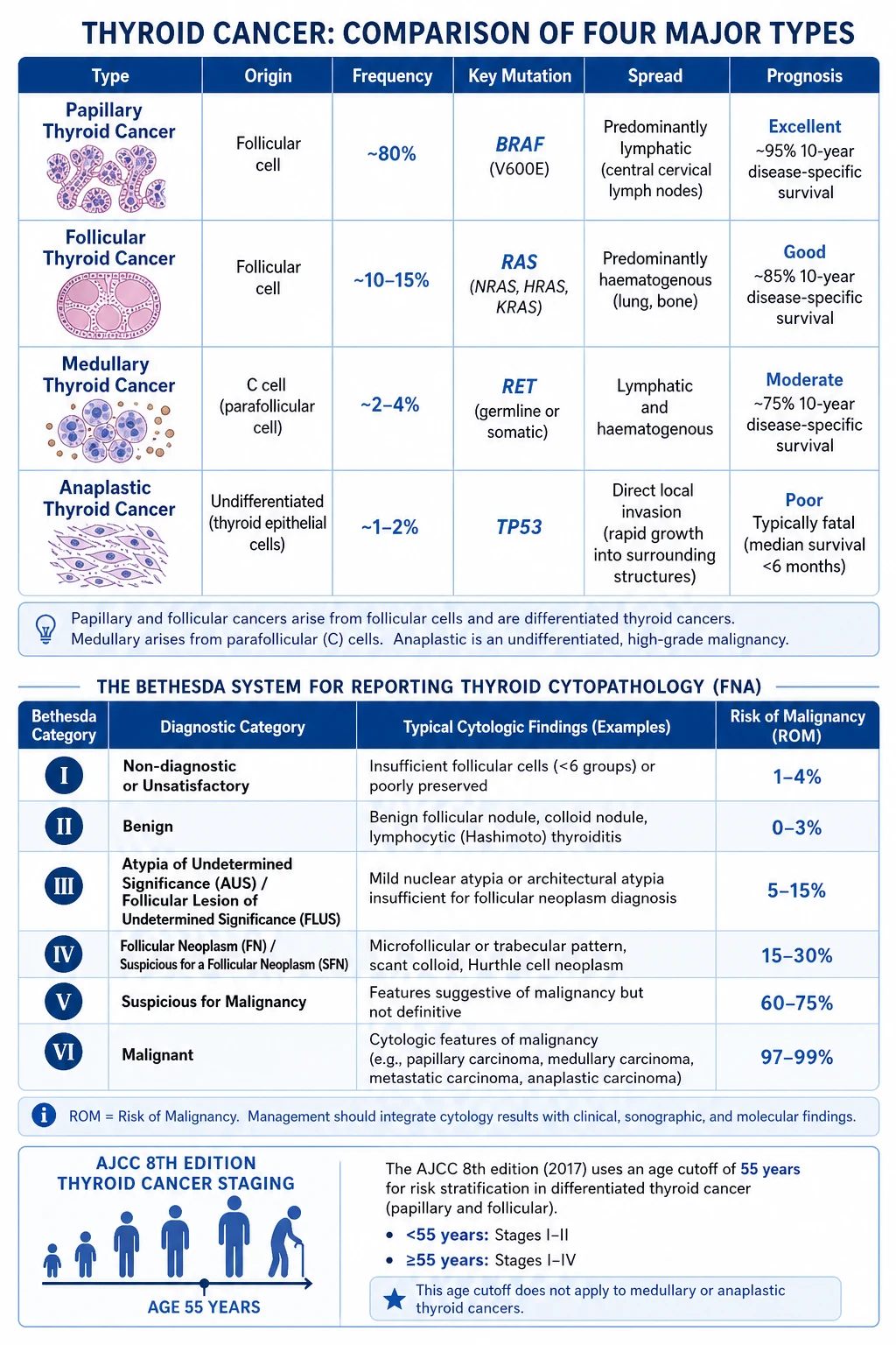
The four histological types are distinguished by origin, frequency, spread, molecular driver, and prognosis — examiners test this matrix relentlessly.[1][2]
| Feature | Papillary | Follicular | Medullary | Anaplastic |
|---|---|---|---|---|
| Origin | Follicular cell | Follicular cell | Parafollicular C cell | Undifferentiated |
| Frequency | 80% | 10% | 5% | 1 to 2% |
| Molecular driver | BRAF V600E, RET/PTC | RAS, PAX8-PPAR-gamma | RET proto-oncogene | TP53, TERT |
| Spread | Lymphatic to cervical nodes | Haematogenous (bone, lung) | Lymphatic plus haematogenous | Direct local invasion |
| Mean age | 30 to 50 | 40 to 60 | 40 to 60 (hereditary younger) | Over 60 |
| Female:male | 3:1 | 3:1 | 1:1 | 1:1 |
| Risk factors | Radiation | Iodine deficiency | MEN-2 / RET | Pre-existing goitre |
| Histology hallmark | Psammoma bodies, nuclear grooves | Capsular / vascular invasion | Amyloid stroma | Pleomorphic spindle / giant cells |
| Tumour marker | Thyroglobulin | Thyroglobulin | Calcitonin, CEA | None useful |
| Prognosis | Excellent (over 95% 10-yr) | Good (about 85% 10-yr) | Moderate (about 75% 10-yr) | Very poor (months) |
Papillary variants: classical, follicular variant, tall cell (aggressive, older patients, poorer prognosis), hobnail, diffuse sclerosing (affects young women, heavy nodal involvement), and solid variant. [1]
Follicular variants: minimally invasive (only capsular invasion, near-normal life expectancy), widely invasive (vascular invasion, poorer prognosis), and the Hurthle (oxyphilic) cell variant which is now classified separately — it is more aggressive, less radioiodine-avid, and carries a worse prognosis than conventional follicular carcinoma.[3]
Medullary carcinoma subtypes: sporadic (75%, the commonest, presents as a solitary thyroid nodule) and hereditary (25%, comprising MEN-2A, MEN-2B, and familial medullary thyroid carcinoma — all autosomal dominant from germline RET mutations).[4]
AJCC TNM 8th edition staging applies to differentiated (papillary and follicular) and medullary cancer; all anaplastic carcinomas are stage IV regardless of size, because they are uniformly aggressive.[1]
Epidemiology & Risk Factors
Thyroid cancer is the most common endocrine malignancy. Female predominance of about 3:1 for the differentiated types reflects the proliferative effect of oestrogen on thyroid tissue; medullary and anaplastic types have an approximately equal sex ratio. The age distribution is bimodal for papillary cancer (peaks in younger women aged 30 to 50 and again in the elderly), while anaplastic cancer is almost exclusively a disease of the elderly (mean age over 65).[2]
The incidence of thyroid cancer has risen steeply over the past three decades — at a faster rate than any other solid tumour. Most of this rise is driven by increased detection of small papillary microcarcinomas through widespread use of high-resolution neck ultrasound, CT, MRI, and PET imaging performed for other indications (incidentalomas). Autopsy series show that occult papillary microcarcinoma is found in up to 30% of thyroid glands, confirming that much of this "cancer" was always present and clinically silent. Mortality from thyroid cancer has remained essentially flat, supporting the conclusion that the incidence rise is largely a detection phenomenon.[2]
Risk factors: [1]
- Radiation exposure — the strongest modifiable risk factor. Childhood head, neck, or mediastinal irradiation (historically used for acne, enlarged thymus, tonsils, haemangioma, or as nuclear fallout — the Chernobyl and Marshall Islands cohorts) increases papillary carcinoma risk up to 30-fold, with a latency of 5 to 30 years. RET/PTC rearrangements predominate in radiation-induced tumours.
- Female sex and reproductive hormones — oestrogen-driven proliferation; pregnancy-related hormones also contribute.
- Family history — a first-degree relative with differentiated thyroid cancer raises risk 4 to 10-fold; about 5% of papillary cancers are familial.
- Iodine deficiency — endemic goitre regions show a relative excess of follicular carcinoma (iodine repletion shifts the subtype mix toward papillary).
- RET proto-oncogene germline mutation — virtually 100% penetrant for medullary carcinoma in MEN-2; the single most important hereditary risk factor in thyroid oncology.[4]
- Pre-existing thyroid disease — long-standing multinodular goitre and Hashimoto thyroiditis modestly raise lymphoma and papillary risk respectively.
- Obesity and metabolic syndrome — modestly associated with increased risk.
Thyroid cancer — the numbers
Pathophysiology
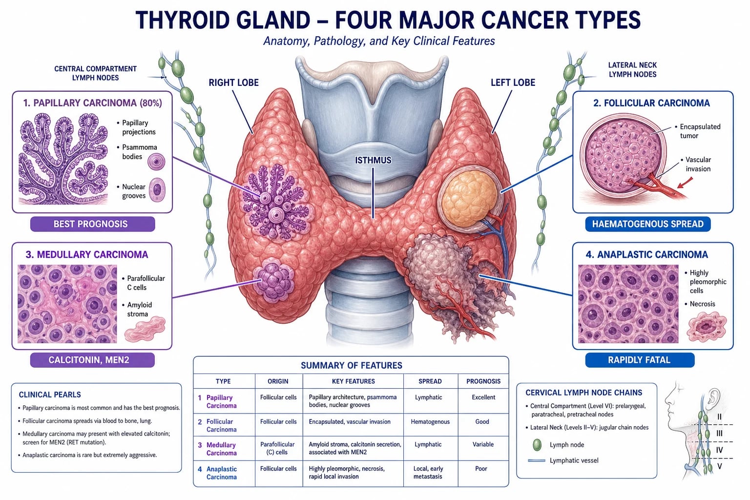
Each thyroid cancer type is driven by a characteristic molecular alteration that activates a specific signalling pathway, and these molecular signatures now also guide targeted therapy.[1][2]
Papillary carcinoma is driven by mutually exclusive alterations in the MAPK pathway: the BRAF V600E mutation (found in 40 to 60% of tumours, especially tall cell and aggressive variants) and RET/PTC rearrangements (10 to 20%, characteristic of radiation-induced and paediatric tumours). RAS mutations occur in 10%. The MAPK activation produces the diagnostic nuclear features: ground-glass or "orphan Annie eye" nuclei (optically clear, overlapping), nuclear grooves, and pseudoinclusions. The tumour forms papillae with fibrovascular cores; psammoma bodies (laminated calcific spherules) are pathognomonic and correspond to the microcalcifications seen on ultrasound. Spread is predominantly lymphatic to cervical nodes (50% of patients have nodal metastases at presentation, frequently to level VI central compartment and then levels II to V laterally), and pulmonary metastases are typical of advanced disease.[1]
Follicular carcinoma is driven by RAS mutation (NRAS, HRAS, KRAS) or the PAX8-PPAR-gamma rearrangement. The tumour forms follicles lined by neoplastic cells that are cytologically indistinguishable from a benign follicular adenoma — this is why FNA cannot make the diagnosis. The diagnosis of carcinoma requires demonstrating capsular invasion (tumour breaching the capsule) and/or vascular invasion (tumour within or beyond the vessel wall of capsular vessels) on the surgical specimen. Spread is haematogenous to bone (especially skull, sternum, spine, pelvis) and lung; nodal metastases are uncommon. Hurthle (oxyphilic) cell carcinoma is now a separate entity — it has abundant mitochondrial-rich granular eosinophilic cytoplasm, is less radioiodine-avid, and behaves more aggressively.[3]
Medullary carcinoma arises from parafollicular C cells (neural-crest origin, interfollicular in location, produce calcitonin). It is driven by a gain-of-function mutation in the RET proto-oncogene — germline in hereditary forms (MEN-2A, MEN-2B, familial MTC) and somatic in about half of sporadic cases. The tumour forms solid nests of polygonal or spindle cells separated by an amyloid stroma (congophilic, apple-green birefringence with Congo red) that represents polymerised calcitonin. It produces calcitonin (the key tumour marker) and carcinoembryonic antigen (CEA), and in advanced disease may secrete serotonin, prostaglandins, or ACTH causing diarrhoea, flushing, or Cushing syndrome. Spread is both lymphatic (cervical and mediastinal nodes) and haematogenous (liver, lung, bone).[4]
Anaplastic carcinoma is undifferentiated — large pleomorphic spindle and giant cells with a very high mitotic rate, often with areas of necrosis. It is thought to arise by dedifferentiation of a pre-existing papillary or follicular carcinoma (a BRAF or RAS driver mutation is often still detectable), with additional TP53, TERT promoter, and PIK3CA mutations driving the aggressive phenotype. It invades locally into trachea, oesophagus, great vessels, and prevertebral fascia, producing airway compromise and dysphagia, and metastasises widely to lung, bone, brain, and liver.[5]
Clinical Presentation
The thyroid nodule is the commonest presentation of all four types. It may be: [1]
- Palpable — detected by the patient or clinician. Nodules over 1 cm may be palpable; smaller ones are usually incidental imaging findings.
- Incidentaloma — discovered on carotid Doppler, neck CT/MRI, PET-CT, or thyroid ultrasound performed for another reason (the modern driver of incidence).
- Presenting as cervical lymphadenopathy — especially papillary carcinoma, where a level II to V node may be the first sign with an occult, sometimes sub-centimetre primary. [1]
Features suggesting malignancy (the red flags examiners reward): [1]
- Rapid growth of a previously stable nodule — anaplastic until proven otherwise.
- Hard, fixed consistency — infiltration of surrounding structures.
- Hoarseness or voice change — recurrent laryngeal nerve involvement; a grave sign indicating locally advanced disease.
- Dysphagia or dyspnoea — oesophageal or tracheal compression or invasion.
- Cervical lymphadenopathy — hard, matted, or fixed nodes, especially in the central compartment or jugular chain.
- Pain radiating to the ear or jaw — unusual for benign disease.
- Age at extremes — under 20 or over 60 years raises malignancy risk in a nodule.
- Male sex — although nodules are commoner in women, a nodule in a man carries a higher probability of malignancy. [1]
Medullary carcinoma specific: may present with the MEN-2 syndrome — concurrent pheochromocytoma (episodic headache, sweating, palpitations, hypertension) and primary hyperparathyroidism (MEN-2A only) — or with diarrhoea, flushing, or Cushing syndrome from peptide secretion in advanced disease. A family history of thyroid cancer, pheochromocytoma, or sudden death should trigger RET testing.[4]
Anaplastic carcinoma specific: rapidly enlarging neck mass over weeks, often in an elderly patient with a long-standing goitre, with pain, dysphagia, dyspnoea, hoarseness, and stridor from rapid local invasion. Patients frequently present with systemic symptoms (weight loss, anorexia). Distant metastases (lung, bone, brain) are present in over half at diagnosis.[5]
Differential Diagnosis
The differential of a thyroid nodule is broad and over 90% benign. The clinician's task is to identify the malignant minority using risk stratification rather than to image or biopsy every nodule.[1]
| Condition | Key distinguishing feature |
|---|---|
| Colloid (adenomatous) nodule | Commonest benign cause; cystic or mixed, spongiform, isoechoic, no suspicious US features; TSH normal; Bethesda II on FNA |
| Multinodular goitre | Multiple bilateral nodules, long history, often with obstructive symptoms; malignancy risk similar per-nodule to solitary nodule |
| Follicular adenoma | Solitary, encapsulated, cannot be distinguished from follicular carcinoma on FNA — requires histology for capsular/vascular invasion; Bethesda IV |
| Hashimoto thyroiditis | Diffusely heterogeneous gland, anti-TPO antibodies, hypothyroid; may harbour a lymphoma or papillary carcinoma |
| Graves disease | Diffuse hyperplasia, suppressed TSH, hyperthyroid; nodules within a Graves gland carry a slightly higher malignancy risk |
| Thyroid cyst | Avascular, anechoic, completely cystic on US; almost always benign |
| Subacute (de Quervain) thyroiditis | Painful, tender gland, raised ESR, transient hyperthyroidism then hypothyroidism; viral aetiology |
| Thyroid lymphoma | Rare; rapid enlargement of a Hashimoto gland in an older woman; B-symptoms |
| Reidel thyroiditis | Rare fibrosing disease; woody-hard gland infiltrating neck structures |
| Metastasis to thyroid | Renal cell, breast, lung, melanoma; usually in known primary, often rapidly growing |
Can't-miss diagnoses: anaplastic carcinoma, medullary carcinoma (screen for MEN-2), thyroid lymphoma, and metastatic disease. Classic mimics: a haemorrhagic cyst (sudden painful enlargement) and de Quervain thyroiditis (painful goitre mimicking anaplastic). [1]
Clinical & Bedside Assessment
The bedside assessment of a thyroid nodule is quick, structured, and decisive in selecting the nodules that proceed to imaging and FNA.[1]
Neck inspection: look for asymmetry, a visible swelling that moves upward with swallowing (thyroid swellings are tethered to the trachea by the pretracheal fascia and so rise on swallowing — this single manoeuvre distinguishes thyroid from other neck masses), skin changes, scars from previous neck radiation, engorged neck veins (retrosternal goitre), and the position of the trachea. [1]
Palpation: stand behind the seated patient. Palpate the thyroid using both hands, identifying the size, consistency (soft, firm, hard), surface (smooth, irregular), mobility (mobile, fixed), tenderness, and any retrosternal extension (inability to feel the lower border). Palpate the cervical lymph node chains systematically — central compartment (level VI), then levels I to V laterally, then the posterior triangle. Hard, fixed, or matted nodes suggest malignant involvement. [1]
Voice assessment: ask the patient to speak — hoarseness indicates recurrent laryngeal nerve palsy and is a red flag for malignant infiltration; it mandates pre-operative flexible nasendoscopy to confirm vocal cord function in any patient booked for thyroidectomy. [1]
General examination: signs of hyperthyroidism (tremor, tachycardia, warm moist palms, eye signs of Graves) or hypothyroidism (bradycardia, dry skin, delayed relaxation of reflexes). In suspected medullary carcinoma, screen for MEN-2 — blood pressure (pheochromocytoma), serum calcium, and a family history of thyroid surgery, pheochromocytoma, or sudden cardiac death. [1]
Bedside rules of thumb: the "rule of 10s" for thyroid nodules (10% of palpable nodules are malignant; 10% of malignant nodules are not papillary) is useful for counselling but does not replace ultrasound and FNA. The decision to biopsy rests on ultrasound risk stratification, not on nodule size alone.[1]
Investigations
The workup is stepwise and risk-stratified: blood test first, then ultrasound, then FNA on the highest-risk lesion, with cross-sectional imaging reserved for staging confirmed cancer.[1]
Step 1 — Blood tests: [1]
- Serum TSH — the first test. A suppressed TSH suggests an autonomous hyperfunctioning nodule (nearly always benign; such nodules do not warrant FNA). A normal or raised TSH proceeds to ultrasound. A raised TSH within the reference range (above about 2.5 mIU/L) is itself a weak, dose-dependent risk factor for malignancy in a nodule.
- Thyroid antibodies (anti-TPO) if autoimmune thyroiditis is suspected.
- Serum calcitonin — basal calcitonin is recommended by the European Thyroid Association for any nodule where medullary carcinoma is suspected (family history, MEN-2, US pattern of hypoechoic solid nodule with coarse calcifications); a basal calcitonin over 100 pg/mL warrants FNA with calcitonin staining and RET testing. Routine calcitonin screening of all nodules is debated; the ATA does not mandate it but the ESMO and European guidelines endorse it.[2][4]
Step 2 — Thyroid ultrasound (the central imaging test): [1]
High-resolution ultrasound with the TI-RADS (Thyroid Imaging Reporting and Data System) provides a structured risk score. Features that increase suspicion: solid composition, hypoechoic echogenicity, taller-than-wide shape, irregular or lobulated margins, microcalcifications, marked hypoechogenicity, extrathyroidal extension, and abnormal cervical nodes. ATR TI-RADS assigns points and a category (TR1 benign through TR5 highly suspicious); FNA is recommended for TR4 nodules at least 1.5 cm and TR5 nodules at least 1 cm. Purely cystic nodules and spongiform nodules are benign and do not need FNA.[1]
Step 3 — Fine-needle aspiration (FNA) cytology — the Bethesda System: [1]
FNA is the definitive diagnostic test for most thyroid nodules and is reported on the Bethesda System for Reporting Thyroid Cytopathology (six categories):[1]
| Bethesda | Category | Cancer risk | Action |
|---|---|---|---|
| I | Non-diagnostic / unsatisfactory | 5 to 10% | Repeat FNA with ultrasound guidance |
| II | Benign | 0 to 3% | Clinical and ultrasound surveillance |
| III | Atypia of undetermined significance / follicular lesion of undetermined significance (AUS/FLUS) | 10 to 30% | Repeat FNA, molecular testing, or diagnostic lobectomy |
| IV | Follicular neoplasm or suspicious for follicular neoplasm (incl. Hurthle cell) | 25 to 40% | Diagnostic lobectomy (FNA cannot distinguish adenoma from carcinoma) |
| V | Suspicious for malignancy | 50 to 75% | Lobectomy or total thyroidectomy |
| VI | Malignant | 97 to 100% | Total thyroidectomy (depending on type and risk) |
Critical limitation of FNA: it cannot distinguish follicular adenoma from follicular carcinoma because both show benign-appearing follicular cells — the diagnosis of follicular carcinoma requires histological assessment of the surgical specimen for capsular and vascular invasion. Bethesda IV (follicular neoplasm) therefore mandates diagnostic lobectomy, with completion thyroidectomy if histology confirms carcinoma.[3]
Step 4 — Additional staging (for confirmed or suspected cancer): [1]
- Contrast-enhanced CT neck, thorax, and mediastinum — for large tumours (over 4 cm), suspected retrosternal extension, invasive disease, or assessment of tracheal, oesophageal, or great-vessel involvement. Especially essential for anaplastic carcinoma staging (whole-body CT plus brain MRI). Iodinated contrast should be avoided if radioiodine ablation is planned, because the stable iodine load saturates the sodium-iodide symporter and renders the tumour non-avid for weeks.
- MRI neck — preferred over CT in younger patients to limit radiation, and for assessment of mediastinal or retrosternal extension.
- Vocal cord assessment — pre-operative flexible nasendoscopy in any patient with hoarseness or a tumour abutting the recurrent laryngeal nerve.
- Diagnostic whole-body I-123 or I-131 scintigraphy — after thyroidectomy, to confirm and quantify radioiodine-avid residual or metastatic disease before ablation. [1]
Tumour markers: [1]
- Thyroglobulin (Tg) — for differentiated thyroid cancer (papillary and follicular) after total thyroidectomy; the cornerstone of surveillance. Only valid after total thyroidectomy, because remaining normal thyroid tissue also produces Tg. Anti-thyroglobulin antibodies interfere with the assay and must be measured alongside; if present, Tg cannot be used reliably. A stimulated Tg (after recombinant human TSH or thyroid hormone withdrawal) below 1 ng/mL post-ablation predicts an excellent response.
- Calcitonin and CEA — for medullary carcinoma. Calcitonin doubling time (less than 6 months is poor, over 24 months is excellent) is a powerful prognostic marker; CEA rises late and tracks tumour burden.
- Calcitonin stimulation test (pentagastrin or calcium-stimulated) — for equivocal basal calcitonin and for screening MEN-2 kindreds. [1]
Management — Resuscitation
Thyroid cancer rarely presents as an emergency. The exceptions are time-critical and dominated by anaplastic carcinoma and advanced local disease:[5]
- Airway compromise from a large anaplastic carcinoma or a rapidly enlarging goitre — urgent surgical and anaesthetic assessment; consider tracheostomy if upper-airway obstruction, or debulking if the tumour is technically resectable; palliative external beam radiotherapy and steroids if not resectable. The "ABC" of anaplastic presentation is securing the airway first.
- Stridor at rest — emergency; the patient may need a helium-oxygen mixture (heliox), nebulised adrenaline, and an emergency surgical airway.
- Superior vena cava obstruction from mediastinal extension — urgent oncology referral for stent or radiotherapy.
- Thyroid storm is a complication of uncontrolled hyperthyroidism, not thyroid cancer — but a hyperfunctioning nodule misdiagnosed as cancer and manipulated surgically can precipitate it. [1]
Management — Definitive & Stepwise
Definitive treatment is type-specific: surgery is the cornerstone for all four types, but the extent of surgery, the role of radioiodine, and the place of systemic therapy differ fundamentally.[1][2]
Surgery for differentiated thyroid cancer (papillary and follicular)
Principle: the extent of surgery depends on cancer type, tumour size, risk stratification, and patient factors. The ATA and ESMO guidelines converge on a risk-stratified approach.[1][2]
- Hemithyroidectomy (thyroid lobectomy plus isthmusectomy) — for low-risk papillary microcarcinoma (unifocal tumour under 1 cm, intrathyroidal, clinically node-negative, no radiation history, no aggressive histology). Removes the ipsilateral lobe and isthmus. Adequate for over 80% of such tumours; avoids lifelong levothyroxine if the contralateral lobe is normal.
- Total thyroidectomy — for tumours over 4 cm, gross extrathyroidal extension, clinically apparent nodal or distant metastases, bilateral disease, multifocal disease, age over 45 (now under debate), and most follicular and Hurthle cell carcinomas. Enables accurate post-operative thyroglobulin surveillance and radioiodine ablation.
- Prophylactic central neck dissection (level VI) — for papillary carcinomas with clinically apparent lateral nodal disease (levels II to V), tumours over 4 cm, or gross extrathyroidal extension. Whether routine prophylactic central neck dissection adds benefit for low-risk tumours remains debated; ATA recommends considering it for intermediate- and high-risk tumours.
- Therapeutic lateral neck dissection (levels II to V) — for clinically or radiologically involved lateral nodes; performed as a selective modified radical neck dissection sparing the sternocleidomastoid, internal jugular vein, and spinal accessory nerve. [1]
Surgery for medullary carcinoma
Total thyroidectomy plus routine central neck dissection (level VI and VII) is the minimum operation for medullary carcinoma, because nodal involvement is common even when not clinically apparent. Therapeutic lateral neck dissection is added if nodes are involved. Before surgery, pheochromocytoma must be excluded (plasma or 24-hour urine metanephrines) in all medullary carcinoma patients, because operating on an undiagnosed pheochromocytoma precipitates a fatal hypertensive crisis — alpha-blockade first if pheo is present. Prophylactic thyroidectomy is offered to children carrying a germline RET mutation: before age 1 year for MEN-2B (highest-risk mutations, codon 918) and before age 5 years for MEN-2A (intermediate-risk), based on the ATA risk tier.[4][11]
Surgery for anaplastic carcinoma
Rarely resectable at presentation because of early local invasion. If caught very early (rare — about 10% of cases), total thyroidectomy with wide en-bloc local excision of involved structures may be attempted. The vast majority receive palliative external beam radiotherapy with or without chemotherapy, with surgery reserved for airway control (tracheostomy, debulking) or as part of a combined-modality protocol in selected stage IVA patients.[5]
Adjuvant therapy after total thyroidectomy
Radioactive iodine (I-131) ablation — given after total thyroidectomy for intermediate- and high-risk differentiated thyroid cancer (tumours over 4 cm, gross extrathyroidal extension, nodal metastases, aggressive histology, distant metastases). It ablates residual thyroid tissue (which improves thyroglobulin surveillance) and treats microscopic residual disease. Standard ablation doses are 30 mCi (1.1 GBq) for low-/intermediate-risk disease and 100 mCi (3.7 GBq) or higher for high-risk or metastatic disease, up to 150 to 200 mCi for bulky metastases. Requires TSH stimulation (recombinant human TSH injection, or thyroid hormone withdrawal to a TSH above 30 mIU/L) and a low-iodine diet for 1 to 2 weeks beforehand. No role for radioiodine in medullary or anaplastic carcinoma — these do not concentrate iodine.[1]
TSH suppression with levothyroxine — suppressive doses of levothyroxine drive TSH below the threshold thought to stimulate tumour growth. Targets depend on risk:[1]
- High-risk disease (gross residual, distant metastases, aggressive histology): TSH below 0.1 mIU/L indefinitely.
- Intermediate-risk disease: TSH 0.1 to 0.5 mIU/L for several years, then relaxed.
- Low-risk disease: TSH at the lower half of the reference range (0.5 to 2.0 mIU/L), to avoid the atrial fibrillation and osteoporosis risk of chronic over-suppression, especially in the elderly. [1]
Typical starting levothyroxine dose is 1.6 micrograms per kilogram per day, adjusted by TSH at 6 to 8 weeks. [1]
Systemic therapy for advanced and radioiodine-refractory disease
For radioiodine-refractory differentiated thyroid cancer (progressive disease on imaging despite radioiodine, or tumours that no longer concentrate iodine), multikinase inhibitors improve progression-free survival:[6][7]
- Lenvatinib (24 mg orally once daily) — the SELECT trial improved progression-free survival to 18.3 months versus 3.6 months with placebo. First-line for progressive disease.[6]
- Sorafenib (400 mg orally twice daily) — the DECISION trial improved PFS to 10.8 months versus 5.8 months with placebo.[7]
For advanced medullary thyroid carcinoma:[8][9]
- Vandetanib (300 mg orally once daily) — the ZETA trial improved PFS in advanced medullary thyroid cancer.[8]
- Cabozantinib (140 mg orally once daily) — the EXAM trial improved PFS in progressive medullary thyroid cancer.[9]
- Selpercatinib and pralsetinib — highly selective RET inhibitors, now licensed for RET-mutant medullary carcinoma.
For BRAF V600E-mutant anaplastic carcinoma:[10]
- Dabrafenib plus trametinib (BRAF plus MEK inhibitor combination) — the ROAR basket trial showed a meaningful response rate and is now licensed for BRAF V600E-mutant anaplastic thyroid cancer, a major advance in a disease that had no effective therapy.[10]
For disease not eligible for targeted therapy, anaplastic carcinoma is treated with external beam radiotherapy plus chemotherapy (paclitaxel-based regimens, sometimes with carboplatin), as tested in the NRG/RTOG 0912 trial.[5]
Specific Subtypes & Scenarios
Papillary microcarcinoma (under 1 cm) is increasingly detected incidentally. It has an excellent prognosis — over 99% disease-specific survival. Hemithyroidectomy is adequate for unifocal, intrathyroidal, node-negative tumours without a radiation history. Active surveillance (no immediate surgery, with serial ultrasound) is a valid option in carefully selected patients, pioneered by the Japanese (Kuma Hospital) cohorts showing very low (under 5%) progression rates over 10 years. Active surveillance is now endorsed by the ATA as an alternative to surgery for very low-risk microcarcinomas.[1]
Follicular carcinoma — because it spreads haematogenously, patients may present with a solitary bone metastasis (pathological fracture of the skull, sternum, or spine) or pulmonary metastases as the first manifestation. Workup of an otherwise unexplained bone metastasis must include consideration of follicular thyroid cancer (check thyroglobulin). Treatment is total thyroidectomy plus radioiodine ablation; radioiodine-avid metastases can be treated with repeated therapeutic doses of I-131. Minimally invasive follicular carcinoma has near-normal life expectancy; widely invasive disease has about a 50% 10-year survival.[3]
Hurthle cell carcinoma — now classified as a separate entity (Hurthle cell carcinoma, oncocytic). More aggressive than conventional follicular carcinoma, less radioiodine-avid (the cells are packed with mitochondria, not colloid), and more likely to metastasise to nodes. Treated with total thyroidectomy; radioiodine is less effective and lenvatinib is increasingly used for advanced disease.[3]
Medullary carcinoma with MEN-2 — hereditary disease demands genetic counselling and cascade RET testing of at-risk relatives. Prophylactic thyroidectomy before C-cell hyperplasia progresses to frank carcinoma can cure the patient of medullary cancer; long-term outcomes confirm biochemical cure in the great majority of children operated before MTC develops, justifying prophylactic surgery in RET carriers.[4][11]
Anaplastic carcinoma arising in a pre-existing goitre — a long-standing goitre that suddenly enlarges, becomes painful, and produces local symptoms is anaplastic carcinoma until proven otherwise; urgent ultrasound, FNA (or core biopsy), and contrast CT are essential. [1]
Thyroid lymphoma — rare; typically a diffuse large B-cell lymphoma arising in a long-standing Hashimoto gland, presenting as rapid painful enlargement in an older woman. Staged with whole-body imaging; treated with chemotherapy (R-CHOP) and radiotherapy, not surgery. [1]
AJCC TNM 8th Edition Staging
The age cutoff was raised from 45 to 55 years in the 8th edition, reflecting data that patients under 55 with distant metastases still have a reasonable prognosis (age is the single most powerful prognostic factor in differentiated thyroid cancer).[1]
Differentiated (papillary and follicular) thyroid cancer: [1]
- Age under 55 years: Stage I = any T, any N, M0 (no distant metastases); Stage II = any T, any N, M1 (distant metastases present). Only two stages — a young patient with distant metastases is still stage II, not IV.
- Age 55 and older: Stage I = T1 to T2 (up to 4 cm, intrathyroidal), N0/Nx, M0. Stage II = T3 (over 4 cm or minimal extrathyroidal extension) or N1a (central nodes), M0. Stage III = T4a (gross extrathyroidal extension into subcutaneous soft tissue, larynx, trachea, oesophagus, recurrent laryngeal nerve) or N1b (lateral nodes), M0. Stage IVA = T4a with any N, M0 (some schema). Stage IVB = T4b (invasion of prevertebral fascia or encasing carotid or mediastinal vessels), any N, M0. Stage IVC = any T, any N, M1 (distant metastases). [1]
Medullary thyroid cancer does not use the age cutoff — staging applies at any age, with T categories similar to differentiated cancer and nodal status driving stage (Stage I = T1 N0 M0; Stage II = T2 to T3 N0 M0; Stage III = T1 to T3 N1a M0; Stage IVA = T4a any N M0 or T1 to T3 N1b M0; Stage IVB = T4b any N M0; Stage IVC = any T any N M1). [1]
Anaplastic thyroid cancer — all cases are stage IV by definition: Stage IVA (T1 to T3a, intrathyroidal, resectable), Stage IVB (T3b to T4a, extrathyroidal extension, or gross residual), Stage IVC (any T with distant metastases). There is no early-stage anaplastic.[1][5]
Complications & Pitfalls
Surgical complications of thyroidectomy (the generic risks examiners test):[1]
- Recurrent laryngeal nerve (RLN) injury — 1 to 2% temporary, 0.5% permanent. Unilateral injury causes hoarseness; bilateral injury causes airway obstruction (a surgical emergency needing re-intubation or tracheostomy). Intra-operative nerve monitoring and visual identification of the RLN reduce injury.
- Superior laryngeal nerve injury — affects cricothyroid muscle; causes loss of high-pitch voice projection (subtle; matters for singers).
- Hypocalcaemia from parathyroid injury or devascularisation — 5 to 10% temporary, 0.5 to 2% permanent. Check calcium 6 to 24 hours post-operatively and at 24 hours; transient hypocalcaemia is common, permanent hypoparathyroidism is the dread complication requiring lifelong calcium and calcitriol.
- Neck haematoma — rare (under 1%) but life-threatening if it compresses the airway; a rapidly expanding neck swelling with stridor in the early post-operative period needs immediate wound opening at the bedside to release the haematoma before returning to theatre.
- Thyroid storm — rare now; precipitated by operating on uncontrolled hyperthyroidism.
- Wound infection, seroma, keloid — minor complications. [1]
Disease-related complications: local invasion causing airway compromise (anaplastic), recurrent laryngeal nerve palsy, dysphagia, SVC obstruction, pathological fracture from bone metastases (follicular), and the hormonal syndromes of medullary carcinoma (diarrhoea, flushing, Cushing). [1]
Classic pitfalls: [1]
- Not checking TSH before FNA — a hyperfunctioning nodule (suppressed TSH) is usually benign and does not need FNA; checking TSH first avoids unnecessary biopsy.
- Treating Bethesda III with surgery first — Bethesda III (AUS/FLUS) should prompt repeat FNA or molecular testing before diagnostic lobectomy.
- Performing total thyroidectomy for Bethesda IV without counselling — the diagnostic lobectomy may reveal a benign follicular adenoma in over half the cases; consent for possible completion thyroidectomy.
- Operating on medullary carcinoma without excluding pheochromocytoma — fatal intraoperative hypertensive crisis.
- Giving iodinated contrast before radioiodine ablation — iodine load saturates the sodium-iodide symporter, rendering the tumour non-avid for weeks.
- Over-suppressing TSH in an elderly low-risk patient — atrial fibrillation and osteoporosis.
- Forgetting post-operative calcium — symptomatic hypocalcaemia is preventable. [1]
Prognosis & Disposition
Prognosis differs dramatically by histological type and is one of the widest ranges in oncology.[1][5]
Papillary carcinoma — excellent: 5-year survival over 95%, 10-year survival over 95%. Even with cervical nodal metastases, prognosis remains very good. Distant metastases (lung, bone) reduce survival but are still potentially curable with radioiodine. Adverse features: older age (over 55), gross extrathyroidal extension, tall cell or diffuse sclerosing variant, BRAF plus TERT co-mutation, large primary (over 4 cm). [1]
Follicular carcinoma — good: 10-year survival approximately 85%. Minimally invasive (capsular invasion only) has near-normal life expectancy; widely invasive (vascular invasion) has about 50% 10-year survival. Distant metastases worsen prognosis but radioiodine-avid disease can be controlled.[3]
Medullary carcinoma — moderate: 10-year survival approximately 75%, highly stage-dependent. Calcitonin doubling time is the single most powerful prognostic marker — under 6 months predicts poor survival, over 24 months excellent. Hereditary cases detected by screening (prophylactic thyroidectomy) have near-normal life expectancy; sporadic advanced disease has poorer outcomes.[4]
Anaplastic carcinoma — very poor: median survival 4 to 6 months; 1-year survival under 20%; almost all patients die from disease, usually from airway compromise. The BRAF V600E-mutant subgroup treated with dabrafenib-trametinib has a markedly improved response and survival.[5][10]
Prognostic factors (differentiated thyroid cancer): age under 55 (most powerful), small tumour size, intrathyroidal (no extrathyroidal extension), no nodal or distant metastases, complete resection (R0), radioiodine avidity, and the absence of aggressive histological variants. The ATA dynamic risk stratification — excellent, indeterminate, or biochemical or structural incomplete response — refines prognosis after the first post-treatment assessment.[1]
Disposition and follow-up: [1]
- After total thyroidectomy and ablation — lifelong levothyroxine with risk-adjusted TSH targets, thyroglobulin (plus anti-Tg antibodies) every 6 to 12 months (undetectable Tg with negative antibodies after ablation is the goal; rising Tg indicates recurrent or metastatic disease and prompts neck ultrasound and diagnostic I-131 scanning).
- After lobectomy — annual TSH to avoid over-replacement, and surveillance ultrasound of the remaining lobe.
- Medullary carcinoma — calcitonin and CEA every 6 to 12 months; doubling times dictate imaging and further treatment.
- Ultrasound surveillance of the neck every 6 to 12 months for the first 2 to 5 years, then annually if stable. [1]
Special Populations
Children and adolescents — thyroid cancer in children is more often papillary, more often associated with cervical and pulmonary metastases, and more often driven by RET/PTC rearrangements than in adults. Despite more aggressive staging, prognosis remains excellent with appropriate treatment. Total thyroidectomy plus prophylactic central neck dissection is favoured in children because of higher nodal involvement. Radiation-induced papillary carcinoma (Chernobyl cohort) predominantly affected children and young adults, with a short latency (4 to 5 years). [1]
Pregnancy — a thyroid nodule discovered in pregnancy is worked up as usual (TSH and ultrasound are safe; FNA is safe in any trimester). Surgery can be performed in the second trimester if the nodule is malignant and growing; otherwise it can be deferred to the post-partum period. Radioiodine ablation is absolutely contraindicated in pregnancy and breastfeeding and must be deferred until after delivery and weaning.[1]
Elderly — anaplastic carcinoma is overwhelmingly a disease of the elderly, presenting with rapid local invasion. In older patients with low-risk papillary cancer, TSH over-suppression carries a significant risk of atrial fibrillation and osteoporotic fracture, so the levothyroxine dose should be relaxed to keep TSH in the lower reference range. [1]
MEN-2 carriers — children with a germline RET mutation require prophylactic thyroidectomy before MTC develops, timed by ATA mutation risk tier (MEN-2B codon 918 before age 1 year; MEN-2A before age 5 years). Cascade testing of at-risk relatives is mandatory.[4][11]
Anticoagulated patients — FNA is safe on warfarin or antiplatelet agents at a therapeutic INR (under 2.5); hold direct oral anticoagulants for 24 to 48 hours around FNA. Surgery requires bridging per the patient's thrombotic risk. [1]
Evidence, Guidelines & Regional Differences
ATA 2015 Guidelines (Haugen et al.)[1] — the most comprehensive guideline for differentiated thyroid cancer and nodules. Key themes: risk-stratified surgery (lobectomy versus total thyroidectomy based on size, histology, and clinical features); risk-stratified radioiodine ablation (low-risk patients do not need ablation); risk-stratified TSH suppression; and the dynamic risk stratification at the first post-treatment assessment that re-assigns patients to excellent, indeterminate, or incomplete response — which then drives further management.
ATA 2015 Revised Medullary Thyroid Carcinoma Guidelines (Wells et al.)[4] — the authoritative MTC guideline. Defines prophylactic thyroidectomy timing by ATA RET mutation risk tier (highest, high, moderate), mandates pheochromocytoma screening before any surgery, and lays out calcitonin-based surveillance.
ATA 2021 Anaplastic Thyroid Cancer Guidelines (Bible et al.)[5] — the first ATA guideline dedicated to anaplastic carcinoma. Establishes a multidisciplinary framework, mandatory BRAF V600E testing (to identify candidates for dabrafenib-trametinib), and stage-appropriate therapy (resection for stage IVA, chemoradiation for IVB, best supportive care or targeted therapy for IVC).
ESMO 2019 Guidelines (Filetti et al.)[2] — the European counterpart, with broader use of routine calcitonin screening of nodules and emphasis on avoiding overtreatment of low-risk papillary microcarcinoma.
AJCC 8th edition (2017)[1] — raised the differentiated cancer age cutoff from 45 to 55 years and declared all anaplastic carcinomas stage IV.
Landmark trials for systemic therapy: [1]
SELECT — lenvatinib in radioiodine-refractory DTC
NEJM 2015
Population: 392 patients with progressive RR-DTC, 2:1 lenvatinib:placebo
Key finding
Median PFS 18.3 months vs 3.6 months (HR 0.21, 99% CI 0.14 to 0.31); response rate 64.8% vs 1.5%. Benefit independent of prior VEGF-targeted therapy.
DECISION — sorafenib in RR-DTC
Lancet 2014
Population: 417 patients with progressive RR-DTC, sorafenib vs placebo
Key finding
Median PFS 10.8 vs 5.8 months (HR 0.59); response rate 12.2% vs 0.5%.
ZETA — vandetanib in medullary thyroid cancer
J Clin Oncol 2012
Population: 331 patients with advanced MTC, vandetanib vs placebo
Key finding
Improved PFS (hazard ratio 0.46); response rate 45% vs 13%. First approved drug for MTC.
EXAM — cabozantinib in medullary thyroid cancer
J Clin Oncol 2013
Population: 330 patients with progressive MTC, cabozantinib vs placebo
Key finding
Improved PFS 11.2 vs 4.0 months (HR 0.28); OS benefit in RET M918T mutation.
ROAR — dabrafenib plus trametinib in BRAF V600E-mutant ATC
Ann Oncol 2022
Population: BRAF V600E-mutant anaplastic thyroid cancer
Key finding
Investigator-assessed response rate 56%; median OS 14.5 months — a transformative result in a previously uniformly fatal disease.
Regional differences: [1]
Exam Pearls & High-Yield Minutiae
Papillary (80%)
best prognosis
- **BRAF V600E** mutation (40 to 60%) or **RET/PTC** rearrangement
- **Psammoma bodies**, nuclear grooves, **orphan Annie eye** nuclei
- **Lymphatic spread** to cervical nodes (50% at presentation)
- **Over 95%** 10-year survival; treat with total thyroidectomy +/- RAI + TSH suppression
Follicular (10%)
haematogenous
- **RAS** or **PAX8-PPAR-gamma** mutation
- **Cannot diagnose on FNA** — needs histology for capsular/vascular invasion
- Spreads via **blood to bone and lung**
- **Bethesda IV** = diagnostic lobectomy; iodine-deficiency association
Medullary (5%)
C cells, calcitonin
- **Parafollicular C cells**, **RET** proto-oncogene
- **Calcitonin** and **CEA** tumour markers; **amyloid stroma**
- 25% **familial (MEN-2)** — screen relatives, **exclude pheo before surgery**
- Total thyroidectomy + central +/- lateral neck dissection; **no role for radioiodine**
Anaplastic (1 to 2%)
rapidly fatal
- **Undifferentiated**; **TP53, TERT** mutations; **elderly**
- **Rapidly growing, invasive** — tracheal compression, stridor
- **All stage IV** in AJCC 8th; median survival **4 to 6 months**
- **Dabrafenib-trametinib** if BRAF V600E; otherwise **palliative radiotherapy/chemo**
MEN-2 — screen before medullary thyroid surgery
MEN2
calcitonin-producing C-cell tumour — the first manifestation in most MEN-2
MUST exclude before thyroid surgery — fatal intraoperative hypertensive crisis if missed
MEN-2A only; check serum calcium and PTH
autosomal dominant; prophylactic thyroidectomy — MEN-2B by age 1, MEN-2A by age 5
Bethesda I to VI — the cancer-risk ladder
BETH
neoplasm — diagnostic lobectomy (FNA cannot rule out carcinoma)
10 to 30% risk — repeat FNA or molecular testing
0 to 3% risk — surveillance
97 to 100% — total thyroidectomy
Classic one-liners examiners reward: [1]
- Papillary carcinoma: BRAF V600E, psammoma bodies, nuclear grooves and pseudoinclusions, lymphatic spread to cervical nodes, best prognosis, most common thyroid cancer.
- Follicular carcinoma: RAS mutation, iodine-deficiency endemic goitre, haematogenous spread to bone and lung, diagnosed on capsular or vascular invasion on histology (not FNA), Bethesda IV.
- Medullary carcinoma: parafollicular C cells, calcitonin, RET proto-oncogene, MEN-2, amyloid stroma, screen for pheochromocytoma before surgery.
- Anaplastic carcinoma: undifferentiated, TP53/TERT, elderly, rapidly fatal, all stage IV in AJCC 8th edition.
- AJCC 8th edition: age cutoff 55 (was 45 in 7th).
- TSH suppression targets: under 0.1 high risk; 0.1 to 0.5 intermediate; lower reference range for low risk.
- Radioiodine: 30 mCi for low-/intermediate-risk ablation; 100 mCi or more for high-risk or metastatic.
- Tumour markers: thyroglobulin for papillary/follicular; calcitonin and CEA for medullary.
- Surgical complications: RLN injury (hoarseness), hypocalcaemia (parathyroids), haematoma (airway emergency). [1]
Exam application bank (NEET-PG / INICET)
One-line answer
Thyroid carcinoma arises from thyroid follicular cells (papillary 80%, follicular 10%) or parafollicular C cells (medullary 5%); anaplastic (1 to 2%) is undifferentiated and near-uniformly fatal. Presents as a thyroid nodule (palpable or incidental on ultrasound); risk factors: radiation exposure in childhood, female sex, family history, iodine deficiency (follicular), RET germline mutation / MEN-2 (medullary). Workup: TSH, ultrasound with TI-RADS risk stratification, FNA cytology using the Bethesda system, plus serum calcitonin when medullary is suspected. Papillary: best prognosis, BRAF V600E, lymphatic spread, psammoma bodies; treated with total thyroidectomy with or without prophylactic central neck dissection, then radioiodine 131I ablation and TSH suppression. Follicular: RAS mutation, haematogenous spread to bone and lung, diagnosed on vascular or capsular invasion. Medullary: cal
Worked stems (answer without another resource)
Stem 1 — Classic presentation. Map symptoms to mechanism; name the first investigation and first treatment step with dose/route if drug therapy is standard. [1]
Stem 2 — Unstable / complicated. List red flags that force immediate resuscitation, theatre, ICU, antidote, or reperfusion — and what you do in the first 15 minutes. [1]
Stem 3 — Atypical group. Elderly, pregnancy, child, or immunocompromised: how presentation and thresholds change. [1]
Stem 4 — Differential trap. Name the three closest mimics and one discriminator for each. [1]
Stem 5 — Disposition. Who goes home with safety-netting, who is admitted, who needs HDU/ICU/theatre, and what follow-up is mandatory. [1]
Rapid viva checklist
- Definition + classification
- Pathophysiology chain
- Bedside signs / criteria
- Score with exact components (if any)
- Emergency bundle
- Definitive therapy with doses
- Complications of disease and of treatment
- Special populations
- Guideline/trial name if classic
- Three exam traps
Coverage self-check
If you cannot answer any stem above from this page alone, re-read the matching section — the page is intended to be self-sufficient for final-prof and NEET-PG/INICET questions on Thyroid Carcinoma.
References
- [1]Haugen BR, Alexander EK, Bible KC, et al. 2015 American Thyroid Association Management Guidelines for Adult Patients with Thyroid Nodules and Differentiated Thyroid Cancer: The American Thyroid Association Guidelines Task Force on Thyroid Nodules and Differentiated Thyroid Cancer Thyroid, 2016.PMID 26462967
- [2]Filetti S, Durante C, Hartl D, et al. Thyroid cancer: ESMO Clinical Practice Guidelines for diagnosis, treatment and follow-up† Ann Oncol, 2019.PMID 31549998
- [3]Grani G, Lamartina L, Durante C, et al. Follicular thyroid cancer and Hürthle cell carcinoma: challenges in diagnosis, treatment, and clinical management Lancet Diabetes Endocrinol, 2018.PMID 29102432
- [4]Wells SA Jr, Asa SL, Dralle H, et al. Revised American Thyroid Association guidelines for the management of medullary thyroid carcinoma Thyroid, 2015.PMID 25810047
- [5]Bible KC, Kebebew E, Brierley J, et al. 2021 American Thyroid Association Guidelines for Management of Patients with Anaplastic Thyroid Cancer Thyroid, 2021.PMID 33728999
- [6]Schlumberger M, Tahara M, Wirth LJ, et al. Lenvatinib in radioiodine-refractory thyroid cancer N Engl J Med, 2015.PMID 25946295
- [7]Brose MS, Nutting CM, Jarzab B, et al. Sorafenib in radioactive iodine-refractory, locally advanced or metastatic differentiated thyroid cancer: a randomised, double-blind, phase 3 trial Lancet, 2014.PMID 24768112
- [8]Wells SA Jr, Robinson BG, Gagel RF, et al. Vandetanib in patients with locally advanced or metastatic medullary thyroid cancer: a randomized, double-blind phase III trial J Clin Oncol, 2012.PMID 22025146
- [9]Elisei R, Schlumberger MJ, Muller SP, et al. Cabozantinib in progressive medullary thyroid cancer J Clin Oncol, 2013.PMID 24002501
- [10]Subbiah V, Kreitman RJ, Wainberg ZA, et al. Dabrafenib plus trametinib in patients with BRAF V600E-mutant anaplastic thyroid cancer: updated analysis from the phase II ROAR basket study Ann Oncol, 2022.PMID 35026411
- [11]Machens A, Lorenz K, Weber F, Dralle H. Long-term outcome of prophylactic thyroidectomy in children carrying RET germline mutations Br J Surg, 2018.PMID 29341155