General Surgery · General Surgery
Trauma Management (ATLS)
Also known as Trauma · ATLS · Primary survey · Polytrauma · Major trauma
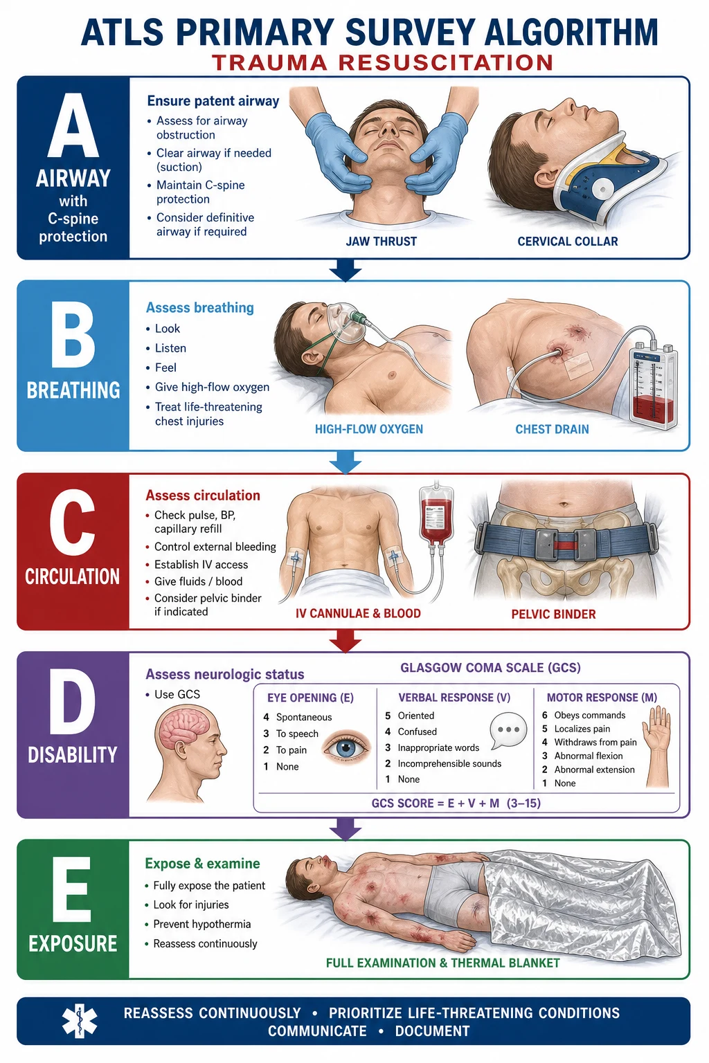
Trauma management follows the ATLS primary survey (cABCDE): catastrophic haemorrhage control first, then Airway with cervical spine protection, Breathing, Circulation, Disability (GCS), Exposure. Life-threatening injuries are identified and treated in order — not all at once. Tension pneumothorax = immediate needle decompression (2nd ICS mid-clavicular or 5th ICS anterior axillary). Massive haemothorax (over 1500 mL initial or over 200 mL/h) = thoracotomy. Cardiac tamponade = Beck's triad. Pelvic fracture = pelvic binder. GCS under 8 = intubate. FAST scan for intraperitoneal blood. Damage control surgery: control bleeding/contamination, temporary closure, ICU, re-operation. Lethal triad: hypothermia + acidosis + coagulopathy. CRASH-2: tranexamic acid 1 g IV within 3 hours.
On this page & tools
Your progress
Saved locally on this device.
Exam tags
Red flags
Overview & Definition
Trauma is physical injury caused by the transfer of mechanical energy to the body from an external force — blunt (road traffic accidents, falls, blast, assault) or penetrating (stab wound, gunshot, impalement). It is the leading cause of death under the age of 45 years worldwide, and the toll is greatest in low- and middle-income countries where pre-hospital systems are still maturing.[1]
The Advanced Trauma Life Support (ATLS) framework provides a systematic, prioritised approach to every injured patient, codified by the American College of Surgeons Committee on Trauma since 1980. Its central premise is simple but counter-intuitive: treat the greatest threat to life first, in a standardised order, regardless of the specific injury. The team does not chase a diagnosis — it identifies and immediately treats life-threatening conditions through the primary survey (cABCDE), and only afterwards takes a history, examines from head to toe, and orders definitive imaging.[1]
The golden hour — the first sixty minutes after injury — is when most preventable deaths occur. ATLS exists to win that hour. Two further ATLS principles govern everything that follows: [1]
- Treat first what kills first. An obstructed airway kills in minutes; an open tibial fracture can wait. The order of the primary survey is the order of lethality.
- Reassessment is continuous. A patient who is stable at minute five may be in refractory shock at minute fifteen. The team leader re-runs cABCDE constantly, not once. [1]
The trimodal distribution of trauma death
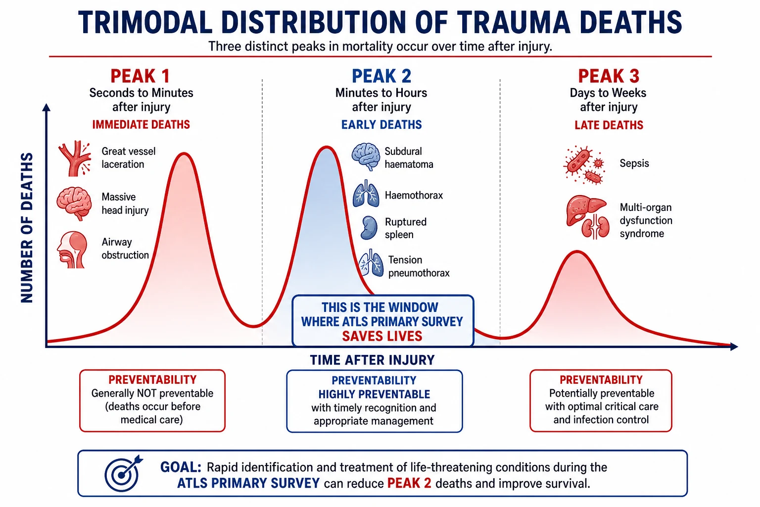
First described by Donald Trunkey in 1983, deaths after trauma cluster into three temporal peaks:[1]
- Immediate (seconds to minutes) — great vessel laceration, massive brain destruction, airway obstruction, high spinal cord transection. Mostly non-survivable regardless of care; addressed by prevention (seatbelts, helmets, road design).
- Early (minutes to several hours) — subdural and extradural haematoma, tension pneumothorax, cardiac tamponade, splenic and liver laceration, pelvic fracture with venous bleeding, exsanguinating extremity injury. This is the golden hour — ATLS primary survey and resuscitation save these lives.
- Late (days to weeks) — sepsis, multi-organ dysfunction syndrome (MODS), venous thromboembolism. Preventable with meticulous critical care, source control, and rehabilitation. [1]
The modern military lesson — bleeding to death from a limb is preventable — inserted the small c before ABCDE: catastrophic external haemorrhage is controlled before anything else, because a dead patient needs no airway. [1]
Classification
By mechanism
| Mechanism | Typical pattern | High-yield clues |
|---|---|---|
| Blunt | Road traffic accident, fall from height, assault, sports | Steering-wheel imprint, seat-belt sign, dashboard injury, bumper fracture |
| Penetrating | Stab wound, gunshot, impalement | Wound track predicts organ involvement; high-velocity gunshot causes cavitation |
| Blast | Explosive device | Primary (barotrauma — tympanic perforation, blast lung), secondary (fragment penetration), tertiary (blunt injury from body displacement), quaternary (burns, inhalation, radiation) |
By severity — the Injury Severity Score (ISS)
The ISS grades anatomical injury severity and predicts mortality. It takes the three most severely injured body regions (using the Abbreviated Injury Scale, AIS 1 to 6), squares the top three, and sums them. Maximum is 75 (or any AIS 6 = unsurvivable = 75 automatically). [1]
Injury Severity Score and mortality risk
ISS over 15 or 16 is the conventional definition of major trauma and triggers trauma-team activation in most systems.[1]
By physiological status — the ATLS "trauma team response" triad
- Stable — normal vital signs, alert. Full assessment and CT imaging.
- Borderline / transient responder — haemodynamically compromised but improves with a fluid bolus. Has ongoing bleeding — needs rapid definitive imaging or operating theatre.
- Unstable / non-responder — immediate life threat, no response to resuscitation. Straight to theatre or emergency intervention (e.g., thoracotomy, pelvic binder, laparotomy) during the primary survey.[1]
Epidemiology & Risk Factors
Trauma is the leading cause of death in people aged 1 to 44 across most of the world. Globally, road traffic accidents account for the largest share, with an estimated 1.19 million deaths annually (WHO). India carries a disproportionate burden — over 150,000 road-traffic fatalities each year, with two-wheeler riders and pedestrians the worst affected.[1]
Risk factors that examiners test: [1]
- Young males aged 15 to 29 — risk-taking behaviour, alcohol, speed.
- Alcohol and recreational drug intoxication (impairs judgement, masks injury, complicates anaesthesia).
- Non-use of seatbelts, helmets, and protective equipment.
- Speeding, reckless overtaking, distraction (mobile phones).
- Occupational exposure — construction (falls from height), agriculture (machinery), industry (crush, blast).
- Violence and conflict — penetrating trauma, blast injury.
- Elderly — falls from standing onto anticoagulants; seemingly trivial mechanisms cause serious intracranial and cervical injury.
- Pregnancy — aortocaval compression, altered physiology masking shock, domestic violence. [1]
Pathophysiology
Haemorrhagic shock — the dominant early killer
Blood loss reduces venous return, stroke volume, cardiac output, and tissue oxygen delivery. The body mounts a sympathetic compensatory response — tachycardia and peripheral vasoconstriction — which maintains blood pressure remarkably well until roughly 30 to 40 per cent of circulating volume is lost. This is why hypotension is a late, pre-terminal sign: a young athlete can be down 1.5 litres and still have a normal systolic pressure. The clinical implication is profound — do not wait for hypotension to diagnose shock. Tachycardia, cool peripheries, prolonged capillary refill, anxiety, and a rising lactate are the early sentinels.[1]
Total blood volume is approximately 70 mL/kg in an adult (5 litres in a 70 kg male), and children have a proportionally higher volume per kilogram. [1]
The ATLS classes of haemorrhagic shock
| Class | Blood loss (% volume) | Volume lost (70 kg) | Heart rate | Systolic BP | Respiratory rate | Urine output | Mental state |
|---|---|---|---|---|---|---|---|
| I | under 15% | under 750 mL | under 100 | Normal | 14 to 20 | over 30 mL/h | Normal |
| II | 15 to 30% | 750 to 1500 mL | 100 to 120 | Normal | 20 to 30 | 20 to 30 mL/h | Anxious |
| III | 30 to 40% | 1500 to 2000 mL | 120 to 140 | Decreased | 30 to 40 | 5 to 15 mL/h | Confused |
| IV | over 40% | over 2000 mL | over 140 | Markedly decreased (under 70) | over 35 | Negligible | Lethargic / unconscious |
Class III is where decompensation begins — by this point surgical haemorrhage control is mandatory.[1]
ATLS classes of haemorrhagic shock — the bedside discriminator
The lethal triad — and the vicious cycle that kills
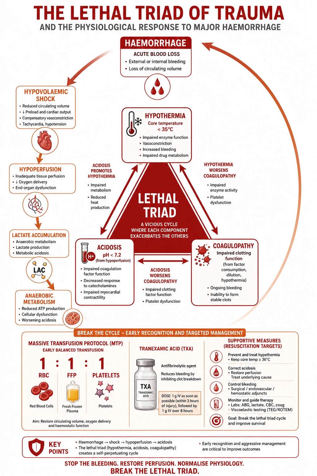
Three derangements develop together in the bleeding trauma patient and reinforce one another:[1]
- Hypothermia (core temperature under 35 degrees) — impairs coagulation factor activity and platelet function. The coagulation cascade is a cascade of enzymes, and every enzyme slows in the cold.
- Acidosis (arterial pH under 7.2) — from tissue hypoperfusion and anaerobic metabolism generating lactate. Acidosis further disables coagulation factors and depresses myocardial function.
- Coagulopathy — from factor consumption at the injury site, dilution by crystalloid resuscitation, hypothermia, and acidosis. Trauma-induced coagulopathy (TIC) is itself an independent predictor of death. [1]
Each element worsens the others — hypothermia worsens coagulopathy, coagulopathy worsens bleeding, bleeding worsens hypoperfusion and acidosis, acidosis worsens coagulation. The cycle predicts mortality. Prevention is the whole rationale of damage control resuscitation: warm the patient, give blood products (not crystalloid), and stop bleeding early.[1]
Clinical Presentation
Trauma presentation is dictated by mechanism and injury pattern, but the ATLS approach deliberately ignores history at the start. The clinician walks in and immediately runs cABCDE, because the patient who is talking cheerfully may be one minute from airway obstruction, and the drunk patient "smelling of alcohol" may be in Class III shock. The following is the structured symptom and sign set the primary survey screens for: [1]
c — Catastrophic external haemorrhage: bright red pulsatile or copious flowing bleeding from a limb, junctional zone, or cavity. [1]
A — Airway (with cervical spine protection): [1]
- Airway obstruction: stridor, gurgling, snoring, cyanosis, use of accessory muscles, inability to speak in sentences, foreign body, blood, vomit or dental fragments in the oropharynx, facial fractures, neck haematoma expanding, burns and soot around the mouth.
- Cervical spine injury is presumed in every blunt trauma patient until cleared clinically (Canadian C-spine rule or NEXUS criteria) or by CT. [1]
B — Breathing: [1]
- Tension pneumothorax: hypoxia, hypotension, trachea deviated away from the affected side, absent breath sounds unilaterally, hyper-resonance to percussion, distended neck veins, tachypnoea.
- Massive haemothorax: hypoxia, hypotension, dull to percussion, absent breath sounds on the affected side, signs of hypovolaemia.
- Open (sucking) pneumothorax: visible chest wall defect with audible air movement; a "sucking chest wound".
- Flail chest: paradoxical (inward) movement of a segment of chest wall during inspiration, severe pain, respiratory distress, underlying pulmonary contusion.
- Pulmonary contusion: hypoxia out of proportion to X-ray changes initially. [1]
C — Circulation: [1]
- Haemorrhagic shock: tachycardia (early), hypotension (late), cool clammy peripheries, prolonged capillary refill (over 2 s), reduced consciousness, oliguria, thready pulse.
- Cardiac tamponade: Beck's triad — hypotension + muffled heart sounds + distended neck veins. Pulsus paradoxus (drop in systolic BP over 10 mmHg on inspiration). Kussmaul's sign (rise in JVP on inspiration).
- Pelvic fracture: unstable pelvis on lateral compression, leg-length discrepancy, scrotal or perineal haematoma, blood at the urethral meatus, Destot's sign (superficial inguinal or scrotal haematoma).
- External bleeding: identify and quantify — scalp, limb, junctional, torso. [1]
D — Disability: [1]
- GCS (Glasgow Coma Scale): Eye 1 to 4, Verbal 1 to 5, Motor 1 to 6. Maximum 15. Under 8 = severe head injury = intubate.
- Pupils: size, symmetry, reactivity. A unilateral fixed dilated pupil suggests uncal herniation from raised intracranial pressure — a neurosurgical emergency.
- Lateralising signs, seizure activity, posturing (decorticate flexion = M3, decerebrate extension = M2). [1]
E — Exposure / Environment: [1]
- Fully expose the patient (remove all clothing — cut it off). Inspect the back (log-roll later). Then prevent hypothermia — warm blankets, warmed fluids, raised ambient temperature. Hypothermia worsens the lethal triad. [1]
Tension pneumothorax
B — breathing
- **Deviated trachea** away from the side
- **Absent breath sounds**, hyper-resonant to percussion
- **Needle decompression** 2nd ICS mid-clavicular immediately
- Clinical diagnosis — do NOT wait for X-ray
Massive haemothorax
B — breathing
- **Dull to percussion**, absent breath sounds
- Over **1500 mL** initial or **over 200 mL/h**
- **Chest drain** 5th ICS mid-axillary (28-32 Fr)
- **Thoracotomy** if criteria met
Cardiac tamponade
C — circulation
- **Beck's triad**: hypotension + muffled HS + distended JVP
- **Pulsus paradoxus**
- **Emergency thoracotomy** (left anterolateral 5th ICS)
- Open pericardium longitudinally, repair cardiac laceration
Atypical presentations examiners test
- Elderly patient on beta-blockers: no tachycardia despite Class III shock — beta blockade masks the heart rate. A "normal" heart rate of 70 in an injured elderly patient is abnormal.
- Pregnant trauma: the mother's cardiovascular reserve masks a 30 to 35% blood loss before vital signs change. Foetal distress (bradycardia on CTG) may be the first sign of maternal hypovolaemia.
- Spinal cord injury with neurogenic shock: bradycardia and hypotension from loss of sympathetic tone — not the tachycardia of hypovolaemia.
- Diabetic or intoxicated patient: reduced GCS may reflect hypoglycaemia or alcohol rather than brain injury — always check finger-prick blood glucose. [1]
Differential Diagnosis
In trauma, the "differential" is identifying which immediately life-threatening injuries are present. The primary survey is precisely this systematic screen. Examiners expect a candidate to reel off the life threats step by step and the action for each: [1]
| Condition | Step | Key sign | Immediate action |
|---|---|---|---|
| Airway obstruction | A | Stridor, cyanosis, silent chest | Jaw thrust, suction, airway adjunct, intubation |
| Tension pneumothorax | B | Tracheal deviation, absent sounds, hyper-resonance | Needle decompression 2nd ICS mid-clavicular, then chest drain |
| Massive haemothorax | B | Dull, absent sounds, hypovolaemia | Chest drain; thoracotomy if over 1500 mL or over 200 mL/h |
| Open pneumothorax | B | Sucking wound | 3-sided occlusive dressing, then chest drain |
| Flail chest | B | Paradoxical movement | Oxygen, analgesia, CPAP; ventilation if respiratory failure |
| Cardiac tamponade | C | Beck's triad | Emergency thoracotomy, open pericardium |
| Haemorrhagic shock | C | Tachycardia, hypotension, cool peripheries | IV access, blood products, control bleeding |
| Pelvic fracture | C | Unstable pelvis, shock | Pelvic binder, external fixation, angiographic embolisation |
| Severe head injury | D | GCS under 8 | Intubation, CT head, neurosurgery |
Clinical & Bedside Assessment
The primary survey IS the assessment
Do NOT take a history, examine the abdomen, or order blood tests before completing cABCDE. The primary survey is simultaneous assessment and treatment — at each step you find a life threat, you fix it before moving on.[1]
The AMPLE history (taken during or after the primary survey)
Once the patient is being resuscitated, the team takes a focused history — ideally from paramedics, witnesses, and the patient: [1]
- Allergies.
- Medications — especially anticoagulants (warfarin, NOACs), antiplatelets, beta-blockers.
- Past medical history / Pregnancy — last menstrual period in any woman of childbearing age.
- Last meal — for anaesthetic aspiration risk.
- Events / Environment — mechanism of injury, time of injury, pre-hospital interventions, entrapment, loss of consciousness. [1]
The secondary survey (only after the primary survey is complete and the patient is stabilised)
A head-to-toe examination that finds every injury the primary survey did not address: [1]
- Scalp and face: lacerations, fractures (Le Fort), periorbital ecchymosis (raccoon eyes).
- Ears and nose: cerebrospinal fluid rhinorrhoea or otorrhoea, haemotympanum, Battle's sign (mastoid ecchymosis) — all suggest basal skull fracture.
- Eyes: pupils, fundoscopy (papilloedema, retinal haemorrhage), contact lenses removed.
- Mouth: dental fractures, tongue bite, airway patency.
- Neck: tracheal position, neck vein distension, posterior element tenderness, step-off, lacerations (penetrating zones I to III).
- Chest: flail segments, paradoxical movement, subcutaneous emphysema, crepitus.
- Abdomen: distension, tenderness, guarding, rigidity, seat-belt sign.
- Pelvis: gentle lateral compression once — do not spring the pelvis repeatedly, it displaces clots.
- Perineum: lacerations, haematoma, urethral bleeding.
- Rectum (PR): sphincter tone (spinal injury), prostate position (high-riding in urethral injury), blood (bowel injury), vaginal examination in women.
- Back (log-roll, four-person technique): spinal tenderness, step-off deformity, contusions, lacerations, penetrating wounds.
- Extremities: deformity, swelling, crepitus, pulses, sensation, compartment tightness, open wounds. [1]
Reassess cABCDE continuously — the secondary survey is interrupted and resumed; it never supersedes the primary survey. [1]
[1]Investigations
During or immediately after the primary survey
- FAST (Focused Assessment with Sonography in Trauma): ultrasound at four views — subxiphoid pericardial, right upper quadrant (Morison's pouch), left upper quadrant (splenorenal), suprapubic (pouch of Douglas). The extended FAST (eFAST) adds bilateral thoracic windows to detect pneumothorax and haemothorax. FAST detects free intraperitoneal fluid or blood (sensitivity rises with volume) and pericardial effusion. It is quick (2 to 3 minutes), repeatable, and bedside. It does NOT identify the specific organ injured and is unreliable for retroperitoneal injury or hollow viscus injury.[1]
- Trauma series X-rays: chest X-ray (pneumothorax, haemothorax, widened mediastinum for traumatic aortic injury, rib fractures), pelvic X-ray (pelvic fractures — immediate binder if unstable), and historically a lateral cervical spine X-ray (CT has now replaced this in most centres).
- Bloods: full blood count, urea and electrolytes, lactate (a marker of tissue hypoperfusion — a rising lactate means ongoing shock), coagulation, group and save / crossmatch (activate the massive transfusion protocol if needed), venous blood gas (pH, base excess — base deficit under minus 5 signals severe shock), amylase or lipase, beta-hCG in every woman of childbearing age.
- ECG: myocardial contusion (new arrhythmia, ST changes), cardiac tamponade (electrical alternans), pre-existing ischaemia.
- Urinary catheter — placed only after excluding urethral injury. Indicators of urethral injury (do NOT catheterise; do a retrograde urethrogram first): blood at the meatus, perineal haematoma, high-riding prostate, scrotal swelling, pelvic fracture.
- Gastric tube — decompresses the stomach, reduces aspiration risk, and (in severe facial injury) is placed orally, not nasally, to avoid intracranial placement through a cribriform plate fracture.
After stabilisation (secondary survey phase)
- CT trauma series (non-contrast head, then contrast-enhanced cervical spine, chest, abdomen and pelvis with IV contrast) — for haemodynamically stable patients only. Provides definitive anatomical imaging. Whole-body CT in major trauma improves survival in some series and is now standard in many trauma centres.
- CT angiography (CTA) — if vascular injury suspected: widened mediastinum on CXR (traumatic aortic injury), pulsatile haematoma, ankle-brachial index under 0.9, expanding haematoma, bruit.
- Diagnostic peritoneal lavage (DPL): now rarely used; largely supplanted by FAST and CT. Reserved for the unstable patient with equivocal FAST where CT is unsafe — a positive DPL (over 100,000 red cells per mL, or bowel content) triggers laparotomy.
- Angiography with embolisation: for pelvic fracture haemorrhage (typically venous, but arterial bleeding from internal iliac branches may need embolisation) and for solid organ injuries managed non-operatively. [1]
Management — Resuscitation (the primary survey, step by step)
The primary survey is the resuscitation. At each step, a life threat is identified and immediately treated before moving on. Continuous reassessment by the team leader is mandatory.[1][1]
c — Catastrophic external haemorrhage
Before airway: stop life-threatening external bleeding. [1]
- Direct pressure with a dressing for most wounds.
- Tourniquet for exsanguinating limb haemorrhage — apply proximal to the wound, tighten until bleeding stops, record the time of application on the patient. Combat application tourniquets (CAT) are the modern standard. Do not release intermittently.
- Junctional tourniquets and pelvic binders for groin and pelvic bleeding; wound packing with haemostatic gauze for cavity wounds.
- This step comes from the military lesson of Iraq and Afghanistan: exsanguinating limb haemorrhage is the leading cause of preventable death. [1]
A — Airway (with cervical spine protection)
- Assess: can the patient speak? Stridor, gurgling, cyanosis, or silent obstruction require immediate intervention.
- Manoeuvres: jaw thrust (the airway manoeuvre of choice in trauma — never head-tilt chin-lift, because the C-spine may be injured). Suction blood, vomit, and debris.
- Adjuncts: oropharyngeal (Guedel) airway if unconscious (gag reflex absent); nasopharyngeal airway if tolerated (and only if no basal skull fracture — never insert an NPA through a suspected cribriform plate fracture).
- Definitive airway — endotracheal intubation with manual inline stabilisation if: GCS under 8, hypoxia despite oxygen, inability to protect the airway, severe maxillofacial trauma, thermal injury with impending airway loss, or need for ventilation. Rapid sequence induction with a criocoid pressure (Sellick manoeuvre, now debated) and two-person laryngoscopy.
- Surgical airway — surgical cricothyroidotomy: if orotracheal intubation is impossible (massive facial trauma, laryngeal fracture, cannot ventilate, cannot intubate). A horizontal incision through the cricothyroid membrane, insertion of a cuffed tube (size 6 in adults). Needle cricothyroidotomy is a temporising bridge in children under 10 to 12 years.
- Cervical spine protection: hard (semirigid) cervical collar applied before any airway intervention, head blocks and tape or sandbags, manual inline stabilisation during laryngoscopy. Maintained until the C-spine is cleared. [1]
B — Breathing
- Oxygen: high-flow 15 L/min via non-rebreather mask with reservoir for every trauma patient. Target SpO2 over 94%.
- Tension pneumothorax: immediate needle decompression — large-bore (14 G or 10/8.5 Fr) cannula in the second intercostal space, mid-clavicular line on the affected side, just above the third rib. Newer ATLS also accepts the 5th intercostal space, anterior axillary line (same site as a chest drain — preferred in muscular or obese patients because the 2nd ICS needle is often too short). Followed by a definitive chest drain (tube thoracostomy) at the 5th intercostal space, mid-axillary line.[1]
- Massive haemothorax: large-bore chest drain (28 to 32 Fr) at the 5th ICS mid-axillary, connected to an underwater seal and a graduated collection chamber. Measure the initial drainage. Over 1500 mL on insertion, or over 200 mL per hour for 2 to 4 hours, or ongoing transfusion requirement = emergency thoracotomy.[1]
- Open pneumothorax (sucking chest wound): apply a 3-sided occlusive dressing (Asherman chest seal or improvised square dressing taped on three sides — the untaped fourth side acts as a one-way flutter valve), then place a formal chest drain at a separate site (not through the wound).
- Flail chest: high-flow oxygen, adequate analgesia (regional block, thoracic epidural — the gold standard for flail segment pain), positive-pressure ventilation (CPAP/BiPAP) for fatigue; intubation and mechanical ventilation for respiratory failure. The underlying pulmonary contusion is the real threat and may worsen over 24 to 48 hours.
C — Circulation (haemorrhage control)
- External bleeding: direct pressure is first-line. Tourniquet for limb exsanguination. Pelvic binder for unstable pelvic fracture — a circumferential sheet or commercial binder around the greater trochanters reduces pelvic volume and tamponades venous bleeding.
- IV access: two large-bore (14 to 16 G) cannulae in the antecubital fossae. Intraosseous access (humeral head, proximal tibia, sternum) if peripheral access fails in under 90 seconds — every fluid and drug given IV can be given IO.
- Fluids: a warmed crystalloid bolus (1 L Hartmann's solution or 0.9% saline) STAT. Reassess — if no response or only transient response, move to blood products (activate the massive transfusion protocol).[1]
- Permissive hypotension: in penetrating torso trauma without traumatic brain injury, target a systolic BP of 80 to 90 mmHg (mean arterial pressure around 65) until haemorrhage is controlled. This prevents clot disruption from excessive fluid. Contraindicated in traumatic brain injury — the injured brain needs cerebral perfusion pressure (CPP = MAP minus ICP), and permissive hypotension risks secondary brain injury.[1]
- Massive transfusion protocol (MTP): deliver blood products in a 1:1:1 ratio of plasma : platelets : red blood cells — the target of the PROPPR trial, which showed that a 1:1:1 ratio achieved earlier haemostasis and fewer deaths from exsanguination at 24 hours compared with 1:1:2. Add calcium (citrate in stored blood binds calcium causing hypocalcaemia), tranexamic acid, and warming.[4]
- Tranexamic acid: 1 g IV bolus over 10 minutes within 3 hours of injury, followed by 1 g infusion over 8 hours (CRASH-2 protocol). Reduces all-cause mortality from bleeding by approximately 10% when given within 3 hours; beyond 3 hours mortality increases. The CRASH-3 trial extended this to isolated traumatic brain injury — TXA is safe and reduces head-injury death when given early (within 3 hours), with the largest benefit in mild-to-moderate TBI.[2][3]
- Calcium — give 10 mL of 10% calcium chloride or calcium gluconate during massive transfusion to maintain ionised calcium over 1.0 mmol/L; hypocalcaemia from citrate chelation impairs cardiac function and coagulation.[1]
- Cardiac tamponade: for trauma arrest or severe instability with Beck's triad, perform an emergency department resuscitative thoracotomy — a left anterolateral thoracotomy through the 5th intercostal space, open the pericardium longitudinally (relieving tamponade), evacuate clot, control the cardiac laceration with sutures or a Foley balloon, and cross-clamp the descending aorta to restore central perfusion. Pericardiocentesis is a temporising measure only in the trauma setting — if the patient is in arrest, open the chest.[1]
D — Disability
- AVPU as a rapid bedside check: Alert, responds to Voice, responds to Pain, Unresponsive. Then formal GCS (Eye 1 to 4, Verbal 1 to 5, Motor 1 to 6).
- GCS under 8 = intubate. This is one of the highest-yield single facts in trauma.
- Pupils: size, symmetry, reactivity.
- Blood glucose: check at the bedside — hypoglycaemia mimics brain injury and is rapidly reversible. Treat with 50 mL of 50% dextrose IV (or 200 mL of 10% dextrose).
- Prevent secondary brain injury: hypoxia (target PaO2 over 8 kPa / 60 mmHg) and hypotension (target SBP over 110 mmHg) double mortality after severe TBI. Even a single episode of hypotension in severe TBI increases mortality. Maintain normocapnia (PaCO2 4.5 to 5.0 kPa — avoid prophylactic hyperventilation which causes cerebral vasoconstriction).
- CT head if GCS under 15, focal neurological signs, vomiting, loss of consciousness, post-traumatic seizure, suspected open or depressed skull fracture, or on anticoagulants. [1]
E — Exposure / Environment
- Fully expose the patient (remove all clothing — cut it off). Inspect the back by log-roll.
- Prevent hypothermia — the lethal triad's anchor. Warm blankets, warmed IV fluids and blood (fluid warmer), raised ambient temperature, bubble wrap or forced-air warmer (Bair Hugger). Aim for core temperature over 35 degrees. [1]
Management — Definitive & Stepwise
Damage control — the central paradigm of modern trauma surgery
Damage control resuscitation (DCR) is the overall philosophy:[1]
- Permissive hypotension (SBP 80 to 90 mmHg in penetrating trauma without TBI) until haemorrhage control.
- Haemostatic resuscitation — blood products in a 1:1:1 ratio rather than clear fluids; minimise crystalloid (which dilutes clotting factors, causes acidosis from Hartmann's, and contributes to abdominal compartment syndrome).
- Early tranexamic acid within 3 hours.
- Damage control surgery for the unstable patient. [1]
Damage control surgery (DCS) is staged operative management for the patient who has, or is developing, the lethal triad:[1]
- Phase 1 — operating theatre (index operation): rapid control of bleeding (packing, ligation, balloon tamponade, vascular shunt) and contamination (staple off or oversew bowel injuries, simple closure) plus temporary abdominal closure (Bogota bag — an opened sterile IV fluid bag sewn to the skin, vacuum-assisted closure dressing, or towel-clip closure). The goal is not to fix every injury — it is to get the patient back to the ICU alive.
- Phase 2 — ICU (24 to 48 hours): rewarm, correct coagulopathy (FFP, cryoprecipitate for fibrinogen over 2 g/L, platelets), correct acidosis (restore perfusion, inotropes, renal support if needed), ventilate, optimise physiology.
- Phase 3 — return to theatre (24 to 48 hours): definitive surgical repair once physiology is restored — formal bowel anastomoses or stomas, definitive vascular repair, fixation of fractures, washout and definitive abdominal closure. The abdomen is re-examined for missed injuries. [1]
Definitive management of specific injuries (for stable patients)
- Head injury: CT head, neurosurgical decompression for extradural or acute subdural haematoma. ICP monitoring (intracranial bolt) for severe TBI (GCS 3 to 8 or CT abnormal with GCS 9 to 12). Target CPP 60 to 70 mmHg. Decompressive craniectomy for refractory intracranial hypertension.
- Cervical spine injury: CT cervical spine (replaces plain films in major trauma). MRI for ligamentous or cord injury. Immobilisation (hard collar, halo, or surgical fixation).
- Chest injury: chest drain for pneumothorax or haemothorax. Thoracotomy for massive haemothorax, great vessel injury, tracheobronchial injury, oesophageal injury, or cardiac injury.
- Abdominal injury: CT for stable patients. Laparotomy for unstable patients with positive FAST or peritonism. Splenic injury: non-operative management (NOM) if stable with CT grading; splenectomy or splenorrhaphy if unstable. Post-splenectomy vaccinations (pneumococcal, meningococcal, Haemophilus influenzae type b) and lifelong penicillin V prophylaxis. Liver injury: packing (damage control), Pringle manoeuvre for portal triad control, selective hepatic artery ligation or angioembolisation. Hollow viscus: primary repair or resection with anastomosis or stoma.
- Pelvic injury: pelvic binder, external fixation, angiographic embolisation for arterial bleeding. Do not spring the pelvis on examination.
- Extremity injury: early fixation of long-bone fractures (within 24 hours) reduces fat embolism, DVT, ARDS, and mortality (early total care versus damage control orthopaedics for the polytrauma patient — the boundary is the patient's physiological reserve). Temporary external fixation followed by definitive intramedullary nailing or plating when stable. [1]
Specific Subtypes & Scenarios
The trauma team
Modern trauma resuscitation is a team sport. A pre-alert from paramedics triggers trauma-team activation. Typical roles:[1]
- Trauma team leader — senior clinician (consultant or senior registrar). Stands back, runs the resuscitation, does NOT perform procedures. Co-ordinates, decides, takes responsibility.
- Airway doctor — anaesthetist or emergency physician. Manages the airway, intubates, manages the C-spine.
- Procedure doctor — surgical registrar or emergency physician. Performs chest drains, central lines, FAST, assists at procedures.
- Right-side doctor — secondary survey, right-sided procedures.
- Primary survey nurse / circulation nurse — IV access, bloods, monitoring, drugs.
- Radiographer — chest and pelvic X-rays, FAST, portable imaging.
- Scribe — documents the timeline, vital signs, drugs, procedures.
- Porter / runner — blood products, equipment. [1]
Good trauma teams practise closed-loop communication ("leader says: give 1 g TXA; receiver says: 1 g TXA given") and the leader calls frequent situation reports. [1]
Specific injury patterns
Head injury: extradural (lenticular/biconvex haematoma, middle meningeal artery, lucid interval), acute subdural (crescent-shaped, bridging veins, worse prognosis), traumatic subarachnoid haemorrhage, intracerebral contusion (frontal and temporal poles, contre-coup), and diffuse axonal injury (rotation-acceleration, poor prognosis, often little on early CT, characteristic on MRI). Indications for intubation and CT: GCS under 13, focal signs, recurrent vomiting, seizure, suspected skull fracture, anticoagulation.[3]
Chest:
- Rib fractures: painful, splinting reduces ventilation; epidural analgesia is the gold standard. The elderly patient with three or more rib fractures is a high-risk admission — deterioration is common over 48 hours.
- Blunt cardiac injury (myocardial contusion): right ventricle most commonly affected; arrhythmia (sinus tachycardia, atrial fibrillation, ventricular ectopics), elevated troponin, ST changes. Echocardiography for wall motion abnormalities.
- Tracheobronchial injury: air leak, subcutaneous emphysema, haemoptysis; bronchoscopy to localise.
- Traumatic aortic injury: widened mediastinum on CXR, left apical cap, depressed left main bronchus, obliterated aortic knuckle; CT angiography is the diagnostic standard; emergency thoracic surgery or endovascular stent graft. [1]
Abdomen: solid organ injury (spleen, liver, kidney — managed by CT grading and nomogram, angioembolisation, or surgery depending on stability), hollow viscus (small bowel, colon, stomach — seat-belt sign raises the index of suspicion for bowel mesenteric injury), diaphragm (typically left-sided, herniation of abdominal contents into the chest), and retroperitoneal organs (kidney, pancreas, duodenum — FAST is unreliable; CT is required). [1]
Pelvic fracture: apply a pelvic binder at the level of the greater trochanters immediately for unstable pelvic ring disruption (Young-Burgess classification — lateral compression, anteroposterior compression or "open book", vertical shear). External fixation within the resuscitation bay. Angiographic embolisation for arterial bleeding. Vertical shear patterns may need skeletal traction. The mortality of an open pelvic fracture remains very high. [1]
Extremity:
- Open fracture: Gustilo-Anderson classification (I to IIIC). Principles: photograph, cover with saline-soaked gauze, intravenous antibiotics within 1 hour (co-amoxiclav or a first-generation cephalosporin plus gentamicin for type III, plus metronidazole for farm/soil contamination), tetanus prophylaxis, splint, surgical debridement within 24 hours, and definitive fixation or external fixation.
- Compartment syndrome: raised pressure within a fascial compartment compromises perfusion. The 5 Ps (often 6) — Pain (disproportionate, on passive stretch — the earliest and most sensitive sign), Pallor, Paraesthesia, Paralysis, Pulselessness (late), Perishing cold. Diagnosis is clinical; compartment pressure within 30 mmHg of diastolic (delta pressure) confirms. Treatment is emergency fasciotomy.
- Fat embolism syndrome: 24 to 72 hours after long-bone fracture. Triad of respiratory distress, neurological deterioration (confusion), and a petechial rash (chest, axillae, conjunctivae). Treatment is oxygen and supportive ventilation; prevented by early fracture fixation.
- Crush injury and rhabdomyolysis: muscle breakdown releases myoglobin and potassium; acute kidney injury from pigment nephropathy. Crush syndrome after extrication — fluid resuscitation before release (to prevent reperfusion surge), aggressive IV fluids, mannitol and bicarbonate for forced alkaline diuresis, treat hyperkalaemia. Check creatine kinase (in the tens of thousands). [1]
Emergency department (resuscitative) thoracotomy
For trauma arrest (no pulse, no organised cardiac activity on FAST): [1]
- Penetrating trauma: within 10 minutes of loss of pulse — left anterolateral thoracotomy through the 5th ICS. Open pericardium longitudinally (avoiding phrenic nerve), cross-clamp descending aorta, internal cardiac massage, control cardiac or great-vessel laceration. Best survival in isolated cardiac stab wounds.
- Blunt trauma: within 10 minutes of loss of pulse — outcomes are very poor; reserved for witnessed arrest in the resuscitation bay. [1]
Survival after ED thoracotomy is approximately 10 to 20% overall — much higher for isolated cardiac stab wounds (over 30%) and very low for blunt trauma (under 2%).[1]
Triage and mass casualty
In a mass casualty incident — when the number of casualties exceeds available resources — the trauma system shifts from "doing the greatest good for each individual" to "doing the greatest good for the greatest number". This requires triage. [1]
Triage sieve (immediate, at scene, physiological): [1]
- Walking / waving — minor (green).
- Waiting / delayed — can wait (yellow).
- Immediate — life threat, salvageable, treat now (red).
- Dead / expectant — non-survivable or unsalvageable given resources (black or white). [1]
The sort is a more detailed secondary physiological triage in hospital using vital signs (respiratory rate, pulse, blood pressure, GCS) and the Triage Revised Trauma Score or START (Simple Triage and Rapid Treatment) algorithm. [1]
Triage priorities — the four colours
RED-YEL-GRN-BLK
life threat, salvageable, treat now
serious but can wait
walking wounded
non-survivable or beyond resources
Complications & Pitfalls
The early complications
- Lethal triad — hypothermia, acidosis, coagulopathy. Each reinforces the other. Prevent by warming, blood products, and early surgical bleeding control.[1]
- Abdominal compartment syndrome — intra-abdominal pressure over 20 mmHg with new organ dysfunction (oliguria from renal venous congestion, raised airway pressures from splinting of diaphragm, hypotension from reduced venous return, abdominal distension). Caused by massive fluid resuscitation, bowel oedema, or retroperitoneal haematoma. Measure bladder pressure. Treat with decompressive laparotomy and leave the abdomen open (vacuum dressing).[1]
- Missed injuries — the secondary survey is designed to catch injuries missed in the primary survey. Up to 10% of injuries are missed on initial assessment — especially in the unconscious or intubated patient. Repeat the secondary survey within 24 hours.
The late complications (the third peak of the trimodal distribution)
- Acute respiratory distress syndrome (ARDS): diffuse alveolar damage from pulmonary contusion, aspiration, sepsis, massive transfusion, or fat embolism. Bilateral infiltrates, refractory hypoxaemia (PaO2/FiO2 under 300), no cardiac cause. Treated with lung-protective ventilation (low tidal volume 6 mL/kg, plateau pressure under 30 cmH2O), prone positioning, and treating the cause.
- Acute kidney injury (AKI): from hypoperfusion (pre-renal), rhabdomyolysis, contrast nephropathy, or abdominal compartment syndrome. Prevent with adequate fluid resuscitation and early IV fluids in crush injury before extrication. Renal replacement therapy if refractory.
- Sepsis and multi-organ dysfunction syndrome (MODS): from infected wounds, perforated bowel, line infection, or pneumonia. The leading cause of late trauma death. Source control, broad-spectrum antibiotics, and ICU support.
- Venous thromboembolism (DVT and PE): trauma is a profoundly prothrombotic state. Pharmacological VTE prophylaxis (low-molecular-weight heparin, e.g., enoxaparin 40 mg subcutaneously once daily) within 24 to 48 hours once bleeding is controlled, plus mechanical prophylaxis (intermittent pneumatic compression). High-risk patients (pelvic fracture, spinal cord injury, major surgery) need both.
- Pressure injuries, contractures, heterotopic ossification, post-traumatic stress disorder — rehabilitation addresses these. [1]
Classic pitfalls
- Taking a history before completing cABCDE — the primary survey always comes first.
- Not protecting the C-spine during airway management — every intubation is a difficult intubation with a collar on; use manual inline stabilisation.
- Missing tension pneumothorax — it is a clinical diagnosis; do not wait for X-ray.
- Over-resuscitating with crystalloid — dilutes clotting factors, causes acidosis and abdominal compartment syndrome. Use blood products early.
- Sending an unstable patient to CT — stabilise first or go to theatre.
- Not giving tranexamic acid within 3 hours — CRASH-2 shows a 10% mortality reduction from bleeding.
- Not checking blood glucose in an unconscious trauma patient — hypoglycaemia mimics brain injury.
- Repeatedly springing the pelvis to check for instability — it disrupts clots and worsens bleeding. Examine once.
- Forgetting the urethra before placing a urinary catheter — blood at the meatus, perineal haematoma, or high-riding prostate mandate a retrograde urethrogram first.
- Not immunising the splenectomised patient — pneumococcal, meningococcal, Haemophilus influenzae type b vaccinations and lifelong penicillin V prophylaxis. [1]
Prognosis & Disposition
Prognosis depends on: injury severity (ISS), age, comorbidities, time to definitive care, and whether the lethal triad develops.[1]
- ISS over 16 = major trauma. Mortality rises sharply.
- ISS over 25 = critical — 30 to 50% mortality without optimal trauma system care.
- GCS is the strongest single predictor of outcome in head injury: GCS 3 to 5 carries 50 to 80% mortality; GCS 13 to 15 carries under 10%.
- Base deficit and lactate clearance over 24 hours: normalisation predicts survival; persistent elevation predicts MODS and death.
- Age over 65 with major trauma doubles mortality for the same ISS, partly because comorbidities and anticoagulants compound injury. [1]
Disposition: all major trauma (ISS over 15) to a major trauma centre with 24/7 trauma surgery, orthopaedics, neurosurgery, intensive care, interventional radiology, and blood bank. Rehabilitation begins on day one — early mobilisation, physiotherapy, occupational therapy, psychological support, and a structured discharge plan. Many survivors of major trauma need months of rehabilitation and have persistent physical and psychological sequelae. [1]
Special Populations
- Elderly: less physiological reserve, comorbidities, anticoagulants increase bleeding risk, higher ISS for the same mechanism, beta-blockers mask tachycardia, and a fall from standing onto warfarin is a high-risk presentation — lower threshold for CT and intensive care.[1]
- Pregnancy: left lateral tilt (15 to 30 degrees) or manual uterine displacement to relieve aortocaval compression from the gravid uterus after 20 weeks. Physiological changes mask shock — blood volume and cardiac output rise by 30 to 50%, so the mother may lose 30 to 35% of her volume before vital signs change. Foetal distress (bradycardia on CTG) may be the first sign of maternal hypovolaemia. Check Rh status — give anti-D immunoglobulin within 72 hours to a Rh-negative mother. Penetrating torso trauma in pregnancy has a high foetal mortality. The primary survey is identical — save the mother first.
- Children: larger head-to-body ratio (more head and cervical spine injuries), flexible rib cage (significant internal injury can occur without rib fractures), compensated shock (children maintain BP until late, then collapse suddenly — monitor mental state and capillary refill, not just BP). Weight-based dosing for fluids and drugs; use the Broslow or Parkland tape for emergency weight estimation. Intraosseous access early when IV access fails.
- Anticoagulated patients: higher bleeding risk for every injury. Reverse warfarin with vitamin K and prothrombin complex concentrate (PCC, faster than FFP). Reverse NOACs: dabigatran with idarucizumab (Praxbind), apixaban and rivaroxaban with andexanet alfa or PCC; PCC is the practical default. Lower threshold for imaging and admission.[1]
Evidence, Guidelines & Regional Differences
Landmark trials
CRASH-2 (2010)
Lancet, n=20,211
- **Tranexamic acid 1 g IV bolus** then 1 g infusion over 8 h
- Given **within 3 h of injury** reduced bleeding mortality by ~10%
- Beyond 3 h: no benefit, possible harm
- Cheap, safe, globally applicable — should be given to every bleeding trauma patient
CRASH-3 (2019)
Lancet, n=12,737
- TXA in **isolated traumatic brain injury**
- **Safe** — no increase in vascular occlusive events
- Reduces head-injury death when given **early (within 3 h)**
- Greatest benefit in mild-to-moderate TBI (GCS 9-15)
PROPPR (2015)
JAMA, n=680
- 1:1:1 (plasma:platelets:RBC) vs 1:1:2 in massive transfusion
- **No difference in 24-h or 30-day mortality** overall
- 1:1:1 achieved **earlier haemostasis** and **fewer deaths from exsanguination at 24 h**
- Established 1:1:1 as the standard target ratio
The European Trauma Bleeding Guideline (6th edition, 2023)
The current international standard for trauma bleeding management:[1]
- Early bleeding control — direct pressure, tourniquet, pelvic binder, pre-hospital if possible.
- Massive transfusion ratio 1:1:1 (plasma : platelets : RBC). Add fibrinogen (cryoprecipitate if fibrinogen under 1.5 to 2.0 g/L) and calcium.
- Tranexamic acid within 3 hours of injury — 1 g bolus over 10 min, then 1 g over 8 h.
- Permissive hypotension in penetrating trauma without TBI — SBP 80 to 90 mmHg until haemorrhage control.
- Damage control surgery for the unstable patient with the lethal triad.
- Whole-blood or plasma-first strategies increasingly supported.
- Viscoelastic testing (ROTEM or TEG) to guide component therapy in complex coagulopathy.
- Target normothermia — active warming throughout. [1]
Regional differences
- ATLS (USA, global): the original framework, now in its 10th edition. Taught worldwide by the American College of Surgeons.
- EMST (Early Management of Severe Trauma): the Australian and New Zealand equivalent, delivered by the Royal Australasian College of Surgeons — same content, regional branding.
- NICE Major Trauma guidelines (UK, NG39, 2016): recommend whole-body CT in major trauma when clinically indicated, contrast-enhanced; pre-alert to a major trauma centre; trauma-team leader on arrival; paediatric major trauma to a paediatric major trauma centre. The UK has a mature regionalised trauma network (since 2012 in London, then nationwide).
- India: road traffic accidents are the leading cause of trauma death. Pre-hospital care is limited — many patients arrive by private vehicle, not ambulance, often hours after injury. Delayed presentation is common. Resource limitations mean trauma series X-rays and FAST may be the only available imaging in district hospitals. Tranexamic acid is cheap and should be given to all bleeding trauma patients. The National Trauma Centre (JPN Apex Trauma Centre, AIIMS Delhi) and a growing network of state trauma centres are maturing the system. The Golden Hour Project and 108 ambulance services are extending pre-hospital care. Helmet and seatbelt legislation varies by state and enforcement is patchy.[1]
The cABCDE order (catastrophic haemorrhage first) is now universal across ATLS, EMST, NICE, the European Resuscitation Council, and the military Tactical Combat Casualty Care (TCCC) framework. The first step is always external bleeding control, then airway with C-spine protection.
Rehabilitation
Trauma care does not end at hospital discharge. Rehabilitation is the third pillar alongside resuscitation and definitive surgery — and the neglected one. Major trauma survivors commonly have persistent functional, psychological, and social impairment. [1]
- Multidisciplinary team: physiotherapy (mobility, chest physiotherapy, splinting), occupational therapy (activities of daily living, home modifications, equipment), clinical psychology, dietetics, social work, vocational rehabilitation.
- Traumatic brain injury: structured neurorehabilitation, cognitive therapy, behavioural management, family support. Recovery continues for 12 to 24 months.
- Spinal cord injury: regional spinal injuries centre, early bladder and bowel care, pressure-area prevention, functional electrical stimulation, vocational retraining.
- Amputation: prosthetic rehabilitation, phantom-limb management, vocational retraining.
- Post-traumatic stress disorder: affects 15 to 30% of major trauma survivors — screen for and treat with trauma-focused cognitive behavioural therapy.
- Major trauma networks in mature systems provide a rehabilitation prescription at discharge — a documented plan of needs and goals reviewed at follow-up. [1]
Exam Pearls
- cABCDE primary survey. Treat life threats in order. Catastrophic external haemorrhage first. Do NOT take history before completing cABCDE.[1]
- Tension pneumothorax = clinical diagnosis. Immediate needle decompression at the 2nd ICS mid-clavicular line (or 5th ICS anterior axillary). Do NOT wait for X-ray. Then chest drain at 5th ICS mid-axillary.[1]
- Massive haemothorax: over 1500 mL initial drainage or over 200 mL per hour = emergency thoracotomy. Chest drain 5th ICS mid-axillary.[1]
- Cardiac tamponade: Beck's triad (hypotension + muffled heart sounds + distended neck veins) = emergency thoracotomy.[1]
- Open pneumothorax: 3-sided occlusive dressing, then chest drain at a separate site.[1]
- GCS under 8 = intubate. GCS: E(1-4) + V(1-5) + M(1-6) = max 15. Always protect C-spine during intubation.[1]
- Shock classes: I under 15%, II 15-30% (HR 100-120, normal BP), III 30-40% (BP falls), IV over 40% (SBP under 70, altered mental). Hypotension is a LATE sign.[1]
- Massive transfusion 1:1:1 (plasma:platelets:RBC). Permissive hypotension SBP 80-90 in penetrating trauma without TBI. Minimise crystalloid.[1][4]
- Pelvic fracture: pelvic binder immediately. Do NOT spring the pelvis repeatedly. Angiographic embolisation for arterial bleeding.[1]
- FAST for intraperitoneal blood. CT for stable patients only. Laparotomy for unstable with positive FAST.[1]
- Damage control surgery: control bleeding and contamination, temporary abdominal closure, ICU rewarming/correction, re-operation 24-48 h. Prevent the lethal triad.[1]
- Lethal triad: hypothermia + acidosis + coagulopathy. Prevent by warming, blood products, early surgery.[1]
- CRASH-2: tranexamic acid 1 g IV within 3 h reduces bleeding mortality by ~10%. CRASH-3: TXA safe in TBI, give early.[2][3]
- Trimodal death: immediate (non-survivable), early (golden hour — cABCDE saves lives), late (sepsis/MODS).[1]
- Never send an unstable patient to CT. Stabilise or go to theatre.[1]
- C-spine protection during ALL airway interventions. Hard collar + head blocks until cleared.[1]
- Compartment syndrome: Pain on passive stretch is the earliest sign. Fasciotomy is the treatment.[1]
- Triage in mass casualty: Red immediate, Yellow delayed, Green minor, Black expectant.[1]
ATLS primary survey — actionable detail (exam detail)
A — Airway with C-spine protection
- Talk to the patient; if voice is normal, airway is temporarily patent.
- Look for obstruction, facial trauma, burns/soot.
- Manouvres: jaw thrust (prefer over head-tilt if C-spine concern), suction, adjuncts (OPA/NPA), definitive airway if GCS ≤8, failure to protect, hypoxia, or anticipated course.
- C-spine: collar + blocks/tape or manual in-line stabilisation until cleared by protocol. [1]
B — Breathing
- Expose chest; RR, SpO2, symmetry, tracheal position, JVP, wounds.
- Tension pneumothorax: hypotension + distended neck veins + absent breath sounds + tracheal deviation — immediate needle/finger thoracostomy then chest drain; do not wait for CXR.[1]
- Open pneumothorax: three-sided dressing then drain.
- Massive haemothorax: chest drain + blood products + surgery if ongoing.
- Flail chest: oxygen, analgesia (consider regional), ventilatory support as needed.
C — Circulation with haemorrhage control
- Stop external bleeding (direct pressure, tourniquet for catastrophic limb bleed).
- Two large-bore IV (or IO); send VBG, FBC, coags, crossmatch, lactate.
- Classes of haemorrhagic shock (ATLS) — know HR/BP/RR/mental status bands.
- Damage control resuscitation: permissive hypotension in selected penetrating torso trauma until haemorrhage control (not in TBI with raised ICP needs), early blood products, limit crystalloid, reverse anticoagulants.
- Massive transfusion protocol (MTP) example ratios approaching 1:1:1 RBC:plasma:platelets; give tranexamic acid 1 g IV over 10 min then 1 g over 8 h if within 3 h of injury (CRASH-2).[1]
- eFAST for free fluid; unstable + positive → theatre, not CT.
D — Disability
- GCS (E4 V5 M6), pupils, lateralising signs, glucose.
- Prevent secondary brain injury: oxygen, BP support, avoid hypo/hyperventilation extremes, early CT when stable, neurosurgical pathway for mass lesion. [1]
E — Exposure / environment
- Full log-roll with spine protection; prevent hypothermia (trauma triad of death: hypothermia–acidosis–coagulopathy). [1]
Adjuncts: CXR, pelvic X-ray, eFAST, urinary catheter (if no urethral injury signs), gastric tube, ECG, ABG/VBG, tetanus, antibiotics for open fractures per protocol. [1]
Secondary survey and disposition
Head-to-toe after primary survey and resuscitation. AMPLE history. Mechanism-based imaging. Never send a haemodynamically unstable patient to CT. Disposition: theatre, angioembolisation, ICU, ward, or major trauma centre transfer. [1]
Worked clinical stems (answer these without another book)
Stem A — Tension. Stab chest, distressed, BP 70, trachea deviated, silent left chest.
Tension pneumothorax — decompress now, then intercostal drain; reassess ABC. [1]
Stem B — Unstable blunt abdomen. RTA, BP 80, positive eFAST.
Haemoperitoneum — MTP, TXA if within 3 h, emergency laparotomy / damage-control surgery; not CT. [1]
Stem C — Pelvic binder. Motorcyclist, unstable pelvis, shock.
Binder correctly applied at greater trochanters; MTP; avoid springing pelvis repeatedly; angio or packing/ex-fix per pathway. [1]
Stem D — GCS 6. Isolated TBI after fall, BP 150/90.
Definitive airway, ventilate carefully, prevent hypotension/hypoxia, urgent CT head, neurosurgery consult; avoid hypotensive resuscitation targets meant for uncontrolled torso bleed. [1]
Stem E — Anticoagulated elder. Fall on warfarin, GCS drop.
ABC; reverse anticoagulation (PCC + vitamin K for warfarin per protocol); early CT head; low threshold for neurosurgical opinion. [1]
Stem F — Pregnancy trauma. 30 weeks, seatbelt mark, maternal BP 100/60.
Left lateral tilt / manual uterine displacement; maternal resuscitation first; Kleihauer if Rh-negative pathway; obstetric + trauma team; CT if needed for mother with shielding discussion — do not withhold life-saving imaging. [1]
OSCE / short-case performance script
- Scene safety / PPE; C-spine manual stabilisation.
- Verbalise ABCDE with hands-on actions.
- Demonstrate needle decompression landmarks (2nd ICS MCL traditional teaching vs mid-axillary finger thoracostomy modern practice — state local ATLS update).
- Call for blood, TXA timing, surgical airway readiness.
- Handover using ATMIST/MIST.
- State clear disposition: theatre vs CT vs transfer. [1]
Extended viva bank (model outlines)
- Trauma triad of death.
- Classes of haemorrhagic shock.
- CRASH-2 TXA dose and window.[1]
- eFAST windows and limitations (cannot exclude hollow viscus).
- Damage control laparotomy sequence (control bleed/contamination, temporary closure, ICU, relook).
- Hard signs of vascular injury in limbs.
- C-spine clearance principles.
- Paediatric vs adult airway and blood volumes (80 mL/kg).
- Blast / mass casualty triage colours.
- When CT is contraindicated.
Common exam traps (fail patterns)
- CT for the unstable patient.
- Forgetting TXA time window.
- Incomplete exposure missing posterior wounds.
- Hypothermia from prolonged exposure.
- Assuming normal BP means not shocked (especially young/pregnant).
- Delayed decompression of tension pneumothorax for imaging. [1]
Self-check coverage map
| Examiner dimension | Covered? |
|---|---|
| Definition & trauma systems | Yes |
| Epidemiology / mechanism | Yes |
| Pathophysiology (shock, triad) | Yes |
| Presentation patterns | Yes |
| Differentials of shock | Yes |
| Primary/secondary survey | Yes |
| Imaging (eFAST vs CT) | Yes |
| Resuscitation + MTP + TXA doses | Yes |
| Cavity-specific management | Yes |
| Special populations | Yes |
| Complications | Yes |
| Disposition / rehab | Yes |
| Evidence (CRASH-2 etc.) | Yes |
| Exam pearls | Yes |
References
- [1]Rossaint R, Bouillon B, Cerny V, et al. The European guideline on management of major bleeding and coagulopathy following trauma: sixth edition Crit Care, 2023.PMID 36859355
- [2]CRASH-2 trial collaborators, Shakur H, Roberts I, et al. Effects of tranexamic acid on death, vascular occlusive events, and blood transfusion in trauma patients with significant haemorrhage (CRASH-2): a randomised, placebo-controlled trial Lancet, 2010.PMID 20554319
- [3]CRASH-3 trial collaborators. Effects of tranexamic acid on death, disability, vascular occlusive events and other morbidities in patients with acute traumatic brain injury (CRASH-3): a randomised, placebo-controlled trial Lancet, 2019.PMID 31623894
- [4]Holcomb JB, Tilley BC, Baraniuk S, et al.; PROPPR Study Group. Transfusion of plasma, platelets, and red blood cells in a 1:1:1 vs a 1:1:2 ratio and mortality in patients with severe trauma: the PROPPR randomized clinical trial JAMA, 2015.PMID 25647203