General Surgery · General Surgery
Umbilical and Epigastric Hernia
Also known as Umbilical and Epigastric Hernia
Umbilical hernias occur through the umbilical ring and are common in infants (95% close spontaneously by 5 years) and adults (associated with obesity, pregnancy, ascites). Epigastric hernias occur through the linea alba between the umbilicus and xiphisternum and always contain preperitoneal fat (often no peritoneal sac). Management: infantile — observe until age 4-5; adult — surgical repair (open or laparoscopic mesh). Richter's hernia (only anti-mesenteric border of bowel in sac) is a particular risk in small umbilical/epigastric hernias.
On this page & tools
Your progress
Saved locally on this device.
Practise this topic
Exam tags
Red flags
Overview
Umbilical and epigastric hernias are ventral abdominal wall hernias occurring through defects in the midline fascia. Together they are among the commonest hernias encountered in surgical practice — only inguinal hernias are more frequent. They span the entire age spectrum: from the healthy newborn whose umbilical ring is still closing, to the multiparous mother, the obese middle-aged adult, and the cirrhotic patient with tense ascites. Understanding them demands a firm grasp of the embryology of the umbilical ring, the layered anatomy of the anterior abdominal wall, and the biomechanics of the linea alba. [1]
These hernias matter to the examiner for two reasons. First, they are rich in applied anatomy and embryology — high-yield material for viva and written questions. Second, they have a spectrum of severity from an entirely benign, self-resolving cosmetic bulge in a baby to a life-threatening strangulated Richter's hernia in a cirrhotic. A single topic must therefore cover paediatric observation, elective adult mesh repair, complex abdominal wall reconstruction, and emergency laparotomy for bowel ischaemia. [1]
Anterior Abdominal Wall Anatomy
A rigorous understanding of the anterior abdominal wall is the foundation on which every ventral hernia — umbilical, epigastric, Spigelian, incisional — is understood, repaired, and tested. [1]
Surface landmarks and regions
The anterior abdominal wall is bounded superiorly by the xiphoid process and costal margins, inferiorly by the inguinal ligaments, pubic tubercles and pubic symphysis, and laterally by the mid-axillary lines. It is divided into nine regions by two horizontal planes (subcostal and intertubercular) and two vertical mid-clavicular (mammillary) lines. The midline furrow corresponds to the linea alba, and the lateral borders of the rectus bellies (the linea semilunaris) run from the pubic tubercle to the ninth costal cartilage. The umbilicus lies at the level of the L3 to L4 disc in the adult, penetrating the linea alba at its widest portion. [1]
The layered anatomy (superficial to deep)
- Skin — attached loosely to the underlying fascia except at the umbilicus, where it is tethered to the deep fascia without subcutaneous fat, producing the dimple. This tethering is why umbilical hernia skin thins and ulcerates under pressure.
- Superficial fascia — two layers: the superficial fatty Camper's fascia and the deeper membranous Scarpa's fascia. Scarpa's fascia is continuous with the dartos, Colles' and the superficial perineal fascia, and is the plane surgeons identify when closing.
- Deep (investing) fascia — a thin layer over the muscles.
- External oblique aponeurosis and muscle — the most superficial of the three flat muscles; its aponeurosis contributes to the anterior rectus sheath along its entire length and forms the inguinal ligament inferiorly.
- Internal oblique muscle — fibres run upwards and medially (opposite to external oblique). Its aponeurosis splits around the rectus above the arcuate line to form both leaves of the sheath; below the arcuate line it passes wholly in front.
- Transversus abdominis — fibres run transversely; aponeurosis passes behind the rectus above the arcuate line and in front of it below.
- Transversalis fascia — the endo-abdominal fascia lining the inner surface of the muscles. The preperitoneal (properitoneal) space lies between it and the peritoneum — the plane exploited by sublay (retrorectus/Rives-Stoppa) mesh repair and totally extraperitoneal (TEP) inguinal repair.
- Extraperitoneal fat — variable, often copious in the obese.
- Parietal peritoneum — the innermost layer; when a true hernia sac is present it is this layer that is stretched out. [1]
The rectus sheath and the arcuate line
The rectus sheath is the fibrous compartment enclosing the rectus abdominis (a vertical strap muscle with three to four transverse tendinous intersections — inscriptions — tethering the anterior sheath and explaining why rectus sheath haematomas are confined vertically). The composition of the sheath changes at the arcuate line (of Douglas), located roughly midway between the umbilicus and pubis: [1]
- Above the arcuate line: the anterior wall is formed by external oblique aponeurosis plus the anterior leaf of the internal oblique aponeurosis; the posterior wall is formed by the posterior leaf of internal oblique aponeurosis plus the transversus abdominis aponeurosis.
- Below the arcuate line: all three aponeuroses pass anterior to the rectus, so the posterior wall consists only of transversalis fascia and peritoneum. The inferior epigastric vessels enter the rectus sheath by crossing this line. [1]
The linea alba
The linea alba (white line) is the midline tendinous raphe formed by the interlacing fibres of the aponeuroses of the three flat abdominal muscles. It extends from the xiphoid process to the pubic symphysis and is the strongest part of the abdominal wall — yet also the site of both epigastric and umbilical hernias, because it is avascular (so repairs heal slowly and dehisce easily) and perforated by small neurovascular foramina through which preperitoneal fat can herniate (the basis of the epigastric hernia). It is widest at the umbilicus (where the umbilical ring penetrates it) and narrowest below, which is why diastasis recti occurs above the umbilicus and why lower midline incisions are mechanically stronger. [1]
Innervation
The abdominal wall is segmentally supplied by the lower six thoracic nerves (T7 to T12) and the iliohypogastric and ilioinguinal nerves (L1). T10 supplies the dermatome around the umbilicus. The nerves run in the neurovascular plane between internal oblique and transversus abdominis, then pierce the posterior rectus sheath to enter the rectus. Iatrogenic injury to the iliohypogastric and ilioinguinal nerves during open mesh placement (especially lateral dissection or deep transfascial sutures) is a leading cause of chronic post-herniorrhaphy pain. [1]
Blood supply
- Superficial: superior and inferior epigastric vessels within the rectus sheath (anastomose around the umbilicus — a portosystemic anastomosis in cirrhosis via the recanalised paraumbilical veins, the Crueveilhier-Baumgarten sign/syndrome).
- Segmental: intercostal and lumbar arteries running with the nerves.
- Inferior: deep circumflex iliac and superficial external pudendal. [1]
The rich anastomotic supply allows wide flap elevation (as in component separation) without devascularising the wall. [1]
Embryology of the Umbilical Ring
[1]Development and closure of the umbilical ring
The umbilical ring is a defect in the anterior abdominal wall through which the umbilical cord passes. After birth, the physiological herniation of the midgut (which occurs at week 6 to 10 of gestation) returns to the abdominal cavity, and the ring begins to contract. Closure is driven by two forces: (1) fibroblast-mediated contraction of the ring itself, and (2) growth of the rectus abdominis, which migrates medially and reinforces the ring. Most closures occur in the first three years of life; by age 5, 95 percent of infantile umbilical hernias have closed. [1]
The strength of closure depends on the initial size of the ring: defects under 0.5 cm almost always close spontaneously, while those over 1.5 cm rarely do — the basis for the surgical threshold. [1]
Cover test (for assessing closure potential)
The Cover test (also called the umbilical cover-up test) is a clinical bedside assessment: with the child supine and relaxed, the examiner's thumb fully occludes the fascial defect while the child strains or cries. If the overlying skin bulges only slightly and the fascial ring is small (under 1 cm), the hernia is likely to close. If the skin balloons out extensively around the thumb (a large fascial defect, over 1.5 cm), spontaneous closure is unlikely and elective repair is planned. This is a classic viva question. [1]
Congenital anomalies at the umbilicus (the differential of an umbilical lesion)
| Anomaly | Origin | Presentation | Key feature |
|---|---|---|---|
| Omphalocoele (exomphalos) | Failure of midgut return to abdomen (week 10) | Sac-covered viscera at birth | Membrane-covered (amnion + peritoneum); associated anomalies (cardiac, Beckwith-Wiedemann) |
| Gastroschisis | Para-umbilical full-thickness wall defect | No covering membrane; bowel exposed | Usually to the right of intact cord; no associated anomalies |
| Patent urachus | Failure of urachal obliteration | Urine leaks from umbilicus at birth | Connection bladder to umbilicus |
| Vitellointestinal duct remnant | Failure of omphalomesenteric duct obliteration | Discharge, Meckel diverticulum, enterocutaneous fistula | Intestinal content/faecal discharge |
| Umbilical granuloma | Excess granulation after cord separation | Red, moist, friable papule at base | Treated with silver nitrate cautery |
| Umbilical (infantile) hernia | Imperfect closure of umbilical ring | Reducible bulge, intact skin | Closes spontaneously in 95 percent by age 5 |
Exam application bank (NEET-PG / INICET)
One-line answer
Umbilical hernias occur through the umbilical ring and are common in infants (95% close spontaneously by 5 years) and adults (associated with obesity, pregnancy, ascites). Epigastric hernias occur through the linea alba between the umbilicus and xiphisternum and always contain preperitoneal fat (often no peritoneal sac). Management: infantile — observe until age 4-5; adult — surgical repair (open or laparoscopic mesh). Richter's hernia (only anti-mesenteric border of bowel in sac) is a particular risk in small umbilical/epigastric hernias.
Worked stems (answer without another resource)
Stem 1 — Classic presentation. Map symptoms to mechanism; name the first investigation and first treatment step with dose/route if drug therapy is standard. [1]
Stem 2 — Unstable / complicated. List red flags that force immediate resuscitation, theatre, ICU, antidote, or reperfusion — and what you do in the first 15 minutes. [1]
Stem 3 — Atypical group. Elderly, pregnancy, child, or immunocompromised: how presentation and thresholds change. [1]
Stem 4 — Differential trap. Name the three closest mimics and one discriminator for each. [1]
Stem 5 — Disposition. Who goes home with safety-netting, who is admitted, who needs HDU/ICU/theatre, and what follow-up is mandatory. [1]
Rapid viva checklist
- Definition + classification
- Pathophysiology chain
- Bedside signs / criteria
- Score with exact components (if any)
- Emergency bundle
- Definitive therapy with doses
- Complications of disease and of treatment
- Special populations
- Guideline/trial name if classic
- Three exam traps
Coverage self-check
If you cannot answer any stem above from this page alone, re-read the matching section — the page is intended to be self-sufficient for final-prof and NEET-PG/INICET questions on Umbilical and Epigastric Hernia.
[1]Classification of Ventral Hernias
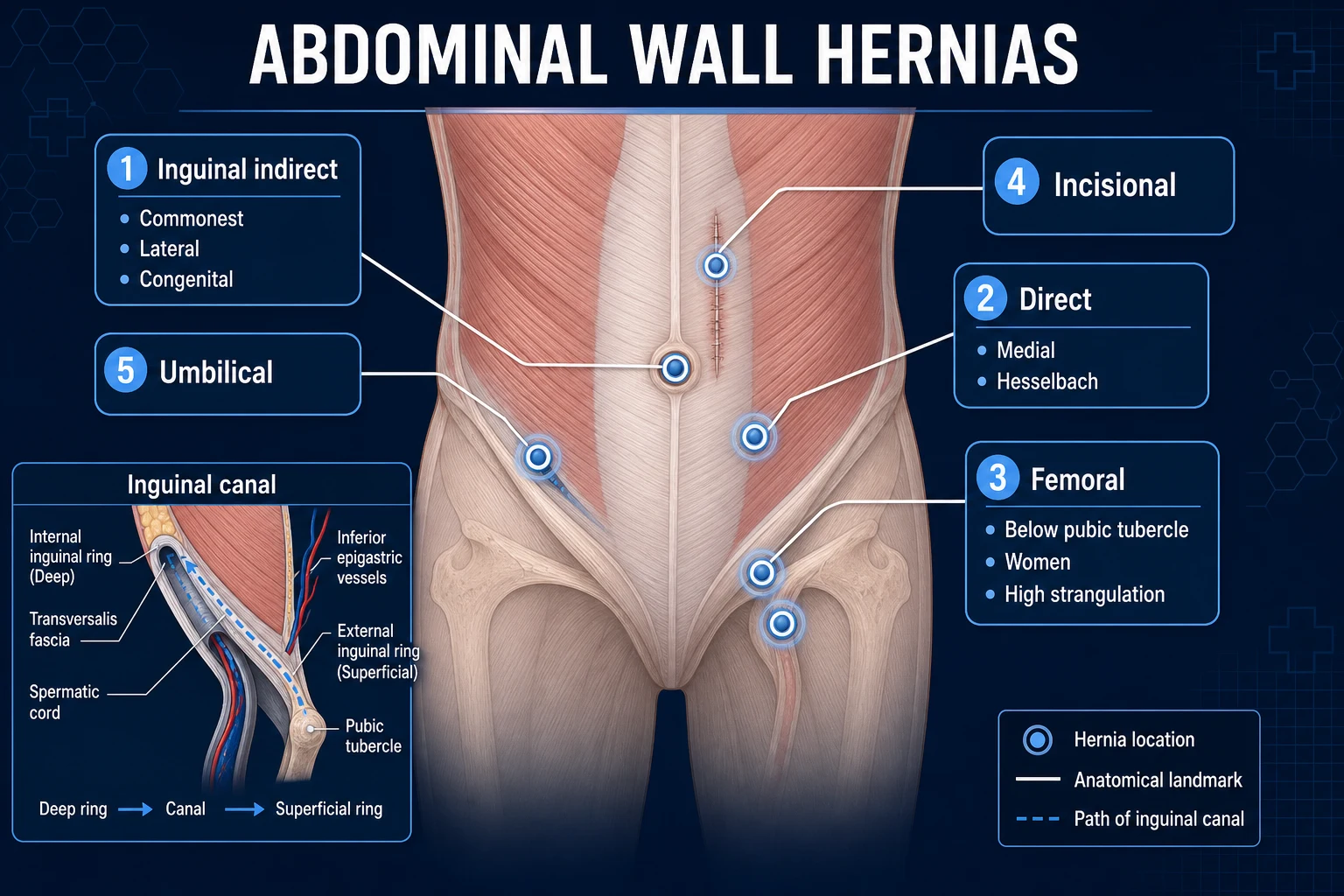
The European Hernia Society (EHS) classifies primary abdominal wall hernias by location: midline (epigastric, umbilical, suprapubic) and lateral (Spigelian, lumbar). Incisional hernias are classified separately by site, width (W1 under 4 cm, W2 4 to 10 cm, W3 over 10 cm), and recurrence. The table below covers all hernia types relevant to the umbilical/epigastric region. [1]
| Type | Age | Defect site | Sac | Management |
|---|---|---|---|---|
| Congenital/infantile | Under 5 years | Umbilical ring | True peritoneal sac | Observe until age 4 to 5; repair if persists or incarcerates |
| Adult umbilical | Over 15 years | Umbilical ring | True peritoneal sac | Surgical repair (mesh for defects over 1 to 2 cm) |
| Epigastric | Any age | Linea alba (umbilicus to xiphoid) | Preperitoneal fat only (often no sac) | Surgical repair if symptomatic |
| Paraumbilical | Adults | Just above/below umbilicus (supra-/infra-umbilical) | True sac | Surgical repair (mesh) |
| Supra-umbilical | Adults | Above umbilicus | True sac | Mesh repair |
| Hypogastric (suprapubic) | Adults | Below umbilicus | True sac | Mesh repair |
| Spigelian | Adults (>50) | Spigelian line (lateral edge of rectus) | Often interparietal (occult) | Surgical repair (lap or open) |
| Incisional | Any age | Previous surgical scar | True sac | Mesh repair (open, lap IPOM, or robotic) |
| Parastomal | Adults | Around stoma | True sac | Modified mesh (Sugarbaker or keyhole) |
| Diastasis recti | Adults/postpartum | Widened linea alba, no true fascial defect | No true hernia | Usually no surgery (cosmetic; functional) |
Special content-based hernia variants
Richter's hernia
- Only the ANTI-MESENTERIC border of bowel enters the sac
- The lumen remains patent, so NO intestinal obstruction
- Bowel can strangulate silently and perforate with minimal warning
- Classic in femoral, small umbilical and small incisional hernias
- Surgical emergency — high mortality if missed
Littre's hernia
- A MECKEL DIVERTICULUM is the hernia content
- May present as a tender, irreducible mass
- Can strangulate, ulcerate or bleed (ectopic gastric mucosa)
- Repair with resection of diverticulum + hernia repair
Sliding (en glissade) hernia
- A retroperitoneal organ (caecum, sigmoid, bladder) forms PART of the sac wall
- No true peritoneal covering on one side of the sac
- Risk of organ injury during sac dissection — reduce, do not excise
- Commoner on the right (caecum) and left (sigmoid) at large defects
Maydl's hernia
- W-shaped loop of bowel in the sac — the CENTRAL loop is strangulated while the ends are viable
- Inspection of only the ends at surgery can MISS an infarcted central loop
- Always reduce and inspect the WHOLE loop before closure
Epidemiology
- Infantile umbilical hernia: present in 10 to 20 percent of all infants; up to 75 percent in premature infants under 1500 g and 40 percent in those of African descent. Equal sex incidence; Down syndrome infants have a markedly higher prevalence.
- Adult umbilical/paraumbilical hernia: female-to-male ratio roughly 3:1; strongly associated with obesity (BMI over 30), multiparity, ascites, chronic cough, chronic constipation and heavy lifting.
- Epigastric hernia: more common in men (roughly 3:1); typically 20 to 50 years of age; multiple in 20 percent of patients.
- Spigelian hernia: rare (under 2 percent of ventral hernias); usually over age 50; the commonest occult interparietal hernia.
- Incisional hernia: occurs in 10 to 20 percent of midline laparotomy incisions (rising to 35 percent in high-risk groups — obesity, smoking, wound infection, immunosuppression); the single largest contributor to complex abdominal wall reconstruction workload. [1]
Pathophysiology
Why midline defects develop
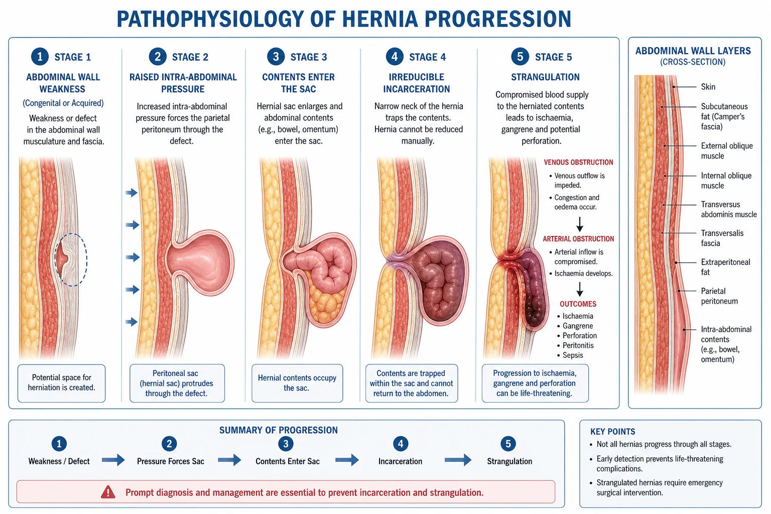
A ventral hernia arises when the balanced tension of the abdominal wall is disrupted by a combination of (1) an intrinsic fascial weakness and (2) chronically raised intra-abdominal pressure. [1]
Intrinsic weakness — the linea alba and umbilical ring are collagen-rich, avascular structures. With ageing, multiparity, obesity and corticosteroid use, the collagen I to collagen III ratio falls, weakening the fascia. Smoking, malnutrition and connective tissue disorders (Marfan, Ehlers-Danlos) accelerate this. The multiperforate nature of the linea alba (for neurovascular bundles) provides ready-made defects through which preperitoneal fat can protrude — the seed of the epigastric hernia. [1]
Raised intra-abdominal pressure — the major extrinsic driver. Causes include: [1]
- Obesity (the single commonest factor in adults) — both increased pressure and mechanical weakening.
- Pregnancy — pressure plus rectus diastasis and hormonal collagen remodelling.
- Chronic cough (COPD, TB), chronic constipation, benign prostatic hyperplasia with straining.
- Ascites (cirrhosis, heart failure, ovarian cancer) — mechanical stretching plus nutritional/collagen impairment.
- Peritoneal dialysis — continuous pressure loading. [1]
Once a small defect forms, peritoneal contents (omentum, small bowel, sometimes colon) enter the sac, the defect edges are slowly widened by peristalsis and straining, and the hernia enlarges irreversibly. Adult umbilical hernias never close spontaneously because the collagen remodelling is complete and there is no growth-driven ring contraction (unlike the infant). [1]
Incarceration, strangulation and the viability cascade
[1]The mechanism of strangulation in a small rigid defect (umbilical, epigastric, femoral) is mechanical: the firm fibrous edge of the ring compresses the venous return of the protruding viscus at the neck of the sac. Venous congestion produces oedema and swelling, which worsens the constriction, occluding the arterial supply and producing transmural ischaemia. The bowel wall becomes dusky, then purple, then black, with serosanguineous (later purulent/feculent) fluid in the sac. Translocation of bacteria leads to peritonitis and systemic sepsis. The whole process can take only 4 to 8 hours in a small rigid-neck defect. [1]
Richter's hernia — pathophysiology in depth
A Richter's hernia is the prototypical small-defect, high-risk hernia. Only the anti-mesenteric border of the bowel enters the sac — typically through a small (1 to 2 cm), rigid, fibrous ring. Because only part of the circumference is trapped, the lumen remains patent and intestinal contents still pass. Therefore: [1]
- No colicky abdominal pain, no distension, no vomiting, no obstipation — the classic four signs of mechanical obstruction are absent.
- The patient presents with a localised, tender, irreducible mass that may progress to systemic sepsis and peritonitis from perforation of the strangulated wall segment.
- This is the hernia most often missed until perforation — the diagnosis hinges on examining every hernial orifice in any patient with unexplained peritonitis or sepsis, particularly elderly women (femoral) and cirrhotic/obese patients (umbilical). [1]
At surgery, the trapped wall segment must be assessed for viability: pink and peristalsing = viable (reduce and repair); dusky that recovers after warm packs = viable; non-viable = resection with primary anastomosis if clean, or stoma if contaminated. [1]
Clinical Presentation
History
- Visible or palpable bulge at the umbilicus or epigastrium, worse with standing, straining, coughing, lifting or at the end of the day; may reduce spontaneously on lying flat or with gentle pressure.
- Discomfort or dragging pain — often a dull ache rather than sharp pain; may be absent (especially with epigastric fat hernias, which may be exquisitely tender despite a tiny defect).
- Symptoms of complications: severe localised pain with nausea, vomiting, distension and constipation suggest obstruction/strangulation. A cirrhotic with clear fluid leaking from the umbilicus has a ruptured umbilical hernia — a surgical emergency.
- Relevant past history: chronic cough, constipation, prostatism, ascites, prior surgery, smoking, steroid use, connective tissue disease; obstetric history in women. [1]
Examination
- Inspection: with the patient standing, look for a bulge, skin changes (thin, red, dusky, ulcerated), scars and striae. Note any stoma.
- Cough impulse: place a finger over the suspected defect and ask the patient to cough — an expansile cough impulse confirms a hernia.
- Palpation: assess the size of the fascial defect (not the bulge — the skin stretches; the defect is the true measure), reducibility, consistency and tenderness. A tense, tender, irreducible mass with overlying skin redness is a strangulated hernia until proven otherwise.
- Auscultation: bowel sounds over the swelling confirm bowel content.
- General examination: signs of cirrhosis (ascites, spider naevi, palmar erythema, jaundice, caput medusae from recanalised paraumbilical veins), COPD (barrel chest, pursed-lip breathing), obesity, pregnancy, and signs of bowel obstruction or peritonitis. [1]
Distinguishing from mimics
Differentials of a periumbilical/epigastric mass
Investigations
Most umbilical and epigastric hernias are clinical diagnoses and require no imaging in the elective setting. [1]
- Ultrasound — first-line when doubt exists; identifies the fascial defect, contents and any diastasis; useful for the obese (where clinical examination is difficult) and for Spigelian hernias (interparietal, occult). A Spigelian hernia lying interparietally between internal and external oblique can be invisible clinically; US or CT is essential.
- CT abdomen — for complex/recurrent/incisional hernias, for loss of domain planning, to characterise contents, identify occult hernias, and in the cirrhotic to map ascites and varices before surgery.
- MRI — occasionally for chronic abdominal wall pain of uncertain cause (nerve entrapment, small occult hernia).
- Pre-operative bloods and anaesthetic assessment: FBC, U&E, LFTs, coagulation, group and save; optimise comorbidities (smoking cessation 4 weeks pre-op, weight loss, diabetic glycaemic control — HbA1c under 69 mmol/mol/8.5 percent).
- Cirrhotic workup: Child-Pugh score and MELD score, ascitic fluid analysis (cell count and culture to exclude spontaneous bacterial peritonitis), variceal screening, coagulation and platelets. [1]
Management Principles
General framework
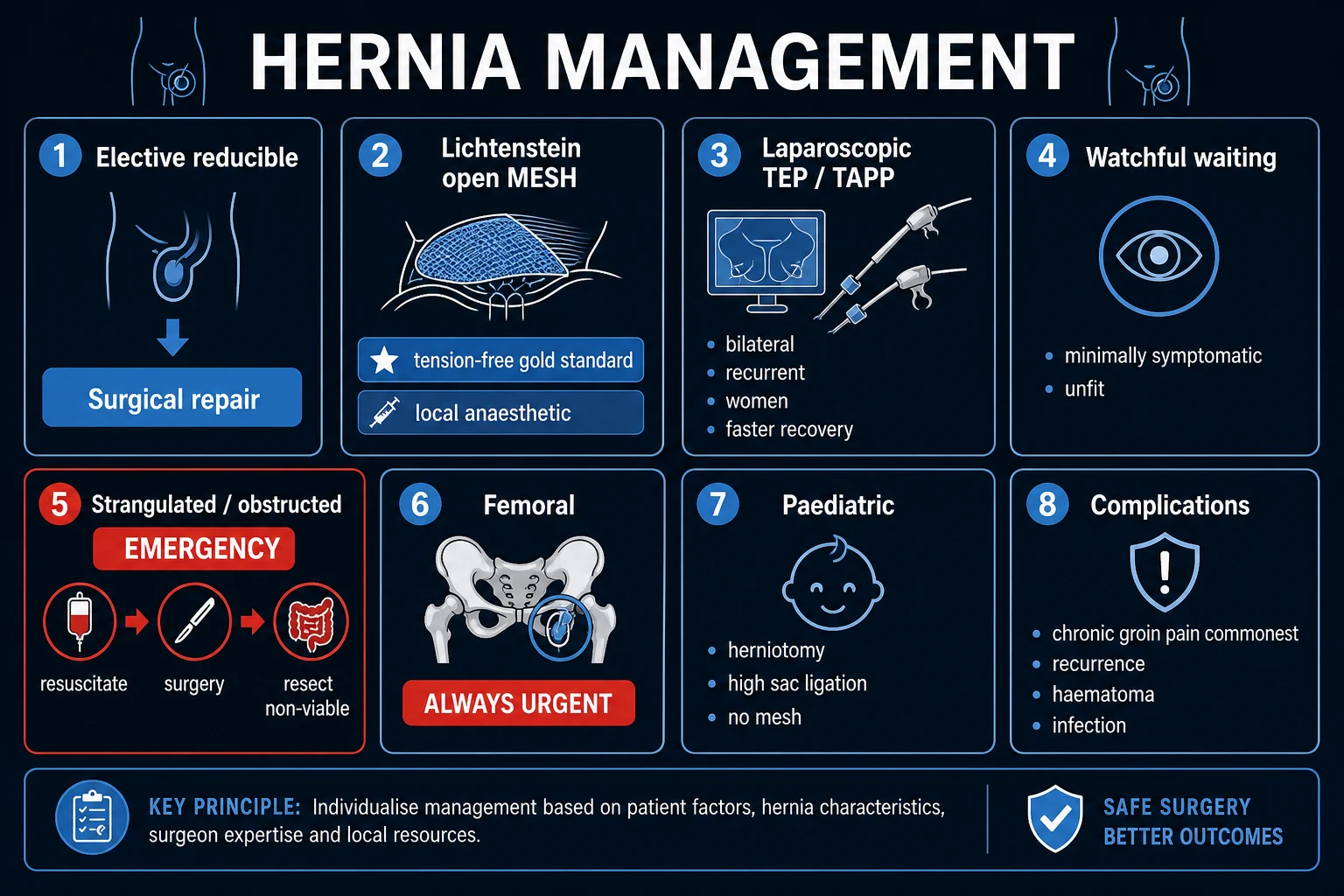
- Confirm the diagnosis and define the defect (size, contents, recurrence).
- Address reversible risk factors (smoking, obesity, COPD, constipation, ascites) — failure to do so guarantees recurrence.
- Choose the operation: observe (paediatric), primary repair (small defects under 1 cm), mesh repair (defects over 1 cm), complex reconstruction (large/recurrent).
- Select the mesh position and type based on defect size, patient comorbidity, and contamination risk.
- Plan for the emergency — recognise and operate on strangulation without delay. [1]
Paediatric Umbilical Hernia
Natural history and indications for surgery
- Present in 10 to 20 percent of infants (up to 75 percent if premature, 40 percent in African descent).
- 95 percent close spontaneously by age 5 — fascial rings under 1 cm close fastest; those over 1.5 cm rarely close.
- Observe until age 4 to 5; surgery indicated for: persistence after age 4 to 5, defect over 1.5 cm, incarceration (rare, under 1 percent), or progressive enlargement.
- The cover test at each clinic visit stratifies likelihood of closure. [1]
Surgical technique (paediatric)
- A small curved transverse infra-umbilical incision in the skin crease (cosmetic — hidden in the umbilical fold).
- Dissect to the sac; the sac is identified, separated from the overlying skin and freed to the fascial level.
- The sac is ligated at the level of the fascial defect and the excess removed (or, if no true sac, simply reduce the preperitoneal fat).
- The fascial defect is closed transversely with interrupted absorbable sutures (2-0 or 0 polydioxanone, PDS).
- No mesh is ever used in children (strong fascia, small defects, foreign-body risk, growing abdomen).
- Skin closed with subcuticular absorbable suture; the umbilical dimple is reconstructed by suturing the umbilical skin to the fascia.
- Day-case surgery; same-day discharge. [1]
Outcomes
Excellent — recurrence under 1 percent, minimal complications. Complication rates rise dramatically in children with connective tissue disorders, mucopolysaccharidoses, and chronic ascites/VP shunts. [1]
Adult Umbilical and Epigastric Hernia
Adult umbilical and epigastric hernias NEVER close spontaneously — surgical repair is indicated whenever they are symptomatic (pain, enlargement, cosmetic concern). Asymptomatic, small, reducible hernias in moribund or extremely high-risk patients may be watched, but the natural history is progressive enlargement and rising complication risk. [1]
| Defect Size | Recommended Repair |
|---|---|
| Under 1 cm | Primary suture repair (small, elective) |
| 1 to 4 cm | Open mesh repair (sublay preferred over onlay) |
| Over 4 cm | Laparoscopic IPOM (or open sublay) |
| Over 10 cm / recurrent / loss of domain | Component separation + mesh (complex reconstruction) |
Why mesh has replaced suture repair
The European Hernia Society 2015 guidelines recommend mesh repair for all primary ventral hernias with defect over 1 cm. The shift is driven by recurrence evidence: [1]
- Arroyo et al (2005) RCT, mesh vs suture for umbilical hernia (1 to 5 cm defects): recurrence 1 percent (mesh) vs 11 percent (suture).
- Abdel-Baki et al (2007) mesh vs Mayo repair: recurrence 0 percent (mesh) vs 20 percent (Mayo).
- The recurrence rate with primary suture repair rises with defect size (up to 50 percent for defects over 3 cm), whereas mesh repair holds recurrence below 5 percent regardless of size. [1]
The principle is the tension-free repair: mesh distributes the load across the abdominal wall rather than concentrating it at a suture line under tension (which is exactly what fails in the Mayo overlap technique). [1]
Detailed Surgical Techniques
Mayo 'vest-over-pants' repair (historical)
Described by William J. Mayo in 1907, this was the standard for over 50 years and is still examined in vivas. [1]
- Transverse elliptical incision around the umbilicus (the umbilicus itself may be excised with the skin flap).
- Dissect the sac circumferentially and open it; reduce contents.
- Create overlapping fascial flaps (the superior flap pulled down over the inferior, like a vest over pants) and suture them together with non-absorbable interrupted sutures (originally silk).
- No mesh. [1]
Why it failed: it is a tension repair under high intra-abdominal pressure, in an avascular linea alba, in patients with collagen weakness. Recurrence 10 to 25 percent; now reserved only for defects under 1 cm in otherwise fit patients, or as a historical comparison. The principle of overlap survives in mesh design (mesh must overlap the defect by at least 4 to 5 cm). [1]
Open sublay (retrorectus / Rives-Stoppa) repair — the preferred open technique
The Rives-Stoppa retrorectus mesh repair is the gold-standard open technique for defects of 2 to 10 cm. It places mesh in a well-vascularised, extraperitoneal plane behind the rectus muscle, where it is incorporated under physiological tension and never contacts bowel. [1]
- Transverse infra-umbilical incision (or vertical if combined with diastasis repair); umbilicus preserved or excised.
- Dissect to the anterior rectus sheath; identify the defect.
- Open the anterior rectus sheath vertically just lateral to the linea alba; develop the retrorectus (pre-muscular) plane between rectus muscle and posterior sheath bilaterally.
- Reduce the sac (do not excise unless needed); close the posterior sheath/peritoneum if possible to make the repair extraperitoneal.
- Place a sheet of macroporous polypropylene (or composite) mesh in the retrorectus space, overlapping the defect by at least 4 to 5 cm in every direction.
- Fix the mesh with absorbable sutures or fibrin sealant (full-thickness transfascial sutures add strength but cause more chronic pain).
- Close the anterior rectus sheath over the mesh (restoring muscle cover); reconstruct the umbilicus.
- Close skin. [1]
Advantages: extraperitoneal mesh (no bowel contact, no adhesions), well-vascularised space (low infection), physiological mechanical advantage (intra-abdominal pressure pushes mesh against the wall, holding it in), low recurrence (under 5 percent). [1]
Disadvantages: more dissection than onlay; not feasible when posterior sheath is deficient (very low defects below arcuate line — needs modification, e.g. enhanced-view totally extraperitoneal, eTEP). [1]
TAR — Transversus Abdominis Release is an extension of the retrorectus repair for very large or recurrent defects: the posterior sheath is incised just lateral to the rectus and the transversus abdominis muscle divided, creating a much larger plane (extending to the retroperitoneum) that allows mesh widths of up to 20 cm and medial advancement of the recti to close the midline. This is the modern workhorse of complex abdominal wall reconstruction. [1]
Open onlay mesh repair
Mesh placed on the anterior rectus sheath after primary closure of the defect. [1]
- Close the fascial defect primarily.
- Develop the prefascial (subcutaneous) plane over the anterior rectus sheath.
- Place mesh on the sheath, overlap 4 to 5 cm, fix with sutures/tackers/glue.
- Close skin over a suction drain. [1]
Advantages: technically easier, faster, no fascial opening, useful in obese patients where the retrorectus plane is difficult. [1]
Disadvantages: higher seroma rate (5 to 15 percent) due to wide subcutaneous dissection, higher infection rate, mesh is outside the physiological pressure zone (mechanical disadvantage — intra-abdominal pressure tends to push it off the wall). Recurrence higher than sublay. [1]
Laparoscopic IPOM (Intraperitoneal Onlay Mesh)
The standard laparoscopic approach for defects over 3 to 4 cm. [1]
Indications: defects over 3 to 4 cm; recurrent hernia after open repair; obesity (BMI over 35); multiple defects; bilateral paraumbilical hernias. [1]
Technique:
- Three ports (camera plus two working ports) — usually left upper quadrant optical entry (Hasson) for camera, with two 5 mm working ports.
- Establish pneumoperitoneum; reduce the hernia contents by traction.
- Divide the falciform ligament (allows flat mesh placement against the upper anterior wall).
- Measure the defect precisely from inside — this is the true defect size (often smaller than the external bulge).
- Choose a composite mesh (anti-adhesive barrier on the bowel-facing side, tissue-ingrowth surface on the wall side) sized to overlap the defect by at least 4 to 5 cm.
- Mark the mesh centre and orientation; insert it through a port; unroll and orient it intraperitoneally.
- Fix the mesh with a double-crown of absorbable tackers plus transfascial sutures (Carter-Thomason or suture-passer) at four to eight points around the periphery — the sutures provide mechanical strength, the tackers provide contour adherence.
- Close all port sites over 5 mm; desufflate. [1]
Advantages: faster recovery, smaller wounds, less wound morbidity, can inspect the entire abdominal wall for occult defects, less postoperative pain than open. Disadvantages: mesh contacts bowel (adhesion, erosion, fistula risk — hence the mandatory composite mesh); requires general anaesthesia; cost of composite mesh; cannot be used in contaminated fields; risk of unrecognised enterotomy at port entry or adhesiolysis (catastrophic if missed — always inspect bowel before closure). [1]
IPOM-plus combines IPOM with defect closure (using a suture-passer to close the fascial defect transcutaneously before placing the mesh) — restores the abdominal wall anatomy and reduces seroma and bulging. [1]
eTEP and robotic approaches
The enhanced-view totally extraperitoneal (eTEP) approach, popularised by Belyansky and Daes, applies the TEP inguinal principle to the ventral wall: a retrorectus/extraperitoneal mesh is placed laparoscopically/robotically without entering the peritoneum, combining the benefits of laparoscopy with those of extraperitoneal mesh placement. Robotic-assisted repair (da Vinci) enhances suturing in deep retrorectus planes and enables TAR to be performed minimally invasively. These are advanced techniques, increasingly the standard in tertiary centres for complex ventral hernia. [1]
Component separation technique (for very large defects over 10 cm)
Used for massive incisional hernias, recurrent hernias with loss of domain, and after damage-control laparostomy. [1]
- Open approach: raise large subcutaneous flaps to expose the lateral abdominal wall to at least the anterior axillary line.
- Divide the external oblique aponeurosis longitudinally 1 to 2 cm lateral to the linea semilunaris (the lateral edge of the rectus), separating external oblique from internal oblique in the relatively avascular plane between them.
- This releases the external oblique laterally and allows medial advancement of the rectus complex — up to 10 cm per side in the upper abdomen, 5 cm at the waist, 3 cm in the suprapubic region.
- Close the midline fascia primarily (the goal of component separation is always fascial closure).
- Reinforce with mesh in the retrorectus or onlay position (component separation alone without mesh has a high recurrence rate).
- The endoscopic/perforator-sparing version preserves the perforator vessels to the skin, dramatically reducing wound morbidity (seroma, necrosis, infection). [1]
Complications: wound breakdown and skin necrosis (large flaps, denervation), seroma, haematoma, recurrence (especially if no mesh), and lateral abdominal wall weakness/bulging from denervation. [1]
Mesh Types and Selection
Mesh selection is a high-yield viva topic and a common MCQ trap. [1]
Synthetic non-absorbable meshes
- Polypropylene (PP) — macroporous (pore size over 1 mm), excellent tissue ingrowth, strong; the workhorse for sublay/onlay. Examples: Prolene, Marlex. Disadvantages: shrinkage (20 to 30 percent), stiffness, and adhesions/fistula if placed intraperitoneally (hence contraindicated in IPOM). Lightweight large-pore PP reduces shrinkage and chronic pain.
- Polyester (PE) — softer, more flexible, macroporous; good conformability; used in some composite meshes (Parietex).
- Expanded polytetrafluoroethylene (ePTFE, Gore-Tex) — microporous, no tissue ingrowth (so fewer adhesions but also less fixation, higher recurrence); used in older IPOM; largely replaced by composites. [1]
Composite meshes (for intraperitoneal use — IPOM)
Combine a tissue-ingrowth layer (macroporous PP/PE on the parietal side) with an anti-adhesive barrier on the visceral side. Examples:
- Proceed — PP + oxidised regenerated cellulose.
- Physiomesh — PP + polyglecaprone (absorbable barrier).
- C-Qur — PP + omega-3 fatty acid coating.
- Parietex Composite — polyester + collagen-hyaluronic acid barrier. [1]
Composite meshes are mandatory whenever mesh will be in contact with bowel (IPOM, when posterior sheath cannot be closed). [1]
Absorbable synthetic meshes
- PHYSIOMESH-type fully absorbable meshes (e.g. TIGR, Phasix) — degrade over 6 to 24 months; used as temporary bridges in contaminated fields where permanent mesh is contraindicated, with a definitive repair later. Weaker than permanent mesh as a sole repair. [1]
Biological meshes
- Human acellular dermis (AlloDerm), porcine dermal collagen (Permacol, Strattice), bovine pericardium (Tutopatch) — decellularised collagen matrices that become vascularised tissue.
- Indications: contaminated/infected fields (where synthetic mesh would fail), enterocutaneous fistula repair, bridging after necrotising fasciitis debridement.
- Evidence: inferior to synthetic mesh for clean elective repairs (higher recurrence and eventration, very expensive) — reserve for genuine contaminated-field need. The RICH trial (Rosen, 2010) of Strattice in contaminated ventral hernia showed reasonable but not superior outcomes. [1]
Mesh selection summary
| Scenario | Mesh choice |
|---|---|
| Elective open sublay/onlay, clean | Macroporous polypropylene |
| Laparoscopic IPOM | Composite mesh (anti-adhesive barrier) |
| Contaminated field (bowel resection, sepsis) | Biological mesh or absorbable synthetic (definitive repair later) |
| Cirrhotic with ascites | Macroporous PP sublay (avoid IPOM — mesh-bowel contact + infection risk) |
| Paediatric | No mesh |
Special Populations
Cirrhotic patients with umbilical hernia
This is the highest-risk elective hernia scenario and a favourite exam topic. [1]
Why cirrhotics get umbilical hernias: ascites raises intra-abdominal pressure, rectus diastasis from muscle wasting widens the linea alba, hypoalbuminaemia impairs collagen healing, and the recanalised paraumbilical veins (Crueveilhier-Baumgarten) stretch the ring. [1]
Risk profile: high rates of incarceration, strangulation, and spontaneous rupture (with fatal peritonitis from infected ascites). Surgical mortality ranges from 5 to 14 percent elective, 50 percent and above emergency. [1]
Management strategy — a stepwise approach:
- Optimise cirrhosis first: aggressive diuretic therapy (spironolactone 100 mg daily + furosemide 40 mg daily, titrated to a 100:40 ratio, max 400 mg/160 mg), salt restriction (under 2 g sodium/day), albumin for hypoalbuminaemia, treat the underlying cause (alcohol abstinence, antivirals).
- Control ascites: if refractory, consider large-volume paracentesis with albumin replacement, and TIPS (transjugular intrahepatic portosystemic shunt) to lower portal pressure before elective surgery.
- Correct coagulopathy: vitamin K, fresh frozen plasma, platelets to INR under 1.5 and platelets over 50 before surgery.
- Child-Pugh A or B (and MELD under 15): elective mesh repair after optimisation — morbidity similar to non-cirrhotics.
- Child-Pugh C (or MELD over 18): very high risk — defer and refer for liver transplant evaluation; emergency surgery only for strangulation/perforation, often as a damage-control procedure.
- Elective technique: open sublay (retrorectus) with macroporous polypropylene mesh — extraperitoneal mesh avoids bowel contact and is more infection-resistant than IPOM. Avoid IPOM in cirrhotics.
- Emergency presentation — spontaneous rupture: ascites leaks through thinned, ulcerated skin, often with evisceration. Surgical emergency: resuscitate, broad-spectrum antibiotics, emergency repair (often primary closure or biological mesh in the contaminated field), with mortality 30 to 50 percent. [1]
Pregnancy and umbilical hernia
- Common due to raised intra-abdominal pressure, rectus diastasis, and hormonal collagen remodelling (relaxin).
- Most close spontaneously postpartum (3 to 6 months after delivery) — repair is deferred unless strangulated.
- Symptomatic but reducible: supportive garment (maternity belt), avoid heavy lifting, monitor.
- Surgical repair during pregnancy: only for incarceration/strangulation — ideally in the second trimester (after organogenesis, before uterine enlargement makes access difficult and preterm labour risk peaks).
- Postpartum repair: mesh if defect over 2 cm persists after 6 months; counsel about recurrence risk in future pregnancy — ideally complete family before definitive repair. [1]
The obese patient
Obesity is the commonest risk factor for both developing and recurring umbilical hernia. Pre-operative weight loss (target BMI under 35, ideally under 30) reduces wound complications, recurrence and anaesthetic risk. Laparoscopic IPOM is preferred for the very obese (small wounds, fewer wound complications). Bariatric surgery may precede or combine with hernia repair in the morbidly obese with a reducible hernia. [1]
The elderly and frail patient
Balance operative risk against the risk of emergency presentation. In the very frail with a small reducible hernia, conservative management with a truss (an external pressure device) may be appropriate — but trusses are uncomfortable, ulcerate the skin over a tense hernia, and do not prevent strangulation. [1]
Emergency Presentations
Incarcerated umbilical hernia
- Irreducible, painful mass at the umbilicus; no or early signs of obstruction (omentum incarceration: no obstruction; bowel incarceration: vomiting, distension).
- Attempt taxis (gentle manual reduction) with the patient in Trendelenburg, adequate analgesia (opioids) and benzodiazepine for muscle spasm — if successful, schedule elective repair within 24 to 48 hours (the hernia will re-incarcerate). Never force taxis in a struggling, septic child or a patient with skin changes — strangulation is likely.
- If unsuccessful or any sign of strangulation: emergency surgery. Do NOT prolong taxis attempts beyond 30 minutes or attempt taxis if there are signs of strangulation. [1]
Strangulated umbilical hernia
A surgical emergency — operate within hours. [1]
- Severe constant pain, irreducible, tender, tense mass, overlying skin redness or dusky (late sign of necrosis).
- Signs of bowel obstruction: vomiting, distension, absolute constipation, hyperactive then absent bowel sounds.
- Systemic sepsis: tachycardia, hypotension, fever, raised lactate, raised WCC — late and ominous. [1]
Management algorithm:
- Resuscitate: oxygen, two large-bore cannulae, IV fluids (crystalloid boluses), nasogastric tube to decompress the stomach, urinary catheter to monitor output, broad-spectrum antibiotics (e.g. co-amoxiclav 1.2 g IV plus metronidazole 500 mg IV, or piperacillin-tazobactam 4.5 g IV).
- Bloods: FBC, U&E, LFTs, amylase, lactate, coagulation, group and save; crossmatch 2 to 4 units if laparotomy anticipated.
- Imaging: erect CXR/AXR for obstruction (but do not delay surgery for imaging if clinically strangulated).
- Surgery: exploration via the hernia incision extended as needed, or midline laparotomy if peritonitis. Assess the bowel: pink and peristalsing → reduce and repair; dusky → warm packs and re-assess in 10 minutes; non-viable (black, no peristalsis, dull serosa) → resect with primary anastomosis (if clean) or stoma (if grossly contaminated). Always check the bowel proximally and distally, and remember the Maydl (W-loop) risk.
- Hernia repair: primary closure or biological mesh if contaminated (synthetic mesh in a contaminated field has a high infection rate); definitive mesh repair at a second stage if needed. [1]
Spontaneous rupture (cirrhotic)
- Ascites leaks through thinned, ulcerated overlying skin — clear or blood-tinged fluid; sometimes bowel eviscerates.
- Medical emergency: peritonitis (from infected ascites), sepsis, electrolyte disturbance, dehydration.
- Management: resuscitation, broad-spectrum antibiotics, emergency surgery (primary closure or biological mesh), aggressive ascites control post-op.
- Mortality: 30 to 50 percent in cirrhotic patients. [1]
Strangulated hernia — the viability timeline
Post-operative Care and Complications
Routine post-operative care
- Day-case surgery for most elective repairs (open and laparoscopic).
- Analgesia: regular paracetamol 1 g QDS plus an NSAID (ibuprofen 400 mg TDS or diclofenac 50 mg TDS if no contraindication) for 5 to 7 days; short course of weak opioid (codeine 30 to 60 mg QDS PRN) for the first 48 hours.
- Early mobilisation and early oral intake.
- Avoid heavy lifting (over 10 kg) and strenuous abdominal exercise for 4 to 6 weeks — the mesh needs time to incorporate.
- Return to work: 1 to 2 weeks (desk/light duties), 4 to 6 weeks (manual labour).
- Wound care: keep dry for 48 hours, then shower; no baths/swimming for 2 weeks. [1]
Complication rates and management
| Complication | Rate | Management |
|---|---|---|
| Seroma | 10 to 20 percent | Observation (most resolve in 6 to 12 weeks); aspiration only if symptomatic or persisting — risk of introducing infection |
| Haematoma | 2 to 5 percent | Compression; evacuation if expanding or infected |
| Surgical site infection (SSI) | 5 to 10 percent | Antibiotics (flucloxacillin or as per culture); wound care; mesh salvage possible in many cases with antibiotics + negative-pressure wound therapy |
| Chronic pain | 5 to 10 percent | Neuropathic from ilioinguinal/iliohypogastric nerve entrapment; trial of nerve block, gabapentin/pregabalin, amitriptyline; mesh removal / neurectomy if refractory |
| Recurrence | Under 5 percent (mesh); 10 to 25 percent (no mesh) | Re-operation with a different, usually mesh-based, technique; investigate modifiable risk factors (smoking, obesity) first |
| Mesh infection | 1 to 3 percent | IV antibiotics; mesh removal often needed for macroporous PP (with resultant hernia recurrence); fistula risk if bowel contact |
| Bowel injury (IPOM) | Under 1 percent | Intra-operative recognition and repair; conversion to open if needed; missed enterotomy is catastrophic |
| Mesh erosion / fistula | Under 1 percent (IPOM) | Presents late with chronic discharge, abscess or bowel obstruction; mesh excision and bowel resection |
| Urinary retention | 1 to 5 percent | Avoid over-distension; short-term catheter |
Seroma — the commonest complication
The post-herniorrhaphy seroma is an inflammatory fluid collection in the dead space left after sac excision. Most resolve without intervention within 6 to 12 weeks. Repeated aspiration risks infection; do not aspirate a sterile seroma unless it is tense, painful, overlying skin threatened, or persisting beyond 3 months. Prevention: meticulous haemostasis, obliteration of dead space, use of defect closure (IPOM-plus), and abdominal binders. [1]
Chronic post-herniorrhaphy pain
Defined as pain persisting beyond 3 months. Causes include neve entrapment (ilioinguinal, iliohypogastric, or the tenth intercostal nerve) by a suture or tacker, neuroma, or mesh-related inflammation. Management is stepwise: reassurance and time, simple analgesia, then neuropathic agents (gabapentin 300 mg TDS titrated, pregabalin, amitriptyline 10 to 25 mg nocte), ultrasound-guided nerve block with local anaesthetic ± steroid, and finally surgical exploration with neurectomy or mesh removal in refractory cases. Prevention at index surgery: avoid full-thickness transfascial sutures near the nerves and use absorbable tackers. [1]
Evidence Base for Mesh Repair
European Hernia Society (EHS) guidelines 2015
- Mesh repair recommended for ALL umbilical/epigastric hernias with defect over 1 cm.
- Sublay (retrorectus) mesh preferred over onlay (lower recurrence, lower seroma).
- Primary suture repair acceptable only for defects under 1 cm.
- Laparoscopic IPOM: alternative for defects over 3 to 4 cm or recurrent hernias; faster recovery but bowel-contact risk.
- Pre-operative optimisation of risk factors (smoking cessation 4 weeks, weight loss, diabetic control) strongly recommended. [1]
Key randomised trials
- Arroyo et al (2005) — mesh vs suture for umbilical hernia (defects 1 to 5 cm): recurrence 1 percent (mesh) vs 11 percent (suture). The landmark trial that ended routine suture repair.
- Abdel-Baki et al (2007) — mesh vs Mayo repair: recurrence 0 percent (mesh) vs 20 percent (Mayo). The Mayo repair abandoned for defects over 1 cm.
- Saleh et al (2016) — laparoscopic IPOM vs open mesh: similar recurrence, less pain and faster recovery with laparoscopy.
- Aslani et al (2010s) and others — biological mesh inferior to synthetic in clean elective repair. [1]
Post-operative Recovery and Follow-up
Expected recovery timeline
- Day 0: surgery (day case for most elective repairs); oral analgesia.
- Days 1 to 7: wound care, analgesia (paracetamol + NSAIDs), light activity; bruising and mild swelling expected.
- Weeks 1 to 2: return to office work; wound heals; seroma may develop (reassure).
- Weeks 2 to 4: return to moderate activity; avoid heavy lifting (over 10 kg).
- Weeks 4 to 6: full recovery; return to manual labour and exercise.
- Months 1 to 3: mesh integrates fully (fibroblastic ingrowth complete by 6 to 8 weeks); scar matures. [1]
Follow-up protocol
- 2-week wound check: assess healing, seroma, infection.
- 6-week full review: confirm recovery, address chronic pain, allow return to full activity.
- Return sooner if: wound infection (increasing redness, discharge, fever), expanding seroma/haematoma, severe worsening pain, or recurrence (bulge returning).
- Long term: recurrence usually presents within 2 years; counsel patients on risk-factor modification (weight loss, smoking cessation, treating chronic cough/constipation) as lifelong prevention. [1]
Prevention and patient counselling
- Weight management — BMI target under 30.
- Smoking cessation — reduces wound complications, mesh infection, and recurrence.
- Treat chronic cough, constipation and prostatism before and after surgery.
- Graduated return to lifting — no maximum lifting for 6 weeks, then progressive.
- Diabetic control — HbA1c under 69 mmol/mol (8.5 percent) reduces SSI four-fold. [1]
Prognosis
- Paediatric: excellent; spontaneous closure in 95 percent, surgical recurrence under 1 percent.
- Adult elective mesh repair: recurrence under 5 percent; chronic pain 5 to 10 percent; full return to activity by 6 weeks.
- Cirrhotic elective (Child-Pugh A/B): morbidity and mortality acceptable after optimisation; recurrence slightly higher.
- Emergency/strangulated: mortality 5 to 14 percent overall, 30 to 50 percent in cirrhotics with rupture; morbidity driven by bowel resection and contamination. [1]
Exam Tips and Common Pitfalls
[1]Adult umbilical hernia — management priorities
UMBILICAL
Adult umbilical hernia is through the umbilical ring — NOT diastasis recti (which has no true fascial defect)
Mesh for ALL defects over 1 cm (EHS 2015); sublay preferred over onlay
Richter's hernia — strangulates without obstruction; cirrhotic rupture = surgical emergency
Taxis with analgesia + Trendelenburg; if successful, elective repair in 24 to 48 h; never force taxis with skin changes
Over 4 cm — consider laparoscopic IPOM with composite mesh; over 10 cm — component separation + mesh
95 percent close by age 5 — observe; NO mesh in children ever
Optimise ascites and coagulopathy first; Child-Pugh C → transplant referral; emergency rupture = 30 to 50 percent mortality
Linea alba widest at umbilicus; arcuate line below which posterior sheath is only transversalis fascia; T10 dermatome at umbilicus
Weight loss, smoking cessation, treat chronic cough — otherwise recurrence is guaranteed
Causes of raised intra-abdominal pressure (hernia risk)
HALF-OFF
Occupational, weight-training
Cirrhosis, heart failure, ovarian cancer, peritoneal dialysis
BPH with straining
The commonest factor in adults
Pressure + collagen remodelling
COPD, TB, bronchiectasis
Chronic straining
Classic viva questions
- "What structures pass through the umbilical ring at birth?" — Two umbilical arteries, one umbilical vein, the urachus and the vitellointestinal duct. Obliteration produces the medial umbilical ligaments, ligamentum teres, median umbilical ligament and a fibrous cord respectively.
- "Why do adult umbilical hernias not close spontaneously?" — Collagen remodelling is complete; no growth-driven ring contraction; persistent raised intra-abdominal pressure.
- "What is a Richter's hernia and why is it dangerous?" — Only the anti-mesenteric border of bowel in the sac; can strangulate without obstructive symptoms (lumen patent); high perforation risk.
- "Which mesh position do you prefer for an elective 3 cm umbilical hernia in a fit adult?" — Open retrorectus (Rives-Stoppa) sublay with macroporous polypropylene mesh — extraperitoneal, well-vascularised, low recurrence.
- "How would you manage a cirrhotic with a strangulated umbilical hernia?" — Resuscitate, antibiotics, INR/platelet correction, emergency surgery with primary closure or biological mesh (avoid synthetic in contaminated field), post-op ascites control.
- "What are the contents of the rectus sheath above and below the arcuate line?" — Above: rectus, pyramidalis, superior and inferior epigastric vessels. Below: rectus, pyramidalis, inferior epigastric vessels — posterior wall is only transversalis fascia. [1]
Common MCQ pitfalls
- Epigastric hernias often have NO peritoneal sac (preperitoneal fat only) — do not expect a reducible viscus.
- Diastasis recti is NOT a true hernia — no fascial defect, no treatment unless symptomatic/cosmetic.
- Spigelian hernia is lateral (Spigelian line — lateral edge of rectus), not midline.
- Mesh is NEVER used in paediatric umbilical hernia repair.
- Omphalocoele has a covering membrane; gastroschisis does not — fundamental exam distinction.
- T10 dermatome = umbilicus; T7 = epigastrium/xiphoid.
- Richter's hernia: no obstruction because lumen patent.
- The Cover test stratifies which infantile umbilical hernias will close spontaneously. [1]
References
- [1]Henriksen NA, Montgomery A, Kaufmann R, Berrevoet F, East B, Fischer J, Hope W, Klassen D, Lorenz R, Renard Y, Garcia Urena MA, Simons MP; European and Americas Hernia Societies. Guidelines for treatment of umbilical and epigastric hernias from the European Hernia Society and Americas Hernia Society. Br J Surg, 2020.PMID 31916607