General Surgery · General Surgery
Urinary Tract Stones
Also known as Urolithiasis · Nephrolithiasis · Kidney stones · Renal colic · Ureteric calculus · Calculus disease · Staghorn calculus
Urinary tract stones (urolithiasis) are crystalline formations arising anywhere from the renal collecting system to the urethra when urine becomes supersaturated with stone-forming solutes. They present with renal colic — sudden, severe pain radiating from loin to groin that makes the patient writhe and roll (unlike the stillness of peritonitis). CT KUB is the gold standard (97% sensitive, sees all stone types). Calcium oxalate is the commonest (75%); uric acid stones are radiolucent and dissolvable; struvite stones form staghorn calculi with urease-producing infection. Stones under 5 mm pass spontaneously in 80%; over 10 mm need intervention (ESWL, ureteroscopy + laser, or PCNL). NSAIDs are first-line analgesia. Obstruction with infection is a urological emergency requiring urgent decompression (JJ stent or nephrostomy). Prevention rests on fluids over 3 L/day, low salt, normal dietary calcium, and targeted metabolic therapy.
On this page & tools
Your progress
Saved locally on this device.
Exam tags
Red flags
Overview & Definition
Urinary tract stones (urolithiasis) are solid crystalline aggregates that form anywhere in the urinary tract — from the renal calyces and pelvis, down the ureter, into the bladder (where they are termed vesical calculi) and rarely the urethra. They arise when urine becomes supersaturated with one or more stone-forming solutes (calcium, oxalate, phosphate, uric acid, cystine, or struvite), permitting crystal nucleation, aggregation, and retention within the collecting system.[1]
The clinical hallmark is renal colic — sudden, severe, colicky pain that radiates from the loin to the groin as a stone descends the ureter. The pain reaches maximal intensity within 30 to 60 minutes and is genuinely one of the most severe pains a human can experience, frequently compared with childbirth. Yet stones may also be incidental and asymptomatic, present with painless haematuria, mimic cystitis with lower urinary tract symptoms, or declare themselves as acute kidney injury from bilateral obstruction — so the diagnosis must never rest on the textbook presentation alone.[1]
Urolithiasis is a systemic metabolic disease expressed in the urinary tract: the stone is the symptom, and the underlying abnormality (low urine volume, hypercalciuria, hyperoxaluria, hypocitraturia, hyperuricosuria, infection, or a tubular transport defect) must be identified and treated or recurrence is guaranteed. Roughly half of all first-time stone formers will form another stone within five years unless preventive measures are taken — a fact that places metabolic evaluation and prevention at the centre of long-term management, not at its periphery.[1]
Classification
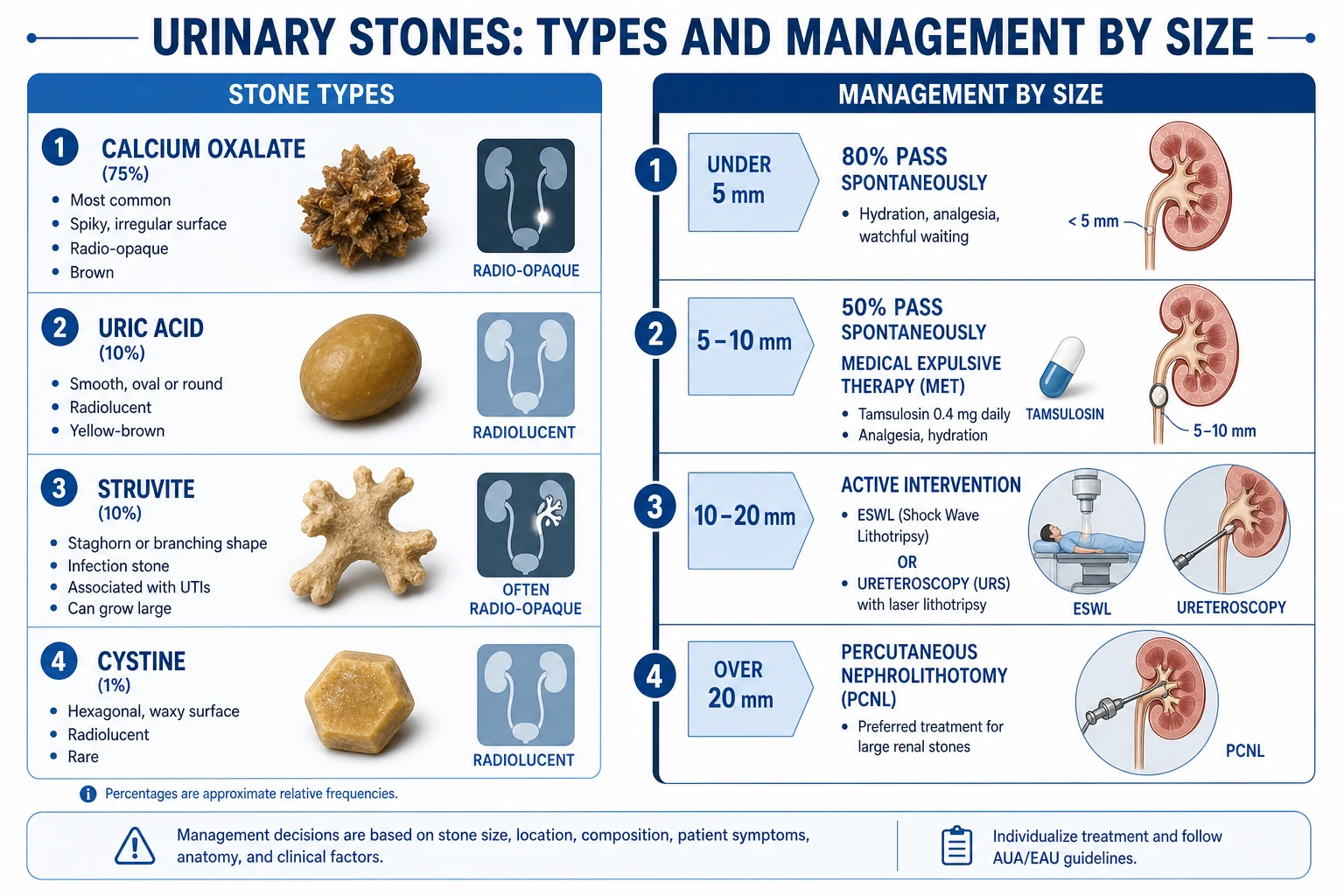
Stones are classified first by composition (which dictates aetiology, radiopacity, and treatment) and second by location (renal, ureteric, vesical). The composition is what an examiner probes, because each composition carries a distinct metabolic story and a distinct management implication.[1]
Calcium oxalate (75%)
the commonest stone
- **Spiky** monohydrate (whewellite) or rough dihydrate (weddellite) surface
- **Radio-opaque** on plain X-ray (visible in about 90%)
- Driven by **hypercalciuria, hyperoxaluria, and hypocitraturia**
- Hard, brown-black; the surface spicules cause haematuria
- Randall plaques on the renal papilla are the precursor lesion
Calcium phosphate (5 to 10%)
brushite / apatite
- Forms in **alkaline urine** (pH over 7.0)
- Classic association: **distal (type 1) renal tubular acidosis** and hyperparathyroidism
- **Brushite** stones are hard and resist ESWL — often need PCNL
- Radio-opaque; may coexist with calcium oxalate
- Look for a persistently high urine pH in the workup
Uric acid (5 to 10%)
dissolvable
- **Radiolucent** on plain X-ray (invisible) but bright on CT
- Forms in **persistently acidic urine** (pH under 5.5) — gout, myeloproliferative disease, chronic diarrhoea
- **Uniquely dissolvable** by alkalinising urine to pH 6.5 to 7.0 with potassium citrate
- Smooth, yellow-brown; pass with relative ease when small
- Family of *acidic urine* rather than hyperuricosuria alone
Struvite (10 to 15%)
infection stones
- Magnesium ammonium phosphate; **staghorn calculi** fill the pelvicalyceal system
- **Urease-producing bacteria** (Proteus, Klebsiella, Pseudomonas, Providencia) split urea to ammonia
- Forms in **alkaline urine** (pH over 7.2)
- Female predominance; recurrent or chronic UTI; neurogenic bladder; long-term catheters
- ESWL is ineffective for large staghorns — PCNL is first-line
Cystine (1%)
genetic
- **Autosomal recessive** cystinuria — defective reabsorption of cystine, ornithine, arginine, lysine (COAL)
- **Hexagonal** crystals in acidic urine
- Semi-opaque (faintly visible); recurrent from childhood
- Alkalinisation (pH over 7.5) and tiopronin for dissolvable fraction
- Every paediatric stone former needs cystine screening
Stone size and the likelihood of spontaneous passage
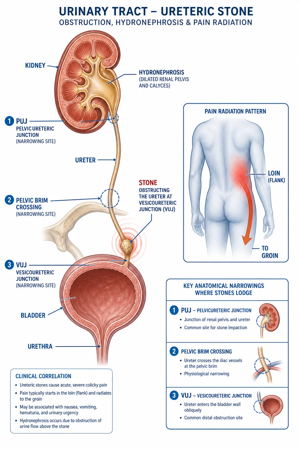
Location matters because the ureter has three anatomical narrowings where stones characteristically lodge: the pelviureteric junction (PUJ), the point where the ureter crosses the pelvic brim at the iliac vessels, and the vesicoureteric junction (VUJ) — the narrowest point and the commonest site of impaction. A stone at the VUJ produces distinctive lower urinary tract symptoms (frequency, urgency, strangury) because of irritation of the bladder trigone.[1]
Epidemiology & Risk Factors
Urolithiasis is one of the commonest diseases of the urinary tract, with a lifetime risk of 10 to 15% in developed countries and a clear upward trend driven by dietary change, obesity, and climate. Peak incidence is between 20 and 50 years, with a male predominance of roughly 2:1 to 3:1 — though this gap is narrowing as dietary and metabolic risk factors rise in women. Recurrence is the rule rather than the exception: about 15% recur at one year, 30 to 40% at five years, and 50% at ten years without preventive measures.[1]
The disease is, at root, a disease of low urine volume — the single most powerful and most modifiable risk factor. Anything that concentrates urine promotes supersaturation. The principal risk factors cluster into metabolic, dietary, anatomical, and infective groups:[1]
- Low fluid intake and dehydration — the master risk factor. Hot climates, outdoor occupations, chronic diarrhoea, and inadequate intake all reduce urine volume below the protective threshold of about 2 L/day.
- Metabolic: hypercalciuria (the commonest metabolic abnormality, often idiopathic or from primary hyperparathyroidism), hyperoxaluria (dietary, or enteric from fat malabsorption in Crohn's disease, ileal resection, or bariatric surgery), hypocitraturia (citrate is the principal inhibitor of crystallisation; low in metabolic acidosis, chronic diarrhoea, thiazide use), hyperuricosuria (gout, myeloproliferative disorders, high purine intake).
- Dietary: high animal protein (acid load increases calcium excretion and lowers citrate), high sodium (sodium and calcium share tubular handling — salt loading drives hypercalciuria), high oxalate foods (spinach, rhubarb, nuts, chocolate, strong tea), and — paradoxically — low calcium intake, which frees dietary oxalate for colonic absorption and raises stone risk.
- Systemic disease: primary hyperparathyroidism, gout, inflammatory bowel disease and Crohn's (enteric hyperoxaluria), distal renal tubular acidosis, medullary sponge kidney, hyperparathyroidism, cystinuria, obesity and metabolic syndrome.
- Anatomical and stasis: BPH with chronic retention, PUJ obstruction, horseshoe kidney, polycystic kidneys, foreign bodies (long-term indwelling catheters, sutures, stents), neurogenic bladder, bladder diverticula. Stasis allows crystals the time to nucleate and grow.
- Drugs: calcium and vitamin D supplements, loop diuretics, acetazolamide, indinavir (forms pure drug stones), sulfonamides, triamterene, excessive vitamin C (metabolised to oxalate).
- Climate and geography: the global stone belt lies in hot, arid regions — the southern United States, the Mediterranean, the Middle East, and north and northwest India.[1][1]
India: the stone belt runs across north and northwest India — Rajasthan, Punjab, Haryana, Gujarat, Delhi and parts of Uttar Pradesh and Maharashtra — attributed to a hot dry climate (chronic dehydration), high-oxalate vegetarian diet (leafy greens, strong tea), and naturally hard water. Endemic bladder stones (vesical calculi) in children were historically common in these regions, linked to a low-phosphate, cereal-based diet and dehydration; modern nutritional improvement has largely eliminated this childhood disease, and adult upper tract stones (predominantly calcium oxalate) are now the main burden. Open stone surgery is still performed in centres without endourological capability; CT KUB may not be available, so ultrasound plus KUB X-ray remains the practical first-line combination in much of the country.[1]
Pathophysiology
Stone formation is the end-result of a four-step process: supersaturation, nucleation, growth, and retention. Normal urine contains dissolved calcium, oxalate, phosphate, uric acid, and sodium held in solution by inhibitors (citrate, magnesium, pyrophosphate, Tamm-Horsfall protein). When the concentration of a lithogenic solute exceeds its solubility product — usually because urine volume is low or solute excretion is high — ions leave solution and a crystal nucleus forms. The nucleus grows by addition of further ions, aggregates with other crystals, and is retained in the collecting system (on the papilla, behind an anatomical narrowing, or in a stagnant region) until it is large enough to obstruct or pass.[1]
Calcium oxalate stones, the dominant variety, arise from the interplay of hypercalciuria, hyperoxaluria, and hypocitraturia and frequently begin on Randall plaques — subepithelial deposits of calcium phosphate at the renal papillary tip that erode through the urothelium and act as a nidus for calcium oxalate overgrowth. Randall plaques are particularly prominent in idiopathic hypercalciuria and in patients with a high body mass index.[1]
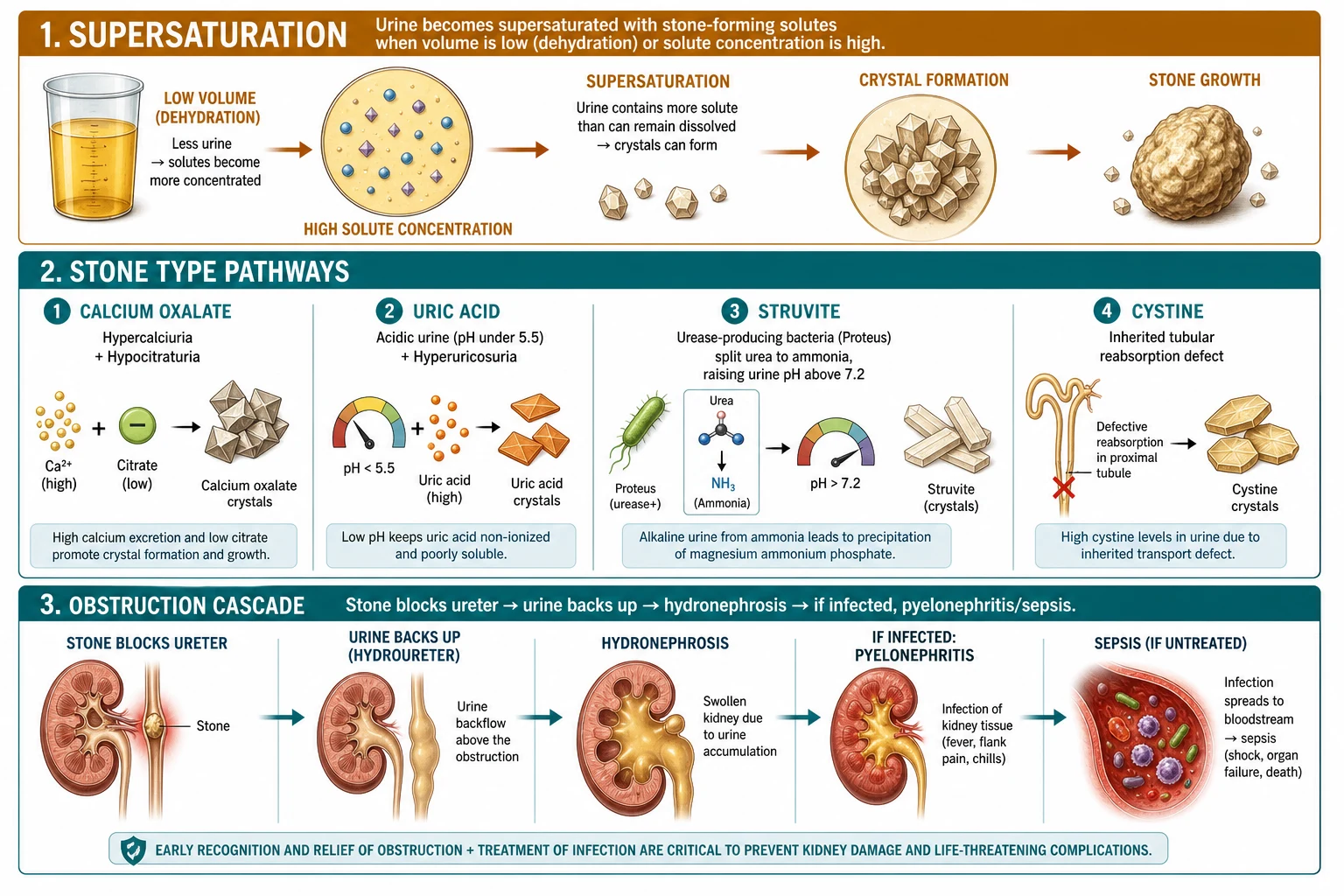
Each composition has a distinctive mechanism, and the urine pH is the single most informative bedside clue:[1]
- Calcium oxalate — driven by hypercalciuria, hyperoxaluria, and hypocitraturia; urine pH is neutral or slightly acidic.
- Calcium phosphate (brushite, apatite) — forms in alkaline urine (pH over 7.0); the classic associations are distal renal tubular acidosis (which fails to acidify urine and leaches calcium from bone) and primary hyperparathyroidism.
- Uric acid — the solubility of uric acid is exquisitely pH-dependent; it is essentially insoluble below pH 5.5 and freely soluble above pH 6.5. Patients with persistently acidic urine (chronic diarrhoea, ileostomy, gout, metabolic syndrome, myeloproliferative disease with high cell turnover) precipitate uric acid stones.
- Struvite — urease-producing bacteria (Proteus mirabilis is the prototype; also Klebsiella, Pseudomonas, Providencia, and some Staphylococcus and Proteus species) hydrolyse urea to two molecules of ammonia and bicarbonate, raising urine pH above 7.2 and precipitating magnesium ammonium phosphate. The fast growth of struvite allows it to fill the entire pelvicalyceal system as a staghorn calculus within weeks.
- Cystine — an autosomal recessive defect in the renal tubular reabsorption of cystine (and the dibasic amino acids ornithine, arginine, lysine) leaves cystine in the urine; it is poorly soluble and crystallises as pathognomonic hexagonal crystals.[1]
The pain pathway: as a stone descends it irritates the ureteric mucosa and provokes intense smooth muscle spasm, which is the proximate cause of colic. The visceral afferent fibres travel with the sympathetic chain through T11 to L2, so the pain is referred to the flank, groin, labia or testicle. A distal ureteric stone at the VUJ also irritates the bladder trigone, producing frequency, urgency, and strangury. Proximal obstruction raises intrarenal pressure, stretches the renal capsule, and produces flank tenderness; if infection is superimposed on obstruction, the pressure forces bacteria and endotoxin into the renal parenchyma and the systemic circulation — the fast route to pyonephrosis and urosepsis.[1]
Clinical Presentation
Classic renal colic
the textbook presentation
- **Sudden onset** of severe, colicky pain, often waking the patient from sleep
- Radiates from the **loin to the groin** and into the testicle or labia (T11 to L2)
- The patient is **restless, writhing, pacing, unable to find a comfortable position** — the cardinal sign
- **Nausea and vomiting** from visceral afferent co-stimulation (gastric plexus, T6 to T10 overlap)
- **Haematuria** — microscopic in 90%, macroscopic in a third
- Duration minutes to hours; the patient may arrive drenched in sweat
Atypical presentations
the traps
- **Isolated testicular or labial pain** from a distal ureteric stone (examine the genitals; exclude torsion)
- **Urinary frequency, urgency, dysuria, strangury** mimicking cystitis
- **Isolated abdominal or back pain** without clear colic
- **Silent obstruction** — incidental hydronephrosis on ultrasound, painless haematuria
- **AKI or anuria** from bilateral obstruction or a single functioning kidney
Infected obstructed system
EMERGENCY
- **Fever, rigors, hypotension, sepsis** with an obstructing stone in situ
- **Pyelonephritis** layered on mechanical obstruction
- **Pyonephrosis** (pus under pressure in the renal pelvis) — glandular destruction of the kidney within hours
- Risk of progression to **urosepsis, septic shock, and death**
- **Emergency decompression** (JJ stent or percutaneous nephrostomy) is the single most important act
The single most useful bedside observation in suspected ureteric colic is the patient's posture. A patient with renal colic is restless and writhing — pacing, curling, rolling, unable to keep still — because visceral pain has no somatic localising value and movement brings no relief. A patient with peritonitis lies perfectly still, motionless, resenting any movement that jars the inflamed parietal peritoneum. This one observation, made within seconds of meeting the patient, separates ureteric colic from appendicitis, a perforated ulcer, or a ruptured ectopic, and it is a favourite viva discriminator.[1]
Symptoms also track the location of the stone as it descends: a stone at the PUJ gives pure flank pain; at the pelvic brim, pain begins to move anteriorly toward the iliac fossa; at the VUJ, pain is felt in the groin and is accompanied by frequency, urgency, dysuria, and strangury (the desperate, dribbling desire to void). This loin-to-groin migration with evolving lower urinary tract symptoms is itself diagnostic.[1]
Differential Diagnosis
Renal colic is a diagnosis of exclusion in any patient with acute flank or abdominal pain, because several of its mimics are surgical or gynaecological emergencies in their own right. The dictum is: prove the stone, do not assume it. A patient with an abdominal aortic aneurysm can present identically, and treating it as a stone can be fatal.[1]
| Condition | Key distinguishing features |
|---|---|
| Ruptured abdominal aortic aneurysm | Older male, known vascular disease, tearing pain radiating to the back, pulsatile abdominal mass, haemodynamic instability; a haematuria does not exclude an AAA — the aneurysm may be the cause of both the pain and an incidental stone seen on CT |
| Acute appendicitis | Migration from central to right iliac fossa, localised peritonism (McBurney point tenderness, Rovsing's sign), the patient lies still, low-grade fever, nausea |
| Diverticulitis | Left iliac fossa pain, older patient, localised peritonism, change in bowel habit; confirmed on CT with contrast |
| Ruptured ectopic pregnancy | Woman of reproductive age, missed period, shoulder-tip pain (diaphragmatic irritation), vaginal bleeding, shock; a negative pregnancy test excludes it |
| Testicular torsion | Sudden scrotal/testicular pain in a boy or young man, high-riding testis, absent cremasteric reflex, onset within six hours is salvageable — every acute scrotum is torsion until proven otherwise |
| Biliary colic and cholecystitis | Right upper quadrant pain radiating to the right shoulder, fatty food intolerance, Murphy's sign, fever |
| Acute pyelonephritis (without stone) | Fever, rigors, flank pain, dysuria and frequency, positive urinalysis with nitrites and leucocytes; no colic, no haematuria of stone calibre |
| Mesenteric ischaemia | Pain out of proportion to examination, metabolic acidosis, elevated lactate, risk factors (atrial fibrillation, vascular disease) |
| Ovarian torsion / ruptured cyst | Sudden pelvic pain in a woman, adnexal mass and tenderness on vaginal examination, free fluid on ultrasound |
Clinical & Bedside Assessment
General appearance comes first and tells you most. The patient with ureteric colic is agitated, pacing, unable to find a comfortable position — the exact opposite of the motionless peritonitic patient. Diaphoresis, pallor, and a look of genuine distress are common.[1]
Abdominal examination is typically unremarkable in uncomplicated colic: the abdomen is soft and non-tender, because the disease is in the ureter, not the peritoneal cavity. There may be mild flank or renal angle tenderness on the affected side. Gross abdominal tenderness, guarding, or rigidity should redirect the diagnosis toward a peritoneal cause (appendicitis, perforation, ectopic). A pulsatile abdominal mass mandates evaluation for AAA before any stone workup proceeds.[1]
Genital examination is mandatory in any male with acute groin or testicular pain: exclude testicular torsion (high-riding lie, absent cremasteric reflex, tender testis) before attributing the pain to referred colic. A digital rectal examination assesses the prostate (BPH, prostatitis, cancer) and excludes rectal causes, and helps evaluate pelvic masses.[1]
Vital signs are the safety net. Fever, hypotension, or a rising tachycardia with a rigid stone in situ means an infected obstructed system — a urological emergency. A patient who looks well and is afebrile with a stone in the ureter has a different clinical problem from one who is febrile, tachycardic, and rigoring with the same stone. Triage the two early.[1]
Investigations
Imaging — the gold standard is CT KUB
The other imaging modalities each have a defined, narrower role:[1]
| Modality | Role, strengths, and limitations |
|---|---|
| KUB X-ray (kidneys, ureters, bladder) | Detects about 80% of stones — the radio-opaque ones (calcium oxalate, calcium phosphate, struvite, cystine semi-opaque). Misses radiolucent uric acid stones and small or overlying-bone stones. Useful for following a known opaque stone and after ESWL, never as the primary diagnostic test. |
| Ultrasound | First-line in pregnancy and children (no radiation). Detects hydronephrosis and large renal stones but misses small ureteric stones (sensitivity 50 to 70%). Point-of-care ultrasound is a reasonable first test in the emergency department in low-pretest-probability patients, reserving CT for non-resolving or atypical cases. |
| Contrast CT (CT urogram) | Reserved for haematuria without a stone (suspecting urothelial tumour), for planning complex PCNL, or when detailed anatomy of the collecting system is needed. |
| IVU (intravenous urography) | Historical — essentially obsolete in modern practice; CT KUB is superior in speed, sensitivity, and information content. |
The NEJM trial by Smith-Bindman and colleagues randomised emergency department patients with suspected nephrolithiasis to ultrasound-first versus CT-first strategies; ultrasound-first did not miss high-risk diagnoses and reduced radiation exposure, though CT gave a higher diagnostic yield. The pragmatic compromise is ultrasound first in pregnancy, children, and recurrent stone formers with typical colic, and CT first in the first episode, in diagnostic uncertainty, or when an alternative diagnosis is being entertained.[4]
Urinalysis
- Haematuria is present in about 90% of ureteric stones (microscopic or macroscopic). A stone-like presentation with completely negative dipstick should prompt reconsideration of the diagnosis (AAA, torsion, ectopic).
- pH is diagnostically rich: a pH under 5.5 points to uric acid stones; a pH over 7.2 with leucocytes and nitrites points to urease-driven struvite; a persistently alkaline pH with a calcium phosphate stone suggests distal renal tubular acidosis.
- Leucocytes and nitrites indicate infection — if present with an obstructing stone, treat as an emergency.
- Crystals on microscopy are pathognomonic when characteristic: hexagonal (cystine), envelope (calcium oxalate dihydrate), coffin-lid (struvite).[1]
Blood tests
- Urea and electrolytes — baseline renal function; a raised creatinine indicates obstruction or acute kidney injury and changes management (lower threshold for decompression).
- Calcium and phosphate — to detect hypercalcaemia (primary hyperparathyroidism); correct for albumin.
- Urate — hyperuricaemia supports a uric acid diathesis.
- Parathyroid hormone (PTH) — if serum calcium is raised, or for recurrent calcium stones, to confirm primary hyperparathyroidism.
- CRP and white cell count — markers of infection and inflammation; a high CRP with fever and an obstructing stone is a surgical emergency.[1]
Stone analysis and metabolic workup
- Stone analysis — if the patient passes or has a stone retrieved, send it for infrared spectroscopy or X-ray diffraction. Composition guides prevention.[1]
- 24-hour urine collection — for all recurrent stone formers (two or more stones) and first-time formers who are young, have a family history, or have a single kidney. Measures volume (aim over 2 L/day), calcium, oxalate, uric acid, citrate, sodium, creatinine, and cystine screen.
- Serum workup — calcium, phosphate, urate, PTH, and (in selected patients) vitamin D, magnesium, and arterial blood gases if distal RTA is suspected.
Management — Resuscitation
Uncomplicated renal colic
Analgesia is the first act, and NSAIDs are first-line. They are at least as effective as opioids for renal colic (a Cochrane review of 20 trials confirmed equivalence), they relieve the prostaglandin E2-mediated ureteric spasm and renal capsule swelling that drives the pain, and they spare the patient the nausea, sedation, and dependence of opioids.[5]
First-line analgesia for renal colic
Hydration is oral in the patient who can tolerate it (encourage 2 to 3 L/day); intravenous fluids are reserved for the patient who is vomiting, dehydrated, or in whom surgery is imminent. There is no evidence that aggressive forced diuresis speeds stone passage, and over-hydration risks distending the renal pelvis and worsening pain. Anti-emetics (metoclopramide 10 mg IV or ondansetron 4 mg IV) treat the vomiting.[1]
Infected obstructed system — the emergency
The emergency bundle is:[1]
- Resuscitate — oxygen, two large-bore cannulae, IV fluids (balanced crystalloid), and broad-spectrum IV antibiotics within one hour (e.g., co-amoxiclav 1.2 g IV or piperacillin-tazobactam 4.5 g IV plus gentamicin if septic).
- Decompress urgently — either a percutaneous nephrostomy (radiologically placed, faster and more reliable in sepsis, drains the pus directly) or a retrograde JJ ureteric stent (cystoscopic, needs anaesthesia, but definitive if stenting is preferred). Both are effective; the choice depends on local expertise and anatomy.
- Defer definitive stone treatment until the patient is afebrile, cultures clear, and sepsis resolves — usually several days to weeks later.[1]
AKI from bilateral obstruction or a single functioning kidney
Bilateral ureteric stones, or a stone obstructing a solitary or transplanted kidney, cause anuria and acute kidney injury — another urological emergency. The management is urgent decompression (stent or nephrostomy), management of hyperkalaemia and acidosis, and definitive stone treatment once renal function has recovered.[1]
Management — Definitive & Stepwise
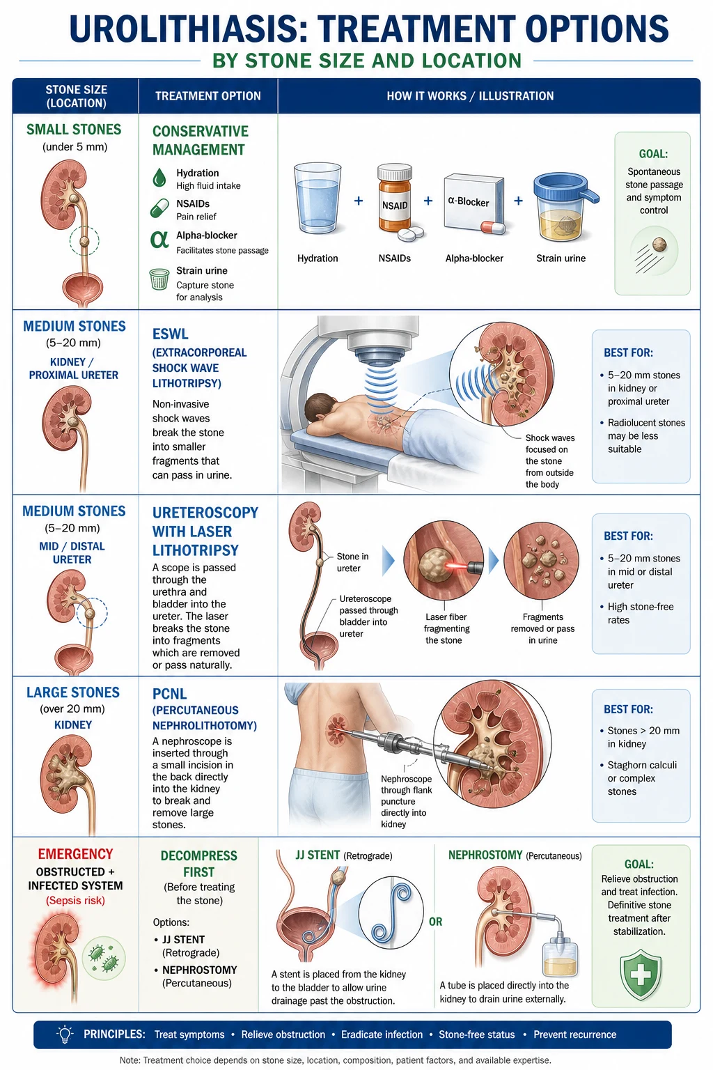
Definitive management is decided by three parameters: stone size, stone location, and the presence or absence of complications (infection, AKI, refractory pain, single kidney). The size thresholds are derived from observational data on spontaneous passage and from randomised trials of medical expulsive therapy and surgical intervention.[1][6]
Management algorithm by stone size and location
Suspected ureteric colic — analgesia (NSAID first-line), urinalysis, CT KUB to confirm size and location
Uncomplicated stone under 5 mm: conservative — analgesia, hydration, MET (tamsulosin 400 mcg OD for up to 4 weeks), strain urine; 80% pass
Uncomplicated stone 5 to 10 mm: MET (tamsulosin) plus close follow-up imaging at 4 weeks; ureteroscopy if no passage or refractory symptoms
Stone over 10 mm, or failure of conservative: intervene — ESWL or ureteroscopy (ureteric) or ESWL/PCNL (renal) by size and location
Renal stone under 20 mm: ESWL or flexible ureteroscopy (RIRS)
Renal stone over 20 mm or staghorn: PCNL first-line
Any stone with obstruction plus infection: emergency decompression first, definitive stone treatment later
Every patient: metabolic workup if recurrent, stone analysis, and structured prevention plan
Conservative and medical expulsive therapy (stones under 10 mm)
The pillars are analgesia, hydration, and time, with medical expulsive therapy (MET) added for stones in the 5 to 10 mm range — particularly in the distal ureter, where alpha-blockade relaxes the ureteric smooth muscle at the VUJ.[1]
- Hydration: oral 2 to 3 L/day; aim for a urine output over 2 L/day.
- Analgesia: diclofenac 75 mg IM/PR or ibuprofen 400 to 600 mg PO; add morphine 5 to 10 mg IV if needed.
- Medical expulsive therapy: tamsulosin 400 mcg orally once daily (an alpha-1a blocker) for up to four weeks. The landmark Lancet multicentre randomised trial by Pickard and colleagues showed that tamsulosin increased the proportion of distal ureteric stones over 5 mm that passed within four weeks (about 80% versus 60% with placebo) and reduced the need for surgical intervention. The benefit is greatest for distal stones over 5 mm; small proximal stones pass regardless, and the trial showed no benefit for stones under 5 mm.[3]
- Strain the urine to capture any passed stone for analysis.
- Safety-net: the patient must return immediately for fever, uncontrollable pain, vomiting, or failure to pass the stone within four weeks. A follow-up imaging study at four weeks confirms passage or prompts intervention.
- Absolute indications for intervention (no trial of conservative): infection with obstruction, AKI, a single kidney, refractory pain, social or occupational reasons (pilots, remote workers).
ESWL (extracorporeal shock wave lithotripsy)
ESWL focuses extracorporeally generated shock waves (electromagnetic, electrohydraulic, or piezoelectric) onto the stone under fluoroscopic or ultrasound guidance, fragmenting it into passable gravel. It is the first-line intervention for renal stones under 20 mm and for upper (proximal) ureteric stones.[2]
ESWL
shock wave lithotripsy
- Renal stones under 20 mm; proximal ureter under 10 mm
- **Outpatient, no anaesthesia** (or light sedation), 30 to 60 minutes
- **Contraindicated in pregnancy, uncontrolled coagulopathy, AAA, obstruction below the stone, morbid obesity, hard (cystine, brushite) stones**
- Stone-free rate 60 to 80% depending on size and density (over 1000 HU resists ESWL)
- Complications: **steinstrasse** (colic from a column of fragments), haematuria, perineal bruising, sepsis
Ureteroscopy (URS) + laser
rigid or flexible
- First-line for **ureteric stones at any location** and for **flexible ureteroscopy (RIRS) for renal stones under 20 mm**
- Rigid scope for distal ureter; flexible for proximal ureter and kidney (retrograde intrarenal surgery)
- **Holmium laser** fragments any stone including cystine and brushite
- Stone-free rate **over 90%** for ureteric stones; one to three days in hospital
- Complications: ureteric injury, stricture, sepsis; usually leaves a **JJ stent** for 1 to 2 weeks
PCNL (percutaneous nephrolithotomy)
for large stones
- **First-line for renal stones over 20 mm and for staghorn calculi**
- **Nephroscope through a percutaneous flank tract** (ultrasound and fluoroscopic guidance) under general anaesthesia
- Stone-free rate **over 90%** for large stones; the only effective option for complete staghorns
- Three to five days in hospital; complication rate 5 to 15% (bleeding, sepsis, injury to adjacent organs)
- **Mini-PCNL** (smaller tract) reduces bleeding for moderate stones
Laparoscopic / open
rarely needed
- Reserved for **complex anatomy** (ectopic kidney, PUJ obstruction), **very large or impacted stones**, or centres without endourology
- **Open ureterolithotomy or pyelolithotomy** is now rare (under 1% of cases)
- Laparoscopic approaches bridge the gap when endoscopic options fail
Stent (JJ ureteric stent)
A double-J ureteric stent bypasses the stone, drains the kidney, and relieves obstruction and pain. Indications are: obstruction with infection (emergency), AKI from bilateral obstruction or a single kidney, refractory pain while awaiting definitive treatment, and post-procedurally after ureteroscopy or PCNL. Stents cause bladder irritation (frequency, dysuria), stent-related flank pain on voiding, and haematuria, and must be changed or removed within months or they encrust and become stones themselves.[2]
Emergency decompression — infected obstruction
As set out above: IV antibiotics plus percutaneous nephrostomy or JJ stent, ideally within six hours, with definitive treatment deferred until sepsis resolves. Nephrostomy is often preferred in sepsis because it can be placed under local anaesthetic in a sick patient and drains pus directly; stenting needs cystoscopy and anaesthesia but is definitive if preferred.[1]
Specific Subtypes & Scenarios
Staghorn calculus (struvite)
A staghorn calculus is a large struvite stone that fills the renal pelvis and extends into the major and minor calyces, branching like a deer's antler. It arises from chronic infection with urease-producing bacteria — Proteus mirabilis the prototype, also Klebsiella, Pseudomonas, Providencia, and some Staphylococcus saprophyticus and Ureaplasma. The split urea raises urine pH above 7.2, precipitating magnesium ammonium phosphate at speed.[1]
The clinical face is one of recurrent or chronic UTI, recurrent stones, haematuria, flank pain, or progressive renal failure — and, because the filling pelvis produces little acute colic, the patient may present only when the kidney is already destroyed. Treatment is PCNL (often in stages for complete staghorns); ESWL alone is ineffective for large staghorns. Untreated staghorns cause progressive renal destruction, xanthogranulomatous pyelonephritis, and renal failure — and carry a measurable mortality. Treat the underlying infection and any anatomical cause (neurogenic bladder, catheter). Partial or total nephrectomy may be needed if the kidney is non-functioning.[2]
Calcium phosphate stones and distal renal tubular acidosis
Calcium phosphate stones (brushite, apatite) form in alkaline urine (pH over 7.0) and are the hallmark of distal (type 1) renal tubular acidosis — the tubule cannot excrete hydrogen ions, so the urine is persistently alkaline and the blood is acidotic; chronic acid buffering leaches calcium from bone, causing hypercalciuria and hypocitraturia. They are also associated with primary hyperparathyroidism. Brushite stones are hard and resist ESWL; many require PCNL. The underlying dRTA is treated with potassium citrate to correct the acidosis and raise citrate.[1]
Uric acid stones
Uric acid stones are radiolucent on plain X-ray (invisible) but dense on CT, and they are the only common stone that can be dissolved medically. They form in persistently acidic urine (pH under 5.5), driven by gout, myeloproliferative disease with high cell turnover, chronic diarrhoea or ileostomy (bicarbonate loss), metabolic syndrome, and a high purine diet. Dissolution therapy with potassium citrate titrated to maintain a urine pH of 6.5 to 7.0 dissolves existing stones over weeks to months and prevents recurrence; allopurinol 100 to 300 mg daily lowers uric acid production in patients with hyperuricaemia. Alkalinise above pH 7.0 with care — calcium phosphate may precipitate.[1]
Cystine stones
Cystinuria is an autosomal recessive defect in the tubular reabsorption of cystine and the dibasic amino acids (cystine, ornithine, arginine, lysine — the mnemonic COAL). Cystine is poorly soluble and forms recurrent stones from childhood. The diagnostic clue is hexagonal crystals on urine microscopy and a positive cyanide-nitroprusside test. Management is aggressive: high fluid intake (over 3 L/day to keep cystine concentration dilute), urine alkalinisation with potassium citrate to a pH over 7.5, alpha-mercaptopropionylglycine (tiopronin) or D-penicillamine to form soluble mixed disulfides, and endourological intervention (ESWL is often ineffective; ureteroscopy and PCNL are needed). Every paediatric stone former must be screened for cystinuria.[1]
Bladder stones (vesical calculi)
Bladder stones form in the setting of urinary stasis — BPH with chronic retention, urethral stricture, neurogenic bladder, foreign bodies (chronic catheters, sutures, forgotten stents), or schistosomiasis. They present with suprapubic pain, haematuria, frequency, urgency, dysuria, and interrupted urinary stream (the stream stops when the stone occludes the bladder neck). Imaging (plain X-ray, ultrasound, CT) confirms the stone. Treatment addresses both the stone (cystolitholapaxy — endoscopic fragmentation, or open cystolithotomy for very large stones) and the underlying cause (TURP for BPH, urethrotomy for stricture, catheter change). Endemic childhood bladder stones from dietary phosphate deficiency are now rare but historically important in the Indian stone belt.[1]
Pregnancy
In pregnancy, ultrasound is the first-line imaging (no radiation), accepting its lower sensitivity. Symptomatic ureteric stones in pregnancy are managed with conservative measures (hydration, analgesia — paracetamol first-line; avoid NSAIDs after 32 weeks and throughout the third trimester; avoid tamsulosin). If intervention is needed (refractory pain, obstruction with infection, AKI), ureteric stent or percutaneous nephrostomy under local anaesthetic is preferred — ESWL is contraindicated in pregnancy. Definitive ureteroscopy is reserved for the refractory case and performed by an experienced endourologist.[1]
Complications & Pitfalls
Of the disease:
- Acute kidney injury — from bilateral ureteric obstruction or a single functioning kidney; presents as anuria, uraemia, hyperkalaemia.
- Pyonephrosis — pus under pressure in an obstructed renal pelvis; the kidney is destroyed within hours; emergency decompression.
- Urosepsis and septic shock — Gram-negative bacteraemia from an infected obstructed system; mortality is significant.
- Chronic kidney disease — from recurrent obstruction, recurrent infection, or bilateral staghorns destroying renal parenchyma.
- Xanthogranulomatous pyelonephritis — a chronic destructive granulomatous inflammation of the kidney, usually around a long-standing obstructing stone (often staghorn); the kidney fills with lipid-laden macrophages and may be mistaken for a tumour; treatment is nephrectomy.
- Hydronephrosis — chronic obstruction dilates the collecting system and atrophies the renal parenchyma.
- Renal failure from bilateral staghorns or recurrent stone disease.[1]
Of interventions:
- ESWL: steinstrasse (a column of fragments obstructing the ureter), haematuria, perineal bruising, sepsis (if an infected stone is fragmented without prophylaxis), perirenal haematoma, hypertension with repeated treatments.
- Ureteroscopy: ureteric injury, ureteric stricture (late), ureteric avulsion (rare, devastating), sepsis, post-operative stent symptoms.
- PCNL: bleeding (requiring transfusion in 1 to 5%), sepsis, injury to colon, liver, spleen, or pleura (hydrothorax with supra-12th rib puncture), prolonged urine leak.[1]
Classic pitfalls:
- Missing an infected obstructed system in a patient "just with a stone" — this is the single most dangerous error; any fever, rigors, or sepsis with a stone in situ is an emergency.
- Treating pain without imaging — assuming colic and missing an AAA, appendicitis, ectopic, or torsion.
- Forgetting the genitals — a distal ureteric stone and testicular torsion can look identical; examine the scrotum.
- Not following up a conservative trial — a stone that fails to pass within four weeks needs intervention; silent obstruction can destroy the kidney.
- Not doing a metabolic workup in recurrent formers — recurrence is guaranteed without prevention; missing cystinuria in a child is a serious error.
- Leaving a stent in too long — forgotten stents encrust and become stones themselves, sometimes irretrievable without open surgery.
- Treating a uric acid stone with surgery when alkalinisation would dissolve it.
- Restricting dietary calcium — paradoxically increases stone risk by freeing oxalate for absorption.[1][1]
Prognosis & Disposition
Stone passage rates depend principally on size and location:[1]
Outcomes by stone size and modality
Recurrence is the natural history of the disease: roughly 15% at one year, 30 to 40% at five years, and 50% at ten years without preventive measures. With structured prevention (fluids over 3 L/day, dietary modification, targeted drugs), recurrence can be halved. This makes the prevention plan part of the treatment, not an afterthought.[1]
Disposition from the emergency department:
- Admit any patient with infection plus obstruction, AKI, refractory pain or vomiting, a single functioning kidney, a staghorn, or social factors preventing safe discharge.
- Discharge with safety-net the patient with an uncomplicated small stone, controlled pain, able to tolerate oral intake, with clear written advice to return for fever, uncontrollable pain, vomiting, or failure to pass the stone — and a defined follow-up appointment at four weeks with imaging.[1]
Special Populations
- Children: metabolic workup is mandatory in every paediatric stone former — cystinuria, primary hyperoxaluria, distal renal tubular acidosis, and inherited hypercalciuria are over-represented. Ultrasound is the first-line imaging to avoid radiation; CT is reserved for diagnostic uncertainty. Alpha-blocker MET is used off-licence. A multidisciplinary approach with paediatric nephrology and urology is the standard.
- Pregnancy: ultrasound-first imaging; avoid NSAIDs in the third trimester and tamsulosin; stent or nephrostomy for obstruction with infection; ESWL contraindicated; ureteroscopy reserved for refractory cases by an experienced endourologist.[1]
- Elderly: atypical presentations are common (confusion, falls, sepsis without localising symptoms); always exclude AAA and mesenteric ischaemia; renal function is more vulnerable to obstruction and to NSAIDs (dose-adjust, monitor).
- Single or transplanted kidney: any obstruction is an emergency — urgent decompression; lower threshold for intervention; nephrotoxic drugs avoided.
- Immunocompromised (corticosteroids, chemotherapy, transplant): higher risk of fungal (candidal) stones and unusual organisms; lower threshold to image and to decompress.
- Anticoagulated patients: bleeding risk modifies analgesia (avoid NSAIDs) and surgical approach; bridge or reverse anticoagulation around ureteroscopy or PCNL as appropriate.
Evidence, Guidelines & Regional Differences
Landmark trials and what they changed:[1]
Pickard Lancet 2015 — Medical Expulsive Therapy in Ureteric Colic
PMID 25998582
Multicentre, randomised, double-blind, placebo-controlled trial in 1167 adults with ureteric colic
Key finding
Tamsulosin 400 mcg OD for up to 4 weeks did NOT improve overall stone passage (the primary outcome), but in a pre-specified subgroup of distal ureteric stones over 5 mm it increased passage and reduced the need for surgery. Nifedipine showed no benefit. Lesson: MET with tamsulosin has a selective role for larger distal ureteric stones, not for all colic.
Smith-Bindman NEJM 2014 — Ultrasound vs CT for suspected nephrolithiasis
PMID 25539110
Pragmatic randomised trial of ultrasound-first vs CT-first in 2759 ED patients with suspected nephrolithiasis
Key finding
Ultrasound-first did not miss high-risk diagnoses and reduced radiation exposure, with no significant difference in serious adverse events, return visits, or pain scores at 7 days. Lesson: ultrasound is a reasonable first test in low-risk and recurrent patients, with CT reserved for diagnostic uncertainty or atypical presentations.
Holdgate Cochrane 2005 — NSAIDs vs opioids for acute renal colic
PMID 15846699
Systematic review of 20 RCTs (over 1600 patients) comparing NSAIDs with opioids for acute renal colic
Key finding
NSAIDs achieved at least equivalent pain relief to opioids with fewer rescue analgesia requirements and fewer adverse effects (nausea, vomiting). Lesson: NSAIDs are first-line for renal colic; opioids are add-on, not first choice.
Guidelines:
- EAU Guidelines on Urolithiasis (European Association of Urology, latest update): the most authoritative international reference. CT KUB is the gold standard; NSAIDs are first-line analgesia; ESWL, ureteroscopy, and PCNL are the three pillars of intervention with clearly defined indications by stone size and location.[1][2]
- EAU/AUA Joint Guideline for the Management of Ureteral Calculi (Preminger et al. 2007): established the size- and location-based algorithm still in use — observation for stones likely to pass, ureteroscopy or ESWL for stones needing intervention, with ureteroscopy favoured for distal and mid-ureteric stones.[6]
Regional differences:
- India and the global stone belt: CT KUB may not be available in all centres; ultrasound plus KUB X-ray is the practical combination. Open ureterolithotomy and pyelolithotomy are still performed where ureteroscopy and PCNL capability are absent. Endemic bladder stones in children are now rare but historically important in the north and northwest stone belt. Emphasis on primary prevention through hydration in hot climates.
- UK and ANZ: CT KUB is universal first-line; ureteroscopy and ESWL widely available; PCNL in tertiary centres.
- US: ultrasound-first strategies increasingly promoted to reduce radiation; ESWL availability variable; ureteroscopy dominant for ureteric stones.[1]
Prevention of Recurrence
Prevention is the second half of treatment — without it, half of patients recur within five years. The strategy is general for all stone types and targeted for the metabolic abnormality.[1]
Stone prevention — the 5 rules (FLUSH)
FLUSH
Over 3 L/day water, distributed across the day and night; aim for over 2.5 L/day of urine output, pale straw colour. The single most effective preventive measure.
Less than 100 mmol (2.3 g) sodium per day; sodium and calcium share tubular handling, so salt restriction reduces calciuria.
Potassium citrate for uric acid (target pH 6.5 to 7.0) and cystine stones (target pH over 7.5); also corrects hypocitraturia in calcium stone formers.
Animal protein (reduces citrate and acidifies urine), oxalate-rich foods (spinach, rhubarb, nuts, chocolate, strong tea), and vitamin C in high doses (metabolised to oxalate).
Do NOT restrict dietary calcium — low calcium increases oxalate absorption and stone risk. Aim for 1000 to 1200 mg/day from food, taken with meals.
Targeted drug therapy for specific metabolic abnormalities:[1]
| Abnormality | Drug | Dose | Rationale |
|---|---|---|---|
| Idiopathic hypercalciuria | Hydrochlorothiazide or indapamide | 25 to 50 mg/day | Thiazides enhance distal calcium reabsorption and reduce urinary calcium |
| Hypocitraturia | Potassium citrate | 10 to 20 mEq twice daily | Citrate complexes calcium and inhibits crystallisation |
| Hyperuricosuria / uric acid stones | Allopurinol | 100 to 300 mg/day | Reduces uric acid production; alkalinisation is the primary therapy |
| Primary hyperparathyroidism | Parathyroidectomy | — | Cures the metabolic driver; indications by PTH, calcium, and stone burden |
| Distal RTA | Potassium citrate | titrated to blood pH | Corrects acidosis, raises citrate, lowers calciuria |
| Enteric hyperoxaluria | Calcium citrate with meals | 500 mg with meals | Binds enteric oxalate; treats the hypocitraturia of chronic diarrhoea |
| Cystinuria | Tiopronin or D-penicillamine | titrated | Forms soluble mixed disulfides with cystine |
India — prevention in the stone belt: the emphasis is on sustained hydration (over 3 L/day in hot weather, with a deliberate bedtime drink to avoid the concentrated morning urine), dietary moderation of oxalate (limit strong tea, spinach, and nuts), normal (not low) dietary calcium, and salt restriction. Public health measures include dietary education and access to safe, low-hardness water. Patients with recurrent calcium oxalate stones are common and benefit from structured follow-up with annual imaging and a 24-hour urine metabolic profile. Thiazide is widely available and under-used for proven hypercalciuria.[1]
Exam Pearls
- CT KUB = gold standard (97%). Renal colic = loin to groin pain. The restless, writhing patient has colic; the motionless patient has peritonitis.[1]
- Calcium oxalate 75% (radio-opaque, spiky). Uric acid = radiolucent (invisible on X-ray) AND dissolvable (alkalinise to pH 6.5 to 7.0). Struvite = staghorn, infection (Proteus, Klebsiella), alkaline urine over 7.2. Cystine = hexagonal crystals, semi-opaque, genetic, recurrent from childhood.[1]
- Stones under 5 mm: 80% pass. 5 to 10 mm: around 50% (tamsulosin helps larger distal stones — Pickard Lancet 2015). Over 10 mm: intervene (ESWL, ureteroscopy, or PCNL).[1][3]
- NSAIDs are first-line for renal colic pain — equal to opioids with fewer side effects (Holdgate Cochrane). Opioids are add-on, not first choice.[5]
- ESWL: kidney and proximal ureter under 20 mm. Ureteroscopy plus laser: ureteric stones at any location. PCNL: kidney over 20 mm and staghorn. Laparoscopic/open: complex or rare.[2]
- Obstruction plus infection = emergency. IV antibiotics plus JJ stent or nephrostomy, ideally within 6 hours. Do not attempt definitive stone treatment during acute infection.[1]
- Prevention: FLUSH — Fluids over 3 L/day, Low salt, Urine alkalinisation, Substances to limit, Hold calcium normal (do NOT restrict dietary calcium).[1]
- Calcium phosphate stones (brushite) and alkaline urine: think distal renal tubular acidosis and hyperparathyroidism.[1]
- Three ureteric narrowings where stones lodge: PUJ, pelvic brim (iliac vessels), and VUJ (narrowest, commonest site).[1]
- Randall plaques: subepithelial calcium phosphate on the renal papilla — the precursor of calcium oxalate stones.[1]
- Staghorn calculus left untreated: progressive renal destruction, xanthogranulomatous pyelonephritis, renal failure, and measurable mortality — PCNL is first-line.[2]
- Every paediatric stone former needs metabolic workup — cystinuria (hexagonal crystals, cyanide-nitroprusside test), primary hyperoxaluria, distal RTA.[1]
- Pregnancy: ultrasound-first; avoid NSAIDs in the third trimester and tamsulosin; ESWL contraindicated; stent or nephrostomy for obstruction with infection.[1]
Exam application bank (NEET-PG / INICET)
One-line answer
Urinary tract stones (urolithiasis) are crystalline formations arising anywhere from the renal collecting system to the urethra when urine becomes supersaturated with stone-forming solutes. They present with renal colic — sudden, severe pain radiating from loin to groin that makes the patient writhe and roll (unlike the stillness of peritonitis). CT KUB is the gold standard (97% sensitive, sees all stone types). Calcium oxalate is the commonest (75%); uric acid stones are radiolucent and dissolvable; struvite stones form staghorn calculi with urease-producing infection. Stones under 5 mm pass spontaneously in 80%; over 10 mm need intervention (ESWL, ureteroscopy + laser, or PCNL). NSAIDs are first-line analgesia. Obstruction with infection is a urological emergency requiring urgent decompression (JJ stent or nephrostomy). Prevention rests on fluids over 3 L/day, low salt, normal dietary
Worked stems (answer without another resource)
Stem 1 — Classic presentation. Map symptoms to mechanism; name the first investigation and first treatment step with dose/route if drug therapy is standard. [1]
Stem 2 — Unstable / complicated. List red flags that force immediate resuscitation, theatre, ICU, antidote, or reperfusion — and what you do in the first 15 minutes. [1]
Stem 3 — Atypical group. Elderly, pregnancy, child, or immunocompromised: how presentation and thresholds change. [1]
Stem 4 — Differential trap. Name the three closest mimics and one discriminator for each. [1]
Stem 5 — Disposition. Who goes home with safety-netting, who is admitted, who needs HDU/ICU/theatre, and what follow-up is mandatory. [1]
Rapid viva checklist
- Definition + classification
- Pathophysiology chain
- Bedside signs / criteria
- Score with exact components (if any)
- Emergency bundle
- Definitive therapy with doses
- Complications of disease and of treatment
- Special populations
- Guideline/trial name if classic
- Three exam traps
Coverage self-check
If you cannot answer any stem above from this page alone, re-read the matching section — the page is intended to be self-sufficient for final-prof and NEET-PG/INICET questions on Urinary Tract Stones.
References
- [1]Turk C, Petrik A, Sarica K, et al. EAU Guidelines on Diagnosis and Conservative Management of Urolithiasis Eur Urol, 2016.PMID 26318710
- [2]Turk C, Petrik A, Sarica K, et al. EAU Guidelines on Interventional Treatment for Urolithiasis Eur Urol, 2016.PMID 26344917
- [3]Pickard R, Starr K, MacLennan G, et al. Medical expulsive therapy in adults with ureteric colic: a multicentre, randomised, placebo-controlled trial Lancet, 2015.PMID 25998582
- [4]Smith-Bindman R, Aubin C, Bailitz J, et al. Ultrasonography vs. CT for suspected nephrolithiasis N Engl J Med, 2014.PMID 25539110
- [5]Holdgate A, Pollock T. Nonsteroidal anti-inflammatory drugs (NSAIDs) versus opioids for acute renal colic Cochrane Database Syst Rev, 2005.PMID 15846699
- [6]Preminger GM, Tiselius HG, Assimos DG, et al. 2007 guideline for the management of ureteral calculi J Urol, 2007.PMID 17993340