General Surgery · General Surgery
Varicose Veins
Also known as Varicosities · Venous insufficiency · GSV varicose veins · Venous ulcer · Chronic venous disease · Chronic venous insufficiency
Varicose veins are permanently dilated, tortuous, elongated superficial leg veins (greater than or equal to 3 mm) caused by incompetent venous valves. Most arise in the great saphenous vein (medial leg, around 80 percent) from saphenofemoral junction incompetence. The disease is graded by the CEAP classification (C0 to C6). Duplex ultrasound is the gold-standard investigation. Endovenous thermal ablation (EVLA or RFA) is the first-line definitive treatment. Venous ulcers (medial gaiter area) are managed with four-layer compression bandaging after confirming an ABPI above 0.8. Never strip if the deep veins are obstructed; biopsy any change in a chronic ulcer to exclude Marjolin ulcer.
On this page & tools
Your progress
Saved locally on this device.
Exam tags
Red flags
Overview & Definition
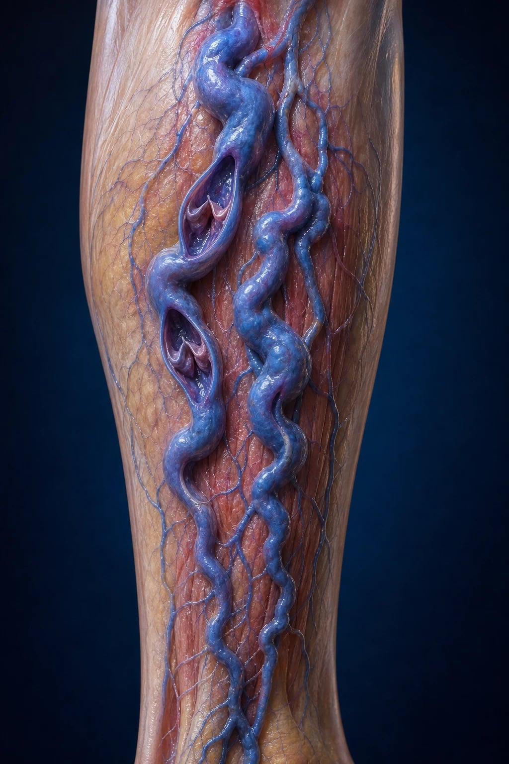
Varicose veins are permanently dilated, tortuous, elongated superficial veins of the leg measuring greater than or equal to 3 mm in diameter, distinct from reticular veins (1 to 3 mm) and telangiectases (less than 1 mm, "spider veins").[4] They are the visible end-point of chronic venous disease, a continuum that runs from symptomless trunk veins through oedema, skin change and finally venous leg ulceration. The Edinburgh Vein Study, a cross-sectional survey of 1,566 adults aged 18 to 64, found that more than half of those with trunk varices reported at least one lower-limb symptom, refuting the once-common idea that varicose veins are merely a cosmetic problem.[2]
The underlying lesion is almost always valvular incompetence. The single commonest site is the saphenofemoral junction (SFJ), where the great saphenous vein joins the common femoral vein in the groin; when its valve fails, high-pressure venous blood refluxes down the GSV. Less often the lesion is at the saphenopopliteal junction (SPJ), the perforator veins (Cockett, Boyd, Dodd), or — in secondary disease — at the deep vein valves themselves after a DVT. The clinical question the examiner always wants answered is not "what is a varicose vein?" but "why has the valve failed in this patient, and what is the safest way to stop the reflux?"[1]
A second distinction examiners reward: primary varicose veins (idiopathic valve and wall degeneration, the great majority) versus secondary varicose veins (post-thrombotic, post-traumatic, or congenital — Klippel–Trenaunay). The distinction changes management: a post-thrombotic limb with deep vein obstruction must never be stripped.[1]
Classification
Varicose veins are classified three ways — by which vein is affected, by aetiology (primary versus secondary), and by the CEAP system, which encodes Clinical class, Etiology, Anatomy and Pathophysiology into a single descriptor. CEAP is the international language of venous disease and the single most examined fact on this topic.[4][5]
GSV (around 80 percent)
medial leg and thigh
- Runs from SFJ in groin to medial malleolus
- **SFJ incompetence** is the commonest single cause of primary varicosities
- Varicosities along the **medial** leg and thigh
- Venous ulcer in the **medial gaiter** area (above medial malleolus)
- Tributaries: anterior accessory, posterior accessory, superficial external pudendal, superficial circumflex iliac, superficial inferior epigastric (the five SFJ tributaries to ligate at surgery)
SSV (around 15 to 20 percent)
posterior calf
- Runs from SPJ in popliteal fossa to lateral malleolus
- **SPJ incompetence** — junction position is variable (mark with duplex pre-op)
- Varicosities along the **posterior** calf
- Venous ulcer near the **lateral** malleolus
- Often missed clinically; the Giacomini vein (intersaphenous) connects GSV and SSV and can carry reflux between systems
Non-saphenous / reticular
cosmetic to mild
- **Reticular veins**: 1 to 3 mm, blue-green, subcutaneous
- **Telangiectasia / spider veins**: less than 1 mm, cosmetic
- CEAP C1 — usually no reflux, no ulcer risk
- Treated by **microsclerotherapy** or laser, not ablation
- Larger non-saphenous varicosities may arise from perforator incompetence alone
CEAP grading
severity
- **C0:** no visible or palpable signs
- **C1:** telangiectases or reticular veins
- **C2:** varicose veins (greater than or equal to 3 mm)
- **C3:** oedema
- **C4a:** pigmentation or eczema; **C4b:** lipodermatosclerosis or atrophie blanche
- **C5:** healed venous ulcer; **C6:** active ulcer
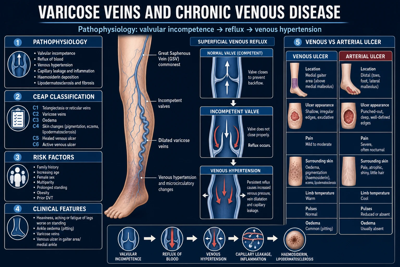
Epidemiology & Risk Factors
Varicose veins are among the commonest chronic diseases in adults. The Edinburgh Vein Study is the definitive population estimate: in 1,566 adults aged 18 to 64, the prevalence of trunk varices was 32 percent in women and 40 percent in men when measured by examination (self-report underestimates by about half).[1] Overall around 20 to 40 percent of adults have some degree of chronic venous disease, with higher rates in industrialised, ageing populations.[3]
Headline numbers — varicose veins
Risk factors fall into three groups — pressure load, wall weakness, and secondary damage:[1][3]
- Age. Prevalence rises with each decade; valves degenerate and the vein wall loses elastic recoil. The Edinburgh study showed prevalence rising from under 20 percent in the twenties to over 50 percent in the sixties.
- Family history. The strongest single risk factor. One affected parent roughly doubles lifetime risk; two affected parents quadruple it, suggesting an autosomal dominant pattern with variable penetrance.
- Female sex and parity. The female excess is largely driven by pregnancy (progesterone relaxes venous smooth muscle; the gravid uterus obstructs pelvic venous return; total blood volume rises 40 percent). Risk rises with each pregnancy. The oral contraceptive pill adds a smaller effect.
- Prolonged standing. Occupational: nurses, teachers, hairdressers, surgeons, factory and retail workers, agricultural labourers. Hydrostatic column pressure at the ankle can exceed 90 mmHg on immobile standing.
- Obesity. Raises intra-abdominal pressure and reduces mobility of the calf pump. The effect is stronger in women.
- Prior deep vein thrombosis. The cause of secondary (post-thrombotic) varicose veins; damaged deep vein valves allow reflux to be transmitted to the superficial system.
- Height and constipation. Taller stature and chronic constipation (low-fibre Western diet) modestly increase risk — the basis of the dietary-fibre hypothesis of varicose veins.
- Congenital. Klippel–Trenaunay syndrome, Parkes Weber syndrome, congenital valve agenesis. [1]
India and the tropics. Squatting and prolonged cross-legged sitting raise intra-abdominal and lower-limb venous pressure; occupations in agriculture, factory line work, street vending, security and cooking demand long hours of standing. Veins present later — often already at C4 to C6 with pigmentation or an established ulcer — because awareness and early access to duplex are limited. Endovenous ablation (EVLA, RFA) is concentrated in metropolitan centres; open surgery (high tie plus stripping) and ultrasound-guided foam sclerotherapy remain the workhorse treatments elsewhere. In the heat, dependent oedema is more pronounced, and fungal superinfection of an ulcer (a frequent confounder for eczema) must be excluded before compression is applied.
Pathophysiology
The unifying mechanism is ambulatory venous hypertension. In health, the superficial veins carry about 10 percent of lower-limb venous return at low pressure; the deep veins, surrounded by muscle, carry the rest. One-way bicuspid valves and the calf pump (the soleus and gastrocnemius squeezing the deep veins on each step) keep venous pressure at the ankle between 20 and 30 mmHg while walking. When valves fail, the system breaks down in a predictable cascade.[1][4]
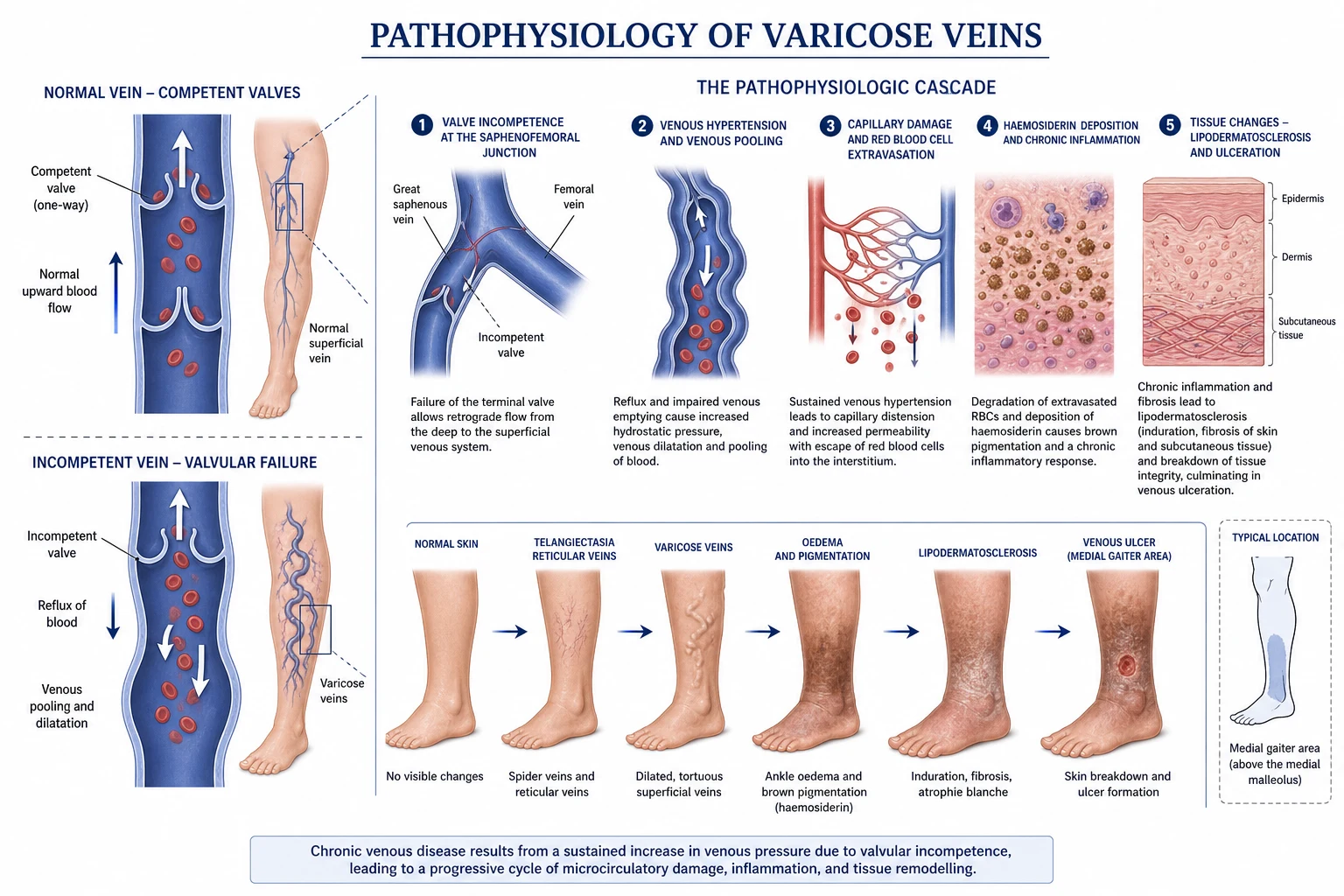
The cascade, step by step:[1]
- Valve incompetence, usually at the SFJ. The valve leaflets no longer meet, so the column of blood between the heart and the ankle is continuous.
- Reflux and venous pooling. On standing, blood flows retrograde down the GSV. The vein fills under hydrostatic pressure — at the ankle this can reach 80 to 90 mmHg.
- Progressive dilation. The vein wall stretches, which separates the next valve's leaflets, which worsens reflux. This positive-feedback loop is why varicose veins progress rather than regress.
- Perforator incompetence. High-pressure deep venous blood is forced outward through incompetent perforator veins (Cockett's below, Boyd's at the calf, Dodd's at the thigh) into the low-pressure superficial system. The superficial veins become "blown out" at these points.
- Capillary leak and the fibrin cuff. Sustained venous hypertension opens endothelial gaps; plasma and red cells extravasate into the dermis. Fibrin precipitates as a pericapillary "cuff" that is thought to obstruct oxygen diffusion.
- Leucocyte trapping hypothesis. Neutrophils and macrophages adhere in the sluggish capillaries, release proteases and free radicals, and drive a chronic inflammatory dermatitis — the basis of venous eczema and lipodermatosclerosis.
- Haemosiderin deposition. Extravasated red cells lyse; haemoglobin breaks down to haemosiderin, which stains the skin the characteristic brown ("brawny") colour of chronic venous insufficiency. Brown leg above a medial malleolus is venous disease until proven otherwise.
- Lipodermatosclerosis. Chronic inflammation and fibrin deposition fibrose the skin and subcutaneous fat into a hard, indurated, tender "inverted champagne-bottle" leg.
- Atrophie blanche. Localised white scarred atrophy at sites of healed micro-infarcts.
- Ulceration. Tissue hypoxia, inflammation and minor trauma finally break the skin — typically at the medial gaiter (above the medial malleolus) for GSV disease, or near the lateral malleolus for SSV disease. [1]
Two specific syndromes follow from this anatomy. Primary varicose veins arise when healthy valves fail idiopathically — the picture above. Post-thrombotic syndrome arises after a DVT damages the deep vein valves; reflux and obstruction combine, the deep system fails, and the superficial veins dilate as a compensatory collateral pathway. Stripping them removes the patient's only escape route — the basis of the absolute rule never strip a post-thrombotic limb.[1]
Clinical Presentation
The Edinburgh Vein Study showed that the symptoms of varicose veins correlate poorly with the visible size of the vein: even small varices can ache, and large ones can be silent. The classical triad is visible tortuous veins, heaviness-aching that is worse on standing and at the end of the day, and skin change.[2]
[1]Symptom distribution (Edinburgh Vein Study)
Atypical presentations that examiners test deliberately: the elderly patient with a non-healing "leg sore" that is in fact a venous ulcer with malignant change (Marjolin); the young pregnant woman with rapidly enlarging varicosities that largely regress post-partum; the post-DVT patient whose new varicosities are collaterals — not to be stripped; the patient with a Klippel–Trenaunay triad of varicose veins, limb hypertrophy and a port-wine stain (congenital). Each demands a different management decision.[1]
Differential Diagnosis
Not every dilated leg vein is a primary varicose vein, and not every leg ulcer is venous. A short structured differential distinguishes the treatable from the dangerous.[1][6]
Primary varicose veins
idiopathic valve failure
- Long history, family history often positive
- SFJ or SPJ incompetence on duplex, deep veins patent
- Full range of endovenous and surgical options available
- Best prognosis after ablation
Secondary (post-thrombotic)
deep vein damage
- Documented prior DVT, or silent DVT on duplex
- Deep vein reflux or obstruction; perforator incompetence
- Compression only — **never strip** (superficial veins are collaterals)
- Higher ulcer recurrence
Klippel–Trenaunay syndrome
congenital
- Triad: varicose veins + limb hypertrophy + capillary malformation (port-wine stain)
- Lateral varicosities present at birth, on the lateral thigh (anomalous embryonal vein)
- Manage conservatively; embolisation of dominant malformation in selected cases
Arterial ulcer
ischaemic
- Lateral malleolus, toes, or pressure points; punched-out edges
- **Painful**, worse at night, relieved by dangling the leg
- Cold, pulseless, hairless leg; shiny skin
- Check ABPI — compression contraindicated if below 0.8
Neuropathic ulcer
diabetic
- Pressure points on the sole (under the metatarsal heads)
- **Painless** despite depth
- Loss of sensation (10 g monofilament), callus ring
- Normal pulses; look for Charcot foot deformity
Reticular / telangiectasia
cosmetic
- Less than 3 mm, no truncal reflux, no ulcer risk
- CEAP C1
- Microsclerotherapy or surface laser only
Less common but exam-worthy mimics include arthrogenic stasis (post-fracture or post-knee-replacement calf pump failure), heart failure and renal failure (bilateral pitting oedema without varices), lymphoedema (non-pitting, dorsum-of-foot involvement, Stemmer's sign positive) and vasculitic ulcer (small, painful, "punched-out", in autoimmune disease). Bilateral leg swelling without skin change is not primary varicose veins — look for a systemic cause.[1]
Clinical & Bedside Assessment
The focused venous examination is performed with the patient standing (varices empty on lying and are missed). Bedside tourniquet tests are now largely historical — replaced by duplex ultrasound — but examiners still ask about them as a test of anatomical reasoning.[1]
Inspection (standing): distribution of varicosities (medial equals GSV, posterior equals SSV, lateral equals non-saphenous or Klippel–Trenaunay), skin change (haemosiderin staining, eczema, lipodermatosclerosis, atrophie blanche), ulcer (site, size, base, edges), corona phlebectatica (fan of intradermal veins on the medial ankle — an early sign), saphena varix (soft, reducible, cough impulse-positive lump at the SFJ). [1]
Palpation: tenderness along a varicose vein (thrombophlebitis), induration (lipodermatosclerosis), pitting oedema, temperature (cellulitis, arterial disease). Percussion/tap test (Cruveilhier–Baumgarten): tap the GSV at the SFJ — a palpable impulse transmitted distally through incompetent valves. [1]
Bedside tourniquet tests (historical, superseded by duplex): [1]
- Trendelenburg test. With the patient supine, elevate the leg to empty the veins. Apply a tourniquet at the SFJ level. Have the patient stand. If varices remain controlled, the SFJ is the point of reflux; release the tourniquet — rapid top-down filling confirms SFJ incompetence. If varices fill while the tourniquet is in place, the reflux is below the tourniquet (perforator disease).
- Perthe's test. Apply a tourniquet at mid-thigh and have the patient walk. If varices enlarge and the leg aches, the deep veins are obstructed and the superficial veins are functioning as collaterals — do not strip. If varices shrink, the deep veins are patent and the perforators are competent enough to allow safe ablation.
- Multiple tourniquet test (modified Trendelenburg). Three tourniquets (SFJ, mid-thigh, below-knee) localise the reflux level. [1]
Arterial check before any compression: palpate femoral, popliteal, posterior tibial and dorsalis pedis pulses; measure ABPI. Compression of a limb with undiagnosed arterial disease is a preventable cause of ischaemia and amputation.[1]
Abdominal and pelvic examination: in any unusual or rapidly progressive presentation, exclude an iliac or pelvic mass (cancer causing secondary varicosities) and check for signs of portal hypertension (caput medusae is paraumbilical, not saphenous). [1]
CEAP clinical class — the examiner's mental scale
Varicose veins
Truncal varices 3 mm or more; no skin change
The full CEAP descriptor adds E (Ec congenital, Ep primary, Es secondary, En none), A (As superficial, Ap perforator, Ad deep, An none) and P (Pr reflux, Po obstruction, Pr,o both, Pn none). A complete CEAP for a typical primary GSV varicose vein with an active ulcer is therefore C6, Ep, As, Pr.[4][5]
Investigations
Duplex ultrasound has replaced every historical bedside test and is the gold standard investigation for chronic venous disease.[1][6]
Duplex ultrasound (venous colour-flow Doppler). Performed standing or with the leg dependent, the patient asked to perform a Valsalva or calf-squeeze. It answers the four questions that decide management:
- Where is the reflux? SFJ, SPJ, GSV, SSV, perforator, or non-saphenous.
- How far does it extend? Length of refluxing segment, diameter of the vein (predicts suitability for ablation; a GSV diameter above 5 to 6 mm ablates well).
- Are the deep veins patent and competent? Critical. Deep vein obstruction from DVT is an absolute contraindication to stripping or ablation of the superficial system in post-thrombotic limbs.
- Are there incompetent perforators? Pathological perforator: outward flow duration of 500 ms or more and diameter 3.5 mm or more underneath a healed or active ulcer.[6]
Hand-held continuous-wave Doppler. A cheap office tool that confirms the direction of flow at the SFJ and SPJ. A positive "reverse flow on Valsalva or on releasing calf compression" indicates reflux and is enough to justify referral for duplex. It cannot localise perforators or assess deep veins.[1]
Venous severity scores. The Venous Clinical Severity Score (VCSS) complements CEAP by tracking change over time — ten items (pain, varices, oedema, pigmentation, inflammation, induration, active ulcer number, duration, size, compression therapy) each scored 0 to 3.[6]
Bloods. Routine bloods are not needed for primary varicose veins. Request a full blood count, glucose and lipids in a patient with an arterial risk factor or a non-healing ulcer; check a coagulation screen and D-dimer if DVT is suspected on duplex. [1]
Other imaging — selective. MR or CT venography for complex pelvic reflux, congenital malformation, or suspected deep obstruction (May–Thurner syndrome, iliac compression). Ascending venography is now rarely diagnostic — it is reserved for planning deep venous stenting. Air plethysmography quantifies reflux and calf-pump function in research or difficult cases. [1]
Always check the ABPI before any compression. A reading below 0.8 means arterial disease; refer for vascular assessment and avoid high-pressure compression.[1]
Management — Resuscitation
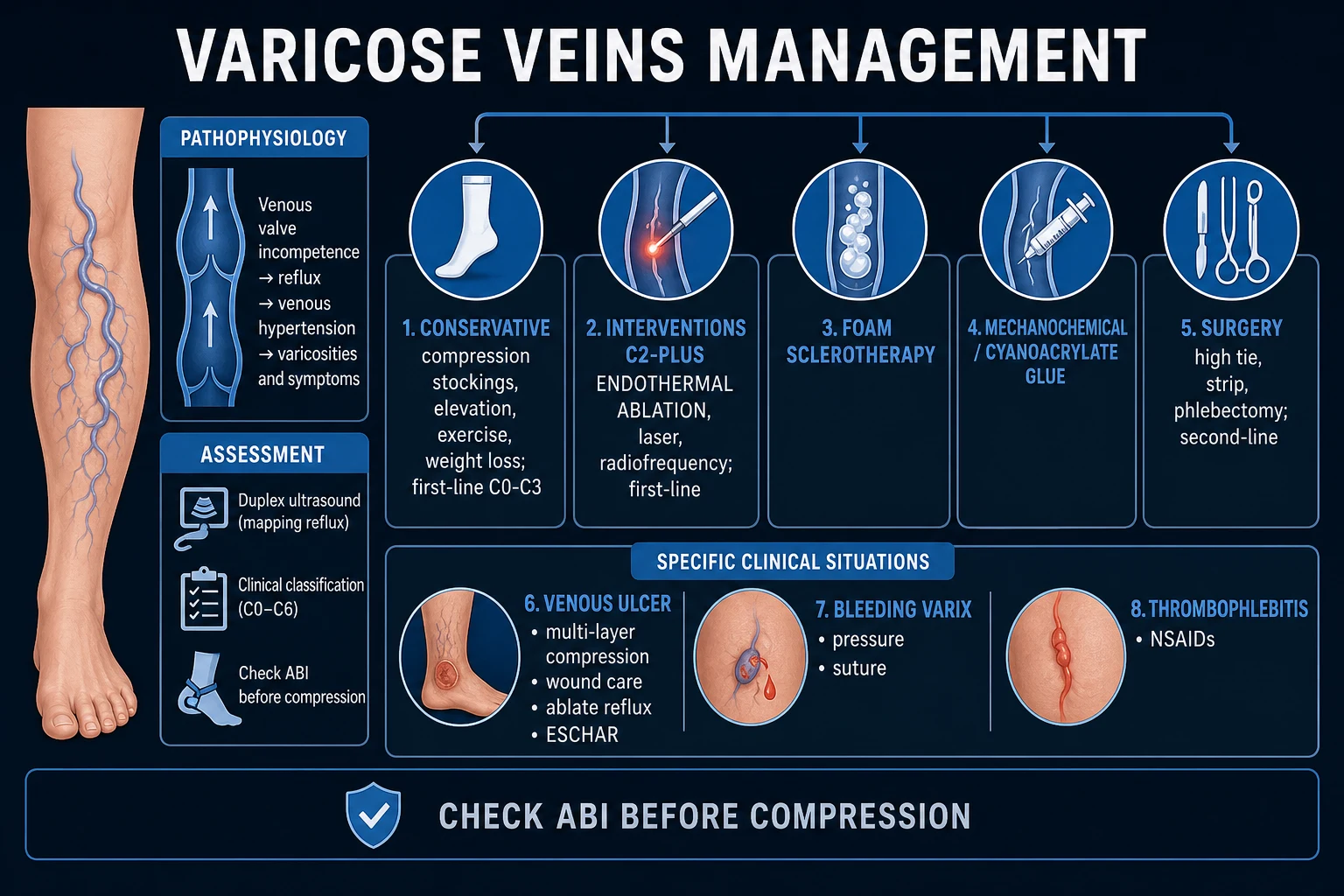
Most varicose veins are chronic, elective problems. Two presentations are time-critical.[1]
Bleeding varix. A trivial skin breach over a high-pressure varix can cause catastrophic venous haemorrhage. The emergency management is simple but must not be omitted: lie the patient flat, elevate the leg above the heart, and apply direct, firm, prolonged pressure (a pad and bandage). Do not apply a tourniquet. Most bleeding stops with elevation and pressure. Once controlled, the varix is suture-ligated locally and the patient referred for definitive ablation; the bleeding will recur if the underlying reflux is left untreated. [1]
Acute superficial thrombophlebitis near the SFJ. Varicophlebitis of a long GSV segment within 3 cm of the SFJ carries a real risk of extension into the deep system as DVT or PE. Image with duplex; if extension is confirmed, treat as DVT with therapeutic anticoagulation (e.g. apixaban 10 mg PO BD for 7 days, then 5 mg PO BD — cite local protocol). Localised distal phlebitis is managed with NSAIDs, compression and ambulation.[1]
Active venous ulcer. The resuscitation phase is wound preparation: clean and debride the ulcer, swab for infection, exclude arterial disease by ABPI, and start four-layer compression bandaging (see below). Do not delay compression for "complete healing" — compression is the treatment that heals the ulcer.[1][9]
Management — Definitive & Stepwise
Definitive treatment follows a ladder: conservative measures for symptoms alone, endovenous ablation as the first-line definitive option for C2 to C6 disease with saphenous reflux, and open surgery reserved for those unsuitable for endovenous techniques. The CLASS trial established that all three definitive modalities (laser, foam, surgery) are clinically equivalent at one and five years; the choice is driven by anatomy, patient preference, local availability and cost.[1][7][8]
The varicose veins treatment ladder
Conservative (symptom control)
Class II compression stockings (18 to 24 mmHg), leg elevation above heart for 30 minutes two to three times a day, weight loss, regular walking and calf-pump exercise, avoidance of prolonged standing. Use if the patient declines intervention, is unfit, or is pregnant.
Endovenous thermal ablation (EVLA or RFA)
First-line definitive treatment for saphenous reflux (NICE GRADE 1B). Day-case, local tumescent anaesthesia, return to work in one to three days. Ablates the GSV or SSV from inside using heat. Five-year closure rates above 90 percent.
Ultrasound-guided foam sclerotherapy
Sclerosant (sodium tetradecyl sulphate or polidocanol) mixed with air in a Tessari 1:4 ratio to form foam, injected under ultrasound. Suits smaller saphenous trunks and tributaries, recurrent varicosities, and patients unfit for thermal ablation. Cheaper; slightly lower closure rates and higher recurrence.
Mechanochemical ablation (MOCA) and cyanoacrylate glue
Non-thermal, non-tumescent alternatives. ClariVein rotates a wire to injure the endothelium while delivering sclerosant; VenaSeal cyanoacrylate glue seals the vein. Useful when tumescent anaesthesia must be avoided. No heat means no risk of thermal nerve injury.
Open surgery (high tie plus stripping plus avulsions)
Reserved for those unsuitable for endovenous, very large varicosities, or recurrent disease. High saphenous ligation at the SFJ with division of all tributaries, invagination stripping of the GSV to just below the knee, and stab-avulsion phlebectomies of tributaries. Higher pain, longer recovery, more nerve injury than ablation.
Treat the ulcer pathway
Active ulcer (C6): four-layer compression bandaging to heal, ABPI permitting. Once healed (C5), ablate the saphenous reflux to prevent recurrence (ESCHAR finding). Pentoxifylline 400 mg orally three times a day as adjunct in slow-healing ulcers. Antibiotics only for clinical infection.
Conservative management
Conservative measures do not abolish reflux and do not prevent progression, but they reduce symptoms and are first-line when intervention is contraindicated (pregnancy, unfit patient, patient preference).[1]
Compression hosiery — British Standard classes
Other conservative measures: leg elevation above the heart for 30 minutes, two to three times a day; weight loss; regular walking, swimming or cycling to drive the calf pump; avoidance of prolonged standing and of constricting garments; heel raises to encourage calf activity. Phlebotonics (diosmin, hesperidin, rutosides — micronised purified flavonoid fraction) are popular in continental Europe and India for short-term symptom relief; the SVS guidelines regard the evidence as low-quality and do not recommend them routinely.[6]
Endovenous thermal ablation (EVLA, RFA) — first-line definitive
Thermal ablation delivers heat to the vein endothelium through a catheter positioned under ultrasound, causing irreversible thermal injury, fibrosis and permanent closure. The vein is accessed at the knee (retrograde) or ankle (antegrade) under local anaesthesia; tumescent anaesthesia (large-volume dilute lignocaine with adrenaline and bicarbonate) is infiltrated around the vein to compress it against the catheter, provide analgesia, and protect surrounding tissue (especially the saphenous and sural nerves) from thermal injury.[7]
- Endovenous laser ablation (EVLA). A laser fibre (810, 980, 1470 or 1927 nm wavelength) delivers energy. Higher wavelengths (1470 nm) target water in the vein wall rather than haemoglobin, reducing bruising and post-procedure pain.
- Radiofrequency ablation (RFA). The ClosureFAST catheter heats a 7 cm segment to 120 degrees Celsius in 20-second cycles. Equivalent outcomes to EVLA; slightly less post-procedure bruising and pain in some trials.
- Outcomes. Five-year closure rates above 90 percent; recurrence of about 5 to 20 percent. Return to work in one to three days. Complications: bruising, transient skin pigmentation (5 to 10 percent), DVT (rare, less than 1 percent), nerve injury (saphenous nerve below the knee, sural nerve for SSV), skin burn (rare with tumescent).[8]
CLASS — Comparison of Laser, foam and Surgery for varicose veins
N Engl J Med 2014 (Brittenden et al.); 5-year outcomes N Engl J Med 2019
Multicentre UK randomised controlled trial of 798 participants with primary varicose veins and great saphenous reflux, allocated to endovenous laser ablation, ultrasound-guided foam sclerotherapy, or surgery (high ligation plus stripping plus phlebectomies).
Key finding
At 1 year all three treatments were clinically effective. Quality of life was similar between groups, with a slightly worse disease-specific quality of life in the foam group than the surgery group. Complications were less frequent after laser than after foam or surgery (4 percent versus 8 percent versus 6 percent). At 5 years, clinical effectiveness remained similar across the three groups; rates of re-intervention and residual reflux favoured laser over foam.
Practice change
Established endovenous thermal ablation as the preferred first-line definitive treatment for saphenous reflux, replacing open surgery for most patients. Underpinned NICE CG168 that lists endovenous ablation before foam and surgery.
Ultrasound-guided foam sclerotherapy
A sclerosant (sodium tetradecyl sulphate 3 percent for the GSV, polidocanol 1 to 3 percent for tributaries) is mixed with air using the Tessari technique (two syringes and a three-way tap, 1 part sclerosant to 4 parts air) to make a foam that displaces blood and contacts a large surface area of the vein wall. Injected under ultrasound guidance, the foam induces endothelial destruction and the vein fibroses shut.[1][7]
Foam is second-line to thermal ablation in NICE CG168 but is first-choice for recurrent varicosities after surgery, for non-saphenous varicosities, for venous malformations, and for patients unfit for thermal ablation. Maximum 10 mL of foam per session to limit the risk of foam embolism.[1][7] Side effects: skin staining (common), transient visual disturbance or migraine (especially with a patent foramen ovale), DVT/PE (rare), anaphylaxis (rare). Contraindications include known right-to-left shunt, recent DVT, pregnancy and local infection.
Mechanochemical ablation (MOCA) and cyanoacrylate glue
MOCA (ClariVein) combines a rotating wire tip that mechanically injures the endothelium with simultaneous delivery of a liquid sclerosant — no heat, no tumescent anaesthesia. Cyanoacrylate glue (VenaSeal) polymerises on contact with blood, instantly sealing the vein. Both avoid the pain of tumescent infiltration and the small risk of thermal nerve injury; they are particularly useful for SSV ablation (where the sural nerve is at risk from heat) and for needle-phobic patients. Closure rates approach those of EVLA at two to three years; cost is higher.[6]
Open surgery (high saphenous ligation, stripping, avulsions)
When endovenous treatment is unsuitable (extremely large or tortuous varicosity, immediate proximity to SFJ with thrombus risk, recurrent disease after previous ablation, or where the technology is unavailable), open surgery remains a durable option.[1]
High saphenous ligation plus stripping (the "Trendelenburg operation"). Under general or regional anaesthesia: a groin crease incision over the SFJ; the GSV is identified and ligated and divided flush with the common femoral vein; all five SFJ tributaries are individually ligated (failure to do so risks recurrence from neovascularisation). The GSV is then stripped from the SFJ to just below the knee using an invagination (Oesch or Myers) stripper — stripping to the ankle is avoided because it injures the saphenous nerve in up to 40 percent of cases. Visible varicosities are removed through 1 to 2 mm stab incisions (avulsion phlebectomies / Muller's technique). Post-op: full-length Class II compression, early ambulation, paracetamol and NSAIDs; return to work in one to two weeks. [1]
Saphenopopliteal junction ligation for SSV. The SPJ is marked pre-op with duplex because its level is variable (anywhere from 2 to 12 cm above the knee crease). A small popliteal fossa incision, the SSV is ligated flush with the popliteal vein. Routine stripping of the SSV is controversial because of sural nerve injury; many surgeons ligate only. [1]
CHIVA (Cure Conservatrice et Hémodynamique de l'Insuffisance Veineuse en Ambulatoire) is a French hemodynamic strategy that preserves the saphenous vein by ligating only the reflux points; evidence is mixed and it requires high-level duplex planning. Ambulatory selective varices ablation (ASVAL) removes varicose tributaries alone, allowing the saphenous trunk to recover — useful for selected C2 disease.[1]
Venous ulcer (C6) — healing then prevention
The two-phase strategy is heal the ulcer with compression, then ablate the reflux to prevent recurrence.[1][9]
Four-layer compression bandaging (Charing Cross system)
Wound care. Cleanse with warm saline; debride slough surgically, enzymatically (in selected cases) or with larval therapy; dressings chosen by exudate level (low-exudate: hydrocolloid; high-exudate: foam or alginate). Topical antimicrobials (silver, honey, cadexomer iodine) only for high-bioburden wounds. Routine systemic antibiotics do not heal venous ulcers and are reserved for clinical infection (cellulitis, increasing pain, systemic sepsis).[10]
Pentoxifylline. The Cochrane review found that pentoxifylline, with or without compression, increases the chance of complete ulcer healing (relative risk 1.7 versus placebo; 1.56 when added to compression). It is used as an adjunct in slow-healing or large ulcers; main adverse effect is gastrointestinal upset.[11]
ESCHAR — Effect of Surgery and Compression on Healing and Recurrence of Venous Ulcers
Lancet 2004 (Barwell et al.)
Multicentre UK randomised controlled trial of 500 legs with chronic venous ulceration and superficial venous reflux. Randomised to compression alone versus compression plus surgical correction of superficial reflux (high tie, stripping, perforator ligation).
Key finding
Surgical correction did NOT improve 24-week ulcer healing rate (65 percent compression-plus-surgery versus 65 percent compression alone). It DID halve 12-month ulcer recurrence (12 percent versus 28 percent, hazard ratio 0.37).
Practice change
Established the two-phase ulcer strategy: heal the ulcer with compression, then ablate the superficial reflux to prevent recurrence. Confirmed that compression alone heals most ulcers and that adding reflux correction is about preventing recurrence, not accelerating healing.
CEAP classification — the four axes
CEAP
C0 (none) through C6 (active ulcer); add s for symptomatic
Ep primary, Es secondary (post-thrombotic), Ec congenital, En none
As superficial, Ap perforator, Ad deep, An none
Pr reflux, Po obstruction, Pr,o both, Pn none
Specific Subtypes & Scenarios
Recurrent varicose veins after previous surgery. Recurrence at five years is 5 to 20 percent after endovenous treatment and 20 to 80 percent after open surgery at 10 to 20 years. Causes include missed SFJ ligation (neovascularisation), residual SSV incompetence, untreated perforators, and progression of disease. A full repeat duplex is mandatory before any re-operation; "operating blind" on a previously dissected groin risks damaging the femoral vein and artery. Endovenous ablation is preferred for recurrence because it avoids the scarred groin.[1]
Post-thrombotic syndrome. The deep vein valves are destroyed by a previous DVT; reflux and obstruction combine. The patient has the skin changes of chronic venous insufficiency (often severe, C4b to C6) but the varicosities are collaterals providing the only venous return. Compression is the mainstay; superficial ablation or stripping is generally contraindicated unless duplex confirms that the deep veins are patent and that the superficial reflux is independently contributing. The exception is selective perforator ablation under duplex guidance in a specialist unit.[1]
Klippel–Trenaunay syndrome. A congenital capillary–lymphatico-venous malformation with the triad of (1) varicose veins (usually lateral, an anomalous embryonal vein, present since birth), (2) limb hypertrophy (soft tissue and bone overgrowth) and (3) a capillary malformation (port-wine stain). Management is conservative (compression, orthotics); embolisation or surgical excision is reserved for selected, well-imaged cases because bleeding and recurrence are common. Parkes Weber syndrome adds a high-flow arteriovenous component.[1]
Pregnancy. Varicosities appear or worsen because of progesterone-mediated venous dilatation, a 40 percent rise in blood volume and iliac compression by the gravid uterus. They usually regress within three to six months of delivery, so definitive treatment is deferred until six months post-partum. During pregnancy, manage with Class II compression stockings, leg elevation and avoidance of prolonged standing; advise against EVLA, RFA, foam sclerotherapy and surgery. Vulval varicosities are managed with a supportive pad and usually resolve post-partum.[1]
Pelvic congestion syndrome and ovarian vein reflux. Pelvic pain, dyspareunia, vulval and thigh varicosities in women may arise from ovarian or internal iliac vein reflux. Suspect when varicosities extend above the inguinal ligament or appear after pregnancy. Diagnosed by transvaginal ultrasound, MR or CT venography; treated by coil embolisation or plugs under interventional radiology.[6]
May–Thurner syndrome. Compression of the left common iliac vein by the overlying right common iliac artery causing left leg venous hypertension, varicosities and a high risk of left iliofemoral DVT. Diagnosed on CT/MR venography; treated by iliac vein stenting. Suspect in any patient with isolated left-leg varicosities and oedema.[1]
Complications & Pitfalls
Disease-related complications:[1]
- Bleeding. Trauma to a high-pressure varix can cause life-threatening haemorrhage. Elevation and direct pressure; suture-ligature once controlled; definitive ablation to prevent recurrence.
- Superficial thrombophlebitis. Tender, erythematous, palpable cord along a varicose vein. NSAIDs and compression for localised phlebitis; therapeutic anticoagulation if the thrombus extends within 3 cm of the SFJ (high DVT risk).
- Venous eczema. Itchy, erythematous, scaly skin in the gaiter area. Treat with emollients, mild topical steroid (e.g. clobetasone 0.05 percent for up to 7 days), and compression. Avoid sensitising topical antibiotics (neomycin).
- Lipodermatosclerosis. Painful fibrosis of the lower leg skin and subcutaneous tissue producing the "inverted champagne bottle" or "piano-leg" contour. Treat with compression; potent topical steroid short-course for the acute inflammatory phase.
- Atrophie blanche. White, scarred, avascular patches at sites of healed micro-infarcts — painful and may ulcerate.
- Venous ulcer. Discussed above.
- Marjolin ulcer. Squamous cell carcinoma arising in a long-standing venous ulcer. Any change in a chronic ulcer (growth, bleeding, rolled everted edges, new pain, foul odour) demands urgent biopsy. Marjolin ulcer is aggressive — wide local excision or amputation depending on staging.
- Chronic venous insufficiency. The end-stage of untreated reflux — irreversible skin and subcutaneous damage even after the reflux is corrected. [1]
Procedure-related complications:[1][7]
- Endovenous ablation: bruising, skin pigmentation, DVT (rare, less than 1 percent), nerve injury (saphenous nerve when the GSV is ablated below the knee; sural nerve for SSV), skin burn, endovenous heat-induced thrombosis (EHIT) extending to the SFJ.
- Foam sclerotherapy: skin staining, transient visual disturbance or migraine (patent foramen ovale), DVT/PE, anaphylaxis.
- Open surgery: groin wound infection, haematoma, lymphorrhoea, lymphocoele, nerve injury (common peroneal at the lateral fibular neck, saphenous below the knee, sural for SSV stripping), DVT/PE, recurrence. [1]
Prognosis & Disposition
Varicose veins are chronic and progressive; untreated, the CEAP class advances over years. Outcomes are best with definitive ablation:[1][8]
- Endovenous ablation. Five-year closure rate above 90 percent; recurrence of 5 to 20 percent; re-intervention rate around 10 percent. Quality of life improves significantly within six weeks.
- Foam sclerotherapy. Closure rate 70 to 90 percent at one year; recurrence higher than thermal ablation; often used as a complement to ablation for residual tributaries.
- Open surgery. Recurrence 5 to 20 percent at five years, rising to 20 to 80 percent at 10 to 20 years (mainly neovascularisation at the SFJ). Long saphenous nerve injury around 5 to 7 percent with knee-level stripping.
- Venous ulcer. With four-layer compression, around 65 to 70 percent heal at six months and 80 percent at one year; without definitive reflux ablation, recurrence at one year is 25 to 30 percent (the ESCHAR control arm). With saphenous ablation added, recurrence falls below 15 percent at one year.[9]
- Quality of life. All definitive treatments improve disease-specific quality-of-life scores; the CLASS trial showed equivalence between laser, foam and surgery at one and five years, with a small advantage to laser on re-intervention.[8]
Disposition: most patients are managed as day cases (endovenous) or outpatients (conservative, foam). An ulcer patient is managed in a community leg-ulcer clinic with weekly compression renewal and wound review, escalating to a vascular service if healing stalls at 12 weeks. A bleeding varix or suspected DVT/PE is an emergency. [1]
Special Populations
Pregnancy. Defer definitive treatment until six months post-partum; manage with Class II compression, elevation and reassurance. Most pregnancy varicosities regress; vulval varicosities nearly always resolve. Foam, EVLA, RFA and surgery are avoided during pregnancy.[1]
Post-DVT / post-thrombotic. Compression is the mainstay. Avoid superficial ablation unless duplex confirms deep vein patency and an independent superficial contribution to reflux.[1]
Elderly and frail. Conservative management if comorbidity makes intervention hazardous. Endovenous ablation under local anaesthesia is well tolerated even in the elderly; open surgery less so. [1]
Diabetic and arterial disease. Always check ABPI; modify compression if below 0.8. A mixed arteriovenous ulcer may require revascularisation before compression. [1]
Children. Varicose veins in children suggest a congenital cause (Klippel–Trenaunay, valve agenesis, Parkes Weber). Refer to a specialist vascular / vascular anomalies unit; routine adult ablation is rarely appropriate. [1]
Patients on anticoagulation. Endovenous ablation can be performed on warfarin or DOACs without interruption in most cases; foam sclerotherapy carries a higher bruising risk. Open surgery requires perioperative anticoagulation bridging.[6]
Evidence, Guidelines & Regional Differences
The modern management of varicose veins rests on a few landmark trials and two practice-defining guidelines. [1]
[1]European guidelines and other deltas. Several European centres use CHIVA and phlebotonics more freely than NICE permits. France and Italy still favour veno-active drugs for symptom relief. The 2020 CEAP update refined the C4 sub-classes, formalised the "n" descriptor (no venous abnormality identified), and introduced the higher-order "C0s/C1s" symptomatic descriptors.[5]
India and resource-limited settings. Open surgery (high tie plus stripping) and ultrasound-guided foam sclerotherapy remain the workhorse treatments because EVLA and RFA equipment is concentrated in metropolitan private centres. Chronic ulcers present late (often C6 with established lipodermatosclerosis). Squatting and cross-legged sitting are added as cultural risk factors. Phlebotonics (diosmin, MPFF) are widely prescribed for symptom relief.
Where the evidence is weak. The place of routine perforator ablation in uncomplicated varicose veins is contested; the SVS recommends against selective perforator treatment in C2 disease. CHIVA has enthusiastic single-centre results but inconsistent replication; it is not recommended by NICE or SVS as routine. The long-term benefit of non-thermal non-tumescent techniques (MOCA, cyanoacrylate) beyond three to five years is still maturing. Phlebotonics have inconsistent trial quality. [1]
Exam Pearls
- SFJ incompetence is the commonest single cause of primary GSV varicose veins.[1]
- Venous ulcer lies in the MEDIAL gaiter area (above the medial malleolus) for GSV disease; arterial ulcer is on the toes, foot, lateral malleolus or pressure points, is painful, and lies in a cold, pulseless, hairless limb.[1]
- CEAP C6 is an active ulcer; C5 is a healed ulcer. Add "s" for symptomatic (e.g. C2s). The full descriptor for a primary GSV ulcer is C6, Ep, As, Pr.[4][5]
- Duplex ultrasound is the gold standard. Always document deep vein patency before any intervention.[1]
- EVLA and RFA are first-line definitive treatment (NICE CG168, SVS GRADE 1B); surgery is third-line.[1][6]
- Never strip a post-thrombotic limb with deep vein obstruction — superficial veins are the only venous return.[1]
- Always measure ABPI before compression; below 0.8 means arterial disease and contraindicates high-pressure compression.[1]
- Marjolin ulcer is squamous cell carcinoma arising in a chronic venous ulcer — any ulcer change demands biopsy.[1]
- Four-layer compression bandaging (Charing Cross system, around 40 mmHg at the ankle) heals 65 to 70 percent of venous ulcers at 6 months; adding saphenous ablation halves recurrence (ESCHAR).[9]
- Trendelenburg test (historical) detects SFJ incompetence; Perthe's test detects deep vein obstruction.[1]
- Haemosiderin staining is the brown pigment of chronic venous hypertension; lipodermatosclerosis is the "inverted champagne bottle" leg.[1]
- CLASS trial: laser, foam and surgery are clinically equivalent at one and five years; laser has fewer complications.[7][8]
- Pentoxifylline 400 mg PO TDS is an adjunct to compression in slow-healing venous ulcers (Cochrane).[11]
- Strip the GSV only to just below the knee — stripping to the ankle injures the saphenous nerve.[1]
- Klippel–Trenaunay triad: varicose veins + limb hypertrophy + port-wine stain; manage conservatively.[1]
Self-test — a 56-year-old woman with a 4 cm ulcer above the medial malleolus and an ABPI of 0.9. What is the first definitive step?
Four-layer compression bandaging to heal the ulcer (ABPI permits high-pressure compression), with duplex ultrasound to map the reflux, and saphenous ablation (EVLA or RFA) once the ulcer is healing to prevent recurrence (ESCHAR). Pentoxifylline 400 mg PO TDS may be added as an adjunct. Never apply high-pressure compression without confirming ABPI; never biopsy a healing ulcer unless change suggests Marjolin.[1][9]
Exam application bank (NEET-PG / INICET)
One-line answer
Varicose veins are permanently dilated, tortuous, elongated superficial leg veins (greater than or equal to 3 mm) caused by incompetent venous valves. Most arise in the great saphenous vein (medial leg, around 80 percent) from saphenofemoral junction incompetence. The disease is graded by the CEAP classification (C0 to C6). Duplex ultrasound is the gold-standard investigation. Endovenous thermal ablation (EVLA or RFA) is the first-line definitive treatment. Venous ulcers (medial gaiter area) are managed with four-layer compression bandaging after confirming an ABPI above 0.8. Never strip if the deep veins are obstructed; biopsy any change in a chronic ulcer to exclude Marjolin ulcer.
Worked stems (answer without another resource)
Stem 1 — Classic presentation. Map symptoms to mechanism; name the first investigation and first treatment step with dose/route if drug therapy is standard. [1]
Stem 2 — Unstable / complicated. List red flags that force immediate resuscitation, theatre, ICU, antidote, or reperfusion — and what you do in the first 15 minutes. [1]
Stem 3 — Atypical group. Elderly, pregnancy, child, or immunocompromised: how presentation and thresholds change. [1]
Stem 4 — Differential trap. Name the three closest mimics and one discriminator for each. [1]
Stem 5 — Disposition. Who goes home with safety-netting, who is admitted, who needs HDU/ICU/theatre, and what follow-up is mandatory. [1]
Rapid viva checklist
- Definition + classification
- Pathophysiology chain
- Bedside signs / criteria
- Score with exact components (if any)
- Emergency bundle
- Definitive therapy with doses
- Complications of disease and of treatment
- Special populations
- Guideline/trial name if classic
- Three exam traps
Coverage self-check
If you cannot answer any stem above from this page alone, re-read the matching section — the page is intended to be self-sufficient for final-prof and NEET-PG/INICET questions on Varicose Veins.
References
- [1]Evans CJ, Fowkes FG, Ruckley CV, Lee AJ. Prevalence of varicose veins and chronic venous insufficiency in men and women in the general population: Edinburgh Vein Study J Epidemiol Community Health, 1999.PMID 10396491
- [2]Bradbury A, Evans C, Allan P, Lee A, Ruckley CV, Fowkes FG. What are the symptoms of varicose veins? Edinburgh vein study cross sectional population survey BMJ, 1999.PMID 9933194
- [3]Robertson L, Evans C, Fowkes FG. Epidemiology of chronic venous disease Phlebology, 2008.PMID 18467617
- [4]Eklöf B, Rutherford RB, Bergan JJ, Carpentier PH, Gloviczki P, Kistner RL, Meissner MH, Moneta GL, Myers K, Padberg FT, Perrin M, Ruckley CV, Smith PC, Wakefield TW; American Venous Forum International Ad Hoc Committee for Revision of the CEAP Classification. Revision of the CEAP classification for chronic venous disorders: consensus statement J Vasc Surg, 2004.PMID 15622385
- [5]Lurie F, Passman M, Meisner M, Dalsing M, Masuda E, Welch H, Bush RL, Blebea J, Carpentier PH, De Maeseneer M, Gasparis A, Labropoulos N, Marston WA, Rafetto J, Santiago F, Shortell C, Uhl JF, Urbanek T, van Rij A, Eklof B, Gloviczki P, Kistner R, Lawrence P, Moneta G, Padberg F, Perrin M, Wakefield T. The 2020 update of the CEAP classification system and reporting standards J Vasc Surg Venous Lymphat Disord, 2020.PMID 32113854
- [6]Gloviczki P, Comerota AJ, Dalsing MC, Eklof BG, Gillespie DL, Gloviczki ML, Lohr JM, McLafferty RB, Meissner MH, Murad MH, Padberg FT, Pappas PJ, Passman MA, Raffetto JD, Vasquez MA, Wakefield TW; Society for Vascular Surgery; American Venous Forum. The care of patients with varicose veins and associated chronic venous diseases: clinical practice guidelines of the Society for Vascular Surgery and the American Venous Forum J Vasc Surg, 2011.PMID 21536172
- [7]Brittenden J, Cotton SC, Elders A, Ramsay CR, Norrie J, Burr J, Campbell B, Bachoo P, Chetter I, Gough M, Earnshaw J, Lees T, Scott J, Baker SA, Francis J, Tassie E, Scotland G, Wileman S, Campbell MK. A randomized trial comparing treatments for varicose veins N Engl J Med, 2014.PMID 25251616
- [8]Brittenden J, Cooper D, Dimitrova M, Scotland G, Cotton SC, Elders A, MacLennan G, Ramsay CR, Norrie J, Burr JM, Campbell B, Bachoo P, Chetter I, Gough M, Earnshaw J, Lees T, Scott J, Baker SA, Tassie E, Francis J, Campbell MK. Five-Year Outcomes of a Randomized Trial of Treatments for Varicose Veins N Engl J Med, 2019.PMID 31483962
- [9]Barwell JR, Davies CE, Deacon J, Harvey K, Minor J, Sassano A, Taylor M, Usher J, Wakely C, Earnshaw JJ, Heather BP, Mitchell DC, Whyman MR, Poskitt KR. Comparison of surgery and compression with compression alone in chronic venous ulceration (ESCHAR study): randomised controlled trial Lancet, 2004.PMID 15183623
- [10]O'Meara S, Cullum NA, Nelson EA. Compression for venous leg ulcers Cochrane Database Syst Rev, 2009.PMID 19160178
- [11]Jull AB, Arroll B, Parag V, Waters J. Pentoxifylline for treating venous leg ulcers Cochrane Database Syst Rev, 2012.PMID 23235582