Haematology · General Medicine
Acute Leukaemia (AML & ALL, including APL)
Also known as Acute myeloid leukaemia · Acute lymphoblastic leukaemia · Acute promyelocytic leukaemia · APL · APML
Acute leukaemia is a clonal malignancy of haematopoietic blasts arrested at an early stage of differentiation, defined by marrow blasts at least 20 percent (or a defining genetic lesion), that crowds out normal haematopoiesis producing anaemia, infection and bleeding. It divides into acute myeloid leukaemia (AML) — the commonest acute leukaemia of adults, driven by recurrent mutations (FLT3, NPM1, CEBPA) and classified by the WHO 2022 / ELN 2022 genetic risk groups — and acute lymphoblastic leukaemia (ALL) — the commonest childhood cancer, divided into B-ALL and T-ALL and transformed in adults by the Philadelphia chromosome t(9;22) BCR-ABL1. Acute promyelocytic leukaemia (APL, FAB M3) is a separate haematological emergency: t(15;17) PML-RARA, a differentiation block at the promyelocyte stage and life-threatening DIC, treated with differentiation therapy — ATRA plus arsenic trioxide — which has made APL the most curable acute leukaemia. AML induction is '7+3' cytarabine plus daunorubicin; unfit AML receives azacitidine plus venetoclax; ALL receives multi-agent induction (vincristine, steroids, asparaginase, anthracycline) plus CNS-directed intrathecal therapy, with TKIs (imatinib/dasatinib) for Ph+ disease and blinatumomab / CAR-T (tisagenlecleucel) for relapse. Watch tumour lysis syndrome, leucostasis, febrile neutropenia and differentiation syndrome.
On this page & tools
Your progress
Saved locally on this device.
Practise this topic
Exam tags
Red flags
Overview & Definition
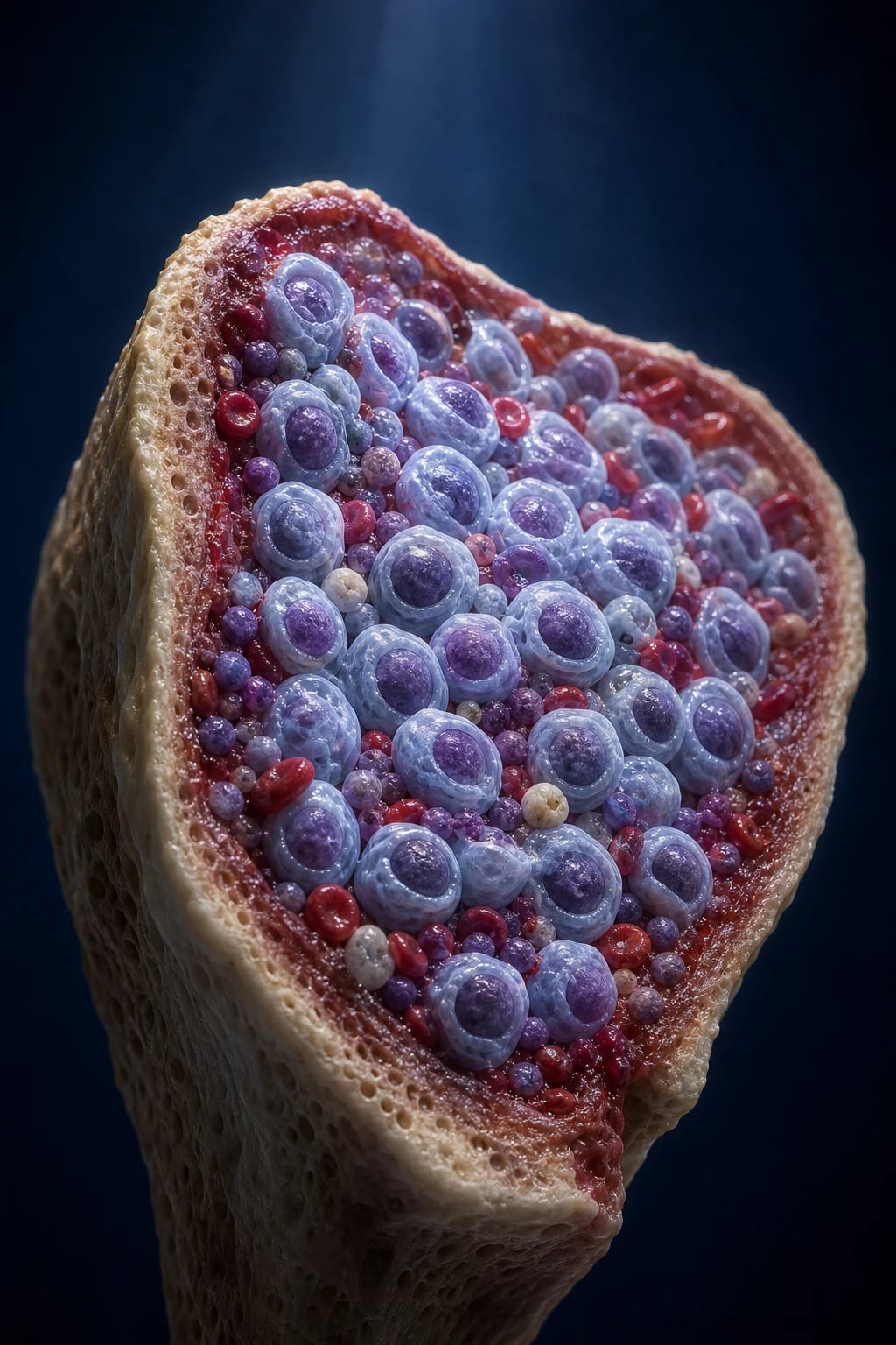
Acute leukaemia is a clonal haematological malignancy arising from a single haematopoietic progenitor cell that has acquired mutations causing two cardinal defects — a block in differentiation (so cells accumulate as immature blasts rather than maturing into functional blood cells) and uncontrolled proliferation (so the clone expands exponentially). The expanding blast mass effaces the bone marrow and spills into the peripheral blood, producing the clinical syndrome of marrow failure.[1]
The diagnostic cornerstone is the blast percentage in the bone marrow: acute leukaemia is defined by marrow (or blood) blasts of at least 20 percent of nucleated cells. This threshold separates acute leukaemia from myelodysplastic syndrome (MDS) (blasts below 20 percent, with dysplasia) and from chronic leukaemia (which is dominated by mature-appearing cells, not blasts). Two defining-genetic exceptions override the 20 percent rule: t(15;17) PML-RARA (APL) is acute leukaemia at any blast count, and other recurrent AML-defining genetic abnormalities (e.g. t(8;21), inv(16), PML-RARA) qualify as AML even with 10 to 19 percent blasts under the WHO 2022 / International Consensus Classification.[1]
Acute leukaemia divides by lineage of the blast into acute myeloid leukaemia (AML) — myeloid precursors, the commonest acute leukaemia of adults — and acute lymphoblastic leukaemia (ALL) — lymphoid precursors, the commonest childhood cancer. A third axis is biological: de novo (no preceding disorder), secondary (arising from antecedent MDS, MDS/MPN or other myeloid neoplasm), and therapy-related (after prior cytotoxic chemo/radiotherapy).[1]
Classification
Acute leukaemia is classified by lineage (myeloid vs lymphoid), by morphology (the FAB system), and — for AML — by defining genetic abnormalities (the WHO 2022 / International Consensus Classification). The modern standard is genetic, because cytogenetic/molecular subtype drives prognosis and treatment far more powerfully than morphology.[1]
FAB classification of AML (M0 to M7)
The French-American-Britist (FAB) morphological classification divides AML by blast morphology and lineage into subtypes M0 to M7. The exam-critical fact is that M3 (acute promyelocytic leukaemia, APL) is a standalone clinical emergency.[1]
FAB subtype
- M0 — AML minimally differentiated
- M1 — AML without maturation
- M2 — AML with maturation (t(8;21) common)
- M3 — acute promyelocytic leukaemia (APL); t(15;17); EMERGENCY
- M4 — myelomonocytic (some inv(16) = M4Eo)
- M5 — monocytic (M5a/M5b); gum hypertrophy, extramedullary
- M6 — erythroleukaemia
- M7 — megakaryoblastic (Down syndrome in children)
Key morphology / clinical hook
- Blasts, no granules
- Blasts, few granules
- Granules + Auer rods
- Promyelocytes with BUNDLES of Auer rods = faggot cells; DIC
- Mixed granulocytic + monocytic; abnormal eosinophils
- Gum hypertrophy, skin (chloroma), high WBC, CNS
- Giant multinucleated erythroid precursors
- Megakaryoblasts; Down-syndrome transient abnormal myelopoiesis (TAM)
WHO 2022 / International Consensus Classification of AML
The WHO 2022 and the ICC reframe AML around genetic drivers rather than morphology. Two-thirds of the value for the exam is to recognise that defining genetic abnormalities outrank the 20 percent rule and that the ELN 2022 genetic risk groups (favourable, intermediate, adverse) drive the treatment decision (consolidate with chemotherapy vs proceed to allogeneic stem-cell transplant).[1]
AML with defining genetic abnormalities
- t(15;17)(q24.1;q21.2) PML::RARA — APL (any blast %)
- t(8;21)(q22;q22.1) RUNX1::RUNX1T1 — core-binding factor, favourable
- inv(16)(p13.1q22) or t(16;16) CBFB::MYH11 — CBF, favourable
- BCR::ABL1 (rare de-novo; more a CML blast crisis)
- NPM1-mutated (without FLT3-ITD) — favourable (used to be)
- CEBPA bZIP-mutated — favourable
- KMT2A (MLL) rearrangements
- MECOM rearranged; monosomy 7, 5q deletion; complex karyotype — adverse
Other categories
- AML, myelodysplasia-related (MDS-like biology, adverse)
- Therapy-related myeloid neoplasm (post chemo/radiotherapy)
- AML not otherwise specified (NOS) — falls back on the 20 percent rule
- Myeloid sarcoma (chloroma) — extramedullary myeloid tumour
Classification of ALL
ALL is classified by immunophenotype (B-ALL versus T-ALL, with sub-stages of maturation) and by cytogenetics/molecular lesions, of which the most important is the Philadelphia chromosome t(9;22)(q34;q11.2) BCR-ABL1, present in about 3 percent of children but around 25 percent of adults (rising with age).[1]
B-ALL (about 85 percent)
- Pro-B, common (CD10+), pre-B, mature-B (Burkitt-like)
- CD19+, CD79a+, CD10+, TdT+
- t(12;21) ETV6-RUNX1 — commonest in children, excellent prognosis
- Hyperdiploidy (good); hypodiploidy (poor)
- KMT2A (11q23) rearrangement (infants, poor)
- BCR-ABL1 (Ph+) — poor without TKI, transformed by imatinib/dasatinib
- BCR-ABL1-like (Ph-like) — poor, mimics Ph+ genetics
T-ALL (about 15 percent)
- Mediastinal mass, older male adolescents/young adults
- Cytoplasmic CD3+, CD7+, CD1a/CD4/CD8 variable, TdT+
- Often presents with high WBC, CNS disease, pleural effusion
- NOTCH1 pathway mutations; cortical subtype better prognosis
Epidemiology & Risk Factors
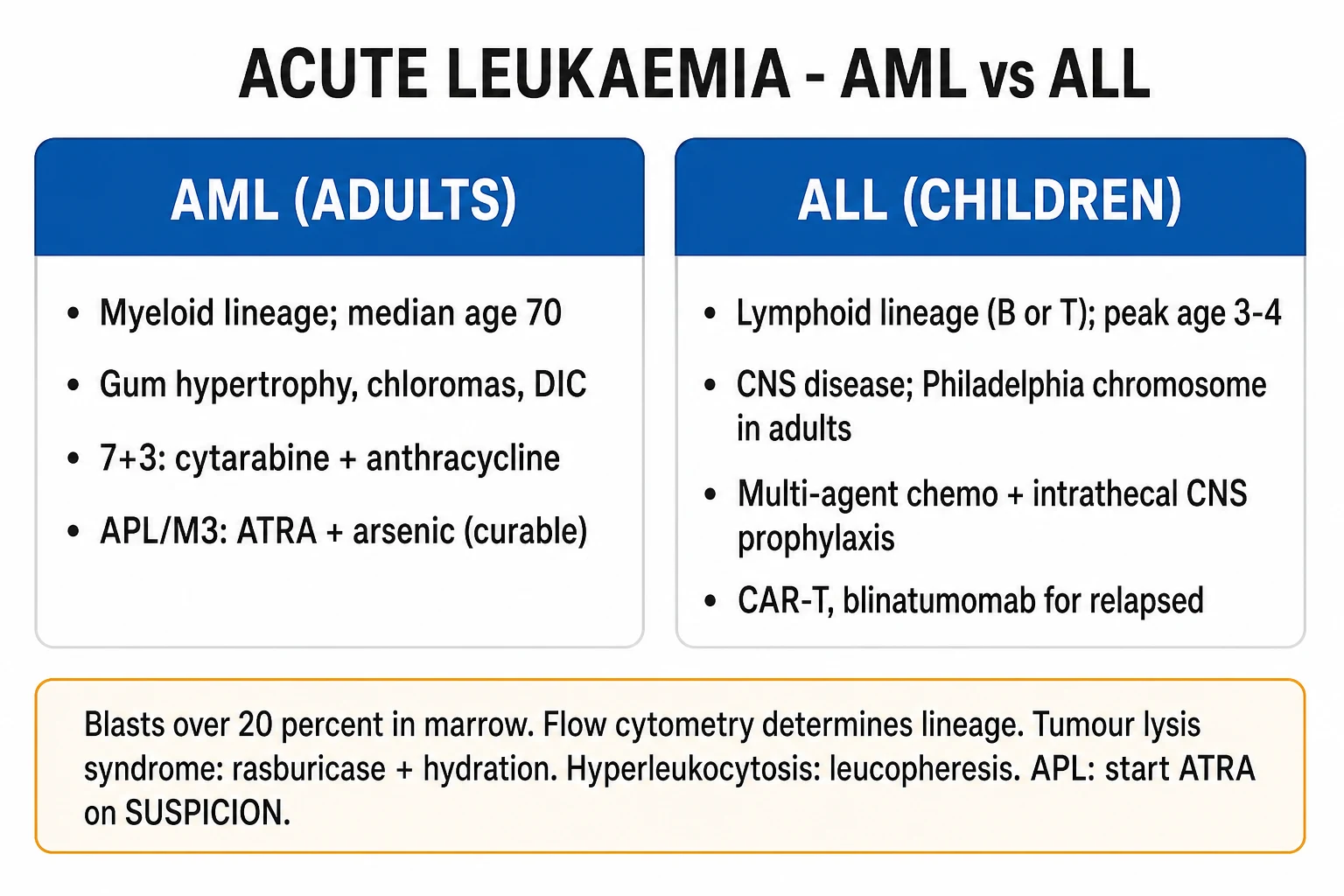
Acute leukaemia is the commonest cancer of childhood, and AML is the commonest acute leukaemia of adults. The two diseases have mirror-image age distributions:[1][1]
ALL
- Commonest childhood cancer; peak age 2 to 5 years
- About 80 percent of childhood acute leukaemia
- In adults ALL is uncommon and outcomes far worse
- Philadelphia chromosome t(9;22) rises from about 3 percent in children to about 25 percent in adults (over 50 percent over age 60)
AML
- Median age about 68 to 70 years; incidence rises steeply with age
- Commonest acute leukaemia of adults
- About 15 to 20 percent of childhood acute leukaemia
- APL (M3) peaks in young adults (median about 40), any age
Acquired risk factors for AML: prior cytotoxic chemotherapy or radiotherapy (therapy-related AML, t-AML), antecedent myelodysplastic syndrome (MDS) or myeloproliferative neoplasm (secondary AML), benzene and petrochemical/solvent exposure, smoking, and ionising radiation. Inherited / germline risk factors: Down syndrome (10 to 20-fold risk of AML — and the unique transient abnormal myelopoiesis, TAM, of the neonate), Fanconi anaemia, Bloom syndrome, ataxia-telangiectasia, Li-Fraumeni syndrome, Kostmann/severe congenital neutropenia, and Shwachman-Diamond syndrome.[1]
Therapy-related AML (t-AML) deserves its own recognition. Two drug classes predispose, with characteristic latency and cytogenetics: alkylating agents (melphalan, cyclophosphamide, chlorambucil) and ionising radiation → myelodysplasia-like picture after 5 to 7 years, typically with monosomy 5 or 7 / complex karyotype (adverse); and topoisomerase-II inhibitors (etoposide, teniposide, doxorubicin, mitoxantrone) → shorter latency (1 to 3 years), often with balanced translocations involving KMT2A (11q23) or RUNX1 (t(8;21)). t-AML carries a poor prognosis.[1]
[1]Pathophysiology
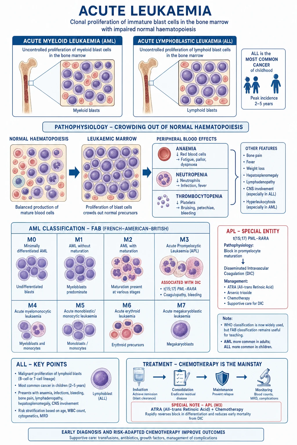
The malignant cell of acute leukaemia is the blast — an immature precursor trapped at an early differentiation stage by a differentiation block, while retaining the capacity for self-renewal and uncontrolled proliferation. Two cooperating classes of mutation drive this, formalised as the two-hit model: class I mutations activate proliferative/survival signalling (e.g. FLT3, RAS, JAK2) and class II mutations impair differentiation (e.g. PML-RARA, RUNX1-RUNX1T1, NPM1, CEBPA). A clinical acute leukaemia usually carries one of each.[1]
As blasts expand they physically displace and paralyse normal haematopoiesis, producing the triad of marrow failure: [1]
- Anaemia — loss of erythroid precursors → fatigue, pallor, dyspnoea, cardiac decompensation.
- Infection — loss of functional neutrophils (neutropenia) → bacterial, fungal and reactivation infections.
- Bleeding — loss of platelets (thrombocytopenia), compounded in APL by DIC → petechiae, gum bleeding, fundal haemorrhage, intracranial bleed. [1]
Molecular pathogenesis of APL — the curable emergency
Acute promyelocytic leukaemia (APL, FAB M3) is the archetype of mechanism-defined therapy. The t(15;17)(q24.1;q21.2) translocation fuses the PML gene on chromosome 15 with the retinoic-acid receptor alpha (RARA) gene on chromosome 17, generating the PML-RARA fusion oncoprotein. This fusion protein dominantly represses transcription of genes needed for myeloid differentiation (by recruiting the N-CoR/SMRT/histone-deacetylase co-repressor complex) so the cell is arrested at the promyelocyte stage. At pharmacological (not physiological) concentrations, all-trans retinoic acid (ATRA) binds the fusion protein, releases the co-repressor and restores differentiation — the blasts mature (over days) into terminal neutrophils and die. Arsenic trioxide (ATO) acts synergistically: it binds PML moiety, degrades the PML-RARA fusion protein directly and drives differentiation plus apoptosis. This is differentiation therapy, and it converted APL — once the most fatal AML — into the most curable acute leukaemia.[2]
The APL coagulopathy is a DIC-like state driven by procoagulant tissue factor and annexin II released from the abnormal promyelocyte granules, plus enhanced fibrinolysis (annexin II upregulates t-PA). The result is prolonged PT and aPTT, low fibrinogen, high D-dimer and thrombocytopenia — with simultaneous bleeding and thrombosis. This coagulopathy is why APL patients die of intracranial haemorrhage in the first days, and why ATRA must be started on suspicion and fibrinogen/platelets aggressively corrected.[2][1]
Mechanism of tumour lysis syndrome (TLS)
Rapid lysis of a large tumour mass (high-burden AML with high WBC, ALL — especially Burkitt-like/T-ALL, or any proliferative leukaemia at treatment onset) releases intracellular contents faster than the body can excrete them: potassium (hyperkalaemia → cardiac arrhythmia), phosphate (hyperphosphataemia, which precipitates with calcium → hypocalcaemia and nephrocalcinosis), and nucleic acids metabolised to uric acid (urate crystallises in the renal tubules → acute urate nephropathy and AKI). The AKI then worsens hyperkalaemia and hyperphosphataemia — a vicious circle. The risk is highest in the first 24 to 72 hours of cytotoxic therapy.[3]
Mechanism of leucostasis (hyperleukocytosis)
When the white-cell count is very high (classically over 100 times 10^9/L in AML, over 400 times 10^9/L in ALL — the threshold differs because myeloblasts are larger and stickier), the blasts form leukocyte aggregates / thrombi in the microvasculature, especially the cerebral (confusion, visual disturbance, stroke, intracranial haemorrhage) and pulmonary (dyspnoea, hypoxia, diffuse infiltrates) beds. Blasts also express adhesion molecules and consume endothelial nitric oxide. The risk is compounded by anaemia (less deformable RBC column) and by APL (bleeding). Cytoreduction is urgent.[1]
Recurrent AML mutations that drive biology and prognosis
The ELN 2022 risk stratification rests on the cytogenetic/molecular profile. The recurrent mutations the examiner expects you to know:[1]
Mutation
- FLT3-ITD (internal tandem duplication)
- FLT3-TKD (D835)
- NPM1 (nucleophosmin)
- CEBPA (bZIP)
- RUNX1-RUNX1T1 (t(8;21)); CBFB-MYH11 (inv 16)
- TP53, ASXL1, RUNX1, monosomy 5/7, complex karyotype
Prognostic impact
- Adverse — proliferative; treated with midostaurin; worse with high FLT3-ITD allelic ratio
- Less adverse than ITD
- Favourable if without FLT3-ITD
- Favourable (bZIP frame)
- FAVOURABLE — core-binding factor AML
- ADVERSE
Clinical Presentation
The clinical syndrome is dominated by marrow failure, modulated by tissue infiltration (extramedullary disease), leucostasis, DIC (APL) and tumour lysis. Most patients present over days to weeks of progressive symptoms.[1]
The marrow-failure triad: [1]
- Anaemia — fatigue, pallor, dyspnoea, exertional limitation, angina or cardiac failure in older patients; on examination, conjunctival/palmar pallor.
- Infection / neutropenia — fever, mucositis, pneumonia, cellulitis, perianal infection, septicaemia; opportunistic organisms (Pseudomonas, gram-negatives, Staphylococcus, Candida, Aspergillus, reactivation of herpes). Febrile neutropenia is a medical emergency (see below).
- Bleeding / thrombocytopenia — petechiae, purpura, epistaxis, gum bleeding, menorrhagia, haematuria, fundal haemorrhage, and catastrophic intracranial bleed. In APL the bleeding is amplified by DIC. [1]
Tissue infiltration / extramedullary disease: [1]
Site / sign
- Gum hypertrophy and infiltration
- Myeloid sarcoma (chloroma)
- Lymphadenopathy and hepatosplenomegaly
- Testicular enlargement (B-ALL, boys)
- CNS involvement — cranial nerve palsy (esp VII), headache, meningism
- Skin infiltrates (leukaemia cutis)
- Mediastinal mass (T-ALL)
- Renal involvement (T-ALL, B-ALL with high tumour burden)
High-yield association
- Monocytic AML (M4/M5)
- Green-coloured extramedullary myeloid tumour; can precede marrow disease; orbital/paraspinal
- More in ALL than AML; splenomegaly in CML-like picture
- Sanctuary site — must be examined in all boys with ALL
- Cranial-nerve palsies = CNS leukaemia; LP for cytology
- Blue nodules, neonates/infants with Down syndrome
- Anterior mediastinum in young men — SVC syndrome
- Massive nephromegaly can cause AKI / tumour lysis at treatment
APL presentation: mucocutaneous bleeding is dominant and often the first symptom — petechiae, gum bleeding, epistaxis, GI/GU bleeding, haematuria, menorrhagia, intracranial haemorrhage; bruising disproportionate to platelet count (because DIC). Low fibrinogen, prolonged PT/aPTT, raised D-dimer. APL patients are the ones who die before reaching the ward: any new diagnosis of acute leukaemia with bleeding should be treated as APL until proven otherwise.[2][1]
Hyperleukocytosis / leucostasis: confusion, somnolence, visual disturbance (retinal vein distension, haemorrhage), dyspnoea, hypoxia, and priapism. A WBC over 100 times 10^9/L in AML is a hyperleukocytosis emergency regardless of symptoms.[1]
Tumour lysis at presentation: oliguria, AKI, nausea/vomiting, cardiac arrhythmia or sudden death from hyperkalaemia, tetany/seizures from hypocalcaemia, lethargy. Most often emerges within hours-to-days of starting therapy in a high-burden leukaemia.[3]
Atypical presentations: elderly — fatigue, anorexia, "off legs", delirium attributed to infection; cytopenias dismissed as "old age". Pregnancy — fatigue and anaemia of pregnancy mask leukaemia; bleeding attributed to obstetric causes. Immunocompromised — overwhelming infection may be the presenting feature, blasts missed on a "septic" film. Down syndrome neonate — transient abnormal myelopoiesis (TAM), a self-limiting blast proliferation that resolves but carries a high later risk of true AML (megakaryoblastic, M7).[1][1]
Differential Diagnosis
The differential splits along two presenting patterns: (a) circulating blasts on the blood film, and (b) pancytopenia (a marrow-failure picture). The task is to distinguish acute leukaemia from its mimics using tempo, history, blood film, marrow and (definitively) immunophenotype and cytogenetics.[1]
Differential
- Acute leukaemia (AML/ALL)
- Chronic myeloid leukaemia (CML) blast crisis
- Myelodysplastic syndrome (MDS) with excess blasts
- Leukaemoid reaction (severe infection, inflammation)
- Aplastic anaemia (also pancytopenia)
- Megaloblastic anaemia (B12/folate)
- Severe infection / sepsis with marrow suppression
- Drug-induced marrow suppression, hypersplenism
Distinguishing features
- Marrow blasts at least 20 percent; Auer rods (AML); flow-cyt clonal; cytogenetics
- Pre-existing CML with BCR-ABL1; baseline leucocytosis, splenomegaly; Ph+ in blast crisis
- Blops below 20 percent, dysplasia, cytopenias, often elderly; MDS history
- Reactive, mature neutrophilia with toxic granulation, Dohle bodies, high LAP score, left shift but NO clonal blasts; resolves with treatment of infection
- Pancytopenia but MARROW IS EMPTY (hypocellular), no clonal blasts; no hepatosplenomegaly
- Macrocytosis, hypersegmented neutrophils, low B12/folate, megaloblastic marrow; reversible
- Transient, infection-driven, recovers; no clonal population
- Drug history (chemo, clozapine, carbimazole); splenomegaly; recovers on withdrawal
The hardest practical distinction is acute leukaemia versus a severe leukaemoid reaction. In overwhelming infection (especially in children) the white count can exceed 50 times 10^9/L with circulating immature forms. The decisive features pointing to leukaemia are: a clonal blast population (flow cytometry), Auer rods (pathognomonic for AML), persistent cytopenias (anaemia/thrombocytopenia — a leukaemoid reaction does not cause them), maturation arrest, no obvious focus of infection, and cytogenetic/molecular abnormality. A leucocyte alkaline phosphatase (LAP) score is high in leukaemoid reaction and low in CML.[1]
Acute leukaemia versus aplastic anaemia — both cause pancytopenia. In aplastic anaemia the marrow is hypocellular (empty) with no clonal blasts, there is no hepatosplenomegaly or lymphadenopathy, and the peripheral blood shows no blasts. In acute leukaemia the marrow is hypercellular with sheets of blasts. The distinction is made on bone-marrow aspirate and trephine biopsy.[1]
AML versus ALL on initial blood film — both show blasts, but AML blasts more often have granules, Auer rods (pathognomonic), and the count may be very high; ALL blasts are smaller, agranular (L1/L2 morphology), high LDH, and the patient is more often a child with adenopathy, mediastinal mass (T-ALL) or bone pain. Definitive separation requires flow cytometry (lineage markers).[1]
Non-malignant causes of pancytopenia to exclude: megaloblastic anaemia (B12/folate deficiency — check levels), severe infection (visceral leishmaniasis/kala-azar, miliary TB, HIV, viral hepatitis), drugs (chemotherapy, clozapine, antithyroid drugs, linezolid), hypersplenism / portal hypertension, systemic lupus erythematosus, paroxysmal nocturnal haemoglobinuria (PNH), and bone-marrow infiltration (metastatic carcinoma, miliary TB).[1]
Clinical & Bedside Assessment
A focused examination in suspected acute leukaemia looks for evidence of marrow failure, tissue infiltration, and the four emergencies (DIC/bleeding, leucostasis, tumour lysis, febrile neutropenia). Examine systematically:[1]
- General — pallor (anaemia), petechiae and purpura (especially over ankles, pressure points, buccal mucosa — thrombocytopenia/DIC), bruising disproportionate to trauma, jaundice (haemolysis, drug, infection).
- Mouth — gum hypertrophy (monocytic AML, M4/M5), mucositis (neutropenia), oral candidiasis, gingival bleeding, aphthae.
- Nodes — cervical, supraclavicular, axillary, epitrochlear, inguinal (lymphadenopathy more in ALL).
- Abdomen — hepatosplenomegaly (infiltration; marked in monocytic AML and ALL; massive in CML), tenderness (typhlitis in neutropenia).
- Skin — chloroma (myeloid sarcoma), leukaemia cutis, infection foci (cellulitis, line sites, perianal).
- Eyes — fundoscopy for retinal haemorrhage (thrombocytopenia, leucostasis — Roth spots with infection).
- Chest — pneumonia, mediastinal mass (T-ALL — stridor/SVC syndrome).
- CNS — cranial-nerve palsy (especially facial nerve) = CNS leukaemia, meningism, confusion (leucostasis, intracranial bleed, hypocalcaemia).
- Genitalia (boys) — testicular enlargement (B-ALL sanctuary site).
- Infection foci — perianal (perianal abscess in neutropenia), central line site, skin, chest, urine, meninges. [1]
Bedside observations that demand escalation (a haematological emergency):[1]
- Fever over 38.3C (single) or over 38.0C sustained over one hour in a neutropenic patient = febrile neutropenia — empirical anti-pseudomonal beta-lactam within one hour (door-to-needle).
- Bleeding with prolonged PT/aPTT and low fibrinogen (suspected APL/DIC) — start ATRA immediately, correct fibrinogen and platelets.
- Confusion, visual change or dyspnoea with WBC over 100 (leucostasis) — leucopheresis, hydroxycarbamide, urgent cytoreduction.
- Oliguria with hyperkalaemia/hyperphosphataemia (tumour lysis) — rasburicase, cardiac monitoring, renal/nephrology. [1]
Febrile neutropenia is an emergency because the neutropenic patient cannot localise or contain infection: a small focus becomes septicaemia within hours, and the mortality of untreated gram-negative bacteraemia in neutropenia approaches 50 percent within 24 to 48 hours. Neutropenia is defined as ANC under 0.5 times 10^9/L (or under 1.0 and falling); fever is over 38.3C single or over 38.0C sustained over one hour. The principle is empirical broad-spectrum antibiotics within one hour of presentation (door-to-needle), after blood cultures but before their results.[1]
Acute leukaemia — high-yield numbers
Investigations
The diagnosis rests on peripheral blood and bone marrow, integrated with flow cytometry, cytogenetics and molecular testing. The diagnostic standard is bone-marrow aspirate plus trephine biopsy.[1]
First-line investigations: [1]
- Full blood count — anaemia, thrombocytopenia, neutropenia; WBC high, normal or low (about 15 percent present with leukopenia — do not be reassured by a normal/low WBC).
- Peripheral blood film — blasts (large nucleus, high nuclear-to-cytoplasmic ratio, prominent nucleoli, scant agranular cytoplasm for lymphoblasts; granules for myeloblasts), Auer rods (needle-shaped azurophilic inclusions — pathognomonic of AML; bundles = faggot cells in APL/M3), pancytopenia. In APL the blasts are abnormal promyelocytes with heavy granulation and multiple Auer rods.
- Coagulation — PT, aPTT, fibrinogen, D-dimer; in APL expect prolonged PT/aPTT, low fibrinogen, high D-dimer (DIC).
- Biochemistry — U&E and creatinine (baseline; AKI from TLS, infiltration, sepsis), urate (high in TLS), LDH (tumour burden, prognostic in ALL), calcium and phosphate (hypocalcaemia/hyperphosphataemia in TLS), LFTs, glucose.
- Group and screen / crossmatch — transfusion support will be needed.
- Viral serology — HIV, hepatitis B and C (affects treatment choice and rituximab/TBI risk); EBV/CMV status in transplant candidates. [1]
Bone marrow: diagnostic criteria. The diagnostic standard is a bone-marrow aspirate plus trephine biopsy. Acute leukaemia requires marrow (or blood) blasts at least 20 percent (or a defining genetic lesion such as PML-RARA, RUNX1-RUNX1T1, CBFB-MYH11). The aspirate provides morphology and material for flow cytometry, cytogenetics (karyotype, FISH) and molecular testing (PCR/NGS); the trephine biopsy provides cellularity (hypercellular, packed marrow in acute leukaemia), architecture and is essential when the aspirate is a "dry tap" (fibrotic or densely packed marrow — common in APL and hairy-cell-like presentations).[1]
Flow cytometry (immunophenotyping) determines lineage and is essential. The key markers:[1]
Lineage
- Myeloid (AML)
- B-ALL
- T-ALL
- Blast / progenitor
- APL (M3)
- Megakaryoblastic (M7)
- Erythroid (M6)
Markers
- MPO+, CD13+, CD33+, CD117+, (CD34, HLA-DR)
- CD19+, CD79a+, CD10+, cCD22+, TdT+
- cytoplasmic CD3+, surface CD3+, CD7+, CD1a/CD4/CD8, TdT+
- CD34, TdT, HLA-DR (pan-blast)
- CD13/CD33+, MPO+, HLA-DR NEGATIVE (key), CD34 weak
- CD41, CD61 (platelet glycoproteins)
- Glycophorin A, haemoglobin
Cytogenetics and molecular testing — the ELN 2022 risk groups. Conventional karyotype and FISH identify the recurring translocations; molecular testing (PCR, NGS) for FLT3 (ITD and TKD), NPM1, CEBPA, RUNX1, TP53, IDH1/2, KIT refines prognosis and matches the patient to a targeted agent (midostaurin/gilteritinib for FLT3, venetoclax, IDH inhibitors). The ELN 2022 risk groups (reproduced in principle):[1]
Favourable
- t(8;21) RUNX1-RUNX1T1
- inv(16) or t(16;16) CBFB-MYH11
- NPM1-mutated without FLT3-ITD (or FLT3-ITD low)
- CEBPA bZIP-mutated
- PML-RARA (APL — cure is the rule)
Intermediate
- NPM1-mutated WITH FLT3-ITD (any)
- FLT3-ITD without NPM1 mutation (wild-type NPM1)
- KMT2A partial tandem duplication
- t(9;11) MLLT3-KMT2A
- Cytogenetic abnormalities not classed favourable/adverse
Adverse
- TP53-mutated
- Complex karyotype (3 or more abnormalities)
- Monosomal karyotype; monosomy 5 or 7, 5q-/7q-
- RUNX1-mutated, ASXL1-mutated
- inv(3) or t(3;3) GATA2/MECOM
- t(6;9) DEK-NUP214
- KMT2A rearrangements (other)
BCR-ABL1 (Philadelphia chromosome) testing in ALL is mandatory for every adult with newly diagnosed ALL (and considered in children). The t(9;22) fusion drives constitutive ABL1 tyrosine-kinase activity; its presence transformed adult ALL from the worst to a treatable prognosis once TKIs (imatinib, dasatinib) were added to chemotherapy. Detected by karyotype, FISH or RT-PCR.[1]
Laboratory criteria of tumour lysis syndrome (TLS). TLS is defined clinically (Cairo-Bishop). Laboratory TLS requires changes within 3 days before to 7 days after therapy in two or more of: uric acid over 476 micromol/L (8 mg/dL) or 25 percent rise, potassium over 6.0 mmol/L or 25 percent rise, phosphate over 1.45 mmol/L (4.5 mg/dL in adults, or age-adjusted) or 25 percent rise, calcium under 1.75 mmol/L or 25 percent fall. Clinical TLS = laboratory TLS plus AKI (rise in creatinine over 1.5 times upper limit) or cardiac arrhythmia or seizure.[3]
DIC investigations (especially APL): PT prolonged, aPTT prolonged, fibrinogen low (under 1.5 to 2.0 g/L), D-dimer high, platelets low, with schistocytes on the film. The ISTH DIC score (5 components: platelets, fibrin-related marker/D-dimer, fibrinogen, PT, underlying disorder) scores 5 or more for overt DIC.[2][1]
Lumbar puncture in ALL is performed at diagnosis in patients at risk of CNS disease (high-risk B-ALL, all T-ALL, CNS symptoms) and as part of CNS-directed intrathecal therapy throughout treatment. It looks for leukaemic blasts in the CSF (CNS disease). In AML an LP is reserved for symptoms or high-risk monocytic disease. A platelet count over 50 times 10^9/L is required before LP; LP is avoided if the patient is hyperleukocytotic/leucostatic because of intracranial-bleed risk.[1]
Baseline supportive workup before treatment: ECG and echocardiogram (anthracycline cardiotoxicity — cumulative dose), hepatitis B (surface antigen and core antibody — chemotherapy/rituximab reactivates HBV) and hepatitis C and HIV, pregnancy test in women of childbearing age, fertility counselling and sperm/oocyte cryopreservation before gonadotoxic chemo, tissue typing of siblings if allogeneic transplant is anticipated, and dental review before mucositis.[1]
Management — Resuscitation
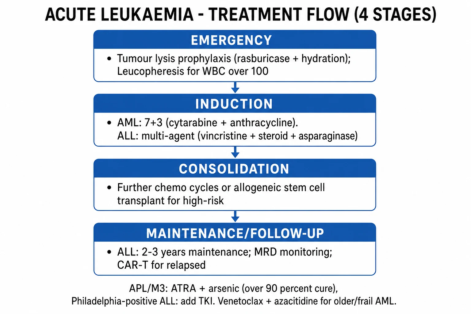
Treat the presenting haemato-oncological emergencies first and set up supportive care, then start definitive chemotherapy. The resuscitation priorities in suspected/confirmed acute leukaemia are: (1) febrile neutropenia, (2) tumour lysis syndrome, (3) leucostasis, (4) APL coagulopathy, and (5) general supportive care (transfusion, hydration, antimicrobial prophylaxis).[1]
Febrile neutropenia
Empirical broad-spectrum anti-pseudomonal beta-lactam within one hour of presentation (door-to-needle), after blood cultures (peripheral and from any central line) but before results. Standard regimens:[1]
- Piperacillin-tazobactam 4.5 g IV every 6 to 8 hours (first-line in most units), or
- Meropenem 1 g IV every 8 hours (if prior resistant organism, septic shock, or ESBL risk), or
- Ceftazidime 2 g IV every 8 hours (alternative). [1]
Vancomycin (or teicoplanin) is added for line infection, severe mucositis, suspected MRSA, haemodynamic instability, or pneumonia. G-CSF (filgrastim 5 micrograms/kg/day SC) is considered in high-risk neutropenia. If fever persists beyond 4 days reassess for fungal infection (Aspergillus — high-resolution CT thorax, galactomannan) and add antifungal cover (liposomal amphotericin B or caspofungin/micafungin). [1]
Tumour lysis syndrome — prophylaxis and treatment
Begin before the first cytotoxic dose in every high-risk patient (proliferative/high-WBC AML, ALL with high burden, Burkitt-like, LDH markedly raised, pre-existing AKI, urate high):[3]
- Aggressive intravenous hydration — isotonic saline 3 L/m2/day (or 2 to 3 L/m2/day), aim for urine output at least 100 mL/m2/hour. Alkalinisation is no longer routine (raises calcium-phosphate precipitation).
- Allopurinol 300 mg orally daily (or 10 mg/kg/day in divided doses; reduce in renal impairment) — for low-to-intermediate TLS risk; inhibits xanthine-oxidase so prevents new urate formation but does NOT clear existing urate.
- Rasburicase 0.15 to 0.2 mg/kg IV once daily for up to 5 days (or a single 0.15 to 0.2 mg/kg dose, repeat as needed) — for high tumour burden / established TLS. It breaks down existing uric acid (recombinant urate oxidase). Check G6PD first — rasburicase causes haemolysis in G6PD deficiency. Send blood on ice (urate continues to degrade in the tube → spuriously low urate). [1]
Established TLS treatment: aggressive hydration, rasburicase, treat hyperkalaemia (calcium gluconate for cardioprotection, insulin-dextrose, salbutamol, potassium binders), hypocalcaemia treated only if symptomatic (correcting asymptomatic hypocalcaemia worsens calcium-phosphate precipitation), renal support / haemodialysis for refractory hyperkalaemia, AKI, fluid overload. [1]
Leucostasis / hyperleukocytosis
Symptomatic leucostasis or a WBC over 100 times 10^9/L in AML (over 400 in ALL) demands urgent cytoreduction:[1]
- Leucopheresis (mechanical white-cell removal) — rapid reduction in blast count; immediate for symptomatic leucostasis.
- Hydroxycarbamide (hydroxyurea) — 50 to 100 mg/kg/day orally for 1 to 3 days to cytoreduce while awaiting definitive chemo.
- Aggressive hydration; avoid transfusing to a high haematocrit (worsens leucostasis) — transfuse red cells only to relieve symptomatic anaemia; platelet count over 20 times 10^9/L to reduce bleeding risk.
- Start definitive chemotherapy urgently (cytoreductive effect within 24 to 48 hours). [1]
Suspected APL — the suspicion-based emergency
Any new diagnosis of acute leukaemia with bleeding or with morphological features of M3 (abnormal promyelocytes, heavy granulation, Auer rods/faggot cells) is APL until proven otherwise. Start management before cytogenetic confirmation:[2][1]
- Start ATRA (all-trans retinoic acid, tretinoin) immediately on suspicion — 45 mg/m2/day in two divided doses orally (or 25 mg/m2/day for children). Do NOT wait for the t(15;17) result. The cost of treating a non-APL patient briefly with ATRA is negligible; the cost of delaying ATRA in true APL is death from intracranial haemorrhage.
- Aggressively correct the coagulopathy — fibrinogen with cryoprecipitate (target fibrinogen over 1.5 to 2.0 g/L) or fibrinogen concentrate; platelet transfusion to keep platelets over 30 to 50 times 10^9/L; fresh-frozen plasma for PT/aPTT correction. Recheck coagulation every 6 to 8 hours until stable.
- Avoid invasive procedures (central lines placed carefully, no intramuscular injections, avoid unnecessary arterial puncture).
- Refer urgently to a haematology centre. Once PML-RARA confirmed, add arsenic trioxide (see definitive management). [1]
Management — Definitive & Stepwise
Definitive therapy is lineage- and risk-adapted and is delivered by a specialist haemato-oncology unit. The regimens below are the high-yield core for the MBBS exam — know the acronym expansion, the agents, the key doses and the rationale.[1]
AML induction — '7+3'
Drug
- Cytarabine (cytosine arabinoside, Ara-C)
- Daunorubicin (anthracycline)
- 7 + 3
- Add for FLT3-mutated: Midostaurin
Dose / route / timing
- 100 to 200 mg/m2/day by continuous IV infusion over DAYS 1 to 7
- 60 to 90 mg/m2 IV daily DAYS 1 to 3 (7-deposit regimen)
- Refers to 7 days cytarabine + 3 days anthracycline
- 50 mg orally every 12 hours days 8 to 21 (RATIFY)
The "7+3" regimen gives cytarabine as a 7-day continuous infusion plus an anthracycline (daunorubicin or idarubicin) for 3 days. Response is assessed at day 14 (marrow cellularity/apoptosis) and day 28 (remission status). Complete remission (CR) is defined as marrow blasts below 5 percent, neutrophils over 1.0 times 10^9/L, platelets over 100 times 10^9/L, no extramedullary disease, and transfusion independence. About 60 to 80 percent of younger adults achieve CR with one or two cycles.[1]
Consolidation follows CR. Favourable-risk AML (CBF, NPM1 without FLT3-ITD) can be consolidated with high-dose cytarabine (HiDAC) — cytarabine 1.5 to 3 g/m2 every 12 hours for 3 days, for 3 to 4 cycles. Intermediate- and adverse-risk AML proceeds to allogeneic haematopoietic stem-cell transplant (allo-HSCT) in first CR if a donor is available and the patient is fit. Measurable (minimal) residual disease (MRD) positivity after induction is a strong trigger for transplant or trial therapy.[1]
Midostaurin (multi-kinase FLT3 inhibitor) — 50 mg orally every 12 hours on days 8 to 21 of induction and consolidation, then maintenance — added to 7+3 for FLT3-mutated AML after the RATIFY trial showed a 22 percent reduction in the risk of death.[4]
AML — older / unfit patient: venetoclax + azacitidine
Patients over about 75 years, or with poor performance status or significant comorbidity, are unfit for intensive 7+3. The standard is lower-intensity therapy with azacitidine plus venetoclax:[3]
- Azacitidine 75 mg/m2/day SC/IV for 7 days of each 28-day cycle (hypomethylating agent).
- Venetoclax — a BCL-2 inhibitor — given as a short ramp-up to 400 mg orally daily (days 1 to 14 or 1 to 28 of the first cycle, then continuous in subsequent cycles). [1]
The VIALE-A trial established this combination: median overall survival 14.7 months (venetoclax + azacitidine) vs 9.6 months (azacitidine + placebo), with a composite remission rate of about 66 percent versus 28 percent — transforming the outlook of unfit AML.[3]
Acute promyelocytic leukaemia — ATRA + arsenic trioxide
APL is treated by differentiation therapy, which has made it the most curable acute leukaemia (over 90 percent disease-free survival in non-high-risk disease):[2][1]
Drug
- ATRA (tretinoin, all-trans retinoic acid)
- Arsenic trioxide (ATO)
- Add for high-risk (WBC over 10): chemotherapy
Dose / route / rationale
- 45 mg/m2/day in two divided oral doses (children 25 mg/m2) — release differentiation block
- 0.15 mg/kg/day IV (or 0.30 mg/kg over 5 days then 0.25 mg/kg) — degrade PML-RARA, synergistic differentiation + apoptosis
- Idarubicin or hydroxycarbamide — cytoreduce the high-WBC patient to prevent leucostasis and differentiation syndrome
Risk stratification by presenting WBC: non-high-risk (WBC up to 10 times 10^9/L) = ATRA + ATO (chemotherapy-free); high-risk (WBC over 10 times 10^9/L) = ATRA + ATO plus chemotherapy (idarubicin, the "AIDA"-style addition) and CNS prophylaxis. The APL0406 trial (Lo-Coco) showed ATRA + arsenic was superior to ATRA + chemotherapy in non-high-risk APL, sparing patients chemotherapy toxicity.[2]
ALL — multi-agent induction with CNS prophylaxis
ALL is treated with paediatric-inspired multi-agent chemotherapy in stages — induction, consolidation, CNS-directed therapy, maintenance — over about 2 to 3 years.[1]
Induction (4 weeks) — typically: [1]
- Vincristine 1.5 mg/m2 IV weekly (capped at 2 mg total per dose) for 4 doses.
- Steroid — prednisolone 60 mg/m2/day orally for 4 weeks, or dexamethasone 10 mg/m2/day (better CNS penetration; preferred in high-risk and T-ALL).
- Asparaginase — pegylated asparaginase 2500 IU/m2 IM/IV (or native Escherichia coli asparaginase 6000 IU/m2 IM thrice weekly). Watch hypersensitivity, pancreatitis, thrombosis, hyperglycaemia, low fibrinogen.
- Anthracycline — daunorubicin 25 to 60 mg/m2 (added in higher-risk / adult regimens; not in standard paediatric low-risk B-ALL). [1]
CNS-directed intrathecal therapy is non-negotiable throughout ALL treatment: intrathecal methotrexate (age-adjusted: 6 mg under 1 year, 8 mg age 1 to 2, 10 mg age 2 to 3, 12 mg over 3 years/adults), with cytarabine and hydrocortisone ("triple intrathecal") in some regimens. Cranial irradiation is now reserved for high-risk CNS disease. The CNS is a sanctuary site that systemic chemotherapy does not reach.[1]
Consolidation / intensification uses cycles of high-dose methotrexate, cytarabine, cyclophosphamide, asparaginase and other agents. Maintenance with daily 6-mercaptopurine and weekly oral methotrexate for 2 to 3 years sustains remission in B-ALL (maintenance is less important in T-ALL). Allogeneic stem-cell transplant is offered for high-risk ALL (Ph+ in some protocols, MRD-positive, hypodiploid, KMT2A-rearranged, induction failure, relapse).[1]
Targeted / biological therapy in ALL
Therapy
- Tyrosine-kinase inhibitor (imatinib/dasatinib) for Ph+ ALL
- Blinatumomab (BiTE — bispecific T-cell engager)
- Inotuzumab ozogamicin (anti-CD22 ADC)
- CAR-T cell therapy (tisagenlecleucel)
Indication / rationale
- t(9;22) BCR-ABL1 — added to chemotherapy; transformed Ph+ ALL from worst- to favourable-prognosis adult ALL
- CD3 x CD19 bispecific — MRD-positive B-ALL, relapsed/refractory B-ALL; now frontline in some high-risk protocols
- CD22-targeted — relapsed/refractory B-ALL
- Anti-CD19 CAR-T — relapsed/refractory B-ALL in children/young adults (ELIANA); watch cytokine-release syndrome and neurotoxicity (ICANS)
Dasatinib (100 mg orally daily) or imatinib (600 mg orally daily) is added to chemotherapy for Ph+ ALL and has transformed the prognosis; some older Ph+ ALL patients are now managed with TKI + steroids/blinatumomab with chemotherapy-sparing intent.[1]
Blinatumomab — a bispecific T-cell engager (BiTE) antibody linking CD3 (T cell) to CD19 (B-ALL blast) — brings T cells into contact with leukaemic B cells and induces their lysis; used for MRD-positive and relapsed/refractory B-ALL and increasingly frontline in high-risk disease.[1]
CAR-T cell therapy (tisagenlecleucel) — the patient's own T cells are genetically engineered to express a chimeric antigen receptor against CD19 and reinfused — produces durable remissions in relapsed/refractory B-ALL in children and young adults (ELIANA). Watch for cytokine-release syndrome (fever, hypotension — tocilizumab, steroids) and immune-effector-cell-associated neurotoxicity syndrome (ICANS).[5]
Supportive care throughout treatment
Domain
- Antimicrobial prophylaxis
- Transfusion
- Growth factors
- Mucositis / nutrition
- Fertility
- VTE / bleeding
- Psychological / social
Detail
- Pneumocystis — co-trimoxazole 960 mg orally three times weekly; antifungal (posaconazole) and antiviral (aciclovir) per protocol; G-CSF for prolonged neutropenia
- Irradiated red cells and platelets (to prevent transfusion-associated graft-versus-host disease); CMV-negative products if CMV-negative; platelet thresholds (prophylactic under 10, bleeding/febrile under 20, APL under 30 to 50)
- G-CSF (filgrastim) shortens neutropenia
- Mouthcare (chlorhexidine, nystatin), soft diet, antiemetics, PEG nutrition if severe mucositis
- Sperm/oocyte cryopreservation before chemo; GnRH analogues debated
- Avoid IM injections; VTE prophylaxis only if high-risk and not thrombocytopenic; transfuse platelets before procedures
- Structured psychological support, financial counselling, fertility and survivorship planning
Specific Subtypes & Scenarios
- Acute promyelocytic leukaemia (APL, M3) — t(15;17) PML-RARA, DIC, the ATRA + arsenic chemotherapy-sparing strategy, curable in over 90 percent of non-high-risk disease. The defining management pearl: start ATRA on suspicion, before genetic confirmation.[2][1]
- Core-binding-factor AML — t(8;21) RUNX1-RUNX1T1 and inv(16)/t(16;16) CBFB-MYH11, the favourable-risk cytogenetic group; treated with intensive chemotherapy + high-dose cytarabine consolidation, with excellent CR rates; allogeneic transplant reserved for relapse.[1]
- Philadelphia-chromosome-positive ALL (Ph+ ALL) — once the worst prognosis adult ALL; transformed by adding a TKI (dasatinib or imatinib) to chemotherapy, with some older patients now managed with TKI + steroids/blinatumomab and chemotherapy-sparing intent.[1]
- Older / frail AML — intensive 7+3 is poorly tolerated over about age 75 or with significant comorbidity; azacitidine + venetoclax (VIALE-A) is the standard lower-intensity option with meaningful remission and survival benefit.[3]
- Childhood ALL — the commonest childhood cancer; paediatric-inspired regimens cure over 85 to 90 percent of children with standard-risk B-ALL (ETV6-RUNX1, hyperdiploidy — excellent prognosis). Outcomes fall steeply with age, high-risk cytogenetics (hypodiploidy, KMT2A, Ph+), and MRD positivity.[1]
- Relapsed / refractory ALL — blinatumomab (anti-CD19 BiTE) and inotuzumab (anti-CD22) induce remission, followed by allogeneic SCT or CAR-T (tisagenlecleucel); CD19-negative relapse (antigen escape) is a key pitfall after CD19-directed therapy.[1][5]
- Therapy-related AML — after alkylating agents/radiotherapy (5 to 7 years latency, monosomy 5/7, complex karyotype, adverse) or topoisomerase-II inhibitors (1 to 3 years, KMT2A/RUNX1 rearrangements); poor prognosis; treated with intensive or venetoclax-based therapy, with early transplant consideration.[1]
Complications & Pitfalls
Treatment and disease complications are heavily examined:[1][2]
- Tumour lysis syndrome — see above; the cardinal pitfall is failing to prophylax high-burden leukaemia before the first cytotoxic dose, and using allopurinol alone (which does not clear existing urate) instead of rasburicase for established TLS.[3]
- APL-associated DIC — catastrophic bleeding (intracranial) and paradoxical thrombosis; the pitfall is delaying ATRA while waiting for cytogenetics, and under-transfusing fibrinogen/platelets.[2][1]
- Differentiation syndrome (formerly retinoic-acid syndrome) — occurs with ATRA and/or arsenic trioxide in APL, typically within the first 3 weeks of treatment: fever, dyspnoea, pulmonary infiltrates, weight gain, pleural/pericardial effusion, renal impairment, hypotension. Mechanism: maturing myeloid cells adhere to endothelium and release cytokines. Treat with dexamethasone 10 mg IV every 12 hours (and transient ATRA hold if severe). Prophylactic dexamethasone is given to high-risk (WBC over 10) APL.[2]
- Leucostasis — see above; the pitfall is not recognising confusion/visual change in a hyperleukocytotic patient as an emergency, or transfusing RBC to a high haematocrit and worsening it.[1]
- Febrile neutropenia and neutropenic sepsis — the single biggest cause of early treatment-related mortality; the pitfall is delaying antibiotics for culture results.[1]
- Anthracycline cardiotoxicity — cumulative-dose congestive heart failure (daunorubicin, doxorubicin); pre-treatment echo and lifetime cardiac surveillance.
- Asparaginase toxicity — pancreatitis, hypersensitivity, thrombosis (including sinus-vein thrombosis), hyperglycaemia, low fibrinogen/coagulopathy.
- Intrathecal-chemotherapy errors — wrong drug into the intrathecal space (vincristine is fatal intrathecally); rigid dispensing protocols prevent this. Always dispense vincristine in a mini-bag for IV only and never in a syringe.
- Pancytopenia from treatment — bleeding, infection, anaemia through 2 to 4 weeks of each cycle (nadir).
- Long-term late effects — second cancers, cardiotoxicity, infertility, endocrine failure, osteoporosis, growth impairment in children, neurocognitive effects (especially after cranial irradiation).
Classic diagnostic pitfalls: delaying ATRA in suspected APL; failing to prevent tumour lysis in a proliferative leukaemia; missing APL on the film (look for abnormal promyelocytes, faggot cells); treating Ph+ ALL without a TKI; relying on a normal/low WBC to exclude leukaemia (15 percent present with leukopenia); confusing a leukaemoid reaction for leukaemia (check LAP, look for cytopenias and clonality); forgetting intrathecal CNS prophylaxis in ALL.[1]
Prognosis & Disposition
Prognosis is determined by age, performance status, cytogenetic/molecular risk (ELN 2022), WBC count, prior myelodysplasia, and response (MRD).[1]
AML — overall 5-year survival is about 30 percent overall but stratifies sharply: younger adults with favourable-risk disease (CBF, NPM1 without FLT3-ITD) can achieve 60 to 70 percent long-term survival; adverse-risk (TP53, complex karyotype, monosomy 5/7) has 5-year survival below 10 to 20 percent even with transplant; older/unfit patients have median survival under 1 year without venetoclax. Age over 60, poor performance status, WBC over 100, secondary/therapy-related AML and antecedent MDS all worsen prognosis.[1]
APL — once uniformly fatal within weeks; with ATRA + arsenic, non-high-risk APL is cured in over 90 percent of cases, making it the most curable acute leukaemia. High-risk (WBC over 10) APL has a worse early death rate (from bleeding/leucostasis) but, with the addition of chemotherapy and modern support, long-term survival exceeds 80 percent.[2][1]
Core-binding-factor AML (t(8;21), inv(16)) — favourable prognosis, long-term survival 60 to 70 percent with intensive chemotherapy and high-dose cytarabine consolidation; KIT mutations in t(8;21) attenuate this.[1]
ALL — childhood standard-risk B-ALL has a 5-year survival over 85 to 90 percent (the success of paediatric-inspired regimens); adult ALL outcomes fall steeply with age to 30 to 40 percent long-term survival, driven by Ph positivity, high WBC, poor-risk cytogenetics and MRD positivity. Adding a TKI has lifted Ph+ adult ALL into a treatable prognosis.[1]
Measurable residual disease (MRD) — multiparameter flow or molecular (PCR for fusion transcript / NGS) detection of residual leukaemia below morphological threshold — is the strongest predictor of relapse and guides consolidation (MRD positivity pushes toward transplant or trial therapy; MRD-negative deep remission allows de-escalation in some protocols).[1]
Disposition — all patients are managed by a haemato-oncology multidisciplinary team in a specialist centre, with urgent inpatient admission at diagnosis and during each treatment cycle (for induction, consolidation, transplant, and supportive care of complications). The safety-net for the discharging clinician is patient education on febrile neutropenia (a single temperature over 38.3C during neutropenia is an emergency — attend immediately, do not take antipyretics at home), bleeding precautions, and recognition of tumour lysis symptoms.[1]
[1]Special Populations
- Pregnancy — diagnostic workup adapts (avoid CT abdomen/pelvis, prefer MRI, shielded single-view X-ray, defer PET-CT where possible). The priority is treating the maternal disease: untreated acute leukaemia is rapidly fatal to both mother and fetus. Chemotherapy can be given in the 2nd and 3rd trimesters (anthracyclines, cytarabine, vincristine relatively safe; avoid methotrexate in the 1st trimester and throughout where possible; asparaginase carries thrombosis risk). In the 1st trimester, the teratogenicity risk is highest — multidisciplinary timing (defer to 2nd trimester where possible, or deliver then treat). APL in pregnancy — ATRA is relatively safe (single case reports of teratogenicity; benefits outweigh); arsenic trioxide is avoided where possible (teratogenic in animals).[1]
- Older / frail — the venetoclax + azacitidine lower-intensity strategy (VIALE-A) has transformed the outlook of unfit AML, with meaningful remission and survival benefit and lower treatment-related mortality than intensive 7+3; best supportive care remains appropriate for the very frail.[3]
- Down syndrome — neonates may have transient abnormal myelopoiesis (TAM), a self-limiting blast proliferation that resolves spontaneously but carries a 10 to 20 percent later risk of true AML (megakaryoblastic, M7), which is itself unusually chemosensitive in Down-syndrome children (lower-dose protocols).[1]
- Paediatric — weight-based dosing, CNS prophylaxis (intrathecal, age-adjusted doses), and attention to long-term late effects (growth, endocrine, neurocognitive, second cancers, cardiotoxicity, infertility) drive paediatric-specific protocols. Survivorship care is a lifelong requirement.[1]
- Immunocompromised (post-transplant, HIV, congenital immunodeficiency) — heightened infection risk (fungal, viral reactivation, PJP) during therapy; aggressive antimicrobial prophylaxis and a lower threshold for empirical treatment.[1]
Evidence, Guidelines & Regional Differences
Guidelines: ELN 2022 (Dohner et al., Blood) is the international standard for AML diagnosis, genetic risk stratification (favourable, intermediate, adverse) and response criteria; NCCN (US) and BSH (UK) provide parallel operational guidance; WHO 2022 / International Consensus Classification unify the genetic classification of myeloid neoplasms. ICMR / national Indian haematology groups adapt these to local epidemiology and resource constraints.[1]
Landmark evidence:[1][2][3][4]
- ELN 2022 (Dohner, Blood 2022) — risk groups (favourable, intermediate, adverse), defining genetic abnormalities, response criteria including CRh, and integration of targeted therapy.[1]
- APL0406 (Lo-Coco, NEJM 2013) — ATRA + arsenic trioxide was superior to ATRA + chemotherapy in non-high-risk APL, establishing the chemotherapy-sparing standard.[2]
- VIALE-A (DiNardo, NEJM 2020) — azacitidine + venetoclax improved overall survival (14.7 vs 9.6 months) and remission over azacitidine alone in unfit AML.[3]
- RATIFY (Stone, NEJM 2017) — midostaurin added to 7+3 improved overall survival in FLT3-mutated AML (22 percent reduction in risk of death), the first targeted therapy to do so.[4]
- ELIANA (Maude, NEJM 2018) — tisagenlecleucel CAR-T produced durable remissions in relapsed/refractory paediatric B-ALL.[5]
Regional differences: flow cytometry, allogeneic transplant, venetoclax, midostaurin, blinatumomab and CAR-T are routine in high-income settings but access-limited in India and other low/middle-income countries, where cost and infrastructure drive treatment toward generic 7+3, ATRA-arsenic for APL, palliative regimens, and limited transplant capacity. APL — universal curability with cheap ATRA/arsenic — is a rare equity success story; CAR-T and blinatumomab remain accessible only in specialist centres.[1]
Controversies: the intensity of therapy in the elderly (intensive 7+3 vs venetoclax-azacitidine — increasingly the latter even for some fit patients); the role of allogeneic transplant in first CR for intermediate-risk AML (driven by MRD); the timing of CAR-T (earlier in relapse vs after salvage); maintenance therapy after transplant; and the cost and equitable access of targeted and cellular therapies.[1]
Exam Pearls
[1]AML — the cytogenetic–molecular associations
APC-F
PML-RARA; M3; DIC; ATRA + arsenic; the curable emergency
t(8;21) RUNX1-RUNX1T1 and inv(16) CBFB-MYH11 — FAVOURABLE prognosis
Favourable-risk AML molecular groups
ADVERSE — proliferative; add midostaurin (RATIFY)
TLS — the four released ions
TLSP
rapid kill of high-burden leukaemia within 24 to 72 h of starting chemo
hyperkalaemia (arrhythmia), hyperphosphataemia (hypocalcaemia, nephrocalcinosis), hyperuricaemia (urate nephropathy, AKI)
Cairo-Bishop: 2 or more electrolyte changes plus clinical end-organ damage
aggressive hydration 3 L/m2/day + rasburicase 0.15 to 0.2 mg/kg for high risk/established TLS (check G6PD); allopurinol only for low/intermediate risk
- Blast threshold — marrow or blood blasts at least 20 percent defines acute leukaemia; PML-RARA qualifies at any percentage.
- Auer rods = AML (never ALL); bundles/faggot cells = APL.
- APL — start ATRA on suspicion, before cytogenetic confirmation; correct fibrinogen and platelets; add arsenic trioxide once confirmed.
- Ph+ ALL — add a TKI; it transformed the prognosis.
- 7+3 = cytarabine (7-day infusion) + daunorubicin (3 days); add midostaurin for FLT3.
- Unfit AML — azacitidine + venetoclax (VIALE-A).
- TLS — rasburicase (not allopurinol alone) for high tumour burden/established TLS; check G6PD.
- Febrile neutropenia — anti-pseudomonal beta-lactam within one hour (door-to-needle).
- Differentiation syndrome — fever, dyspnoea, infiltrates, weight gain with ATRA/arsenic; dexamethasone.
- Childhood ALL — commonest childhood cancer; over 85 percent cure with paediatric-inspired regimens + CNS prophylaxis.
- Vincristine is FATAL if given intrathecally — dispense in a mini-bag for IV use only. [1]
Exam application bank (NEET-PG / INICET)
One-line answer
Acute leukaemia is a clonal malignancy of haematopoietic blasts arrested at an early stage of differentiation, defined by marrow blasts at least 20 percent (or a defining genetic lesion), that crowds out normal haematopoiesis producing anaemia, infection and bleeding. It divides into acute myeloid leukaemia (AML) — the commonest acute leukaemia of adults, driven by recurrent mutations (FLT3, NPM1, CEBPA) and classified by the WHO 2022 / ELN 2022 genetic risk groups — and acute lymphoblastic leukaemia (ALL) — the commonest childhood cancer, divided into B-ALL and T-ALL and transformed in adults by the Philadelphia chromosome t(9;22) BCR-ABL1. Acute promyelocytic leukaemia (APL, FAB M3) is a separate haematological emergency: t(15;17) PML-RARA, a differentiation block at the promyelocyte stage and life-threatening DIC, treated with differentiation therapy — ATRA plus arsenic trioxide — whi
Worked stems (answer without another resource)
Stem 1 — Classic presentation. Map symptoms to mechanism; name the first investigation and first treatment step with dose/route if drug therapy is standard. [1]
Stem 2 — Unstable / complicated. List red flags that force immediate resuscitation, theatre, ICU, antidote, or reperfusion — and what you do in the first 15 minutes. [1]
Stem 3 — Atypical group. Elderly, pregnancy, child, or immunocompromised: how presentation and thresholds change. [1]
Stem 4 — Differential trap. Name the three closest mimics and one discriminator for each. [1]
Stem 5 — Disposition. Who goes home with safety-netting, who is admitted, who needs HDU/ICU/theatre, and what follow-up is mandatory. [1]
Rapid viva checklist
- Definition + classification
- Pathophysiology chain
- Bedside signs / criteria
- Score with exact components (if any)
- Emergency bundle
- Definitive therapy with doses
- Complications of disease and of treatment
- Special populations
- Guideline/trial name if classic
- Three exam traps
Coverage self-check
If you cannot answer any stem above from this page alone, re-read the matching section — the page is intended to be self-sufficient for final-prof and NEET-PG/INICET questions on Acute Leukaemia (AML & ALL, including APL).
References
- [1]Dohner H, Wei AH, Appelbaum FR, et al. Source identification and toxicity apportionment of polycyclic aromatic hydrocarbons in surface soils in Beijing and Tianjin using a PMF-TEQ method PLoS One, 2022.PMID 35771809
- [2]Lo-Coco F, Avvisati G, Vignetti M, et al. Retinoic acid and arsenic trioxide for acute promyelocytic leukemia N Engl J Med, 2013.PMID 23841729
- [3]DiNardo CD, Jonas BA, Pullarkat V, et al. PCNA activates the MutLγ endonuclease to promote meiotic crossing over Nature, 2020.PMID 32814343
- [4]Stone RM, Mandrekar SJ, Sanford BL, et al. Midostaurin plus Chemotherapy for Acute Myeloid Leukemia with a FLT3 Mutation N Engl J Med, 2017.PMID 28644114
- [5]Maude SL, Laetsch TW, Buechner J, et al. Email Reminders Increase the Frequency That Pet Owners Update Their Microchip Information Animals (Basel), 2018.PMID 29385095