Haematology · General Medicine
Anaemia of Chronic Disease (Anaemia of Inflammation)
Also known as Anaemia of chronic disease · Anemia of chronic disease · Anaemia of inflammation · Anaemia of chronic inflammation · Functional iron deficiency
Anaemia of chronic disease (anaemia of inflammation) is the commonest anaemia in hospitalised patients and the second commonest worldwide after iron deficiency. It arises from chronic immune activation (infection, autoimmune disease, malignancy, chronic kidney disease) driving hepcidin-mediated iron sequestration (iron trapped in macrophages, reduced gut absorption), suppressed erythropoiesis, and a moderately shortened red-cell lifespan. Classically a normocytic, normochromic anaemia (MCV 80 to 100), occasionally mild microcytic; iron profile shows low serum iron, LOW TIBC/transferrin (vs HIGH in iron deficiency), normal or raised ferritin (an acute-phase reactant) and low transferrin saturation. Treat the underlying cause; IV iron when oral iron is ineffective (hepcidin blocks absorption); ESA in CKD and cancer.
On this page & tools
Your progress
Saved locally on this device.
Exam tags
Red flags
Overview & Definition
Anaemia of chronic disease — now more accurately called anaemia of inflammation — is the anaemia of the unwell patient: a hypoproliferative, typically normocytic anaemia that arises within weeks of any sustained inflammatory, infectious, malignant or renal insult and tracks the activity of the underlying disease. It is not a single disease but a stereotyped haematological response to chronic immune activation, and its importance at the bedside is diagnostic more than therapeutic. Recognising the iron-study pattern — low serum iron with a LOW TIBC and a normal or raised ferritin — distinguishes it from iron-deficiency anaemia (low iron, HIGH TIBC, low ferritin) at a glance, prevents a pointless course of oral iron, and points the clinician towards the hidden chronic disease that is driving it.[1][2]
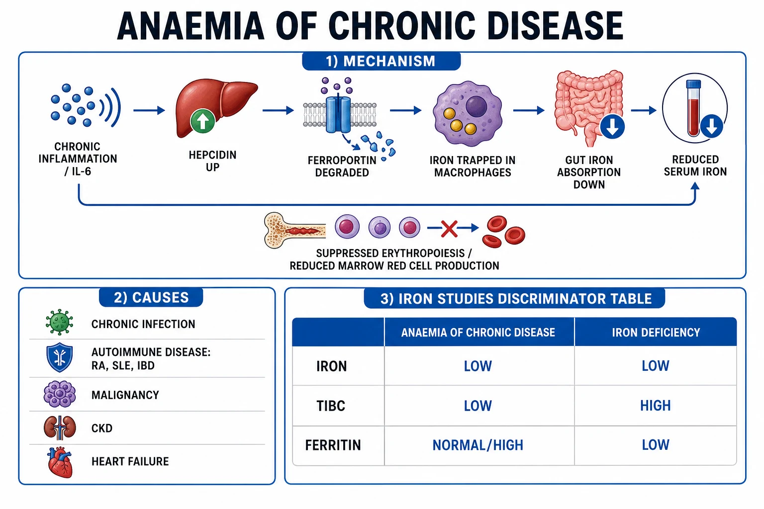
The mechanism, once mysterious, is now understood to revolve around a single peptide hormone: hepcidin. Chronic inflammation releases interleukin-6, which switches on hepatic hepcidin; hepcidin binds the iron-export channel ferroportin on macrophages and duodenal enterocytes and triggers its internalisation and degradation. The body's iron reserves are intact or increased, but iron is locked inside storage cells and cannot reach the marrow — a state termed functional iron deficiency, or iron-restricted erythropoiesis despite adequate stores. Inflammatory cytokines further suppress erythroid progenitors, blunt the erythropoietin response, and modestly shorten red-cell survival, completing a four-hit mechanism that produces a normocytic, hypoproliferative anaemia in the face of plentiful iron.[1][2][3]
Two clinical realities shape every consultation. First, the anaemia is usually mild to moderate (haemoglobin rarely below 80 g/L); a very low haemoglobin should prompt a search for an additional cause — bleeding, true deficiency, haemolysis, or marrow failure. Second, anaemia of chronic disease frequently coexists with true iron deficiency in inflammatory bowel disease, rheumatoid arthritis and chronic kidney disease, where blood loss, gut inflammation and uraemia stack on top of hepcidin-driven sequestration. Recognising the mixed picture is part of the diagnosis.[2]
Classification
Anaemia of chronic disease is classified along two axes: the morphology of the red cells and the underlying cause driving the inflammation. Both are examinable, and the morphological classification is the one that secures the diagnosis from a full blood count.[1]
By morphology, anaemia of chronic disease is normocytic normochromic in the great majority of cases (MCV 80 to 100 fL). In perhaps a quarter of long-standing cases the mean cell volume drifts down into the mild microcytic range (MCV 70 to 80 fL), but values below 70 fL are unusual and should prompt a search for coexisting iron deficiency or thalassaemia. The reticulocyte count is low — a hypoproliferative picture — and the red cells are normochromic (normal MCH) rather than the markedly hypochromic cells of established iron deficiency. The anaemia is typically mild to moderate (Hb 80 to 110 g/L); a Hb below 80 g/L is a red flag for an additional process.[1][2]

By cause, the same pathophysiology is shared across a wide range of triggers, which group into four exam-friendly categories: [1]
Infection
- Chronic bacterial: tuberculosis, osteomyelitis, abscess, infective endocarditis
- Chronic viral: HIV, chronic hepatitis B/C
- Chronic fungal disease, parasitic infection (malaria)
- Onset within weeks of infection; resolves with control of source
Autoimmune
- Rheumatoid arthritis (the classic exam example)
- Inflammatory bowel disease (Crohn, UC)
- Systemic lupus erythematosus, systemic vasculitis
- Polymyalgia rheumatica, giant cell arteritis, sarcoidosis
Malignancy
- Solid tumours and metastatic cancer (cancer cachexia)
- Haematological malignancy (lymphoma, myeloma, leukaemia)
- Cytokine-driven (IL-6, TNF-alpha from tumour)
- May coexist with marrow infiltration or blood loss
CKD, cardiac, metabolic
- Chronic kidney disease — low EPO plus inflammation (mixed)
- Chronic heart failure — cytokine and congestion drive
- Chronic obstructive pulmonary disease, obstructive sleep apnoea
- Obesity, type 2 diabetes (chronic low-grade inflammation)
- Often coexists with true iron deficiency
Anaemia of chronic disease — the numbers that decide a question
Epidemiology & Risk Factors
Anaemia of chronic disease is the second commonest anaemia worldwide after iron deficiency and the single commonest anaemia in hospitalised patients, where up to a half of medical inpatients have some element of inflammation-driven anaemia by the time they are discharged. Its prevalence tracks the prevalence of chronic inflammatory disease in a population: in rheumatoid arthritis roughly 30 to 60 percent of patients develop anaemia during their disease course; in inflammatory bowel disease 20 to 40 percent; in chronic kidney disease the proportion rises with declining GFR, exceeding 50 percent in stage 4 to 5 CKD; and in advanced heart failure the figure is similar. Cancer-related anaemia, much of which is inflammation-driven, affects a third of patients at diagnosis and the great majority during chemotherapy.[1][2]
India — the regional delta that examiners test. India carries a disproportionate burden of every major driver of anaemia of chronic disease: the world's largest tuberculosis burden (an estimated 2.6 million incident cases per year, with anaemia present in over half of new diagnoses), rheumatoid arthritis prevalence of around 0.5 to 1 percent with high rates of untreated active disease, endemic chronic infections (leprosy, filariasis, malaria, visceral leishmaniasis, recurrent urinary and biliary sepsis), and a rapidly rising burden of CKD and diabetes. Anaemia of chronic disease is often overlooked against the background of nutritional iron deficiency, which affects over half of Indian women and children; the ICMR and the National Anaemia Action Plan emphasise that ferritin should always be read alongside an inflammatory marker (CRP) in the Indian setting, because a ferritin in the 50 to 200 range that would suggest iron deficiency in a healthy person is fully consistent with coexisting inflammation.
The risk factors are simply the underlying causes: any chronic infection, autoimmune disease, malignancy, CKD, chronic heart failure, or chronic inflammatory state. Additional contributors include advanced age, frailty, obesity, smoking, and chronic critical illness, all of which sustain a low-grade inflammatory cytokine milieu. A specific iatrogenic contributor is prolonged intensive-care stay, where the cytokine storm of critical illness, frequent blood sampling, and blunted erythropoietin response produce a characteristic "anaemia of the ICU" that is mechanistically an anaemia of inflammation.[2]
Pathophysiology
The whole of anaemia of chronic disease follows from a single insight: iron is abundant in the body but unavailable to the marrow. Four overlapping mechanisms produce this state, and a candidate who can recite them in order has the topic. They are hepcidin-driven iron sequestration, suppression of erythropoiesis by inflammatory cytokines, blunted erythropoietin response, and a moderately shortened red-cell lifespan.[1][2]
Mechanism one — hepcidin and ferroportin. This is the central, rate-limiting mechanism. Chronic inflammation activates macrophages and T-cells, which release interleukin-6 (IL-6) — the dominant hepcidin-stimulating cytokine — alongside IL-1-beta and lipopolysaccharide. IL-6 binds the hepatocyte IL-6 receptor and signals through the JAK2 / STAT3 pathway to switch on the HAMP gene, the gene encoding hepcidin. Hepcidin, a 25-amino-acid peptide hormone, is exported to the bloodstream and binds ferroportin, the only known cellular iron exporter, which is expressed on the surface of duodenal enterocytes, splenic and hepatic macrophages, hepatocytes, and placental cells. Binding of hepcidin to ferroportin triggers ferroportin internalisation and lysosomal degradation. With ferroportin gone, iron exported from gut absorption and recycled from senescent red cells cannot leave these cells: it accumulates inside macrophages and enterocytes, plasma iron falls, and the marrow is starved of its substrate despite plentiful body stores. This is functional iron deficiency.[2][3]
Mechanism two — direct suppression of erythropoiesis. Inflammatory cytokines act directly on the bone marrow. TNF-alpha and interferon-gamma inhibit the proliferation of early erythroid progenitors (burst-forming unit-erythroid, BFU-E) and reduce their survival, while IL-1 suppresses colony-forming unit-erythroid (CFU-E) growth. The result is fewer red-cell precursors reaching maturity, which is why the reticulocyte count is inappropriately low for the degree of anaemia — a hypoproliferative marrow that cannot compensate for even the mild red-cell shortfall.[1]
Mechanism three — blunted erythropoietin (EPO) response. A healthy kidney responds to anaemia by secreting more EPO, which stimulates the marrow to make red cells. In inflammation this response is blunted: the kidney produces less EPO than expected for the degree of anaemia (a "relative EPO deficiency"), partly because inflammatory cytokines and reactive oxygen species suppress EPO gene transcription in renal peritubular fibroblasts. In chronic kidney disease this is compounded by absolute loss of EPO-producing cells as nephrons die, which is why CKD-related anaemia sits at the intersection of low EPO and anaemia of inflammation.[1][2]
Mechanism four — shortened red-cell lifespan. Red-cell survival is moderately shortened (from the normal 110 to 120 days down to around 80 to 90 days). Activated splenic macrophages phagocytose slightly damaged red cells more aggressively, and the structural changes induced by inflammatory cytokines on the red-cell membrane accelerate removal. The marrow would normally compensate for this shorter lifespan by increasing output, but with erythropoiesis already suppressed it cannot, and the balance tips into anaemia.[1][2]
The four-hit mechanism of anaemia of chronic disease
HITS
IL-6 raises hepatic hepcidin via JAK-STAT3
Ferroportin internalised; iron locked in macrophages and enterocytes
TNF-alpha, IFN-gamma, IL-1 suppress erythroid progenitors
Activated macrophages remove cells early (80 to 90 days)
Two further details close the molecular story. First, hepcidin also explains the high ferritin of inflammation: ferritin is both a storage protein and an acute-phase reactant whose synthesis is upregulated by IL-6 and IL-1 independently of iron loading, so serum ferritin rises in inflammation even when body iron is normal or low. Second, the transferrin level (and therefore TIBC) falls in inflammation because transferrin is a negative acute-phase reactant: its hepatic synthesis is down-regulated by cytokines, which is the opposite of iron deficiency where transferrin rises. These two acute-phase behaviours — high ferritin, low transferrin — are the biochemical signature of the disease and the entire basis of its differentiation from iron deficiency.[1][2]
Clinical Presentation
The presentation of anaemia of chronic disease is dominated by the underlying disease, not the anaemia. The anaemia itself is usually mild to moderate and develops insidiously over weeks, so the patient attributes the early symptoms to the chronic illness and rarely presents with anaemia alone. The clinician's job is to recognise the haematological component of a patient who is already known to have — or is being investigated for — a chronic inflammatory, infectious, malignant or renal condition.[1]
The anaemic symptoms, when present, are the standard cluster of fatigue, exertional dyspnoea, reduced exercise tolerance, pallor and lightheadedness. Because the onset is gradual and the haemoglobin is usually only modestly reduced (80 to 110 g/L), symptoms are often mild; patients with heart failure or CKD may notice a clear worsening of their exercise tolerance as the haemoglobin falls. Angina, syncope, or resting dyspnoea are red flags that the anaemia is severe enough — or the underlying cardiovascular reserve compromised enough — to warrant specific treatment or transfusion.[2]
The features of the underlying disease are the more prominent findings. In rheumatoid arthritis the patient has symmetrical small-joint swelling, morning stiffness and deformity; in inflammatory bowel disease there is diarrhoea, abdominal pain, weight loss and perianal disease; in SLE there may be malar rash, arthralgia, alopecia and renal impairment; in chronic infection there is fever, weight loss, night sweats or a chronic focus (cavity, abscess, endocarditis); in malignancy there may be weight loss, anorexia, a mass or unexplained symptoms; in CKD there is hypertension, oedema and uraemic features. The anaemia is one clue among many and rarely the presenting one.[1]
General examination in isolated anaemia of chronic disease shows pallor (conjunctival, palmar crease, mucous membranes) and the signs of the underlying disease. There is no organomegaly, no jaundice, no lymphadenopathy, no bony tenderness and no neurological deficit attributable to the anaemia itself. The presence of any of these should redirect the diagnosis: splenomegaly and jaundice suggest haemolysis or a primary haematological disorder; profound pallor with bruising or infection suggests marrow failure; a mass or visceromegaly points to malignancy or infiltration.[1]
Atypical presentations examiners probe deliberately: the elderly patient with worsening heart failure and a "normal" ferritin in whom anaemia of chronic disease coexists with iron deficiency; the ICU patient with falling haemoglobin after two weeks in whom the anaemia of critical illness has set in; the postpartum woman with active rheumatoid arthritis who fails to respond to oral iron and is labelled "non-compliant" when the real cause is hepcidin; and the patient with treated malignancy but persistent anaemia where chemotherapy-induced marrow suppression stacks on top of inflammation. In every case the anaemia disproportionate to a single cause is the clue to a mixed picture.[2]
Differential Diagnosis
Anaemia of chronic disease sits at the centre of the normocytic anaemias and, in its microcytic form, the microcytic anaemias. The differentials are best organised by mean cell volume, because the FBC result is usually the first test back and frames the rest of the work-up.[1]
Iron deficiency anaemia
- The single most important differential — distinguished by iron studies
- LOW iron, HIGH TIBC, LOW ferritin (under 30 mcg/L strongly suggests deficiency)
- Low transferrin saturation; marrow iron ABSENT on Prussian blue stain
- Caused by bleeding, menorrhagia, hookworm, coeliac; microcytic with high RDW
Thalassaemia trait
- Microcytic (MCV often under 75 fL) but with HIGH or normal red cell count
- Iron, TIBC and ferritin all NORMAL
- Raised HbA2 in beta trait (over 3.5 percent); normal electrophoresis in alpha trait
- Mentzer index (MCV/RBC) over 13; do NOT give iron
Sideroblastic anaemia
- Microcytic, hypochromic with a DIMORPHIC film
- Ringed sideroblasts on Prussian-blue marrow stain; ferritin and transferrin saturation HIGH
- Causes: hereditary (X-linked ALAS2), myelodysplasia, alcohol, isoniazid, lead, copper deficiency
- Some forms respond to pyridoxine
Aplastic / marrow failure anaemia
- Normocytic with pancytopenia (low RBC, WBC, platelets)
- Low reticulocytes; dry tap on marrow aspirate; hypocellular trephine
- Causes: idiopathic, drugs, viruses (parvovirus, hepatitis), toxins, PNH
- Differentiated by full blood count and bone marrow biopsy
Anaemia of CKD
- Normocytic; usually coexists with anaemia of inflammation
- Low EPO for the degree of anaemia; ferritin may be normal or high
- Falls with GFR below 60; near-universal in dialysis patients
- Treatment: ESA plus IV iron; see KDIGO 2026 targets
Haemolytic anaemia
- Normocytic or macrocytic with HIGH reticulocyte count
- Raised bilirubin and LDH; low haptoglobin; positive direct antiglobulin test in AIHA
- Smear shows spherocytes, schistocytes or sickle cells depending on cause
- Distinguished from ACD by the high reticulocyte and haemolysis markers
The single highest-yield discriminator in the whole topic is the direction of TIBC/transferrin: it is LOW in anaemia of chronic disease and HIGH in iron deficiency, because transferrin is a negative acute-phase reactant (cytokines suppress its hepatic synthesis) while iron deficiency upregulates it in an attempt to capture scarce iron. Combined with the ferritin (high in ACD, low in iron deficiency), this pair resolves the great majority of normocytic and microcytic anaemias without recourse to bone marrow sampling.[1][2]
Clinical & Bedside Assessment
There is no pathognomonic bedside sign for anaemia of chronic disease. The clinician looks for (1) the severity of the anaemia, (2) the underlying disease driving it, and (3) features that argue for an additional cause — bleeding, haemolysis, marrow failure, or coexisting deficiency.[1]
General inspection assesses pallor (conjunctivae, palmar creases, oral mucosa), hydration, nutritional status and cachexia (which suggest malignancy, advanced CKD or chronic infection), and the tempo of presentation (acute decline suggests bleeding or haemolysis; chronic fatigue suggests inflammation). Vital signs reveal the haemodynamic significance of the anaemia: tachycardia, postural drop, tachypnoea and a flow murmur are expected with Hb below 80 to 90 g/L; resting hypotension or confusion suggests decompensation and the need for transfusion.[2]
A systemic examination hunts for the underlying cause. The musculoskeletal system: symmetrical small-joint swelling, deformity, rheumatoid nodules and morning stiffness point to rheumatoid arthritis; proximal muscle tenderness and temporal artery tenderness suggest polymyalgia/giant cell arteritis; a hot swollen mono-articular joint suggests septic arthritis or gout. The abdomen: hepatosplenomegaly suggests lymphoma, myeloproliferative disease, chronic infection (malaria, kala-azar) or portal hypertension; renal enlargement or bruit suggests polycystic or renovascular CKD; a palpable mass suggests malignancy. The skin and mucous membranes: malar rash, oral ulcers and photosensitivity suggest SLE; purpura and bruising suggest marrow failure; pigmented palmar creases suggest Addison disease (a rare cause of anaemia). The chest: crackles, oedema and a gallop suggest heart failure; consolidation suggests chronic infection; a murmur with splinter haemorrhages and Osler nodes suggests infective endocarditis. A rectal and pelvic examination (where indicated) screens for occult gastrointestinal and pelvic malignancy.[1]
The bedside reasoning is straightforward: a normocytic anaemia in a patient with a known chronic inflammatory disease is anaemia of chronic disease until iron studies prove otherwise. A patient without an obvious chronic disease who presents with this pattern needs a work-up for occult infection, autoimmunity, malignancy and CKD — the anaemia may be the first clue to a hidden diagnosis.[2]
Investigations
The investigation pathway is staged: (1) full blood count and film, (2) iron studies and inflammatory markers, (3) a work-up for the underlying cause, and (4) specialist tests when the picture is mixed or atypical. The diagnosis is fundamentally one of pattern recognition on iron studies, supported by the clinical context.[1][2]
Full blood count. Anaemia of chronic disease is a normocytic normochromic anaemia in the majority (MCV 80 to 100 fL, MCH 27 to 32 pg), with mild microcytosis (MCV 70 to 80 fL) in perhaps a quarter of long-standing cases. The red cell count is normal or low (unlike thalassaemia, where it is high). The reticulocyte count is low (under 2 percent, or an absolute reticulocyte count under 25 to 50 thousand per microlitre) — a hypoproliferative picture indicating the marrow is not compensating. The white cell count and platelet count are usually normal; thrombocytosis may accompany active inflammation (reactive thrombocytosis), and pancytopenia should redirect the diagnosis to marrow failure or infiltration. The RDW is usually normal or only mildly raised (unlike iron deficiency, where it rises early).[1]
Blood film. The red cells are normocytic and normochromic — unremarkable. There is no specific morphological marker of ACD; the film excludes other causes (microcytosis of iron deficiency, target cells of thalassaemia, schistocytes of microangiopathy, blasts of leukaemia). Mild anisocytosis and rouleaux may be present when inflammation is severe.[1]
Iron studies — the discriminator. This is the single most testable element of the topic. The pattern is: [1]
Anaemia of chronic disease
- Serum iron LOW
- TIBC / transferrin LOW (negative acute-phase reactant)
- Transferrin saturation LOW (under 20 percent)
- Ferritin NORMAL or HIGH (acute-phase reactant)
- Soluble transferrin receptor NORMAL
- Marrow iron PRESENT but trapped (Prussian blue positive)
Iron deficiency anaemia
- Serum iron LOW
- TIBC / transferrin HIGH
- Transferrin saturation LOW (under 15 percent)
- Ferritin LOW (under 30 mcg/L strongly suggests; under 100 with inflammation)
- Soluble transferrin receptor HIGH
- Marrow iron ABSENT (Prussian blue negative)
Thalassaemia trait
- MCV under 75 fL with HIGH red cell count
- Serum iron NORMAL
- TIBC NORMAL
- Transferrin saturation NORMAL
- Ferritin NORMAL or slightly HIGH
- HbA2 over 3.5 percent (beta); normal electrophoresis (alpha)
Sideroblastic anaemia
- Dimorphic film; ringed sideroblasts on marrow
- Serum iron HIGH or normal
- TIBC LOW or normal
- Transferrin saturation HIGH
- Ferritin HIGH
- Causes: hereditary (ALAS2), myelodysplasia, alcohol, isoniazid, lead
Iron studies — the discriminating numbers
Inflammatory markers. C-reactive protein (CRP) is the most useful: it is raised (typically above 10 mg/L) in active inflammation and confirms that the iron-study pattern is being driven by inflammation rather than coexisting iron deficiency alone. Erythrocyte sedimentation rate (ESR) is also raised but lags CRP and is less specific. Plasma viscosity may also be elevated. A normal CRP in the presence of a typical iron-study pattern does not exclude ACD (chronic low-grade inflammation may not raise CRP) but should prompt consideration of mixed disease.[2]
Work-up for the underlying cause. This is directed by the clinical context but typically includes renal function (urea, creatinine, eGFR) for CKD; liver function and hepatitis serology; HIV testing; urinalysis for nephritis; autoantibodies (rheumatoid factor, anti-CCP, ANA, anti-dsDNA, ANCA) for autoimmune disease; serum protein electrophoresis and free light chains for myeloma; faecal calprotectin for IBD; and chest radiograph, tuberculosis screening (IGRA) and age-appropriate cancer screening (mammography, colonoscopy, prostate-specific antigen) where indicated. The history and examination should focus this work-up rather than requesting every test on every patient.[1]
Specialist tests for the mixed picture. When anaemia of chronic disease coexists with true iron deficiency — common in IBD, RA, CKD and heart failure — several tools separate the components. The soluble transferrin receptor (sTfR) is raised in true deficiency (it reflects marrow iron demand and is not an acute-phase reactant) and normal in pure ACD. The sTfR/log(ferritin) ratio improves discrimination. The reticulocyte haemoglobin content (CHr) and the percentage of hypochromic red cells are early markers of iron-restricted erythropoiesis. Bone marrow aspiration with Prussian blue staining remains the historical gold standard — showing increased storage iron in macrophages with a reduced number of sideroblasts in ACD — but is now rarely required because non-invasive tests suffice. Hepcidin assays (serum or urine) are promising but not yet in routine clinical use.[1][2]
Bone marrow biopsy is reserved for atypical presentations: pancytopenia, a very low or very high MCV, reticulocytopenia out of proportion, suspicion of marrow infiltration (myelofibrosis, malignancy) or failure to respond as expected. In ACD the marrow shows normocellular or mildly hypercellular erythropoiesis with increased iron stores (Prussian blue positive) — iron is present but cannot be used.[1]
Management — Resuscitation
Anaemia of chronic disease is rarely a haematological emergency. The anaemia is usually mild to moderate and develops gradually, allowing time for physiological compensation and a measured diagnostic work-up. Resuscitative intervention — red-cell transfusion — is reserved for the symptomatic, decompensating or severely anaemic patient, not for routine correction of the haemoglobin number.[1]
The trigger for transfusion is clinical, not numerical. Transfuse if the patient has active, severe symptoms attributable to anaemia — exertional angina, resting dyspnoea, syncope, confusion, or worsening heart failure — or if the haemoglobin is below 70 g/L (below 80 g/L in older patients, those with cardiovascular disease, or acutely bleeding patients). In stable, asymptomatic patients with Hb 70 to 90 g/L, transfusion is not indicated; the focus should be on diagnosing and treating the underlying cause and considering iron or ESA therapy. A single unit of leucodepleted red cells raises the haemoglobin by roughly 10 to 15 g/L in an adult without ongoing loss; reassess after each unit.[1]
The risks of unnecessary transfusion in chronic disease are real: transfusion-transmitted infection, alloimmunisation (especially relevant if future transplant or chronic transfusion is likely), transfusion-associated circulatory overload (TACO), transfusion-related acute lung injury (TRALI), and iron overload with repeated exposure. The benefit is transient (red cells last 80 to 120 days), and the underlying mechanism — hepcidin-driven iron trapping — is not addressed. The default is therefore restrictive: transfuse only what is needed to relieve symptoms or stabilise the patient, then turn to definitive management.[2]
Management — Definitive & Stepwise
The cornerstone of management is to treat the underlying disease: control the infection, suppress the autoimmune inflammation, treat the malignancy, manage the CKD, optimise the heart failure. As inflammation settles, hepcidin falls, iron is released from macrophages, and the haemoglobin recovers — usually over 4 to 8 weeks. Specific haematological therapy is added only when the anaemia is symptomatic, persistent, or the underlying cause cannot be fully controlled.[1][2]
Step one — treat the underlying cause. This is always the first intervention and is often the only one needed. Antitubercular therapy, antiretroviral therapy, antibiotics for chronic infection, DMARDs and biologics for rheumatoid arthritis, corticosteroids and immunosuppressants for SLE and vasculitis, anti-TNF or anti-integrin biologics for IBD, cancer therapy, renin-angiotensin blockade, SGLT2 inhibitors and dialysis for CKD, and GDMT for heart failure all reduce the inflammatory cytokine load, lower hepcidin, and allow the anaemia to recover. A patient whose anaemia does not improve despite good control of the underlying disease should be re-evaluated for an additional cause.[1]
Step two — iron therapy, almost always intravenous. Oral iron is often ineffective in anaemia of chronic disease because raised hepcidin blocks its absorption (degrading ferroportin on enterocytes), and it can worsen inflammation and produce gut side effects that compound the underlying disease. Intravenous iron bypasses the hepcidin-blocked gut and repletes iron directly. It is preferred when there is coexisting iron deficiency, active inflammation, CKD, heart failure with iron deficiency, IBD, or when oral iron is intolerant or ineffective. Ferric carboxymaltose 500 to 1000 mg IV (over 15 minutes, single or repeated dose up to a calculated maximum based on body weight and Hb) is the most widely used preparation in the UK, India and Australia; alternatives include iron isomaltoside 1000 mg (monoferric), low-molecular-weight iron dextran, iron sucrose (multiple smaller doses), and ferric derisomaltose. A test dose is no longer recommended for most modern preparations, but resuscitation facilities should be available because of the small risk of hypersensitivity. The benefit in heart failure with iron deficiency (with or without anaemia) was confirmed in the FAIR-HF trial, where intravenous ferric carboxymaltose improved symptoms, functional capacity and quality of life regardless of baseline anaemia.[2][4]
Step three — erythropoiesis-stimulating agents (ESAs). ESAs are indicated for anaemia of CKD (after correction of iron deficiency) and chemotherapy-induced anaemia in cancer (where the goal is to avoid transfusion). They are not indicated for anaemia of chronic disease in the absence of CKD or cancer — trial evidence does not support their routine use in autoimmune disease, heart failure, or chronic infection. Epoetin alfa starting dose 50 to 100 IU per kg subcutaneously three times weekly (or 20 000 IU weekly), darbepoetin alfa 0.45 micrograms per kg every 1 to 2 weeks, or methoxy polyethylene glycol-epoetin beta monthly, are titrated to a target Hb of 100 to 115 g/L (below 115 in cancer). Targets above 115 g/L are avoided because they raise the risk of thrombosis, cardiovascular events and stroke — the lesson of the TREAT trial, in which darbepoetin alfa targeting Hb of 13 g/dL in diabetic CKD did not reduce cardiovascular or renal events and doubled the risk of stroke (5.0 percent vs 2.6 percent). Iron status should be repleted (TSAT under 30 percent or ferritin under 500 suggests functional deficiency) before and during ESA therapy.[2][5][6]
Step four — investigate and treat coexisting deficiency. Ferritin under 100 micrograms per litre (or under 300 with a TSAT under 20 percent in CKD or inflammation) suggests coexisting iron deficiency requiring iron repletion — usually intravenous because of hepcidin. Concurrent B12 and folate deficiency should be excluded and treated. Hypothyroidism (a contributor to anaemia in autoimmune disease) should be sought and corrected. Occult gastrointestinal bleeding (common in IBD, malignancy, NSAID use) should be excluded with endoscopy.[2]
Oral iron
- Often INEFFECTIVE — hepcidin blocks absorption
- Examples: ferrous sulfate 200 mg tid, ferrous fumarate 305 mg tid
- Side effects: nausea, constipation, abdominal pain, worsened inflammation
- Reserve for confirmed coexisting deficiency with low hepcidin
- Avoid in active IBD; IV iron preferred
Intravenous iron
- Preferred when inflammation active, deficiency coexists, or oral iron intolerant
- Ferric carboxymaltose 500 to 1000 mg IV over 15 min (FAIR-HF evidence)
- Iron isomaltoside 1000 mg; iron sucrose 100 to 200 mg over several visits
- Bypasses hepcidin; rapid repletion; benefits HF with iron deficiency
- Risk: hypersensitivity (rare); phosphochrome-needed monitoring for FCM hypophosphataemia
ESA (CKD, cancer)
- Epoetin alfa 50 to 100 IU/kg SC three times weekly
- Darbepoetin alfa 0.45 mcg/kg every 1 to 2 weeks
- Target Hb 100 to 115 g/L; do NOT exceed 115
- Replete iron first (TSAT under 30% or ferritin under 500)
- Avoid in active stroke, uncontrolled hypertension, history of malignancy cure
Transfusion
- Only for symptomatic or decompensating anaemia; Hb below 70 g/L
- Leucodepleted; one unit raises Hb by 10 to 15 g/L
- Risk: TACO, TRALI, alloimmunisation, iron overload, infection
- Benefit transient — does not treat the mechanism
- Restrictive strategy is default
Regional dosing notes. UK (NICE / BSH): ferric carboxymaltose up to 1000 mg single IV dose, repeated to calculated total; iron isomaltoside 1000 mg in a single 20-minute infusion; iron sucrose 100 to 200 mg per visit. US (FDA): similar preparation availability; ferric carboxymaltose 750 to 1000 mg; warning on hypophosphataemia. Australia: ferric carboxymaltose 1000 mg or iron polymaltose; extensive use in heart failure post-FAIR-HF. India (ICMR / ISHBT): ferric carboxymaltose 500 to 1000 mg widely available; oral ferrous sulfate, ferrous ascorbate, and carbonyl iron remain first-line for uncomplicated iron deficiency; IV iron expanding in private oncology and nephrology practice. ESA dosing is broadly similar across regions; KDIGO 2026 endorses a target Hb of 100 to 115 g/L in CKD and stresses individualisation.[6]
Specific Subtypes & Scenarios
Anaemia of CKD is the most important subtype and the commonest reason a patient with anaemia of chronic disease receives specific treatment. It blends absolute EPO deficiency (loss of EPO-producing renal fibroblasts as nephrons die) with anaemia of inflammation (uramic oxidative stress, chronic indwelling lines, dialysis-related inflammation). The KDIGO 2026 guideline recommends investigating anaemia when Hb falls below 110 g/L in adults or below 105 g/L in children, and treating with ESA plus IV iron when the diagnosis is confirmed. Iron is given first to correct functional deficiency (target TSAT up to 30 percent, ferritin up to 500), then ESA is added with a starting dose aimed at a Hb rise of 10 to 20 g/L per month to a target of 100 to 115 g/L. Hyporesponsiveness to ESA is common and prompts re-evaluation for ongoing inflammation, infection, hyperparathyroidism, aluminium toxicity, or pure red-cell aplasia.[2][6]
Anaemia of heart failure is now recognised as a specific entity. Roughly 30 to 50 percent of patients with chronic heart failure are anaemic, and the cause is usually a combination of anaemia of inflammation, iron deficiency (often absolute), haemodilution, and renal dysfunction. The key trial is FAIR-HF (2009): in 459 patients with NYHA class II to III heart failure and iron deficiency (ferritin under 100, or 100 to 300 with TSAT under 20 percent, with or without anaemia), intravenous ferric carboxymaltose improved symptoms, NYHA class, exercise capacity and quality of life compared to placebo. IV iron is now recommended for heart-failure patients with iron deficiency irrespective of anaemia in European (ESC) and UK (NICE) guidelines; oral iron is not effective in this setting.[2][4]
Anaemia of cancer and chemotherapy is a mixed picture of anaemia of inflammation, marrow suppression from chemotherapy, renal impairment, and blood loss. The treatment is ESA (epoetin or darbepoetin) when Hb is below 100 g/L, with IV iron to optimise response and a target Hb avoiding levels over 120 g/L because of thromboembolic risk. ESA is avoided in patients with active malignancy being treated with curative intent, particularly head-and-neck and breast cancer, where trial data suggested reduced survival. The TREAT trial in diabetic CKD also flagged the stroke risk of high Hb targets.[2][5]
Anaemia of autoimmune disease. Rheumatoid arthritis is the classic exam example: 30 to 60 percent of patients with active RA are anaemic, the pattern is the textbook low-iron-low-TIBC-high-ferritin, and the anaemia improves as disease activity is controlled with DMARDs or biologics. In SLE, anaemia of chronic disease coexists with autoimmune haemolysis, iron deficiency from NSAID-induced GI bleeding, and uraemic anaemia from lupus nephritis. In IBD, anaemia is common (20 to 40 percent) and is typically mixed iron deficiency plus anaemia of inflammation; oral iron worsens gut inflammation, so IV iron is preferred.[1][2]
Anaemia of chronic infection. Tuberculosis, osteomyelitis, infective endocarditis, HIV, chronic hepatitis and abscess all produce anaemia of chronic disease through the same hepcidin mechanism. Treatment of the infection reverses the anaemia. HIV adds direct marrow suppression (B19 parvovirus, drug effects) and the anaemia of chronic disease is one component of a multifactorial picture. Malaria, leishmaniasis and helminthic infestations (hookworm causing bleeding on top of inflammation) are common Indian contributors.[1]
Anaemia of critical illness (ICU). Patients in ICU for over a week develop a characteristic anaemia driven by sustained inflammation, frequent blood sampling, blunted EPO response, and reduced red-cell lifespan — essentially anaemia of inflammation in its purest form. Treatment is restrictive transfusion (Hb below 70 g/L) and minimisation of phlebotomy; ESAs and iron are not routinely used.[2]
Complications & Pitfalls
The disease-related complications of anaemia of chronic disease are the complications of chronic anaemia in the setting of underlying disease: fatigue and reduced quality of life, exacerbation of underlying disease (worsening heart failure, increased angina, reduced renal perfusion), and impaired response to therapy (cancer patients with anaemia tolerate chemotherapy less well). Severe untreated anaemia in cardiovascular disease carries an increased risk of decompensation, hospitalisation and death.[2]
The iatrogenic complications arise from mismanagement. The classic error is misdiagnosis of ACD as iron deficiency — both have low serum iron — leading to a pointless course of oral iron that fails to work, exposes the patient to gut side effects, may worsen inflammation, and delays diagnosis of the underlying disease. The single test that prevents this error is the TIBC (low in ACD, high in IDA) and the ferritin (high or normal in ACD, low in IDA).[1]
The opposite error is over-treatment with ESA. ESAs targeted to a high Hb (above 115 g/L) increase the risk of thrombosis, stroke, cardiovascular events, and (in cancer) reduced survival — the lesson of TREAT, CHOIR, CREATE and the oncology ESA trials. ESAs are not indicated in pure anaemia of chronic disease without CKD or cancer.[2][5]
Pitfalls to avoid include: (1) interpreting a "normal" ferritin as excluding iron deficiency in an inflamed patient (it does not — ferritin is an acute-phase reactant); (2) treating the number (Hb) rather than the patient (asymptomatic mild anaemia usually needs no treatment); (3) using oral iron where hepcidin blocks its absorption; (4) missing coexisting iron deficiency, B12 deficiency, hypothyroidism, or occult blood loss in a patient with a "typical" ACD picture; (5) failing to investigate the underlying cause, missing an occult malignancy, infection or autoimmune disease; (6) inappropriate transfusion in a stable patient; (7) failing to reassess response and look for additional causes if the anaemia does not improve with control of inflammation.[1][2]
Exam application bank (NEET-PG / INICET)
One-line answer
Anaemia of chronic disease (anaemia of inflammation) is the commonest anaemia in hospitalised patients and the second commonest worldwide after iron deficiency. It arises from chronic immune activation (infection, autoimmune disease, malignancy, chronic kidney disease) driving hepcidin-mediated iron sequestration (iron trapped in macrophages, reduced gut absorption), suppressed erythropoiesis, and a moderately shortened red-cell lifespan. Classically a normocytic, normochromic anaemia (MCV 80 to 100), occasionally mild microcytic; iron profile shows low serum iron, LOW TIBC/transferrin (vs HIGH in iron deficiency), normal or raised ferritin (an acute-phase reactant) and low transferrin saturation. Treat the underlying cause; IV iron when oral iron is ineffective (hepcidin blocks absorption); ESA in CKD and cancer.
Worked stems (answer without another resource)
Stem 1 — Classic presentation. Map symptoms to mechanism; name the first investigation and first treatment step with dose/route if drug therapy is standard. [1]
Stem 2 — Unstable / complicated. List red flags that force immediate resuscitation, theatre, ICU, antidote, or reperfusion — and what you do in the first 15 minutes. [1]
Stem 3 — Atypical group. Elderly, pregnancy, child, or immunocompromised: how presentation and thresholds change. [1]
Stem 4 — Differential trap. Name the three closest mimics and one discriminator for each. [1]
Stem 5 — Disposition. Who goes home with safety-netting, who is admitted, who needs HDU/ICU/theatre, and what follow-up is mandatory. [1]
Rapid viva checklist
- Definition + classification
- Pathophysiology chain
- Bedside signs / criteria
- Score with exact components (if any)
- Emergency bundle
- Definitive therapy with doses
- Complications of disease and of treatment
- Special populations
- Guideline/trial name if classic
- Three exam traps
Coverage self-check
If you cannot answer any stem above from this page alone, re-read the matching section — the page is intended to be self-sufficient for final-prof and NEET-PG/INICET questions on Anaemia of Chronic Disease (Anaemia of Inflammation).
Prognosis & Disposition
The prognosis of anaemia of chronic disease is the prognosis of the underlying disease. The anaemia itself is usually mild, reversible with control of inflammation, and rarely the direct cause of death — but it is a powerful independent prognostic marker across nearly every chronic disease in which it appears. In heart failure, anaemia (and iron deficiency) is associated with roughly a two-fold increase in mortality independent of ejection fraction. In CKD, anaemia accelerates left ventricular hypertrophy, decompensation and progression to dialysis. In cancer, anaemia predicts reduced survival and poorer tolerance of chemotherapy. In rheumatoid arthritis, anaemia tracks disease activity and responds to DMARD control. In chronic infection, anaemia resolves with effective treatment.[1][2]
The disposition depends on the severity of the anaemia and the underlying disease. Outpatient management is appropriate for the typical patient with mild to moderate anaemia (Hb above 80 g/L), stable vital signs, and an identifiable underlying cause being managed. Inpatient care is reserved for severe anaemia (Hb below 70 to 80 g/L), symptomatic decompensation, rapid decline, diagnostic uncertainty with red flags (pancytopenia, organomegaly), or an underlying disease requiring admission. Critical care is rarely required for the anaemia itself but may be needed for the underlying disease (sepsis, heart failure, malignancy).[2]
The safety-net for discharge is clear: a patient sent home with anaemia of chronic disease must have (1) a documented plan for the underlying cause, (2) a clear reason for any iron or ESA therapy, (3) a date for repeat full blood count and iron studies (typically 4 to 8 weeks), and (4) explicit safety-net advice to return if symptoms worsen, bleeding occurs, or new symptoms develop. Patients on ESA must have blood pressure, Hb and iron status monitored every 2 to 4 weeks during titration, then monthly; on IV iron, phosphate should be checked 1 to 2 weeks after ferric carboxymaltose because of the risk of severe hypophosphataemia.[2][6]
Special Populations
Chronic kidney disease. Anaemia of CKD is the prototype "anaemia of chronic disease plus". The two mechanisms — uraemic inflammation (anaemia of inflammation) and absolute EPO deficiency — are inseparable. The KDIGO 2026 guideline recommends investigation when Hb is below 110 g/L in adults, correction of iron deficiency with IV iron (TSAT under 30 percent or ferritin under 500), then ESA titrated to a target Hb of 100 to 115 g/L with monthly monitoring. Hyporesponsiveness (failure of Hb to rise despite adequate dose) prompts a search for infection, hyperparathyroidism, aluminium toxicity, malnutrition, occult blood loss, or pure red-cell aplasia.[2][6]
Chronic heart failure. Iron deficiency (absolute and functional) is present in 30 to 50 percent of heart-failure patients and is a stronger predictor of outcome than anaemia itself. IV ferric carboxymaltose (500 to 1000 mg) is recommended by the European Society of Cardiology for symptomatic heart failure with iron deficiency (ferritin under 100, or 100 to 300 with TSAT under 20 percent) with or without anaemia, on the basis of FAIR-HF, CONFIRM-HF and AFFIRM-AHF. Oral iron is ineffective in this setting. ESAs are not routinely recommended in heart failure because of the increased thrombotic risk demonstrated in the RED-HF trial.[4]
Pregnancy. Anaemia of chronic disease is uncommon as a primary diagnosis in pregnancy but may coexist with physiological haemodilution, iron deficiency and folate deficiency. Ferritin thresholds shift (a ferritin under 30 in the third trimester suggests deficiency); CRP rises physiologically in late pregnancy. Treat the underlying disease (autoimmune flare, infection) and replete iron — IV iron in the second and third trimester if oral is ineffective; avoid ESA in pregnancy unless on dialysis. Transfusion for symptomatic severe anaemia or haemorrhage.[2]
Elderly patients. Anaemia of chronic disease is the commonest cause of a normocytic anaemia in older adults, driven by the high prevalence of CKD, heart failure, cancer, chronic infection, and inflammatory disease in this group. Interpretation is more difficult because ferritin rises with age and inflammation, masking iron deficiency; treat the underlying disease, investigate aggressively for occult blood loss and malignancy, and use IV iron when deficiency coexists. Restrictive transfusion thresholds (Hb 70 to 80 g/L) are appropriate; frail patients with cardiovascular disease may need a higher threshold.[1]
Children. Anaemia of chronic disease in children is most often secondary to chronic infection (recurrent UTI, tuberculosis, congenital infection), juvenile idiopathic arthritis, inflammatory bowel disease, or chronic kidney disease. Iron studies follow the adult pattern. Treatment is of the underlying cause; weight-based IV iron dosing (e.g. ferric carboxymaltose 15 mg/kg, maximum 1000 mg) for confirmed deficiency; weight-based ESA for CKD. The work-up for occult infection and malignancy is essential.[1]
Immunocompromised patients. In HIV, organ transplant, and chemotherapy, anaemia of chronic disease stacks on top of direct marrow suppression, drug effects, opportunistic infection (B19 parvovirus, CMV, MAC) and haemolysis. Treatment of the underlying cause, IV iron for confirmed deficiency, and ESA in the setting of cancer or HIV-related anaemia (with Zidovudine) are the mainstays.[2]
Evidence, Guidelines & Regional Differences
The modern understanding of anaemia of chronic disease rests on a single molecular discovery: the hepcidin-ferroportin axis, established by Nemeth and colleagues in 2004 in a paper that showed hepcidin binds ferroportin and triggers its internalisation and degradation, completing the homeostatic loop between iron availability and inflammation. This transformed anaemia of chronic disease from a descriptive diagnosis into a mechanistically understood disease and opened the door to hepcidin-targeted therapies.[3]
The clinical reviews that frame the topic are Weiss and Goodnough 2005 (the definitive NEJM review of ACD as iron-restricted anaemia) and Weiss, Ganz and Goodnough 2019 (the Blood update that reframes the disease as anaemia of inflammation and adds CKD, heart failure, COPD and obesity to the list of causes, with a forward look to hepcidin antagonists).[1][2]
The landmark treatment trials are: [1]
FAIR-HF (2009)
- Anker et al, NEJM; 459 patients with NYHA II-III HF and iron deficiency
- Ferric carboxymaltose vs placebo; benefit irrespective of anaemia
- Improved symptoms, NYHA class, 6-min walk, quality of life
- Established IV iron in HF with iron deficiency with or without anaemia
TREAT (2009)
- Pfeffer et al, NEJM; 4038 patients with type 2 diabetes and CKD
- Darbepoetin alfa to target Hb 13 g/dL vs placebo (rescue below 9)
- No reduction in cardiovascular or renal composite endpoints
- Doubled stroke risk (5.0% vs 2.6%) — established upper Hb target
KDIGO 2026
- Babitt et al, Kidney Int; updates the 2012 guideline
- Target Hb 100 to 115 g/L in CKD; ESA cautiously
- IV iron first to optimise functional iron deficiency
- HIF-PHIs (daprodustat, roxadustat) approved in some regions as ESA alternative
Regional guideline differences are explicit. The KDIGO 2026 guideline is the global reference for CKD anaemia; NICE (NG203) in the UK mirrors its targets; NKF-KDOQI in the United States endorses similar Hb targets and stresses individualisation; ERBP in Europe adds cautions on ESA in active malignancy; the Australian KHA-CARI guidelines accept the same targets and emphasise IV iron; and Indian guidelines (ICMR, ISHBT) align with KDIGO while emphasising the diagnostic challenge of distinguishing ACD from the very high background prevalence of nutritional iron deficiency, recommending CRP-alongside-ferritin as standard.[6]
Controversies and emerging therapies. Hepcidin antagonists (spiegelmers, antibodies, minihepcidins) and hepcidin production inhibitors are in development and may eventually redistribute endogenous iron for erythropoiesis in pure ACD, but none is yet in routine use. Hypoxia-inducible factor prolyl hydroxylase inhibitors (HIF-PHIs) such as daprodustat, roxadustat, vadadustat and molidustat are oral ESA alternatives now licensed in some regions (Japan, China, EU) for CKD anaemia; they mimic the hypoxia response, transiently raise endogenous EPO and improve iron handling, with non-inferiority to ESAs in cardiovascular outcomes in the ASCEND, PRO2TECT and INNO2VATE trials. Their role in pure anaemia of chronic disease (without CKD) is still being defined.[2][6]
Exam Pearls
The iron studies mnemonic — ferritin, TIBC, and iron studies
ITFH
in BOTH ACD and iron deficiency
in ACD (HIGH in iron deficiency) — the discriminator
in ACD (LOW in iron deficiency); acute-phase reactant
drives it all — IL-6-induced, degrades ferroportin
Causes of anaemia of chronic disease
ACID
rheumatoid arthritis, IBD, SLE, vasculitis
low EPO plus inflammation; cardiac failure too
TB, osteomyelitis, HIV, endocarditis, abscess
solid tumours, lymphoma, myeloma, leukaemia
Quick self-test
A 64-year-old woman with rheumatoid arthritis has Hb 92 g/L, MCV 85 fL, serum iron low, TIBC low, ferritin 340 micrograms per litre, CRP 42 mg/L. What is the diagnosis, and which single iron-study feature distinguishes it from iron deficiency? [1]
Diagnosis: anaemia of chronic disease (anaemia of inflammation). [1]
Discriminator: the LOW TIBC (transferrin) — it is high in iron deficiency. The normal or high ferritin (here 340, with active inflammation raising CRP) is the second clue. Treat the rheumatoid arthritis; consider IV iron only if coexisting deficiency is confirmed.[1]
References
- [1]Weiss G, Goodnough LT. Anemia of chronic disease N Engl J Med, 2005.PMID 15758012
- [2]Weiss G, Ganz T, Goodnough LT. Anemia of inflammation Blood, 2019.PMID 30401705
- [3]Nemeth E, Tuttle MS, Powelson J, Vaughn MB, Donovan A, Ward DM, Ganz T, Kaplan J. Hepcidin regulates cellular iron efflux by binding to ferroportin and inducing its internalization Science, 2004.PMID 15514116
- [4]Anker SD, Colet JC, Filippatos G, Willenheimer R, Dickstein K, Drexler H, Luscher TF, Bart B, Banasiak W, Niegowska J, Kirwan BA, Mori C, von Eisenhart Rothe B, Pocock SJ, Poole-Wilson PA, Ponikowski P; FAIR-HF Trial Investigators. Ferric carboxymaltose in patients with heart failure and iron deficiency N Engl J Med, 2009.PMID 19920054
- [5]Pfeffer MA, Burdmann EA, Chen CY, Cooper ME, de Zeeuw D, Eckardt KU, Feyzi JM, Ivanovich P, Kewalramani R, Levey AS, Lewis EF, McGill JB, McMurray JJ, Parfrey P, Parving HH, Remuzzi G, Singh AK, Solomon SD, Toto R; TREAT Investigators. A trial of darbepoetin alfa in type 2 diabetes and chronic kidney disease N Engl J Med, 2009.PMID 19880844
- [6]Babitt JL, Berns JS, Bozkurt B, Cheung Khedairy RS, Cuevas Y, Effa EE, Eisenga MF, Fishbane S, Ginzburg YZ, Haase VH, Hedayati SS, Kim S, Moura-Neto JA, Nagler EV, Rossignol P, Sahay M, Tanaka T, Yee-Moon Wang A, Wheeler DC, Robinson KA, Wilson LM, Wilson RF, Earley A, Akl EA, Tonelli M. Executive Summary of the KDIGO 2026 Clinical Practice Guideline for the Management of Anemia in Chronic Kidney Disease (CKD) Kidney Int, 2026.PMID 41485807