Haematology · General Medicine
Antiphospholipid Syndrome (APS)
Also known as Antiphospholipid syndrome · APS · Hughes syndrome · Lupus anticoagulant · Anticardiolipin · Catastrophic APS
Antiphospholipid syndrome (APS, Hughes syndrome) is an acquired autoimmune thrombophilia defined by persistent antiphospholipid antibodies (lupus anticoagulant, anticardiolipin, anti-beta-2-glycoprotein-I) causing venous and arterial thrombosis and pregnancy morbidity (recurrent early miscarriage, late fetal death, severe pre-eclampsia/HELLP, placental insufficiency). It is primary (no autoimmune disease) or secondary (most often to SLE). Diagnosis needs one clinical criterion (thrombosis or pregnancy morbidity) plus one laboratory criterion, the antibody persistent on two occasions at least 12 weeks apart (revised Sapporo 2006). The lupus anticoagulant paradox — it prolongs the APTT in vitro yet thromboses in vivo — is the signature concept. Treat thrombosis with lifelong anticoagulation (warfarin preferred; avoid DOACs in high-risk APS); pregnancy uses aspirin plus LMWH; catastrophic APS needs combined anticoagulation plus corticosteroids plus plasma exchange plus IVIG.
On this page & tools
Your progress
Saved locally on this device.
Practise this topic
Exam tags
Red flags
Overview & Definition
Antiphospholipid syndrome (APS, Hughes syndrome) is an acquired, autoimmune, prothrombotic disorder defined by the persistent presence of antiphospholipid antibodies (aPL) in association with either vascular thrombosis or pregnancy morbidity. First described by Graham Hughes in 1983 in patients with systemic lupus erythematosus (SLE) who had a circulating anticoagulant, recurrent thrombosis and recurrent miscarriage, it is now recognised as the most common acquired thrombophilia and the leading treatable cause of recurrent pregnancy loss.[2]
Two ideas define the syndrome and decide almost every exam answer. First, the antibodies are not innocent bystanders — they are pathogenic drivers of clot, binding phospholipid-binding plasma proteins (chiefly beta-2-glycoprotein-I and prothrombin) on endothelial and platelet surfaces and igniting thrombosis in any vessel, venous or arterial, large or small. Second, the same antibodies can strike the placenta, producing a characteristic trio of obstetric outcomes — three or more unexplained early miscarriages, late fetal death, and severe pre-eclampsia or placental insufficiency demanding premature delivery. APS is therefore the only thrombophilia that is simultaneously a vascular and an obstetric disease, and the only recurrent-miscarriage cause with a specific effective treatment.[1]
The clinical skill is recognising whom to test — thrombosis at a young age, in an unusual site, or recurrent miscarriage — and interpreting the lupus anticoagulant paradox: an antibody that prolongs clotting times in the laboratory but causes thrombosis in the patient. Management hinges on lifelong anticoagulation with warfarin preferred over direct oral anticoagulants (DOACs) in high-risk disease, a modified pregnancy regimen of heparin plus aspirin (warfarin is teratogenic), and aggressive combined immunomodulation for the rare but lethal catastrophic APS.[2][3]

Classification
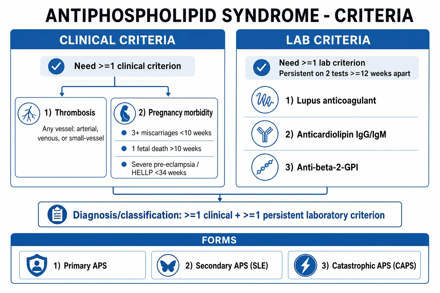
APS is classified along two axes. The first is definite versus probable APS, decided by the revised Sapporo classification criteria (Miyakis 2006), which require one clinical criterion plus one laboratory criterion, the antibody being persistent on two or more occasions at least 12 weeks apart. The second axis separates primary APS (no underlying autoimmune disease — the majority of cases) from secondary APS (most often to SLE, but also Sjögren syndrome, rheumatoid arthritis, idiopathic thrombocytopenic purpura, certain infections — HIV, hepatitis C, syphilis — and some drugs — chlorpromazine, procainamide, quinine). Antibodies that appear transiently after infection or drug exposure are not APS; persistence is the dividing line.[1]
Primary APS
- No underlying autoimmune disease; roughly **50 to 60 percent of all APS**
- Female predominance; any age, peak young adults
- Antibody profile and treatment identical to secondary
- Higher cumulative clot risk than secondary-without-SLE in some series
Secondary APS (SLE)
- **Underlying SLE in most secondary cases**; also Sjögren, RA, ITP
- Up to **30 to 40 percent of SLE patients** carry aPL; about half thrombose over time
- Add **hydroxychloroquine**; control lupus activity
- Higher risk of cardiac valve lesions (Libman-Sacks) and thrombocytopenia
Catastrophic APS (CAPS)
- **Under 1 percent of APS** but **30 to 50 percent mortality**
- Widespread **microvascular thrombosis** with multi-organ failure within a week
- **Triggered** by infection, surgery, withdrawal of anticoagulation, malignancy, obstetric complications
- Treated with **anticoagulation plus steroids plus plasma exchange plus IVIG**
Seronegative APS
- Clinical phenotype strongly suggestive of APS with **repeatedly negative** standard antibodies
- Possible antibodies to non-criteria antigens (e.g. phosphatidylserine, vimentin, PT-complex)
- Specialist referral; treated empirically on a case-by-case basis
Revised Sapporo 2006 classification criteria (reproduced verbatim)
Definite APS requires at least one clinical AND at least one laboratory criterion. There is no upper time limit between the clinical event and the positive laboratory test. Coexisting autoimmune disease is permitted. Antibodies must be persistent — present on two or more occasions at least 12 weeks apart, and no more than five years apart from the clinical event.[1]
Revised Sapporo 2006 — the numbers that matter
Clinical criteria (need one): [1]
- Vascular thrombosis — one or more clinical episodes of arterial, venous, or small-vessel thrombosis in any tissue or organ, confirmed by objective validated imaging or histology. Histopathologically, thrombosis must be present without significant inflammation in the vessel wall (this excludes vasculitis).
- Pregnancy morbidity, defined as any one of:
- (a) One or more unexplained deaths of a morphologically normal fetus at or beyond 10 weeks of gestation; OR
- (b) One or more premature births of a morphologically normal neonate before 34 weeks because of (i) eclampsia or severe pre-eclampsia (standard definitions) or (ii) recognised features of placental insufficiency (abnormal Doppler, oligohydramnios, intrauterine growth restriction, abnormal fetal heart tracing); OR
- (c) Three or more unexplained consecutive spontaneous abortions before 10 weeks, with maternal anatomic or hormonal abnormalities and parental chromosomal causes excluded.[1]
Laboratory criteria (need one): [1]
- Lupus anticoagulant (LA) present in plasma, on two or more occasions at least 12 weeks apart, detected by the International Society on Thrombosis and Haemostasis (ISTH) algorithm — a phospholipid-dependent clotting test that is prolonged, does not correct on a mixing study, and does correct on confirmation with excess phospholipid.
- Anticardiolipin (aCL) antibody of IgG and/or IgM isotype in medium or high titre — over 40 GPL units or MPL units, or above the 99th percentile — on two or more occasions at least 12 weeks apart.
- Anti-beta-2-glycoprotein-I antibody of IgG and/or IgM isotype in units above the 99th percentile, on two or more occasions at least 12 weeks apart, by standardised ELISA.[1]
The 2023 ACR/EULAR classification criteria (Barbhaiya 2023) build on Sapporo with a weighted score that captures additional aPL specificities and obstetric phenotypes; they are intended chiefly for research entry criteria but reinforce the core principle: persistent LA, high-titre aCL/anti-beta-2-GPI and a compatible clinical event define the disease.[4]
Epidemiology & Risk Factors
Antiphospholipid antibodies are common in the general population — detectable in roughly 1 to 5 percent of healthy adults and in 3 to 10 percent of blood donors — yet most of these people never develop APS. Antibody positivity alone is not the syndrome; the syndrome requires the clinical picture plus persistent antibodies. This is the single most common over-diagnosis error: labelling a patient with APS on the basis of a single low-titre antibody, often after a viral illness.[2]
- Incidence and prevalence of APS: estimated annual incidence around 1 to 2 new cases per 100,000 and prevalence around 40 to 50 per 100,000, although true figures are uncertain because testing is inconsistent. APS accounts for a meaningful share of young-adult thrombosis — it underlies roughly 10 to 15 percent of all deep vein thrombosis (DVT) and 10 to 25 percent of strokes in patients under 50.[2]
- Secondary APS and SLE: up to 30 to 40 percent of SLE patients carry antiphospholipid antibodies; roughly half of antibody-positive SLE patients thrombose over a 20-year follow-up, making SLE the single most important associated condition and the one every examiner expects you to name.
- Age and sex: APS predominates in young women (reflecting SLE epidemiology and the obstetric presentation), with a female-to-male ratio of roughly 3.5 to 1 in primary APS and higher in secondary. It nonetheless occurs in either sex and at any age; the male with idiopathic DVT should be tested.
- Thrombotic risk is antibody-dependent: lupus anticoagulant positivity carries the highest thrombotic risk of the three antibodies (odds ratios for thrombosis consistently two- to four-fold higher than aCL). Triple-positive patients (LA, aCL and anti-beta-2-GPI all positive) carry the greatest recurrence risk and are the group in whom DOACs must be avoided.[3]
- Catastrophic APS is rare — under 1 percent of APS — but has 30 to 50 percent mortality, the emergency end of the spectrum.[5]
- Other risk factors that amplify risk in antibody carriers: oestrogen-containing oral contraception and hormone replacement, pregnancy and the puerperium, surgery, immobility, smoking, malignancy, nephrotic syndrome, and active lupus. The clinician's job in an asymptomatic carrier is risk attenuation — stop oestrogens, treat hypertension, control SLE.
Pathophysiology
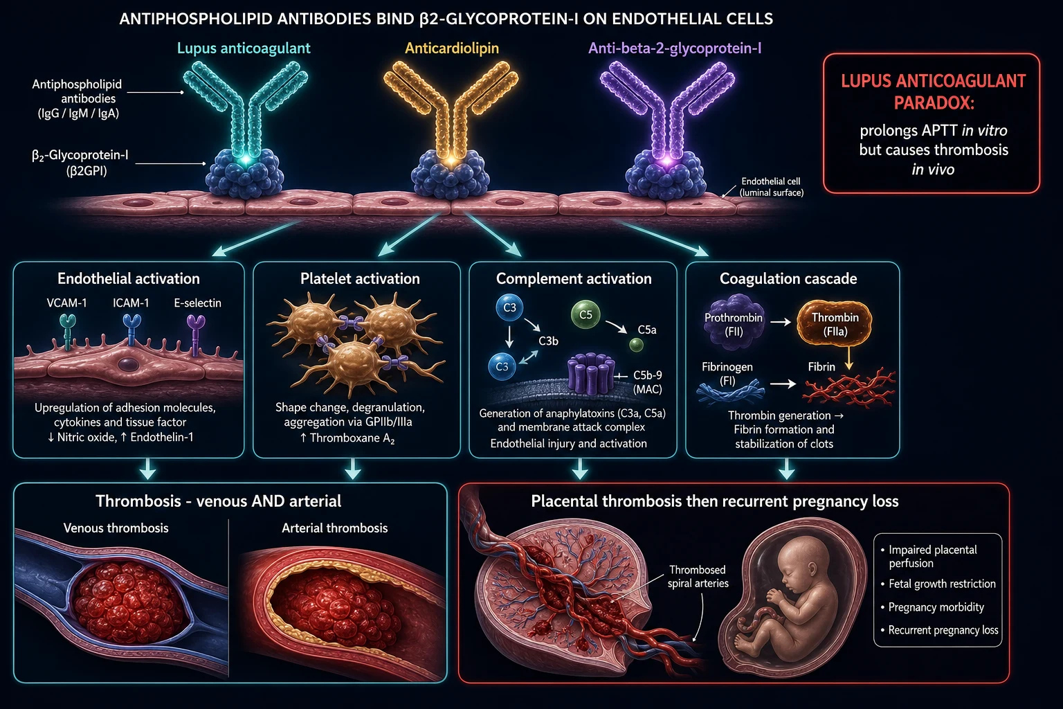
Antiphospholipid antibodies are directed not against phospholipid itself but against phospholipid-binding plasma proteins, principally beta-2-glycoprotein-I (beta-2-GPI, apolipoprotein H) and prothrombin. Beta-2-GPI is the dominant antigen: it normally has mild anticoagulant and anti-platelet effects, but when aPL binds it docked on the surface of endothelial cells, platelets, monocytes and trophoblasts, the antibody-antigen complex is recognised by cell-surface receptors ( Toll-like receptors, apolipoprotein E receptor 2, annexin A2) and activates those cells.[2]
The activated cell then drives a cascade of prothrombotic events: [1]
- Endothelial activation — upregulation of tissue factor, adhesion molecules (E-selectin, ICAM-1, VCAM-1) and von Willebrand factor, converting the vessel wall from anticoagulant to procoagulant.
- Platelet activation — cross-linking of Fc-gamma receptors and glycoprotein Ib/IX promotes platelet aggregation and thromboxane A2 release.
- Monocyte activation — tissue-factor and cytokine (interleukin-1, tumour necrosis factor) expression amplifies the coagulation cascade.
- Complement activation — particularly C5a and the membrane attack complex, which recruit neutrophils and amplify endothelial injury; complement is now recognised as central to both thrombosis and placental damage.
- Coagulation cascade amplification — interference with the protein C/S anticoagulant pathway, annexin A5 shield disruption, and direct activation of factor X and prothrombinase complexes. [1]
The net result is a systemic prothrombotic state and thrombosis in any vessel. Two consequences are especially exam-relevant. First, the lupus anticoagulant paradox: the antibody interferes with phospholipid-dependent coagulation tests in vitro — the APTT and the dilute Russell viper venom time (dRVVT) are prolonged, fail to correct on a mixing study because the inhibitor remains in the mixture, yet correct when excess phospholipid is added — but in the living patient the antibody is prothrombotic. Misreading a prolonged APTT as a bleeding tendency and withholding anticoagulation is a classic and dangerous error.[2]
Second, placental disease is thrombotic and complement-driven: aPL binds beta-2-GPI expressed on trophoblasts, triggering complement deposition, trophoblast injury, defective placentation and placental microvascular thrombosis — the mechanism that underpins recurrent early miscarriages, late fetal deaths and the severe pre-eclampsia and placental insufficiency that define obstetric APS. This is why aspirin plus heparin — both anti-thrombotic and complement-modulating — rescues pregnancy, and why warfarin (teratogenic) cannot be used.[2]
Clinical Presentation
APS presents in one of three broad ways — thrombosis, pregnancy morbidity, or (rarely) catastrophic multi-organ failure. A fourth, "asymptomatic antibody carrier," is not APS by definition but is the population in whom primary prevention matters. [1]
Venous thrombosis is the commonest thrombotic manifestation (about two-thirds of thrombotic events). Lower-limb DVT and pulmonary embolism predominate. APS is unusual, however, in that almost any venous bed can be involved: cerebral venous sinus thrombosis, portal vein thrombosis, hepatic vein thrombosis (Budd-Chiari syndrome), renal vein thrombosis, retinal vein occlusion, adrenal vein thrombosis (causing haemorrhagic adrenal infarction and Addisonian crisis). Recurrent DVT at multiple sites despite adequate anticoagulation should trigger aPL testing.[2]
Arterial thrombosis is where APS truly separates itself from the inherited thrombophilias, which are overwhelmingly venous. Stroke or transient ischaemic attack in a young patient (under 50) is the classical arterial event and a favourite exam stem — APS accounts for a substantial minority of "cryptogenic" young strokes. Other arterial events include myocardial infarction, mesenteric and renal artery thrombosis, peripheral arterial occlusion, and retinal artery occlusion. Arterial events tend to recur arterially and venous events venously.[2]
Pregnancy morbidity takes the three Sapporo forms: three or more unexplained consecutive miscarriages before 10 weeks (the classic recurrent early loss); one or more unexplained fetal deaths at or beyond 10 weeks of a morphologically normal fetus; or severe pre-eclampsia, eclampsia or placental insufficiency requiring delivery before 34 weeks.HELLP syndrome and late-onset intrauterine growth restriction are also associated. A history that combines recurrent early loss and a late stillbirth is highly suggestive. [1]
Non-criteria features (present in APS, supportive but not diagnostic) include: [1]
- Haematological — mild thrombocytopenia (around 30 percent of patients; usually 100 to 150 x 10⁹ per litre and rarely bleeding), rare autoimmune haemolytic anaemia.
- Skin — livedo reticularis and livedo racemosa (broken, branching cyanosis — livedo racemosa is the more irregular, non-uniform form more strongly associated with APS), cutaneous ulceration, splinter haemorrhages, necrotising vasculitis-like lesions.
- Cardiac — cardiac valve thickening and Libman-Sacks verrucous endocarditis (mostly aortic and mitral valves), accelerated atherosclerosis, intracardiac thrombi.
- Neurological — besides stroke, migraine, cognitive dysfunction, seizures, chorea, transverse myelitis, and the "Sneddon syndrome" triad of livedo plus stroke plus hypertension.
- Renal — APS nephropathy: vaso-occlusive lesions of the intrarenal vessels producing hypertension, proteinuria and chronic kidney disease, histologically thrombotic microangiopathy and fibrous intimal hyperplasia. [1]
Catastrophic APS (CAPS) is defined by rapid development of multiple thromboses over days, leading to multi-organ failure. The clinical picture is of microvascular thrombosis affecting kidneys, brain, lungs, skin, heart and gut simultaneously, with thrombocytopenia, schistocytes and frequently DIC overlap. Diagnostic criteria include involvement of three or more organs/systems/tissues, development simultaneously or within a week, histopathological confirmation of small-vessel occlusion, and aPL positivity. CAPS is usually triggered — by infection (about half of cases), surgery, withdrawal of anticoagulation, obstetric complications, or malignancy — and it carries a 30 to 50 percent mortality.[5]
Differential Diagnosis
The key distinctions are from inherited thrombophilia, from other acquired hypercoagulable states, and from other causes of recurrent miscarriage or of a prolonged APTT. [1]
Inherited thrombophilia
- Genetic: factor V Leiden, prothrombin G20210A, antithrombin, protein C/S deficiency
- Predominantly venous thrombosis; no arterial stroke phenotype (except homozygous or combined defects)
- No persistent antibodies; normal APTT
- Warfarin-induced skin necrosis (protein C/S); heparin resistance (antithrombin deficiency)
Other acquired thrombophilia
- Malignancy (Trousseau) — migratory thrombophlebitis; exclude with age-appropriate cancer screen
- Heparin-induced thrombocytopenia — falling platelets on heparin, 4Ts score, anti-PF4 ELISA
- Myeloproliferative neoplasm (JAK2 V617F), paroxysmal nocturnal haemoglobinuria — splanchnic (Budd-Chiari) thrombosis
- Pregnancy, oestrogen, nephrotic syndrome, sepsis — transient prothrombotic states
Other recurrent miscarriage
- Parental chromosomal abnormalities (balanced translocation — karyotype both partners)
- Uterine anomalies (septate uterus on hysteroscopy), cervical insufficiency
- Endocrine: uncontrolled diabetes, untreated hypothyroidism, antithyroid antibodies
- Crucially, APS is the only recurrent-miscarriage cause with a specific effective treatment (aspirin plus heparin)
Prolonged APTT mimics
- Factor deficiency (haemophilia A/B, von Willebrand disease) — APTT corrects on mixing study
- Specific inhibitor (acquired anti-factor VIII) — does not correct, often presents with bleeding
- Lupus anticoagulant — does not correct, corrects with excess phospholipid; thrombosis not bleeding
- Heparin contamination — prolonged thrombin time; check sample timing
When CAPS is suspected, also differentiate sepsis with disseminated intravascular coagulation, thrombotic microangiopathy (haemolytic uraemic syndrome, thrombotic thrombocytopenic purpura — ADAMTS13 under 10 percent), and HELLP syndrome — all share microvascular thrombosis and thrombocytopenia, but only CAPS has the antiphospholipid antibody background, the small-vessel occlusion on histology without vasculitis, and the combined immunomodulation-plus-anticoagulation response. [1]
Clinical & Bedside Assessment
There is no pathognomonic bedside sign of APS. Assessment is that of the thrombotic event combined with a structured hunt for clues, complications and an underlying autoimmune disease. [1]
Venous event: examine for calf swelling, tenderness, erythema, warmth; measure the calf-circumference difference (over 3 cm is significant); apply the Wells score for DVT. For suspected pulmonary embolism, look for tachycardia, tachypnoea, hypoxia, pleuritic pain, and right-heart strain; apply the Wells or revised Geneva score and obtain D-dimer and CT pulmonary angiography. [1]
Arterial event: a focal neurological deficit (face/arm weakness, aphasia, visual field loss) suggests stroke — urgent CT or MRI brain; abdominal pain and bloody stools suggest mesenteric thrombosis; flank pain and new hypertension suggest renal artery or vein thrombosis; sudden limb ischaemia is an emergency. [1]
Skin: inspect for livedo reticularis and racemosa (reticulate cyanosis, especially over the thighs, knees, arms), cutaneous ulcers, splinter haemorrhages, digital gangrene, and cutaneous necrosis. [1]
Cardiac: auscultate for new murmurs of Libman-Sacks endocarditis (mitral or aortic regurgitation from verrucous vegetations) or valve thickening; an echocardiogram confirms. [1]
Autoimmune screen: look for SLE features — malar (butterfly) rash, discoid rash, oral or nasal ulcers, photosensitivity, arthritis, serositis, alopecia, Raynaud phenomenon — which mark secondary APS and change management (add hydroxychloroquine). Check for lymphadenopathy and organomegaly to exclude occult malignancy. [1]
Obstetric history: document the number, gestation and outcome of every pregnancy — miscarriages (with weeks of gestation and whether fetal heart was seen), fetal losses, pre-eclampsia or HELLP, gestation at delivery, indications for premature delivery, birthweights, and any neonatal deaths. The Sapporo obstetric criteria are quantitative, so precise records matter. [1]
General examination: blood pressure (renal involvement, APS nephropathy), urine dipstick (proteinuria), and signs of other risk factors (smoking, obesity, varicose veins). [1]
Investigations
Diagnosis of definite APS needs one clinical plus one laboratory criterion, the antibody persistent on two or more occasions at least 12 weeks apart (revised Sapporo). Always exclude transient causes (recent infection, drugs) and repeat abnormal results before labelling a patient. A single positive result is never APS.[1][2]
The diagnosis of lupus anticoagulant follows the ISTH four-step algorithm: (1) screen — a prolonged phospholipid-dependent clotting test (APTT, dRVVT, kaolin clotting time, dilute prothrombin time); (2) mix — failure of the prolonged test to correct on a 1:1 mixing study with normal pooled plasma (proves it is an inhibitor, not a factor deficiency); (3) confirm — correction of the prolongation when excess phospholipid is added (proves the inhibitor is phospholipid-dependent); (4) exclude — rule out heparin contamination and specific factor inhibitors. [1]
| Test | What it detects | Diagnostic role and pitfall |
|---|---|---|
| Lupus anticoagulant (dRVVT, APTT-based, mixing, phospholipid correction) | Functional inhibitor — prolonged APTT/dRVVT that does not correct on mixing, does correct with excess phospholipid | Highest thrombotic predictive value of the three; most specific. Distorted by acute thrombosis, heparin and DOACs — retest when stable |
| Anticardiolipin IgG/IgM | Antibody to cardiolipin-beta-2-GPI complex | Medium/high titre — over 40 GPL/MPL units or above the 99th percentile. Low titres and IgM-only are often transient and non-specific |
| Anti-beta-2-GPI IgG/IgM | Antibody to the key antigen | Adds specificity, especially when LA is negative; above the 99th percentile |
| ANA, anti-dsDNA, anti-Smith, complement (C3/C4) | Underlying SLE (secondary APS) | A positive ANA alone is not APS; define the autoimmune substrate |
| Full blood count, film | Often mild thrombocytopenia; schistocytes if CAPS/TMA | Severe thrombocytopenia (under 50 x 10⁹ per litre) suggests CAPS, TTP or an alternative |
| Renal and liver function, urinalysis | APS nephropathy; HELLP overlap; Budd-Chiari | Hypertension and proteinuria are clues to APS nephropathy |
| Coagulation screen (PT, APTT, thrombin time, fibrinogen) | Prolonged APTT that fails to correct on mixing | The first clue in many patients; run a mixing study and phospholipid confirmation |
| Imaging (compression Doppler for DVT, CT pulmonary angiography for PE, MRI/MRV for cerebral sinus, echocardiography for valve lesions) | Objectively confirms the thrombotic event and any valve vegetations | Site and extent guide intensity and duration |
| Other thrombophilia screen (factor V Leiden, prothrombin G20210A, antithrombin, protein C/S, homocysteine, JAK2) | Co-inheritance or alternative diagnosis | Test protein C/S before starting warfarin (warfarin lowers them) |
Timing and interpretation: antibodies can be transiently positive after infection (especially viral, syphilis, HIV, hepatitis C) and some drugs (chlorpromazine), so confirm persistence at least 12 weeks later. The 12-week persistence rule exists precisely to exclude these transient positives. Do not diagnose APS from a single positive result, and do not test in the middle of an acute thrombotic event if you can wait — acute-phase and heparin effects can give false negatives. When the APTT is prolonged, always run a mixing study and, if it does not correct, a phospholipid correction step — this sequence is the signature of lupus anticoagulant and the most examinable laboratory algorithm in the disease.[1]
Management — Resuscitation
APS is not an emergency in itself — the emergency is the event it has caused, with one specific, life-threatening exception (catastrophic APS). The immediate approach is to treat the thrombosis or pregnancy complication while arranging confirmation of APS. [1]
- Acute VTE: weight-based low-molecular-weight heparin (LMWH) — for example enoxaparin 1 mg per kg subcutaneously every 12 hours — then transition to warfarin once the patient is stable and the INR is in range. Unfractionated heparin is an alternative if rapid reversibility is needed (renal failure, recent surgery). Massive PE with haemodynamic instability → thrombolysis (alteplase) or surgical/catheter embolectomy by the pulmonary embolism response team.
- Acute arterial event (e.g. large-vessel ischaemic stroke): urgent imaging (CT then MRI), intravenous thrombolysis (alteplase) if within the time window and no contraindication, mechanical thrombectomy if eligible, then therapeutic heparin and vascular-team input. APS itself is not a contraindication to thrombolysis.
- Catastrophic APS (CAPS) — a medical emergency requiring combined therapy without delay: therapeutic anticoagulation (heparin/LMWH unless bleeding contraindicates), high-dose corticosteroids (e.g. methylprednisolone 1 g intravenously daily for three to five days), plasma exchange (3 to 6 sessions over 5 to 8 days, replacing with fresh-frozen plasma), and intravenous immunoglobulin (IVIG) 0.4 g per kg daily for four to five days. Identify and aggressively treat triggers — infection is the commonest, also surgery, withdrawal of anticoagulation, malignancy, and obstetric complications. Provide critical-care organ support (renal replacement therapy, ventilation, vasopressors). Refractory cases may need rituximab (anti-CD20 B-cell depletion) or the terminal complement inhibitor eculizumab (anti-C5), particularly when there is a strong complement signal.[2][5]
Management — Definitive & Stepwise
Once the acute event is controlled, APS management is lifelong and risk-stratified. The commonest exam error is choosing a DOAC or stopping anticoagulation after a finite course. APS thrombosis recurs, often in the same vascular bed, and the recurrence rate is higher than for non-APS thrombosis.[1][2]
| Scenario | Regimen | Rationale |
|---|---|---|
| Thrombotic APS — first venous event | LMWH overlap then warfarin lifelong, target INR 2.0 to 3.0 | Higher recurrence than non-APS VTE; lifelong therapy |
| Thrombotic APS — arterial event or recurrent venous on INR 2 to 3 | Warfarin, target INR 3.0 to 4.0; consider adding low-dose aspirin 75 mg daily | Arterial events recur at lower INR; tighter control needed |
| High-risk / triple-positive APS | Warfarin (avoid DOACs) | TRAPS trial: rivaroxaban had excess thrombosis vs warfarin |
| Asymptomatic aPL carrier, no thrombosis | No routine anticoagulation; primary prevention — hydroxychloroquine and aspirin in high-risk (SLE) profiles; avoid oestrogens, treat modifiable risk | Antibody positivity alone is not APS; risk is antibody- and context-dependent |
| Obstetric APS, no prior thrombosis | Aspirin 75 mg daily (start pre-conception) plus prophylactic LMWH (enoxaparin 40 mg subcutaneously daily) throughout pregnancy and 6 weeks postpartum | Warfarin teratogenic; combination reduces pregnancy loss dramatically |
| Obstetric APS with prior thrombosis | Aspirin plus treatment-dose LMWH (enoxaparin 1 mg per kg twice daily or 1.5 mg per kg daily) throughout pregnancy; switch to warfarin postpartum | Prior thrombosis needs full anticoagulant intensity |
| SLE, antibody-positive, no thrombosis | Aspirin 75 mg plus hydroxychloroquine 200 to 400 mg daily; avoid oestrogen-containing contraception | Primary prevention of first thrombosis |
| Refractory obstetric APS (loss despite aspirin plus LMWH) | Add IVIG 0.4 g per kg daily for 4 to 5 days or plasma exchange; consider hydroxychloroquine and low-dose prednisolone in first trimester | Rescues a subset of refractory cases; evidence limited |
Anticoagulant choice — the DOAC trap. The TRAPS trial (Pengo 2018) randomised patients with high-risk (triple-positive) APS to rivaroxaban versus warfarin and was stopped early because rivaroxaban was associated with excess arterial thrombotic events versus warfarin. Subsequent observational data on apixaban are not robust enough to overturn this. Warfarin remains the preferred anticoagulant for definite APS, especially triple-positive or arterial disease. DOACs are reasonable only in low-risk APS (single positive antibody, isolated provoked VTE) where warfarin is truly not feasible, and even then only with specialist input.[3]
Hydroxychloroquine reduces thrombosis risk and is recommended in SLE-associated APS (200 to 400 mg daily by mouth, weight-based to avoid retinal toxicity) and is increasingly used in refractory or recurrent cases regardless of SLE status. Statins have pleiotropic anti-inflammatory effects and are sometimes added. Rituximab (anti-CD20, e.g. 375 mg per m squared weekly for four doses) is reserved for refractory thrombosis or autoimmune cytopenias. Eculizumab (terminal complement inhibitor) is an emerging option in refractory CAPS and renal transplant in APS. [1]
Pregnancy protocol — the structured plan. This is a viva-classic and must be reproduced precisely: [1]
- Pre-conception counselling — confirm APS diagnosis with persistent antibodies; switch from warfarin to aspirin plus LMWH before conception (warfarin is teratogenic, especially weeks 6 to 12).
- Aspirin 75 mg daily — started at confirmation of a positive pregnancy test, ideally pre-conception; continued throughout pregnancy.
- LMWH — commenced once fetal heart is confirmed (around 6 to 7 weeks) to minimise early-pregnancy bleeding; prophylactic enoxaparin 40 mg subcutaneously daily if no prior thrombosis, treatment-dose enoxaparin 1 mg per kg twice daily if prior VTE; continued to delivery.
- Anticoagulant monitoring — anti-Xa levels in renal impairment or at extremes of weight; full blood count for heparin-induced thrombocytopenia; bone-density awareness with prolonged heparin.
- Fetal surveillance — serial growth scans and umbilical artery Doppler from 20 to 24 weeks; biophysical profile in the third trimester; planned delivery at 37 to 39 weeks (earlier if pre-eclampsia or growth restriction).
- Peripartum — stop LMWH 24 hours before planned delivery or induction to allow neuraxial anaesthesia (ASRA guidance: 12 hours after last prophylactic LMWH, 24 hours after last treatment-dose); resume heparin postpartum.
- Postpartum — switch to warfarin (safe in lactation) with LMWH overlap until INR is in range for at least two consecutive days; continue warfarin for a minimum of 6 weeks postpartum, longer if prior thrombosis (lifelong). Aspirin usually continued.
- Refractory obstetric APS — IVIG 0.4 g per kg daily for 4 to 5 days, plasma exchange, or hydroxychloroquine plus low-dose prednisolone in the first trimester for selected cases. [1]
With this regimen, live-birth rates in obstetric APS exceed 70 percent, transforming a previously bleak prognosis.[2]
Specific Subtypes & Scenarios
- Primary APS — no underlying autoimmune disease; the majority of cases; managed with lifelong anticoagulation as above. The antibody profile and prognosis are similar to secondary APS once a thrombosis has occurred.
- Secondary APS (SLE) — add hydroxychloroquine 200 to 400 mg daily; screen all SLE patients for antiphospholipid antibodies at diagnosis; control disease activity; treat hypertension and dyslipidaemia; avoid oestrogen-containing contraception and hormone replacement; vaccinate (influenza, pneumococcus, COVID-19) and treat infections promptly (triggers of CAPS).
- Catastrophic APS (CAPS) — under 1 percent of APS but 30 to 50 percent mortality; combined anticoagulation plus corticosteroids plus plasma exchange plus IVIG; treat triggers aggressively; consider rituximab or eculizumab if refractory; survivors need lifelong warfarin at higher INR targets.
- Obstetric APS — recurrent miscarriage, late fetal loss, severe pre-eclampsia/HELLP, placental insufficiency; aspirin plus heparin dramatically improves live-birth rate (from under 20 percent untreated to over 70 percent). Distinguish from "non-criteria obstetric APS" (early losses not meeting Sapporo) where benefits of treatment are less certain.
- Triple-positive APS (LA, aCL and anti-beta-2-GPI all positive) — highest recurrence risk; warfarin at higher INR, not DOACs; consider hydroxychloroquine.
- Seronegative APS — clinical phenotype with repeatedly negative standard antibodies; antibodies to non-criteria antigens may explain some; specialist input, treatment individualised.
- Microvascular APS / Sneddon syndrome — livedo racemosa plus stroke plus hypertension; managed as APS with anticoagulation plus antiplatelet and aggressive vascular risk-factor control. [1]
Complications & Pitfalls
| Complication or pitfall | Notes |
|---|---|
| Recurrent thrombosis | Higher than non-APS; arterial recurs arterially, venous venously. The 10-year recurrence risk approaches 30 percent despite anticoagulation in some series. |
| Major bleeding | From anticoagulation, especially if thrombocytopenic; rare acquired hypoprothrombinaemia from anti-prothrombin antibodies (Lupus anticoagulant-hypoprothrombinaemia syndrome) — check factor II |
| Post-thrombotic syndrome / CTEPH | Long-term sequelae of DVT and PE; chronic thromboembolic pulmonary hypertension after PE |
| Fetal loss and prematurity | Despite treatment, residual pregnancy loss and preterm birth persist |
| APS nephropathy | Chronic kidney disease from vaso-occlusive intrarenal lesions; hypertension |
| Cardiac valve damage | Libman-Sacks endocarditis and valve thickening; rarely need valve replacement (high thrombotic risk of mechanical valves) |
| Pitfall — misreading prolonged APTT as bleeding | Lupus anticoagulant is prothrombotic; do a mixing study, do not withhold anticoagulation |
| Pitfall — DOACs in high-risk APS | Excess thrombosis vs warfarin — use warfarin |
| Pitfall — single positive antibody | Persistence over 12 weeks is mandatory; transient positivity follows infections and drugs |
| Pitfall — warfarin in pregnancy | Teratogenic — switch to LMWH plus aspirin pre-conception |
| Pitfall — protein C/S tested on warfarin | Warfarin lowers both — test before starting, or stop and bridge |
| Pitfall — overlooking CAPS | Rapid multi-organ failure in a known APS patient is CAPS until proven otherwise; treat empirically with combined therapy |
Prognosis & Disposition
APS requires lifelong anticoagulation after a thrombotic event, and the recurrence risk is higher than for non-APS thrombosis — arterial events recur arterially and venous events venously. The 10-year prospective cohort of 1000 APS patients (Cervera 2015) showed a substantial rate of further thrombosis and a measurable mortality despite treatment; triple-positive status conferred the highest future risk.[5]
With treatment (aspirin plus heparin), most obstetric APS pregnancies reach live birth (over 70 percent), transforming a previously bleak prognosis. Catastrophic APS mortality is 30 to 50 percent; survivors need aggressive long-term anticoagulation and close surveillance, and have a high risk of further thrombosis and chronic organ damage. Quality of life is affected by the burden of lifelong anticoagulation (monitoring, bleeding risk, dietary and drug interactions of warfarin), recurrent pregnancy loss, and the chronic uncertainty of autoimmune disease. [1]
Disposition is outpatient anticoagulation under a haematologist or thrombosis clinic with combined obstetric-haematology care in pregnancy, rheumatology input for SLE-associated disease, and a low threshold to hospitalise any rapidly deteriorating patient (suspect CAPS). All patients should carry a medical-alert indicating APS and anticoagulation. [1]
Special Populations
- Pregnancy + APS: stop warfarin pre-conception (teratogenic, especially weeks 6 to 12) and use low-dose aspirin plus prophylactic or treatment-dose LMWH throughout pregnancy and for 6 weeks postpartum; DOACs avoided (insufficient safety data); plan delivery around neuraxial anaesthesia and anticoagulation timing (ASRA guidance); combined obstetric-haematology care with serial fetal surveillance.
- SLE: screen all SLE patients for antiphospholipid antibodies at diagnosis and re-test if thrombosis or pregnancy loss; aspirin plus hydroxychloroquine for antibody-positive patients; avoid oestrogen-containing contraception; lifelong anticoagulation after any thrombosis; treat hypertension, dyslipidaemia and active lupus.
- Children and adolescents: APS in the young is often post-infectious (transient) — confirm persistence; weigh the lifelong anticoagulation decision carefully; school and sport counselling.
- Elderly: first-presentation APS is rare beyond 60 — look hard for malignancy and other acquired causes; balance bleeding risk against thrombotic risk in anticoagulation.
- Anticoagulated / peri-operative: bridge warfarin with LMWH for surgery; never discontinue anticoagulation in APS without a plan (withdrawal is a CAPS trigger); coordinate with the surgical, anaesthetic and haematology teams.
- Renal impairment: LMWH dose-adjust by anti-Xa or use unfractionated heparin; eculizumab dosing requires meningococcal vaccination.
- Asymptomatic antibody carriers: no routine anticoagulation; address modifiable thrombotic risk (avoid oestrogens, stop smoking, control blood pressure and diabetes); aspirin considered in high-risk profiles (SLE, triple-positive). [1]
Evidence, Guidelines & Regional Differences
Diagnosis rests on the revised Sapporo classification (Miyakis 2006) — one clinical plus one lab criterion, antibody persistent at least 12 weeks apart. The 2023 ACR/EULAR classification criteria (Barbhaiya) retain the core structure but introduce a weighted score that adds criteria aPL specificities (IgA anti-beta-2-GPI, "lupus anticoagulant ratio"), adjusts for the strength of the laboratory result, and refines the obstetric phenotypes; they are intended chiefly for research entry criteria but are increasingly used clinically.[1][4]
The TRAPS trial (Pengo 2018, Blood) randomised triple-positive APS patients to rivaroxaban versus warfarin and was stopped early for an excess of events with rivaroxaban — this single trial cemented warfarin as preferred in high-risk APS and is the most examinable randomised evidence in the disease. The subsequent ASTRO-APS (apixaban) and RAPS (rivaroxaban) observational data are not robust enough to overturn it.[3]
Obstetric management follows ASH (American Society of Hematology) 2018, BSH (British Society for Haematology), and ACOG/RCOG guidance: low-dose aspirin plus prophylactic or treatment-dose LMWH in pregnancy, with 6 weeks of postpartum anticoagulation. The 1000-patient Euro-Phospholipid cohort (Cervera 2015) remains the definitive prospective description of morbidity and mortality.[5]
[1] [1]There is genuine regional delta on three points. First, the target INR for arterial APS varies — 2 to 3 versus 3 to 4 — and some centres add aspirin rather than intensify warfarin to limit bleeding. Second, IVIG and plasma exchange for refractory obstetric APS are used variably (more in Europe and Japan). Third, hydroxychloroquine and complement-directed therapy (eculizumab) are increasingly used in refractory and SLE-associated disease, with the strongest uptake in tertiary autoimmune centres.
Exam Pearls
[1]The lupus anticoagulant paradox — remember ISTH four steps
SMCE
a phospholipid-dependent test (APTT, dRVVT) is prolonged
fails to correct on 1:1 mixing with normal plasma — it is an inhibitor, not a factor deficiency
the prolongation corrects when excess phospholipid is added — it is phospholipid-dependent
rule out heparin contamination and specific factor inhibitors
When to suspect APS
APLAS
unexplained arterial or venous thrombosis in a young patient
3 or more miscarriages under 10 weeks, or late fetal loss, or severe pre-eclampsia
prolonged APTT that does not correct on mixing
stroke or MI in a young person without risk factors
secondary APS — screen all lupus patients
Exam application bank (NEET-PG / INICET)
One-line answer
Antiphospholipid syndrome (APS, Hughes syndrome) is an acquired autoimmune thrombophilia defined by persistent antiphospholipid antibodies (lupus anticoagulant, anticardiolipin, anti-beta-2-glycoprotein-I) causing venous and arterial thrombosis and pregnancy morbidity (recurrent early miscarriage, late fetal death, severe pre-eclampsia/HELLP, placental insufficiency). It is primary (no autoimmune disease) or secondary (most often to SLE). Diagnosis needs one clinical criterion (thrombosis or pregnancy morbidity) plus one laboratory criterion, the antibody persistent on two occasions at least 12 weeks apart (revised Sapporo 2006). The lupus anticoagulant paradox — it prolongs the APTT in vitro yet thromboses in vivo — is the signature concept. Treat thrombosis with lifelong anticoagulation (warfarin preferred; avoid DOACs in high-risk APS); pregnancy uses aspirin plus LMWH; catastrophic A
Worked stems (answer without another resource)
Stem 1 — Classic presentation. Map symptoms to mechanism; name the first investigation and first treatment step with dose/route if drug therapy is standard. [1]
Stem 2 — Unstable / complicated. List red flags that force immediate resuscitation, theatre, ICU, antidote, or reperfusion — and what you do in the first 15 minutes. [1]
Stem 3 — Atypical group. Elderly, pregnancy, child, or immunocompromised: how presentation and thresholds change. [1]
Stem 4 — Differential trap. Name the three closest mimics and one discriminator for each. [1]
Stem 5 — Disposition. Who goes home with safety-netting, who is admitted, who needs HDU/ICU/theatre, and what follow-up is mandatory. [1]
Rapid viva checklist
- Definition + classification
- Pathophysiology chain
- Bedside signs / criteria
- Score with exact components (if any)
- Emergency bundle
- Definitive therapy with doses
- Complications of disease and of treatment
- Special populations
- Guideline/trial name if classic
- Three exam traps
Coverage self-check
If you cannot answer any stem above from this page alone, re-read the matching section — the page is intended to be self-sufficient for final-prof and NEET-PG/INICET questions on Antiphospholipid Syndrome (APS).
References
- [1]Miyakis S, Lockshin MD, Atsumi T, et al. International consensus statement on an update of the classification criteria for definite antiphospholipid syndrome (APS) J Thromb Haemost, 2006.PMID 16420554
- [2]Knight JS, Branch DW, Ortel TL. Antiphospholipid syndrome: advances in diagnosis, pathogenesis, and management BMJ, 2023.PMID 36849186
- [3]Pengo V, Denas G, Zoppellaro G, et al. Rivaroxaban vs warfarin in high-risk patients with antiphospholipid syndrome Blood, 2018.PMID 30002145
- [4]Barbhaiya M, Costenbader KH, Guan H, et al. The 2023 ACR/EULAR Antiphospholipid Syndrome Classification Criteria Arthritis Rheumatol, 2023.PMID 37635643
- [5]Cervera R, Serrano R, Pons-Estel GJ, et al. Morbidity and mortality in the antiphospholipid syndrome during a 10-year period: a multicentre prospective study of 1000 patients Ann Rheum Dis, 2015.PMID 24464962