haematology · haematology
Aplastic Anaemia
Also known as Aplastic anemia · Bone marrow failure · Hypoplastic anaemia · Acquired aplastic anaemia · Severe aplastic anaemia · SAA
Aplastic anaemia is a life-threatening bone marrow failure syndrome defined by pancytopenia (anaemia + neutropenia + thrombocytopenia) with a markedly hypocellular marrow and no abnormal infiltrate. Most cases are acquired and idiopathic (immune-mediated); recognised triggers include drugs (chloramphenicol, carbamazepine, NSAIDs), toxins (benzene, radiation), and viruses (seronegative hepatitis, EBV, parvovirus B19, HIV); inherited forms include Fanconi anaemia and dyskeratosis congenita. Presents with fatigue, bleeding and infection in a well-looking patient without organomegaly. Diagnose with full blood count, film, reticulocyte count and a trephine biopsy (cellularity under 25 percent). Severe AA (Camitta criteria): cellularity under 25 percent plus at least two of neutrophils under 0.5, platelets under 20, reticulocytes under 20 (all x 10^9/L). Treat young patients (under 40) with an HLA-matched sibling donor by allogeneic haematopoietic stem cell transplant; everyone else with horse anti-thymocyte globulin plus ciclosporin, plus eltrombopag in selected cases. Supportive care: transfusions, neutropenic-sepsis antibiotics, infection prophylaxis.
On this page & tools
Your progress
Saved locally on this device.
Exam tags
Red flags
Overview & Definition
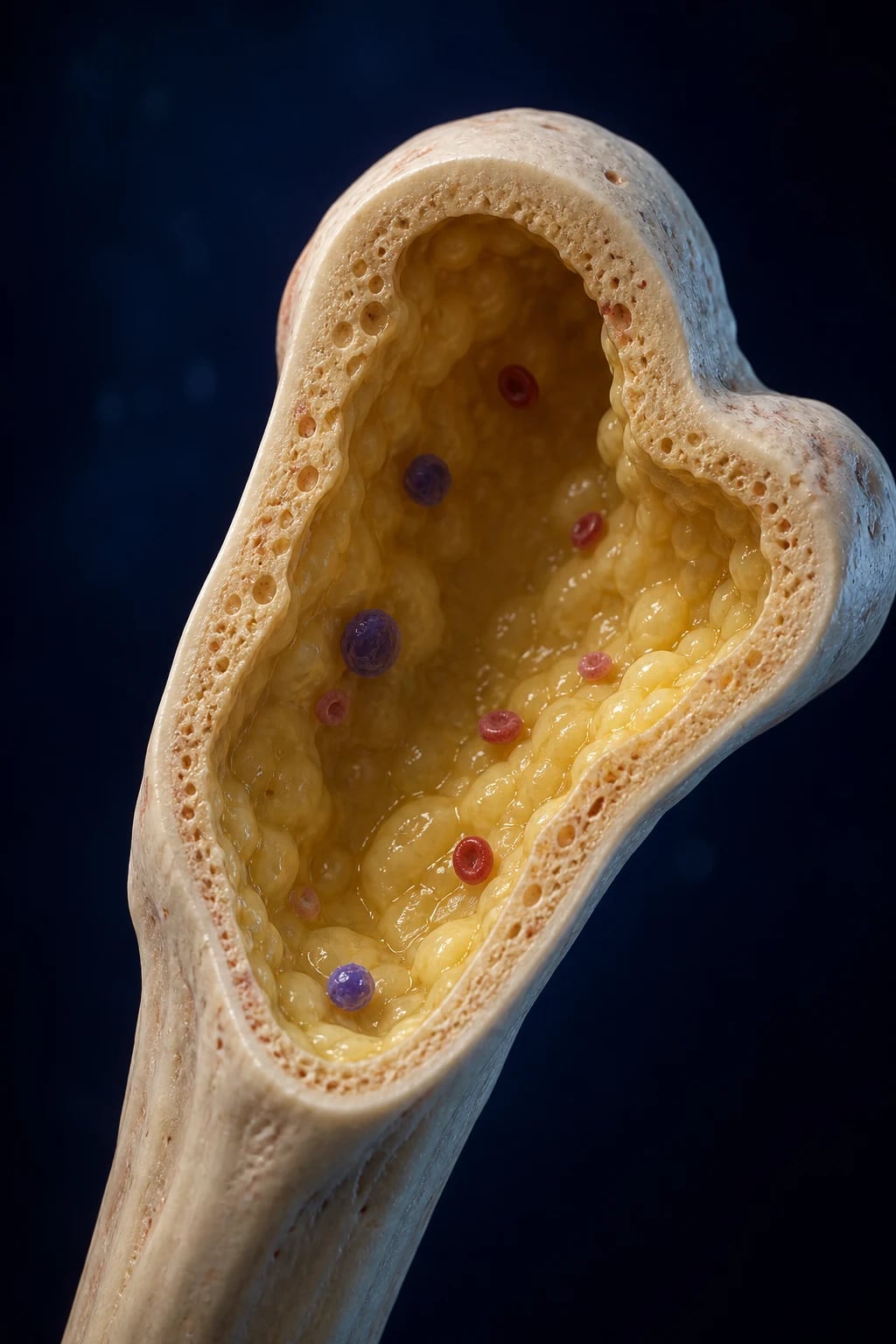
Aplastic anaemia is a life-threatening bone marrow failure syndrome defined by two coexisting findings:[4]
- Peripheral-blood pancytopenia — anaemia, neutropenia and thrombocytopenia together.
- A markedly hypocellular bone marrow with fatty replacement, in the absence of abnormal infiltrates (no leukaemic blasts, no carcinoma, no fibrosis) and without dysplasia. [1]
The marrow cellularity is under 25 percent (or 25 to 50 percent with under 30 percent residual haematopoietic cells) in severe disease. The syndrome is not a malignancy — there is no clonal neoplastic population — but it can evolve into clonal disorders (paroxysmal nocturnal haemoglobinuria, myelodysplastic syndrome, acute myeloid leukaemia) over years.[1]
The name is something of a misnomer: although "anaemia" appears in the title, the defining feature is failure of all three haematopoietic lineages — the triad of pancytopenia. Pure red cell aplasia (a selective failure of erythroid precursors alone) and amegakaryocytic thrombocytopenia (isolated platelet-lineage failure) are distinct entities, not variants of aplastic anaemia. The distinction matters because the work-up, treatment and prognosis differ.[1]
The clinical skill in aplastic anaemia is not the diagnosis (it is bone-marrow biopsy) but three things: (i) recognising pancytopenia and not dismissing the well-looking patient; (ii) distinguishing aplastic anaemia from its mimics (especially hypoplastic MDS, megaloblastic anaemia, aleukaemic leukaemia, and hypersplenic causes); and (iii) stratifying by severity (Camitta criteria) to choose between haematopoietic stem cell transplant and immunosuppressive therapy.[1][4]
Classification
Aplastic anaemia is classified along three axes — severity, acquired versus inherited, and aetiology.[4]
By severity (Camitta/modified criteria — examinable verbatim): [1]
| Severity | Marrow cellularity | Peripheral blood criteria |
|---|---|---|
| Severe AA (SAA) | under 25 percent (or 25 to 50 percent with under 30 percent residual haematopoiesis) | At least 2 of: neutrophils under 0.5 x 10^9/L, platelets under 20 x 10^9/L, reticulocytes under 20 x 10^9/L |
| Very severe AA (vSAA) | Same as SAA | Neutrophils under 0.2 x 10^9/L plus the other criteria |
| Non-severe AA | Hypocellular marrow | Cytopenias that do not meet severe criteria |
By aetiology: [1]
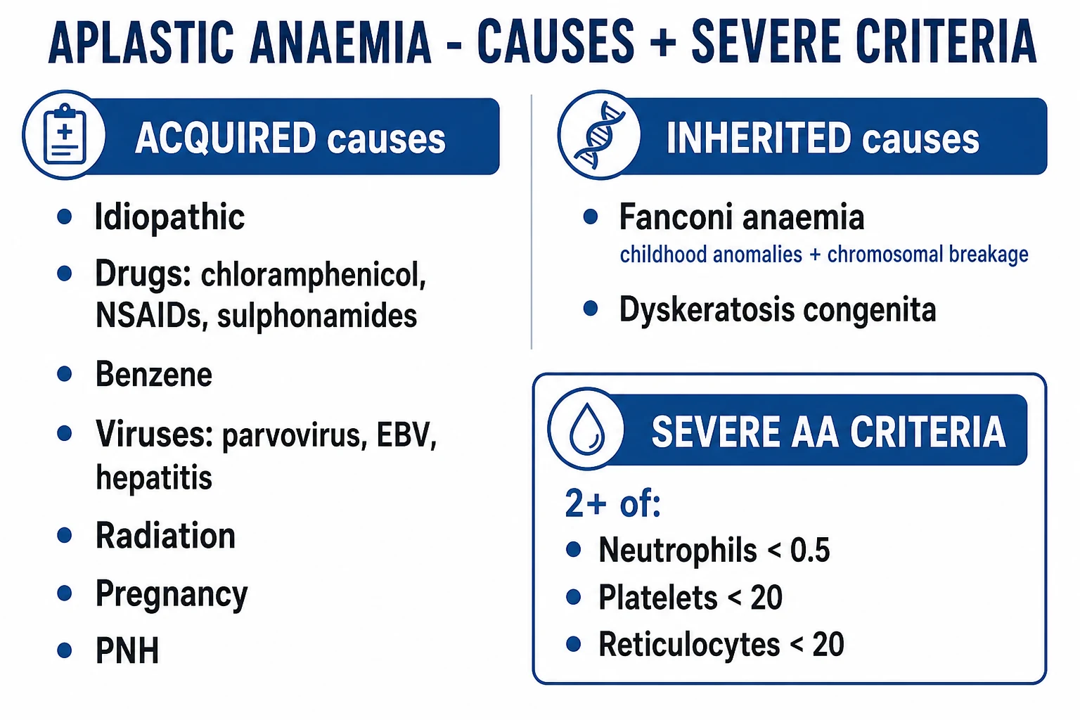
Acquired (commonest in adults)
- Idiopathic / autoimmune — about 60 to 70 percent of cases; T-cell-mediated destruction of CD34+ stem cells
- Secondary: idiosyncratic drugs (chloramphenicol, carbamazepine, phenytoin, valproate, NSAIDs, sulphonamides, gold, penicillamine, ticlopidine)
- Toxins: benzene and aromatic solvents, insecticides, radiation
- Viruses: seronegative hepatitis, EBV, CMV, HIV, parvovirus B19
- Pregnancy-associated (rare)
- Connective tissue disease: SLE, graft-versus-host disease
Inherited / constitutional
- Fanconi anaemia (FANC genes; autosomal recessive; chromosomal breakage; café-au-lait spots, radial ray anomalies)
- Dyskeratosis congenita (DKC1, TERC, TERT, RTEL1; telomere shortening; nail dystrophy, oral leukoplakia, reticular pigmentation)
- Shwachman-Diamond syndrome (SBDS; pancreatic exocrine insufficiency, neutropenia)
- GATA2 deficiency (MonoMAC/Emberger syndrome; MDS/AML risk)
- Congenital amegakaryocytic thrombocytopenia (MPL)
- Pearson syndrome (mitochondrial)
Epidemiology & Risk Factors
The incidence is about 2 per million per year in the West, with a two- to three-fold higher incidence in East and South Asia (China, Thailand, Indonesia, India) — attributed to environmental exposure (agricultural chemicals, benzene, infectious triggers) and possibly genetic susceptibility (HLA-DR15 over-representation, which is also associated with a better response to ciclosporin).[3]
There are two age peaks: a larger peak at 15 to 25 years (adolescents and young adults) and a smaller peak over 60 years (which overlaps diagnostically with hypoplastic MDS). The bimodal distribution reflects the fact that younger patients often carry inherited marrow-failure predisposition (short telomeres, FANC mutations) that manifests under environmental or infectious stress, while older patients accumulate somatic clones.[4]
Acquired risk factors — high-yield: [1]
| Risk factor / trigger | Detail and exam pearl |
|---|---|
| Idiopathic (commonest) | About 60 to 70 percent; thought to be autoimmune — a T-cell attack on CD34+ stem cells |
| Drug — idiosyncratic | Chloramphenicol (the classic exam answer, now rare), carbamazepine, phenytoin, valproate, NSAIDs (phenylbutazone, indometacin), sulphonamides, gold salts, penicillamine, chlorpropamide, ticlopidine, methimazole |
| Drug/toxin — dose-related | Benzene and aromatic solvents, insecticides/pesticides, cytotoxic chemotherapy (alkylators, antimetabolites), ionising radiation |
| Viral | Seronegative hepatitis (hepatitis-associated AA, typically young males 1 to 6 months after hepatitis), Epstein-Barr virus, cytomegalovirus, HIV, parvovirus B19 (especially immunocompromised) |
| Pregnancy | Rare; may remit after delivery but can recur in subsequent pregnancies |
| Autoimmune | Systemic lupus erythematosus, eosinophilic fasciitis, thymoma-associated, graft-versus-host disease |
| Inherited syndromes | Fanconi anaemia, dyskeratosis congenita, Shwachman-Diamond syndrome, GATA2 deficiency — short telomeres and stem-cell defects lower the threshold for marrow failure |
Aplastic Anaemia — epidemiology at a glance
Pathophysiology
Three mechanisms contribute; the dominant one in acquired disease is immune-mediated destruction.[3]
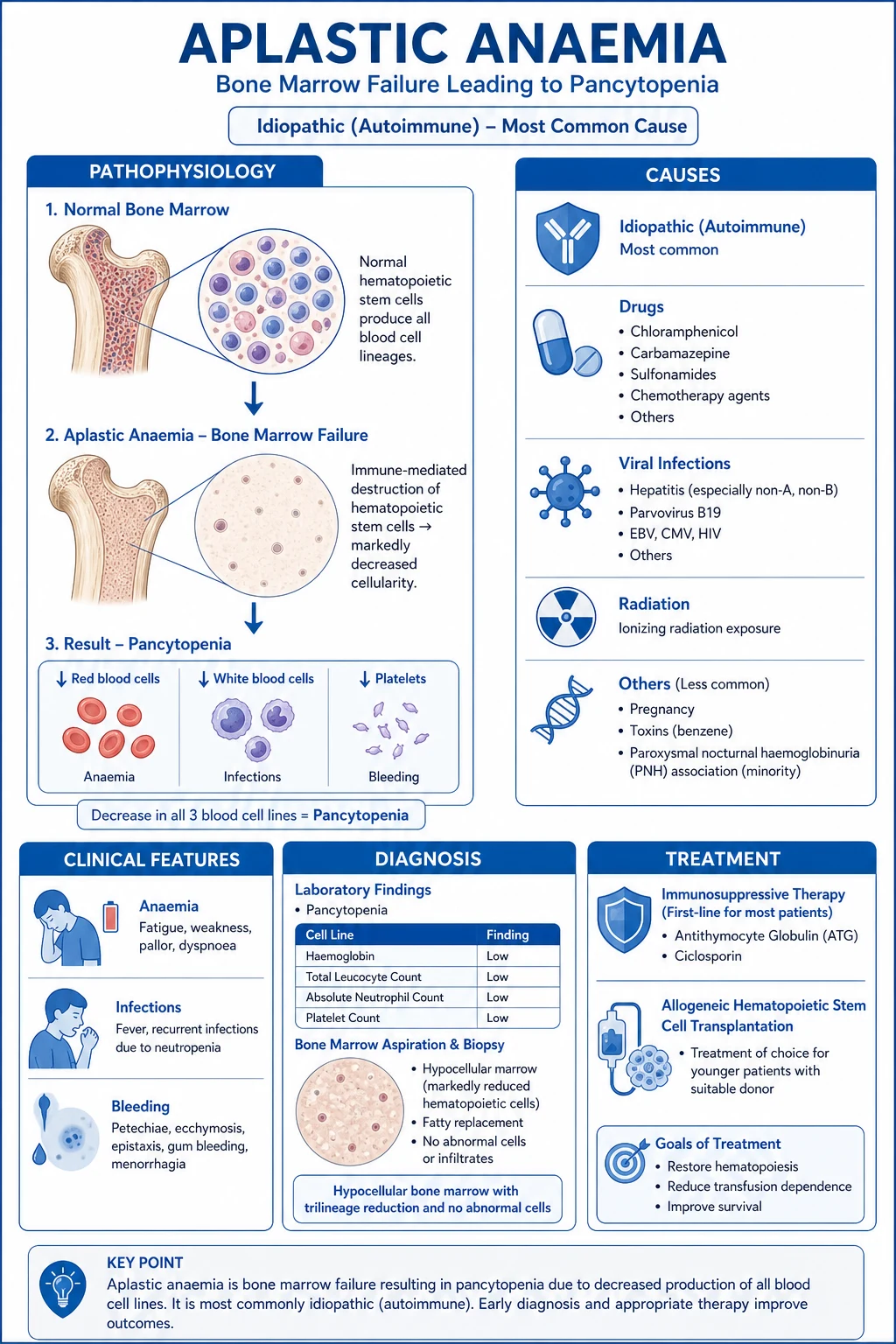
1. Immune-mediated stem-cell destruction (the core mechanism). An oligoclonal expansion of cytotoxic CD8+ T-helper-1 lymphocytes produces interferon-gamma, tumour necrosis factor-alpha and interleukin-2. These cytokines:[3]
- Induce apoptosis of CD34+ haematopoietic stem and progenitor cells via the Fas–Fas-ligand pathway — the death-receptor cascade that activates caspase-8 and downstream executioner caspases.
- Drive stem cells into cell-cycle arrest (loss of quiescence), exhausting the proliferative reserve — the stem cells are forced out of their protective G0/G1 resting state and into cycling, where they are vulnerable to cytokine-induced apoptosis.
- Down-regulate c-kit (the stem-cell factor receptor) and activate indoleamine 2,3-dioxygenase (IDO), which depletes tryptophan in the marrow microenvironment, starving the very cells needed for recovery.
- The transcription factor T-bet, a master regulator of Th1 differentiation, is over-expressed in the T cells of aplastic anaemia patients, sustaining the pathogenic cytokine programme. [1]
This explains why immunosuppression works: removing the autoimmune T-cell clone allows the surviving (often small) pool of stem cells to repopulate the marrow. The proof of concept is the clinical response rate of 60 to 70 percent to anti-thymocyte globulin plus ciclosporin, and the experimental observation that T-cell depletion of the marrow in vitro restores haematopoiesis.[1]
2. Intrinsic stem-cell defect. A subset of apparently acquired disease carries short telomeres from germline mutations in telomere biology genes — TERT, TERC, DKC1, RTEL1, NHP2, NOP10, CTC1, PARN — impairing stem-cell self-renewal. Telomeres are the protective caps at chromosome ends; when they become critically short, the cell enters senescence or apoptosis (the Hayflick limit). In the marrow, this means progressive depletion of the stem-cell pool over time. These patients respond less well to IST and carry a higher risk of clonal evolution (especially monosomy 7 MDS/AML) because stressed, short-telomere stem cells are prone to acquiring cytogenetic abnormalities. About 10 percent of apparently acquired aplastic anaemia carries a clinically significant telomerase mutation, and these mutations may be hypomorphic (partially functional), explaining why the family history is often unremarkable.[1]
3. Abnormal marrow microenvironment and stromal dysfunction — a less prominent contributor but relevant for the inherited syndromes. Mesenchymal stromal cells, endothelial cells and the extracellular matrix provide the "niche" that maintains stem-cell quiescence; defects here can contribute to marrow failure independently of the stem cells themselves. [1]
Clonal evolution and the PNH link. A small glycosylphosphatidylinositol-deficient clone (caused by a PIG-A mutation — absent CD55 and CD59 on the cell surface) is detectable by flow cytometry in up to half of acquired aplastic anaemia cases, even without overt haemolysis. This is one of the most important conceptual points in the disease: PNH and aplastic anaemia are two faces of the same immune attack. The PNH clone survives because its absent GPI-anchored proteins make it invisible to the T-cell-mediated destruction — it is an immune-evasive clone. Over years, this clone (and other clones — monosomy 7, trisomy 8, del(13q)) can expand into overt paroxysmal nocturnal haemoglobinuria, myelodysplastic syndrome or acute myeloid leukaemia. This is why lifelong surveillance for clonal evolution is mandatory.[5]
Histological hallmarks of aplastic anaemia on trephine biopsy: fatty marrow with scattered islands of lymphocytes and plasma cells, markedly reduced haematopoietic cells (cellularity under 25 percent), no significant fibrosis, and no abnormal infiltrate. The aspirate is often a "dry tap" or dilute because there are so few cells to aspirate — which is why the trephine biopsy is the critical diagnostic specimen, not the aspirate alone.[4]
Clinical Presentation
The symptoms reflect which cell line is lowest, but all three cytopenias often coexist at presentation.[4]
Anaemia (low haemoglobin): The most common presenting symptom is progressive fatigue — patients describe feeling "washed out," breathless on minimal exertion, and unable to keep up with their usual activities. Physical signs include pallor (of the conjunctivae, palmar creases and nail beds), exertional dyspnoea, palpitations, and — in older patients or those with rapid haemoglobin falls — angina and cardiac failure. The onset is usually gradual enough for physiological adaptation, so patients may present with remarkably low haemoglobin (40 to 60 g/L) and yet be ambulant. [1]
Neutropenia (neutrophils under 0.5 x 10^9/L): The most dangerous cytopenia in the short term. Fever is the cardinal sign — in a neutropenic patient, fever above 38.0 degrees Celsius constitutes neutropenic sepsis and is a medical emergency requiring empirical broad-spectrum antibiotics within one hour. Other features include oral mucositis (painful mouth ulcers), perianal infection (often missed on examination), recurrent bacterial infections (skin, respiratory, urinary), severe or overwhelming sepsis, and invasive fungal infection (aspergillosis — look for pleuritic chest pain, haemoptysis and a halo sign on CT; candidiasis — look for hepatosplenic lesions on imaging). The absence of pus formation is itself a clue — neutrophils are needed to form pus, so infections in neutropenic patients may be indolent in appearance but rapidly progressive in reality.[4]
Thrombocytopenia (platelets under 20): The bleeding phenotype depends on the platelet count and the presence of additional risk factors (fever, infection, medications). Petechiae (pinpoint, non-blanching macules) and purpura (larger areas) appear especially over the lower limbs (due to dependent capillary pressure), pressure points (waistband, blood-pressure cuff site) and buccal mucosa. Mucosal bleeding includes epistaxis (often bilateral and hard to control), gingival bleeding, menorrhagia (can be life-threatening), haematuria, and melaena. The most feared complication is intracranial haemorrhage — rare but catastrophic, presenting with severe headache, altered consciousness, or focal neurological deficit.[4]
Onset is usually insidious over weeks to months, but severe bleeding or sepsis can precipitate a sudden presentation. Patients and their families may attribute early symptoms (tiredness, easy bruising) to stress or minor illness until a blood test reveals the severity. [1]
Examination is the key clue: the patient is often well-appearing despite profound cytopenias, with pallor, petechiae and purpura — but characteristically no hepatosplenomegaly and no lymphadenopathy. This is the most important bedside discriminator from leukaemia: a leukaemic patient with equivalent cytopenias looks unwell, often has organomegaly, and may have tissue infiltration. The presence of splenomegaly or lymphadenopathy in a patient thought to have aplastic anaemia should prompt an immediate search for an alternative diagnosis (leukaemia, lymphoma, hypersplenism, megaloblastic anaemia, visceral leishmaniasis).[4]
Atypical and scenario-specific presentations: [1]
- Adolescents/young adults — the "well patient with frightening bloods"; a classic clue to bone marrow failure rather than leukaemia. The clinical contrast between how well the patient looks and how bad the blood count is can be striking.
- Elderly — may be misclassified as hypoplastic MDS; presentation overlaps with myelodysplasia and cytogenetic assessment is essential. The distinction has major treatment implications: MDS may need hypomethylating agents or supportive care, while aplastic anaemia needs IST or HSCT.
- Hepatitis-associated AA — typically young males, develops 1 to 6 months after an episode of seronegative hepatitis (negative for hepatitis A, B, C, E and all known hepatotropic viruses). The liver injury resolves but the marrow fails. Often fulminant and poorly responsive to IST — early HSCT is considered if a donor exists.[1]
- Pregnancy — rare; risk of severe haemorrhage and infection; managed supportively during pregnancy, with IST considered postpartum. May remit after delivery but can recur in subsequent pregnancies.
- Parvovirus B19 — classically pure red cell aplasia with giant proerythroblasts in the marrow (the virus directly infects and lyses erythroid progenitor cells through the P-antigen receptor); in the immunocompromised host can cause a more generalised aplasia because the virus cannot be cleared by neutralising antibodies.
- Aplastic-PNH syndrome — overlapping marrow failure and haemolysis (jaundice, dark urine), venous thrombosis (hepatic, portal, cerebral, dermal). The thrombosis of PNH is characteristically in unusual sites — Budd-Chiari syndrome (hepatic vein), portal/mesenteric/splenic vein thrombosis, cerebral venous sinus thrombosis — and should trigger a PNH work-up in any patient with aplastic anaemia.
Differential Diagnosis
The differential of pancytopenia is broad; the most efficient approach is to classify by marrow cellularity, because this immediately separates the close mimics from the rest.[4]
Hypocellular marrow causes (the close differentials from aplastic anaemia): [1]
- Hypoplastic myelodysplastic syndrome (MDS) — the single hardest distinction in bone marrow failure. Dysplastic morphology (micromegakaryocytes, hypogranular neutrophils, nuclear budding), clonal cytogenetics (monosomy 5/7, trisomy 8, del(20q)), and raised CD34+ blasts all favour MDS. Cytogenetics and next-generation sequencing increasingly inform this distinction, but some cases remain genuinely ambiguous ("hypoplastic MDS vs aplastic anaemia"). The clinical implication is large: MDS is managed differently (hypomethylating agents, supportive care, HSCT in selected cases) and carries a different prognosis.
- Aleukaemic leukaemia — acute leukaemia presenting with a low white cell count (no peripheral blasts); marrow is usually hypercellular with over 20 percent blasts but can occasionally be hypocellular.
- Advanced hairy-cell leukaemia — pancytopenia with dry tap on aspirate; marrow shows hairy cells (villose cytoplasmic projections), annexin-A1 and BRAF V600E positive on immunohistochemistry, and there is usually massive splenomegaly.
- Late myelofibrosis — tear-drop cells (dacryocytes) on the blood film, massive splenomegaly, and marked reticulin and collagen fibrosis on trephine — the marrow is hypocellular but fibrotic (fibrosis is absent in classic aplastic anaemia).
- Severe infection — miliary tuberculosis, visceral leishmaniasis (kala-azar), brucellosis. These are particularly relevant in endemic regions (India, South Asia) and should be excluded with appropriate cultures and serology.
- Paroxysmal nocturnal haemoglobinuria (aplastic-PNH syndrome) — see below; the marrow may be hypocellular early in the disease course. [1]
Normo-/hypercellular marrow causes of pancytopenia: [1]
- Megaloblastic anaemia (B12 or folate deficiency) — macro-ovalocytes, hypersegmented neutrophils (5 or more lobes), megaloblastic marrow (giant metamyelocytes, nuclear-cytoplasmic asynchrony), high LDH and indirect bilirubin, low B12 or folate. The key point: it causes pancytopenia with a hypercellular marrow, and it responds rapidly to supplementation within days to weeks. A common exam trap is to confuse megaloblastic anaemia with aplastic anaemia — the marrow cellularity and the film morphology are the decisive discriminators.
- Sepsis with marrow suppression — temporary, resolves with treatment of the underlying infection.
- Hypersplenism — portal hypertension (cirrhosis, portal/splenic vein thrombosis), storage disorders (Gaucher, Niemann-Pick); splenomegaly is dominant, marrow is hypercellular with maturation arrest.
- Systemic lupus erythematosus and other autoimmune cytopenias — may present with pancytopenia from autoantibodies or from secondary marrow involvement; ANA and anti-dsDNA are positive.
- Disseminated (miliary) tuberculosis — particularly in endemic regions; may cause marrow infiltration and secondary haemophagocytic syndrome.
- Infiltrative malignancy — metastatic carcinoma (breast, lung, prostate, stomach), lymphoma, myelophthisis (marrow replacement). The film may show nucleated red cells and immature myeloid precursors (leukoerythroblastic picture).
- Gaucher disease, visceral leishmaniasis — storage or parasitic infiltration of the marrow; organisms visible on marrow aspirate in kala-azar (Leishman-Donovan bodies). [1]
How to distinguish aplastic anaemia from its key mimics — exam high-yield: [1]
Aplastic anaemia
- Pancytopenia + hypocellular marrow under 25 percent
- No blasts, no dysplasia, no abnormal infiltrate
- No organomegaly; well-looking patient
- Cytogenetics normal; FLAER/flow may show small PNH clone
Hypoplastic MDS
- Hypocellular marrow with dysplasia
- Clonal cytogenetics — monosomy 5/7, trisomy 8, del(20q)
- Raised CD34+ blasts (under 20 percent)
- Older patient; next-generation sequencing helps
Acute leukaemia
- Marrow hypercellular with over 20 percent blasts
- Peripheral blasts usually present
- Lymphadenopathy/splenomegaly common
- Symptoms often more acute; organ infiltration
Megaloblastic anaemia
- Macro-ovalocytes, hypersegmented neutrophils on film
- LDH high; B12 or folate low
- Marrow hypercellular, megaloblastic (giant metamyelocytes)
- Rapid response to B12/folate replacement
Hypersplenism
- Splenomegaly dominant
- Marrow hypercellular with maturation arrest
- Isolated cytopenias common (e.g. thrombocytopenia)
- Cause: portal hypertension, storage disease, lymphoma
PNH
- Haemolysis — high LDH, indirect bilirubin, low haptoglobin
- Dark urine (haemoglobinuria)
- Venous thrombosis — hepatic, portal, cerebral
- FLAER-negative granulocytes; absent CD55/CD59
Clinical & Bedside Assessment
Bedside examination must include:[4]
- Vital signs — fever in a neutropenic patient is neutropenic sepsis until proven otherwise and requires immediate empirical antibiotics. Do not wait for blood cultures to return before starting treatment — the window for effective intervention is the first hour.
- Skin — pallor (conjunctival, palmar, nail-bed), petechiae and purpura (especially lower limbs, pressure points, buccal mucosa). Look carefully at the lower legs and buccal mucosa — these are dependent and traumatised areas where petechiae first appear.
- Fundoscopy — retinal haemorrhage (flame-shaped or dot-blot), a sign of severe thrombocytopenia and a bleeding risk. Roth spots (white-centred haemorrhages) suggest additional infection.
- Lymph nodes, liver and spleen — characteristically absent in aplastic anaemia; their presence argues for leukaemia, lymphoma, hypersplenism, or megaloblastic anaemia. Carefully examine the cervical, axillary and inguinal nodes, and palpate and percuss for hepatosplenomegaly. [1]
Look for stigmata of inherited marrow-failure syndromes (especially in younger patients — these are missed diagnoses with major treatment implications):[4]
- Fanconi anaemia — short stature, café-au-lait spots, radial ray anomalies (absent thumb, triphalangeal thumb, radial hypoplasia), microcephaly, renal anomalies (horseshoe kidney, duplicated collecting system), hypogonadism, strabismus. The physical features may be subtle — café-au-lait spots can be mistaken for ordinary pigmentation, and thumb anomalies may have been surgically corrected in childhood. A careful family history of early-onset cancers, congenital anomalies or unexplained cytopenias is essential.
- Dyskeratosis congenita — the classic triad of dystrophic nails (ridged, split, atrophic), reticular skin pigmentation (neck, chest, axillae — a lace-like pattern), and oral leukoplakia (whitish patches on the tongue and buccal mucosa). These appear in childhood to early adolescence. Also look for premature greying and dental abnormalities.
- Shwachman-Diamond — skeletal anomalies (metaphyseal chondrodysplasia, rib abnormalities), growth failure, and steatorrhoea from pancreatic exocrine insufficiency (ask about greasy, foul-smelling stools from childhood).
- GATA2 deficiency — warts (persistent, extensive HPV infection due to monocytopenia), opportunistic infection (atypical mycobacteria, fungal), lymphoedema (Emberger syndrome), and a family history of MonoMAC (monocytopenia, dendritic cell deficiency, B-cell and NK-cell deficiency). [1]
Search for an underlying trigger: jaundice/hepatomegaly (recent hepatitis — ask about a febrile illness with elevated liver enzymes in the preceding 1 to 6 months), thyroid goitre (autoimmune polyglandular syndrome), pregnancy, occupational history (benzene exposure in petrochemical workers, pesticide use in agricultural workers, radiation exposure), and drug history (chloramphenicol — still used in some regions; antiepileptics; NSAIDs; sulphonamides; gold).[3]
Look for PNH clues: jaundice (haemolytic — check for an indirect bilirubin out of proportion to the anaemia), dark urine (haemoglobinuria) — classically worse in the morning after sleep (hence "nocturnal"), abdominal pain/Budd-Chiari (ascites, abdominal-wall collaterals, hepatomegaly), and neurological deficit (cerebral venous sinus thrombosis — headache, papilloedema, seizures). [1]
The well-looking patient with severe pancytopenia and absent organomegaly is a classic bedside clue to aplastic anaemia. Conversely, a patient with the same blood counts who looks unwell, is febrile with localising signs, or has organomegaly is far more likely to have leukaemia, lymphoma or infection.[4]
Signs requiring immediate action:
- Fever with neutropenia → neutropenic sepsis bundle (cultures and empirical antibiotics within 1 hour).
- Uncontrolled bleeding, severe headache or altered consciousness → suspected intracranial haemorrhage — urgent platelet transfusion, CT imaging, neurosurgical consultation.
- Retinal haemorrhage with visual change → urgent platelet support and ophthalmology review. [1]
Investigations
First-line tests:[4]
- Full blood count with differential — confirms pancytopenia: low haemoglobin, low neutrophils (absolute neutrophil count under 0.5 in severe AA), low platelets, low reticulocytes. The MCV is often normal or mildly raised (macrocytosis is common in marrow failure and in telomere biology disorders). A high MCV with pancytopenia should always prompt consideration of megaloblastic anaemia, but a normal MCV does not exclude it.
- Peripheral blood film — no abnormal cells, no blasts (the key finding that excludes leukaemia). May show anisocytosis, macrocytes, and mild poikilocytosis. The absence of blasts, dysplastic neutrophils, and nucleated red cells is itself diagnostic information. In megaloblastic anaemia, you would see macro-ovalocytes and hypersegmented neutrophils; in leukaemia, you would see blasts.
- Reticulocyte count — inappropriately low for the degree of anaemia (the marrow is not compensating). Absolute reticulocytes under 20 x 10^9/L is part of the severe criteria. A high reticulocyte count in the setting of pancytopenia suggests haemolysis or recovery rather than aplastic anaemia.
- Bone marrow aspirate AND trephine biopsy — the diagnostic cornerstone. The trephine is essential because the aspirate can be misleadingly dilute (a "dry tap" or blood-only aspirate is common when the marrow is severely hypocellular). On the trephine: cellularity under 25 percent (or 25 to 50 percent with under 30 percent residual haematopoietic cells), no dysplasia, no abnormal infiltrate, no significant fibrosis. The cellularity should be assessed on the trephine biopsy, not the aspirate, because the aspirate is prone to dilution artefact. [1]
Severe AA — Camitta criteria (reproduced verbatim, examinably verbatim):[4]
Marrow cellularity under 25 percent (or 25 to 50 percent with under 30 percent residual haematopoietic cells), AND at least two of the following:
- Neutrophils under 0.5 x 10^9/L
- Platelets under 20 x 10^9/L
- Reticulocytes under 20 x 10^9/L [1]
Very severe AA: meets the severe criteria AND neutrophils under 0.2 x 10^9/L. Non-severe AA: hypocellular marrow with cytopenias that do not meet severe criteria. [1]
The severity grading is not academic — it directly determines treatment: severe and very severe AA need definitive therapy (HSCT or IST), while non-severe AA may be observed or managed supportively. A patient with very severe AA has a worse prognosis than one with severe AA and should be fast-tracked to definitive therapy.[1]
Second-line / aetiological work-up: [1]
- Cytogenetics (conventional karyotyping plus FISH for chromosomes 5, 7, 8, 20) — normal in idiopathic AA; clonal abnormalities (monosomy 7, trisomy 8, del(20q)) suggest MDS or clonal evolution. A normal karyotype supports the diagnosis of aplastic anaemia; an abnormal karyotype with a hypocellular marrow shifts the diagnosis toward hypoplastic MDS.
- Flow cytometry for a PNH clone (FLAER test on granulocytes and monocytes; CD55/CD59 on red cells) — detect a small PNH clone in up to half of acquired cases. FLAER (fluorescent aerolysin) binds to GPI-anchored proteins; GPI-deficient cells (PNH clone) fail to bind. This is more sensitive than CD55/CD59 alone and is now the preferred method.[5]
- Exclude triggers and mimics: liver function tests (hepatitis-associated AA); viral serology (hepatitis A/B/C/E, EBV, CMV, HIV, parvovirus B19 PCR); vitamin B12 and folate (to exclude megaloblastic anaemia); serum copper (deficiency causes a reversible pancytopenia with neurological signs and myelodysplastic features — easily missed and treatable); autoimmune screen (ANA, anti-dsDNA, rheumatoid factor — SLE can cause secondary aplastic anaemia); serum immunoglobulins.
- Telomere length assay (flow-FISH) and directed germline gene testing (TERT, TERC, DKC1, RTEL1, SAMD9/SAMD9L, GATA2, FANC genes) when an inherited disorder is suspected — i.e., young patients, family history of cytopenias/cancers, physical stigmata, or poor response to IST. Telomere length below the first percentile for age is highly suggestive of a telomere biology disorder.
- Chromosomal breakage test (DEB or MMC) — diepoxybutane (DEB) or mitomycin C (MMC) induced chromosomal breakage testing is the diagnostic test for Fanconi anaemia: Fanconi cells show increased chromosomal breakage and radial figures when exposed to cross-linking agents. This should be performed in all young patients with aplastic anaemia, because undiagnosed Fanconi anaemia changes the HSCT conditioning regimen (must avoid radiation and alkylators) and carries implications for family members.
- HLA typing of the patient and full siblings at diagnosis — early donor identification informs the first-line choice between HSCT and IST. HLA-A, -B, -C, -DRB1, -DQB1 (10/10 match) is the minimum for sibling assessment.
- Imaging — chest X-ray or CT if tuberculosis or fungal infection suspected (halo sign, air-crescent sign for invasive aspergillosis); abdominal ultrasound for splenomegaly (which would argue against aplastic anaemia). Not diagnostic of aplastic anaemia itself.
- Iron studies and ferritin before the transfusion programme — iron overload is a late complication of repeated transfusions and needs chelation when ferritin exceeds 1000 micrograms/L.
Aplastic Anaemia — key numbers to memorise
Management — Resuscitation
ABCDE assessment first. The two immediate threats are bleeding (severe thrombocytopenia) and infection (neutropenia).[1]
Neutropenic fever — emergency bundle (do not delay antibiotics for investigation):[4]
- Blood cultures from every lumen of any central line and a peripheral site — before antibiotics if possible, but do not delay antibiotics beyond 1 hour from presentation.
- Empirical broad-spectrum IV antibiotics within 1 hour per local policy — typically piperacillin-tazobactam 4.5 g IV 8-hourly, or ceftazidime 2 g IV 8-hourly / cefepime 2 g IV 8-hourly as monotherapy. Add vancomycin 1 g IV 12-hourly (or 15 to 20 mg/kg) or teicoplanin 400 mg IV 12-hourly for suspected line infection, haemodynamic instability, severe mucositis, or known MRSA colonisation. The rationale is to cover Gram-negative bacilli (Pseudomonas, E. coli, Klebsiella — the most rapidly lethal organisms in neutropenia) and Gram-positive cocci (Staphylococcus, Streptococcus viridans — increasingly common with central lines).
- Reassess at 48 to 72 hours; escalate to antifungal cover (liposomal amphotericin B 3 mg/kg/day, caspofungin 70 mg loading then 50 mg/day, voriconazole 6 mg/kg loading then 4 mg/kg 12-hourly) for persistent fever — which raises suspicion of invasive fungal infection (aspergillosis, candidiasis). A galactomannan assay (serum or BAL) and high-resolution CT chest should be considered.
- Reverse barrier nursing, strict hand hygiene, neutropenic diet (avoid raw foods, unpasteurised dairy). G-CSF (granulocyte colony-stimulating factor) may be considered in severe sepsis, though its routine use in aplastic anaemia is debated. [1]
Severe anaemia: transfuse leucodepleted packed red cells to a target haemoglobin over 70 g/L (or over 80 to 90 g/L if symptomatic, pregnant, or with cardiovascular disease). Each unit raises haemoglobin by approximately 10 g/L in an adult without active bleeding. [1]
Severe thrombocytopenia / bleeding: [1]
- Prophylactic platelet transfusion when platelets under 10 x 10^9/L (or under 20 if febrile or with active bleeding). One adult therapeutic dose raises the count by approximately 20 to 40 x 10^9/L.
- Therapeutic transfusion for active bleeding or before invasive procedures — aim for platelets over 50 x 10^9/L for active bleeding or major procedures, and over 100 for neurosurgery or ophthalmic procedures.
- Tranexamic acid 1 g IV 8-hourly for mucosal bleeding (avoid in haematuria — risk of clot obstruction in the urinary tract).
- Avoid NSAIDs, intramuscular injections, rectal examinations, hard tooth-brushing, razors in thrombocytopenic patients — these can precipitate life-threatening bleeding. [1]
Use CMV-safe (leucodepleted) blood products in patients who may proceed to HSCT, to prevent CMV transmission and alloimmunisation. Alloimmunisation (especially anti-HLA antibodies) makes future platelet transfusions ineffective — platelet refractoriness — and complicates HSCT conditioning.[1]
Prophylaxis during cytopenias: co-trimoxazole 480 mg orally daily (or 960 mg three times weekly) for Pneumocystis jirovecii; aciclovir 200 to 400 mg orally daily for HSV/VZV; posaconazole 300 mg orally daily or itraconazole for antifungal cover; iron chelation with deferasirox 20 mg/kg/day orally when ferritin exceeds 1000 micrograms/L. [1]
Rapid referral to a haematology centre with HSCT capability for severe/very severe disease — definitive therapy must start within days, not weeks. Untreated severe aplastic anaemia has a 70 percent one-year mortality from infection or bleeding.[2]
Management — Definitive & Stepwise
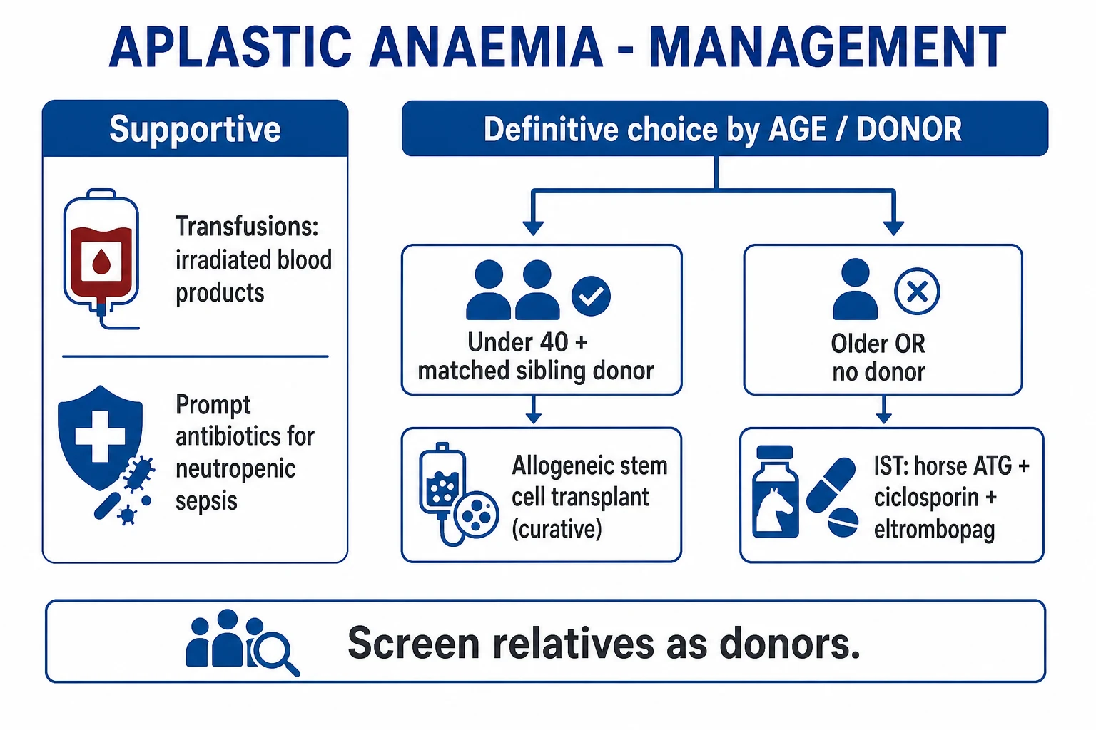
Definitive treatment is stratified by severity and age:[1][4]
Decision rule: [1]
- Severe or very severe AA AND age under 40 years AND an HLA-matched sibling donor available → allogeneic haematopoietic stem cell transplant (HSCT) as first-line curative therapy. This offers the best chance of long-term cure (80 to 90 percent survival in young patients).
- Severe or very severe AA in patients over 40, OR no matched sibling donor → immunosuppressive therapy (IST): horse anti-thymocyte globulin plus ciclosporin. This is not curative but achieves a haematological response in 60 to 70 percent, allowing transfusion independence.
- Non-severe AA — observation plus supportive care; treat with IST only if transfusion-dependent or progressing to severe criteria. [1]
The age cut-off of 40 reflects transplant-related mortality: HSCT-related complications (GVHD, infection, conditioning toxicity) rise sharply with age, and IST becomes the safer option for older patients. The age cut-off is a guideline, not absolute — fit patients up to 50 with a matched sibling may be considered for HSCT, and frail 35-year-olds may be better served by IST.[1]
Immunosuppressive therapy (IST) — drug, dose, route, timing, rationale, monitoring: [1]
| Agent | Dose & route | Timing | Rationale & monitoring |
|---|---|---|---|
| Horse anti-thymocyte globulin (hATG) | 40 mg/kg/day IV over 4 days (days 1 to 4), via central line, with test dose and premedication (antihistamine, paracetamol, hydrocortisone) | Days 1 to 4 of the regimen | Depletes the autoreactive T-cell clone by polyclonal antibody-mediated lysis. Horse ATG is superior to rabbit ATG (response rate 68 percent vs 37 percent at 6 months — Scheinberg NEJM 2011).[7] Monitor for serum sickness (fever, rash, arthralgia, proteinuria around days 7 to 14) and anaphylaxis (test dose is mandatory). |
| Ciclosporin | 5 mg/kg/day orally in two divided doses; target trough whole-blood level 200 to 400 micrograms/L | Started on day 1; continued at least 6 months after maximal response, then slow taper over 1 to 2 years | Inhibits T-cell activation by blocking calcineurin, preventing interleukin-2 transcription and T-cell proliferation. Monitor renal function (creatinine — reduce dose if rising), blood pressure, trough levels (under-dosing leads to relapse, over-dosing to nephrotoxicity), LFTs; watch for gingival hypertrophy, tremor, hirsutism, hypertension. |
| Methylprednisolone | 1 mg/kg/day (short course) | Days 1 to 14 typically | Reduces serum sickness from ATG; not used long term (no benefit and significant toxicity). |
| Eltrombopag (TPO-receptor agonist) | 150 mg orally daily for 6 months (adults); avoid in East Asian ancestry or reduce to 75 mg | Added to ATG + ciclosporin in selected protocols (RACE trial) for treatment-naïve severe AA; also used alone for refractory disease | Stimulates residual megakaryocytes and stem cells via the thrombopoietin (MPL) receptor; improves trilineage haematopoiesis (not just platelets). Improves complete response rate in treatment-naïve and refractory severe AA.[6] Monitor LFTs (hepatotoxicity) and for clonal evolution (concern about increased risk of cytogenetic abnormalities with prolonged use). |
Response assessment at 3 to 6 months:[1]
- Complete response (CR) — haemoglobin over 100 g/L, neutrophils over 1.5 x 10^9/L, platelets over 100 x 10^9/L, transfusion-independent.
- Partial response (PR) — no longer meeting severe AA criteria, transfusion-independent.
- No response — consider second-line therapy. [1]
Responses typically begin at 3 months (first sign is usually a rising neutrophil count and reticulocyte count) and are maximal by 6 months. Patients who have not responded by 6 months should be considered for second-line therapy. Responders remain at risk of relapse (10 to 30 percent) and clonal evolution (10 to 15 percent at 10 years), so lifelong surveillance is mandatory.[5]
Refractory or relapsed severe AA — second-line options:[2]
- Matched unrelated donor HSCT (younger patients with a suitable donor — outcomes approaching matched sibling HSCT with modern conditioning).
- Eltrombopag monotherapy for refractory disease (response in approximately 40 percent of refractory patients).[6]
- A second course of IST (rabbit ATG if first course was horse ATG; alemtuzumab — anti-CD52 — in selected cases).
- Haploidentical HSCT (half-matched family donor — post-transplant cyclophosphamide conditioning) is an emerging option for patients without a matched donor.
Allogeneic HSCT — conditioning and procedure:[1][5]
- Donor preference: HLA-matched sibling first (lowest GVHD risk, best outcomes); matched unrelated donor if no sibling and the patient is young (8/8 or 10/10 HLA match).
- Conditioning: fludarabine plus cyclophosphamide plus ATG (with or without low-dose total-body irradiation 2 Gy or alemtuzumab for in vivo T-cell depletion). Avoid heavy alkylators and total-body irradiation in inherited marrow-failure syndromes (Fanconi anaemia — extreme radiosensitivity and chromosomal fragility; dyskeratosis congenita — sensitivity to alkylators and radiation).
- Source: peripheral blood stem cells (faster engraftment) or bone marrow (lower chronic GVHD in some studies).
- Outcomes: long-term survival about 80 to 90 percent in young patients with matched sibling donors; 70 to 80 percent with matched unrelated donors; lower in older or heavily pretreated patients.
- Late complications: graft-versus-host disease (acute — skin rash, diarrhoea, cholestatic jaundice in the first 100 days; chronic — sicca syndrome, scleroderma-like skin changes, bronchiolitis obliterans after 100 days), veno-occlusive disease/sinusoidal obstruction syndrome (weight gain, tender hepatomegaly, jaundice — from conditioning toxicity), infections, secondary malignancy (solid tumours, post-transplant lymphoproliferative disorder), infertility, endocrine failure (growth hormone deficiency, hypothyroidism, gonadal failure).[5]
PNH clone surveillance — flow cytometry every 6 to 12 months; start eculizumab (anti-C5 monoclonal antibody, 1200 mg IV every 2 weeks after loading) or ravulizumab (longer-acting anti-C5, every 8 weeks) if overt haemolysis or thrombosis develops. Both require meningococcal vaccination and prophylactic penicillin because they block complement-mediated defence against Neisseria. [1]
Specific Subtypes & Scenarios
Inherited/congenital aplastic anaemia — these syndromes are missed at the candidate's peril, because they change the conditioning regimen and carry family implications:[4]
- Fanconi anaemia — the commonest inherited marrow-failure syndrome. Autosomal recessive (FANC gene family — at least 22 genes involved in DNA cross-link repair). The diagnostic test is the DEB (diepoxybutane) chromosomal breakage test: Fanconi cells show increased chromosomal breakage, gaps and radial figures when exposed to DEB or mitomycin C. Physical stigmata: short stature, café-au-lait spots, radial ray anomalies (absent or hypoplastic thumbs, radial hypoplasia), microcephaly, renal anomalies, hypogonadism. Bone marrow failure typically develops in the first decade. HSCT is the only cure for the marrow failure, but conditioning must avoid total-body irradiation and alkylators (extreme chromosomal fragility — use fludarabine-based reduced-intensity conditioning). Lifelong cancer surveillance is needed (head and neck, gynaecological, skin cancers).[2]
- Dyskeratosis congenita — a telomere biology disorder (X-linked DKC1, or autosomal dominant TERC/TERT, or autosomal recessive RTEL1, NHP2, NOP10, CTC1, PARN, TCAB1). The classic mucocutaneous triad — dystrophic nails, reticular skin pigmentation, oral leukoplakia — appears in childhood. Marrow failure develops in the teens to twenties. Telomere length is markedly shortened (below first percentile for age). Pulmonary fibrosis and hepatic cirrhosis are non-haematological complications of the short-telomere syndrome. HSCT conditioning should be reduced-intensity with fludarabine; patients are sensitive to radiation and alkylators.
- Shwachman-Diamond syndrome — autosomal recessive (SBDS gene). Features: pancreatic exocrine insufficiency (steatorrhoea, failure to thrive from infancy — distinguish from cystic fibrosis by normal sweat chloride), neutropenia (often the first haematological sign), skeletal dysplasia (metaphyseal chondrodysplasia), and marrow failure. MDS/AML risk is increased. HSCT is the only cure for marrow failure.
- GATA2 deficiency — includes MonoMAC syndrome (monocytopenia, dendritic cell deficiency, B-cell and NK-cell deficiency) and Emberger syndrome (primary lymphoedema with MDS/AML). Presents with persistent warts (HPV), opportunistic infection (atypical mycobacteria, fungal), and progression to MDS/AML. HSCT is curative for the immunodeficiency and marrow failure.
- Congenital amegakaryocytic thrombocytopenia (MPL gene) — presents with isolated thrombocytopenia at birth (absent megakaryocytes in the marrow), progressing to trilineage marrow failure. HSCT is the only cure.
Hepatitis-associated AA — young males, 1 to 6 months after seronegative hepatitis (negative for all known hepatotropic viruses — A, B, C, E); fulminant course; poor response to IST (response rate lower than idiopathic); early HSCT if a donor is available. The mechanism is thought to be immune cross-reactivity between liver and marrow antigens, but the exact trigger is unknown.[1]
Pregnancy-associated AA — rare; support with transfusions during pregnancy (leucodepleted, CMV-safe); defer IST until postpartum where possible (ciclosporin is relatively safe in pregnancy — category C, but ATG is generally avoided); HSCT deferred until delivery. Risk of miscarriage, preterm birth, and maternal haemorrhage. May remit postpartum but can recur in subsequent pregnancies. Multidisciplinary obstetric–haematology–anaesthesia care is essential.[4]
Post-EBV / post-mononucleosis AA — rare; usually self-limited or responds to IST. The virus infects and lyses haematopoietic progenitors in the immunocompromised host. [1]
Drug-induced AA — withdraw the offending drug (chloramphenicol, carbamazepine, NSAIDs, sulphonamides, gold, penicillamine, ticlopidine); supportive care; IST for severe cases. Dose-related causes (benzene, chemotherapy, radiation) may recover after withdrawal, though benzene exposure carries a long-term leukaemia risk.[3]
Parvovirus B19 — pure red cell aplasia in immunocompetent hosts (the virus infects and lyses erythroid progenitors via the P-antigen receptor, causing a transient reticulocytopenia); generalised aplasia in the immunocompromised (who cannot mount a neutralising antibody response to clear the virus). Marrow shows giant proerythroblasts with viral inclusions. Treat with intravenous immunoglobulin (IVIG 0.4 g/kg/day for 5 days) — provides neutralising antibodies — and reduce concomitant immunosuppression where possible. [1]
Aplastic-PNH syndrome — overlapping marrow failure and haemolysis; treat marrow failure with IST, and haemolysis/thrombosis with complement inhibition (eculizumab, ravulizumab). The two conditions coexist because the PNH clone (GPI-deficient) survives the immune attack that causes aplastic anaemia, and over time may expand. [1]
Refractory severe AA — second-line options: matched unrelated donor HSCT, eltrombopag, repeat IST (rabbit ATG if first was horse). Haploidentical HSCT is an emerging option for patients without a matched donor.[2]
Complications & Pitfalls
Bleeding: severe thrombocytopenia → mucocutaneous bleeding (epistaxis, gingival, menorrhagia, gastrointestinal, urinary), intracranial haemorrhage (rare but fatal — the most feared bleeding complication), retinal haemorrhage (may threaten vision). Manage with platelet transfusion (aim over 50 for active bleeding), tranexamic acid 1 g IV 8-hourly for mucosal bleeding, and general haemostatic measures. Avoid antiplatelet drugs and NSAIDs. Menorrhagia can be controlled with norethisterone 5 mg orally three times daily or a levonorgestrel intrauterine system. [1]
Infection: neutropenia → bacterial sepsis (Gram-negative bacilli — Pseudomonas, E. coli, Klebsiella — the most rapidly lethal; Gram-positive cocci — Staphylococcus, Streptococcus viridans — increasingly common with central lines) and invasive fungal infection (aspergillosis — pulmonary infiltrates, pleuritic pain, haemoptysis; candidiasis — hepatosplenic, bloodstream) — the leading early cause of death in aplastic anaemia.[4]
Iron overload: cumulative transfusions → hepatic (cirrhosis, hepatitis), cardiac (dilated cardiomyopathy, arrhythmia) and endocrine (diabetes, hypothyroidism, hypogonadism) dysfunction. Each unit of packed red cells delivers approximately 200 mg of iron, and the body has no physiological mechanism for excreting excess iron. Manage with deferasirox (oral, 20 mg/kg/day — monitor renal function and LFTs) or deferoxamine (subcutaneous infusion 40 mg/kg over 8 to 12 hours — less convenient but effective). Start chelation when ferritin exceeds 1000 micrograms/L or after approximately 20 units of red cells. [1]
Alloimmunisation and platelet refractoriness: from repeated transfusions — the patient develops anti-HLA or anti-platelet antibodies that rapidly clear transfused platelets. Diagnosed by a post-transfusion platelet increment less than expected (1-hour corrected count increment under 7500). Manage with HLA-matched or antigen-negative platelets and, in severe cases, intravenous immunoglobulin. [1]
Clonal evolution: development of MDS, AML (especially monosomy 7, trisomy 8), or overt PNH over years — the cumulative risk is approximately 15 percent at 10 years after IST. Higher risk with short telomeres (telomere biology disorders), older age at IST, and addition of eltrombopag (some studies suggest a slight increase in cytogenetic abnormalities — monitoring is essential). This is why lifelong surveillance with blood counts, cytogenetics and PNH flow cytometry is mandatory.[5]
Ciclosporin toxicity: nephrotoxicity (the dose-limiting toxicity — monitor creatinine, reduce dose if it rises by more than 30 percent from baseline), hypertension, gum hypertrophy, tremor, hirsutism, hepatotoxicity, hyperkalaemia, hypomagnesaemia, and opportunistic infection (PJP — hence co-trimoxazole prophylaxis; BK virus nephropathy). [1]
ATG toxicity: serum sickness (a Type III hypersensitivity reaction — fever, rash, arthralgia, myalgia, proteinuria around days 7 to 14 after infusion; managed with corticosteroids), anaphylaxis (immediate hypersensitivity — test dose is mandatory; have adrenaline, antihistamines and oxygen ready), and viral reactivation (CMV, EBV, HBV — prophylactic antivirals and monitoring). [1]
Post-HSCT complications: graft-versus-host disease (acute — skin rash, diarrhoea, cholestatic jaundice; chronic — sicca syndrome, scleroderma-like skin, bronchiolitis obliterans), veno-occlusive disease/sinusoidal obstruction syndrome (weight gain, tender hepatomegaly, jaundice, ascites — from sinusoidal endothelial injury by conditioning), infections (bacterial, viral, fungal — particularly in the first 100 days), graft failure/rejection (primary — no engraftment; secondary — loss of donor cells after initial engraftment), secondary malignancy (solid tumours, post-transplant lymphoproliferative disorder), infertility, and endocrine failure (growth hormone deficiency, hypothyroidism, gonadal failure).[5]
Classic pitfalls:
- Misclassifying hypoplastic MDS as aplastic anaemia (or vice versa) — cytogenetics, CD34 counts and next-generation sequencing are decisive. The distinction changes the treatment from IST to hypomethylating agents or supportive care.
- Failing to recognise neutropenic sepsis and delaying antibiotics beyond 1 hour — the single most preventable cause of early death.
- Under-transfusing platelets in active bleeding — the target is over 50 x 10^9/L for active bleeding, not just over 20.
- Missing an occult PNH clone at diagnosis — leads to missed haemolysis, thrombosis, and delayed complement-directed therapy.
- Overlooking inherited marrow-failure syndromes in young patients — leads to inappropriate conditioning (radiation, alkylators) and avoidable toxicity, and misses the opportunity for family counselling and genetic testing.
- Premature taper of ciclosporin → relapse. The taper must be slow (over 1 to 2 years) after maximal response.
- Using non-leucodepleted blood products → alloimmunisation and CMV transmission, complicating future HSCT. [1]
Prognosis & Disposition
Untreated severe AA is fatal within 6 to 12 months from infection or bleeding — this is the baseline against which all treatments are measured.[1]
- HSCT (HLA-matched sibling donor): long-term survival about 80 to 90 percent in young patients; best outcomes in children and adolescents (over 90 percent survival in children with matched siblings). The curative potential of HSCT is its great advantage — a successful transplant eliminates the disease and the risk of clonal evolution (though it introduces GVHD and other transplant-related risks).
- IST (horse ATG + ciclosporin): overall response 60 to 70 percent at 6 months; 5-year survival 70 to 85 percent; 10-year survival approximately 70 percent. Relapse rate 10 to 30 percent (hence the slow ciclosporin taper). Clonal evolution (MDS/AML/PNH) 10 to 15 percent at 10 years — this is the major long-term threat after IST.[5]
- Adding eltrombopag to first-line IST (RACE trial) improved the complete response rate at 6 months from approximately 10 percent to over 50 percent (response defined as haematological improvement).[6]
Worse prognosis: age over 40 (higher transplant mortality, lower IST response), very severe AA (neutrophils under 0.2) — the single most powerful adverse prognostic factor, short telomeres (germline telomerase mutations — worse IST response, higher clonal evolution), hepatitis-associated AA (poor IST response), no response at 6 months, infection at presentation (a marker of severe immunocompromise).[1]
Better prognosis: young age, no infection at diagnosis, absence of clonal cytogenetic abnormality (normal karyotype), higher baseline reticulocyte and lymphocyte counts (indicating more residual stem-cell reserve), presence of a PNH clone (paradoxically — patients with a small PNH clone respond better to IST, perhaps because the clone proves the immune mechanism). [1]
Lifelong follow-up is required for clonal evolution (PNH/MDS/AML) and late effects of HSCT (secondary malignancy, endocrine, chronic GVHD). This is not a disease that is "cured and forgotten" — even after successful HSCT or IST response, surveillance continues indefinitely.[5]
Disposition: severe AA requires admission to a haematology centre; referral to a transplant-capable unit at diagnosis (HLA typing of patient and siblings done early); survivors need long-term haematology follow-up with annual blood counts, PNH flow cytometry every 6 to 12 months, and cytogenetics if counts change. [1]
Special Populations
-
Children — higher response to IST and HSCT; matched sibling HSCT is preferred first-line if available (survival over 90 percent in children with matched siblings). Conditioning regimens should avoid total-body irradiation to preserve growth, development and fertility — fludarabine-based reduced-intensity conditioning is preferred. Always exclude inherited marrow-failure syndromes before transplant (DEB test for Fanconi, telomere length for dyskeratosis congenita) because they change the conditioning and prognosis.[2]
-
Pregnancy — rare; supportive transfusions are the mainstay during pregnancy (leucodepleted, CMV-safe; aim haemoglobin over 80 g/L, platelets over 20, over 50 near term or for delivery). IST deferred until postpartum where possible (ciclosporin is relatively safe — category C — but ATG is generally avoided in pregnancy). HSCT deferred until delivery. Risks: miscarriage, preterm birth, maternal haemorrhage (especially postpartum with thrombocytopenia). Multidisciplinary care (obstetrics, haematology, anaesthesia) with delivery planned in a unit with haematology and transfusion support. May remit postpartum.[4]
-
Elderly (over 40 to 50) — less likely to respond to IST (response rate falls with age); higher transplant-related mortality; comorbidities favour IST over HSCT. If HSCT is needed, reduced-intensity conditioning is used. Differentiation from hypoplastic MDS is critical in this age group — the two conditions overlap and the wrong diagnosis leads to the wrong treatment. [1]
-
Immunocompromised host (HIV, post-transplant) — exclude parvovirus B19 (PCR) and drug-related marrow suppression (ganciclovir, valganciclovir, trimethoprim-sulfamethoxazole, linezolid); IST modified (ciclosporin alone may be tried); higher infectious risk requires aggressive prophylaxis and early treatment of infection. [1]
-
Inherited marrow-failure syndromes — avoid total-body irradiation and alkylator-heavy conditioning; HSCT is the only cure for the marrow failure but does not correct the non-haematological manifestations (Fanconi — cancer risk; dyskeratosis congenita — pulmonary fibrosis, hepatic cirrhosis). Chromosomal breakage testing (DEB) and telomere length should be performed in young patients with suggestive features or family history. Family counselling and genetic testing are essential.[4]
-
Patients with established PNH clone — complement-directed therapy (eculizumab 1200 mg IV every 2 weeks, ravulizumab every 8 weeks) for haemolysis or thrombosis; IST for the marrow-failure component. Vaccinate against Neisseria meningitidis and give prophylactic penicillin (anti-C5 therapy blocks complement-mediated defence against encapsulated organisms). [1]
-
Resource-limited settings (relevant for India/South Asia) — incidence is 2 to 3 fold higher; donor registries are smaller and transplant access is limited — IST is the realistic first-line for most patients; emphasis on supportive care, infection prevention and iron chelation. Access to eltrombopag and transplantation is variable and often determined by economics rather than clinical need.[2]
Evidence, Guidelines & Regional Differences
- Bacigalupo A. 'How I treat acquired aplastic anemia' (Blood 2017) — the practical framework that most clinicians follow: IST for older patients and those without donors; HSCT for young patients with matched sibling donors. Emphasises the immune mechanism and the rationale for IST.[1]
- Killick SB et al, BCSH Guidelines for the diagnosis and management of adult aplastic anaemia (British Journal of Haematology 2016) — the UK diagnostic criteria, severity grading and treatment recommendations. Defines the work-up (FBC, film, reticulocytes, trephine biopsy, cytogenetics, PNH flow, telomere length, DEB test in young patients) and the treatment algorithm (HSCT under 40 with matched sibling; IST otherwise).[4]
- Scheinberg P et al (NEJM 2011): horse ATG is superior to rabbit ATG for first-line treatment of severe AA (6-month response: 68 percent vs 37 percent; survival: 96 percent vs 76 percent) — horse ATG remains first choice. This is a frequently tested exam point.[7]
- Olnes MJ et al (NEJM 2012) and the subsequent RACE trial: eltrombopag improves trilineage haematopoiesis in refractory and treatment-naïve severe AA — now incorporated into first-line IST for selected patients. The RACE trial showed a significant improvement in complete response rate when eltrombopag was added to horse ATG plus ciclosporin.[6]
- Young NS & Maciejewski J (NEJM 1997): the foundational description of immune-mediated destruction of CD34+ stem cells as the core pathophysiology — established the cytokine cascade (IFN-gamma, TNF-alpha) and the Fas–FasL apoptosis pathway. Still widely cited as the mechanism reference.[3]
- Wingard JR et al (JCO 2011): long-term outcomes after allogeneic HSCT, including late deaths from secondary malignancy and chronic GVHD — informs the need for lifelong surveillance after transplant.[5]
Controversy: the boundary between hypoplastic MDS and aplastic anaemia is debated; some cases are genuinely ambiguous on morphology alone. Cytogenetics, flow cytometry (CD34 counts) and next-generation sequencing (mutations in ASXL1, RUNX1, TP53 favour MDS) increasingly inform classification and prognosis, but a grey zone remains.[1]
Guideline bodies: British Society for Haematology (BCSH), European Society for Blood and Marrow Transplantation (EBMT), Asian Hematology Alliance, Indian Society of Hematology and Blood Transfusion. All agree on the Camitta severity criteria and the age-and-donor treatment algorithm, but differ on access to eltrombopag and transplantation in resource-limited settings. [1]
Exam Pearls
Causes of pancytopenia — mnemonic 'PAINT'
PAINT
Aplastic anaemia (hypocellular marrow); Paroxysmal nocturnal haemoglobinuria
Idiopathic autoimmune, drugs (chloramphenicol), radiation, benzene
Leukaemia, lymphoma, myelofibrosis, miliary TB, visceral leishmaniasis
B12 and folate deficiency (megaloblastic); copper deficiency
Chemotherapy, radiation, idiosyncratic drugs; hypersplenic sequestration
- Pancytopenia (anaemia + neutropenia + thrombocytopenia) with a HYPOCELLULAR marrow and NO abnormal cells is aplastic anaemia until proven otherwise. The trephine biopsy (not the aspirate) is the diagnostic specimen.
- Most common form is idiopathic (autoimmune); classical drug cause is chloramphenicol (idiosyncratic, now rare); classical virus is seronegative hepatitis.
- Severe AA (Camitta criteria): marrow cellularity under 25 percent AND at least two of neutrophils under 0.5, platelets under 20, reticulocytes under 20 (all x 10^9/L). Very severe AA adds neutrophils under 0.2.
- First-line for a young patient (under 40) with an HLA-matched sibling donor: allogeneic HSCT. First-line for older patients or no donor: horse ATG plus ciclosporin.
- Horse ATG is superior to rabbit ATG (Scheinberg NEJM 2011) — a frequent exam point. Dose: 40 mg/kg/day IV x 4 days.[7]
- Ciclosporin 5 mg/kg/day orally, target trough 200 to 400 micrograms/L, tapered over 1 to 2 years to limit relapse. Side effects: nephrotoxicity, hypertension, gum hypertrophy, tremor, hirsutism.
- Eltrombopag (TPO-receptor agonist) 150 mg orally daily for 6 months — is an adjunct to IST and a treatment for refractory disease. Monitor LFTs and for clonal evolution.[6]
- Always check a PNH clone (FLAER / CD55-CD59 flow cytometry) at diagnosis — aplastic anaemia, PNH and MDS can overlap. A small PNH clone is found in up to half of cases.[5]
- Hepatitis-associated AA: young male, seronegative hepatitis, poor response to IST — consider early HSCT.[1]
- Inherited marrow-failure syndromes: Fanconi (café-au-lait, radial ray, chromosomal breakage — DEB test) vs dyskeratosis congenita (nail dystrophy, leukoplakia, reticular pigmentation, short telomeres). Always test in young patients.
- Absent hepatosplenomegaly in a well-looking patient with severe pancytopenia is a bedside clue to aplastic anaemia rather than leukaemia or hypersplenism.
- Clonal evolution to MDS/AML (especially monosomy 7) and PNH is a recognised late complication (15 percent at 10 years after IST) — lifelong surveillance.[5]
- Parvovirus B19 causes pure red cell aplasia with giant proerythroblasts; treat with intravenous immunoglobulin.
- Telomere shortening (TERT/TERC/DKC1 mutations) identifies a subset with worse outcome and higher clonal risk — relevant to apparently 'acquired' disease. About 10 percent of apparently acquired AA has a clinically significant telomerase mutation.[3]
- Serum copper deficiency causes a reversible pancytopenia with neurological signs — easily missed, easily treated (copper supplementation). Check in patients with risk factors (bariatric surgery, parenteral nutrition, zinc excess).
- Neutropenic fever = sepsis until proven otherwise — cultures and empirical IV antibiotics (piperacillin-tazobactam or ceftazidime/cefepime) within 1 hour. Do not wait for results.
- Platelet transfusion thresholds: prophylactic under 10 (under 20 if febrile or bleeding); therapeutic over 50 for active bleeding or procedures.
Exam application bank (NEET-PG / INICET)
One-line answer
Aplastic anaemia is a life-threatening bone marrow failure syndrome defined by pancytopenia (anaemia + neutropenia + thrombocytopenia) with a markedly hypocellular marrow and no abnormal infiltrate. Most cases are acquired and idiopathic (immune-mediated); recognised triggers include drugs (chloramphenicol, carbamazepine, NSAIDs), toxins (benzene, radiation), and viruses (seronegative hepatitis, EBV, parvovirus B19, HIV); inherited forms include Fanconi anaemia and dyskeratosis congenita. Presents with fatigue, bleeding and infection in a well-looking patient without organomegaly. Diagnose with full blood count, film, reticulocyte count and a trephine biopsy (cellularity under 25 percent). Severe AA (Camitta criteria): cellularity under 25 percent plus at least two of neutrophils under 0.5, platelets under 20, reticulocytes under 20 (all x 10^9/L). Treat young patients (under 40) with an
Worked stems (answer without another resource)
Stem 1 — Classic presentation. Map symptoms to mechanism; name the first investigation and first treatment step with dose/route if drug therapy is standard. [1]
Stem 2 — Unstable / complicated. List red flags that force immediate resuscitation, theatre, ICU, antidote, or reperfusion — and what you do in the first 15 minutes. [1]
Stem 3 — Atypical group. Elderly, pregnancy, child, or immunocompromised: how presentation and thresholds change. [1]
Stem 4 — Differential trap. Name the three closest mimics and one discriminator for each. [1]
Stem 5 — Disposition. Who goes home with safety-netting, who is admitted, who needs HDU/ICU/theatre, and what follow-up is mandatory. [1]
Rapid viva checklist
- Definition + classification
- Pathophysiology chain
- Bedside signs / criteria
- Score with exact components (if any)
- Emergency bundle
- Definitive therapy with doses
- Complications of disease and of treatment
- Special populations
- Guideline/trial name if classic
- Three exam traps
Coverage self-check
If you cannot answer any stem above from this page alone, re-read the matching section — the page is intended to be self-sufficient for final-prof and NEET-PG/INICET questions on Aplastic Anaemia.
References
- [1]Bacigalupo A. How I treat acquired aplastic anemia Blood, 2017.PMID 28096088
- [2]Piekarska A, Pawelec K, Szmigielska-Kaplon A, et al. The state of the art in the treatment of severe aplastic anemia: immunotherapy and hematopoietic cell transplantation in children and adults Front Immunol, 2024.PMID 38646536
- [3]Young NS, Maciejewski J. The pathophysiology of acquired aplastic anemia N Engl J Med, 1997.PMID 9134878
- [4]Killick SB, Bown N, Cavenagh J, et al. Guidelines for the diagnosis and management of adult aplastic anaemia Br J Haematol, 2016.PMID 26568159
- [5]Wingard JR, Majhail NS, Brazauskas R, et al. Long-term survival and late deaths after allogeneic hematopoietic cell transplantation J Clin Oncol, 2011.PMID 21464398
- [6]Olnes MJ, Scheinberg P, Calvo KR, et al. Eltrombopag and improved hematopoiesis in refractory aplastic anemia N Engl J Med, 2012.PMID 22762314
- [7]Scheinberg P, Nunez O, Weinstein B, et al. Horse versus rabbit antithymocyte globulin in acquired aplastic anemia N Engl J Med, 2011.PMID 21812672