Haematology · Haematology
Asplenia and Post-Splenectomy Care
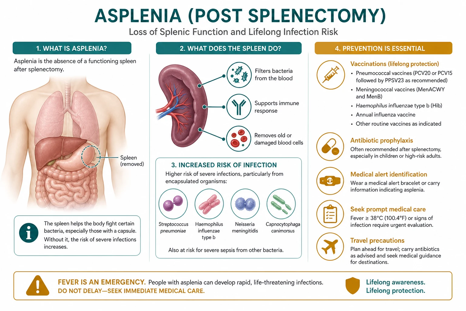
Asplenia (surgical, congenital, or functional from sickle cell) predisposes to overwhelming post-splenectomy infection (OPSI) from encapsulated organisms (Strep pneumoniae, Neisseria meningitidis, Haemophilus influenzae). Management: vaccination (PCV13, PPSV23, MenACWY, MenB, Hib) at least 2 weeks before elective splenectomy or 2 weeks after emergency; lifelong daily antibiotic prophylaxis (phenoxymethylpenicillin 250 to 500 mg BD, or amoxicillin); patient education and alert card; prompt empirical antibiotics for fever (ceftriaxone 2 g IV/IM).
On this page & tools
Your progress
Saved locally on this device.
Exam tags
Red flags
Overview & Definition
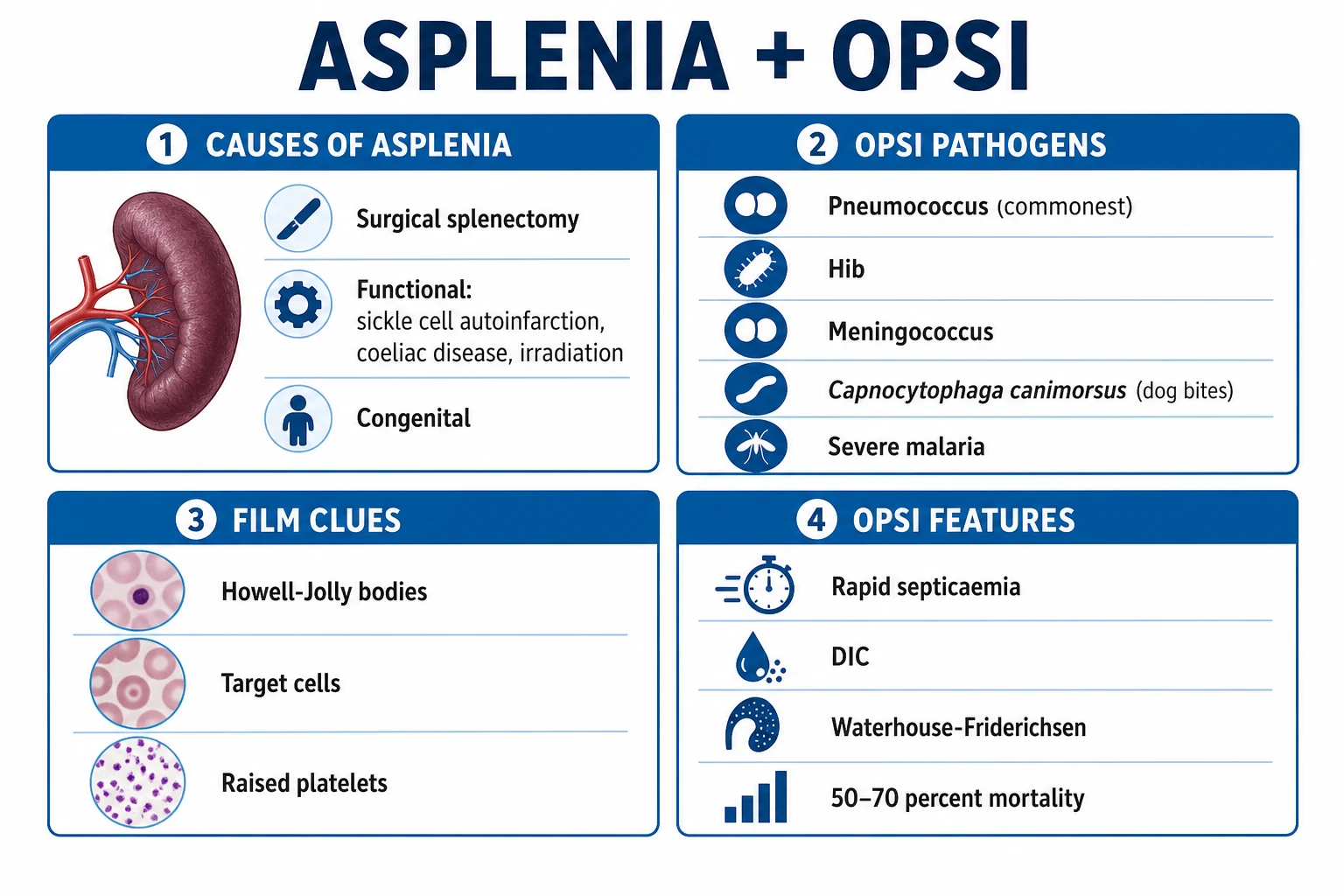
The spleen is the largest lymphoid organ in the body, and its loss — whether by surgical removal, congenital absence, or functional silence — converts an otherwise well patient into one who can be killed within hours by bacteria that a normal host clears without ever noticing. Asplenia is the complete absence of splenic function. It has two forms: anatomical (surgical) asplenia, in which the organ has been physically removed (most often for trauma, immune thrombocytopenia, hereditary spherocytosis, or hypersplenism from a haematological disease); and functional asplenia (hyposplenism), in which the organ is present but its tissue has been destroyed or replaced — classically by repeated infarction in sickle cell disease (so-called autosplenectomy), but also in severe coeliac disease, autoimmune disease, amyloidosis, advanced HIV, and after bone marrow transplant.[1][3]
The unifying clinical consequence is overwhelming post-splenectomy infection (OPSI) — a fulminant, bacteraemic, often meningitic sepsis caused by encapsulated organisms, with a case-fatality of 50 to 70 percent despite modern intensive care. The dominant pathogen is Streptococcus pneumoniae (responsible for 50 to 90 percent of cases), followed by Haemophilus influenzae type b, Neisseria meningitidis, and the dog-bite organism Capnocytophaga canimorsus. The tragedy of OPSI is that almost all of it is preventable: the three pillars of vaccination, antibiotic prophylaxis, and patient education reduce lifetime risk from around 5 percent to under 1 percent. The reason this topic rewards close study is that every step — which vaccines, in what order, how far apart, what antibiotic dose, what standby pack, what to do when a fever appears at midnight — is examinable, and getting any one of them wrong can be fatal.[1][4]
Classification
Asplenia is best classified by mechanism, because mechanism determines onset, reversibility, and the urgency of preventive care. The first division is between anatomical loss (the organ is gone) and functional loss (the organ is present but inert, so the blood film still shows Howell-Jolly bodies and the pitted-erythrocyte count is high). A third, smaller category is congenital absence, which is rare but important because it is frequently missed until a child presents with catastrophic sepsis.[3]
Anatomical (surgical) asplenia
- Trauma — the commonest indication; blunt splenic injury, often emergency splenectomy
- Immune thrombocytopenia (ITP) refractory to medical therapy
- Hereditary spherocytosis / pyruvate kinase deficiency (haemolytic anaemia)
- Hypersplenism from portal hypertension, thalassaemia, or Gaucher disease
- Splenic tumour (haemangioma, lymphangioma) or involvement by lymphoma
- Staging laparotomy for Hodgkin lymphoma (now historical)
- Total pancreatectomy with splenectomy for pancreatic tail tumours
Functional asplenia (hyposplenism)
- Sickle cell disease — the single commonest cause worldwide; autosplenectomy by age 5
- Severe / refractory coeliac disease (splenic atrophy from autoimmune destruction)
- Autoimmune connective-tissue disease (SLE, rheumatoid arthritis)
- Infiltrative disease — amyloidosis, sarcoidosis
- Advanced HIV / AIDS
- Post bone marrow transplant with chronic graft-versus-host disease
- Therapeutic splenic irradiation
Congenital asplenia
- Isolated congenital asplenia — sporadic or autosomal dominant (RPSA / Hox genes); often missed
- Ivemark syndrome (asplenia with heterotaxy and congenital heart disease)
- Polysplenia / heterotaxy syndromes — multiple small spleens with little function
- Present in infancy with severe sepsis; absent Howell-Jolly bodies history initially
Epidemiology & Risk Factors
The lifetime incidence of OPSI in an asplenic adult who receives no specific prevention is around 5 percent, with an incidence rate of roughly 0.23 to 0.42 per 100 person-years. After splenectomy, the risk is highest in the first two to three years, but it never returns to baseline — OPSI has been reported decades after surgery, which is why antibiotic prophylaxis, once started, is increasingly continued for life rather than for a fixed period. Several factors multiply the risk and should be sought deliberately at every review.[4][5]
The risk is concentrated in children younger than 16, in adults older than 50, in patients splenectomised for an underlying haematological malignancy (lymphoma, thalassaemia), in those with concurrent immunosuppression (chemotherapy, transplant, advanced HIV), and — most powerfully — in those who are inadequately vaccinated. The indication for splenectomy matters: splenectomy for trauma carries the lowest long-term OPSI risk (the patient is otherwise well), while splenectomy for haematological disease carries the highest, because the underlying condition itself impairs immunity. Sickle cell disease is a special case: the combination of functional asplenia, auto-infarction, and impaired complement/alternative-pathway activity makes OPSI the leading cause of death in children with sickle cell disease under the age of five in unvaccinated populations.[3]
Pathophysiology
To understand why a small organ can be lethal to lose, it helps to follow what the spleen actually does to blood. Blood enters through the splenic artery, branches into trabecular arteries, and then percolates through two functionally distinct compartments: the white pulp (lymphoid tissue organised around central arterioles, rich in T- and B-cells) and the red pulp (the cords of Billroth and venous sinuses, lined by macrophages). The marginal zone between them is where the immunological work against encapsulated bacteria is done.[3]
Four functions are lost in asplenia, and each maps onto a clinical consequence. First, filtration: the red-pulp macrophages physically remove bacteria, immune complexes, and abnormal or aged red cells from the circulation. Without the spleen, Howell-Jolly bodies (nuclear DNA remnants), Pappenheimer bodies (iron-containing granules), target cells and pitted erythrocytes appear in the peripheral film — these are the morphological signature of splenic absence, not of any single disease. Second, opsonisation and antibody production: the marginal-zone B-cells are the body's specialist responders to T-cell-independent polysaccharide antigens — exactly the kind that form the capsule of pneumococcus, meningococcus and Hib. They produce IgM opsonising antibodies rapidly and, in asplenia, this first-line response is crippled. Third, complement activity: the spleen is a major site of alternative-pathway C3b generation, so loss of the spleen reduces opsonisation of organisms even when antibody is present. Fourth, platelet and cellular pooling: the spleen normally sequesters about one-third of the body's platelets, so splenectomy predictably raises the platelet count (post-splenectomy thrombocytosis).[1][3]
Anatomy and physiology of the spleen — why it cannot be replaced
The spleen sits in the left upper quadrant, weighs about 150 to 200 g in an adult, and receives roughly 5 percent of the cardiac output through the splenic artery. Structurally it is built around three zones — the white pulp, the red pulp, and the marginal zone between them — and each does a distinct job. The white pulp is organised like a lymph node around a central arteriole: a peri-arteriolar lymphoid sheath (PALS) of T-cells, surrounded by B-cell follicles, all clothed by the marginal zone. This is where adaptive immune responses are generated, including the T-cell-dependent response that conjugate vaccines exploit. The red pulp is a sponge of cords of Billroth separated by venous sinuses, lined by littoral cells and packed with macrophages — this is the mechanical filter of the blood, the only tissue in the body that performs this function.[3]
Blood flows through the spleen by two routes. The closed circulation (fast) carries most flow directly from arterioles into venous sinuses. The open circulation (slow) spills blood into the cords, forcing red cells to squeeze through narrow gaps (1 to 3 micrometres) between littoral cells to re-enter the sinuses. This slow transit is the basis of pitting (removal of intra-erythrocytic inclusions such as Howell-Jolly bodies and malarial pigment) and of culling (removal of aged or abnormal red cells such as spherocytes). Asplenic patients lose both, which is why the film shows pitted cells and inclusions and why malaria is uniquely severe.[3]
The marginal-zone B-cells are the immunological jewel of the spleen. They are the only population in the body that mounts a rapid T-cell-independent IgM response to capsular polysaccharide antigens — the very antigens that coat pneumococcus, Hib and meningococcus. Within hours of bacteraemia these cells release IgM that opsonises the organism and fixes complement (C3b), allowing the red-pulp macrophages to clear it. The liver (Kupffer cells) can clear organisms from the blood only when they are already heavily opsonised; the spleen is unique in clearing lightly opsonised organisms at low inoculum. This is the precise deficit that converts a trivial bacteraemia in a normal host into OPSI in an asplenic one. It also explains the vaccine logic: a conjugate vaccine (PCV13, MenACWY) chemically links the polysaccharide capsule to a protein carrier, switching the response from T-cell-independent (lost without the spleen) to T-cell-dependent (preserved, generated in lymph nodes elsewhere), which is why conjugate vaccines remain effective in asplenic patients while plain polysaccharide vaccines (PPSV23) are blunted.[2][3]
The consequence of all four failures is speed. In an asplenic host, the doubling time of pneumococcus in blood is short, there is no splenic filter to slow it, and the IgM/complement first response is weak. The result is a sepsis that can move from a vague flu-like prodrome to refractory septic shock, disseminated intravascular coagulation (DIC), adrenal haemorrhage (Waterhouse-Friderichsen syndrome), and cutaneous purpura fulminans within as little as 12 to 24 hours. This tempo is the entire reason the preventive bundle exists — by the time OPSI declares itself, antibiotics are often too late.[3][6]
Clinical Presentation
Between infections, the asplenic patient is usually asymptomatic, and the only clue may be the scar, the medical-alert bracelet, or the blood film. The clinical question that matters is what OPSI looks like, because recognising it is the difference between life and death.[1]
OPSI classically begins as a non-specific flu-like illness: fever (often high, with rigors), malaise, myalgia, headache, and sometimes a sore throat or gastrointestinal upset. The trap is that the early phase looks trivial, and patients — even educated ones — may take a single standby antibiotic dose and wait. Within hours the picture evolves to severe sepsis: hypotension unresponsive to fluids, tachycardia, tachypnoea, confusion or agitation, oliguria, and lactic acidosis. Meningism may appear (meningococcal or pneumococcal meningitis), and a rash may develop — petechial in meningococcaemia, or the rapidly spreading, sharply demarcated purpura fulminans of overwhelming pneumococcal or meningococcal sepsis. Purpura fulminans is DIC made visible in the skin: large confluent purpuric patches that progress to bullae, digital ischaemia, and distal gangrene, often with laboratory DIC (prolonged PT/APTT, falling fibrinogen, rising D-dimer).[4][6]
Atypical presentations deserve special attention because examiners test them deliberately. In the elderly patient, fever may be blunted and the only sign may be confusion, falls, or hypothermia. In the immunocompromised (post-transplant, chemotherapy), the source may be opportunistic (Capnocytophaga, Babesia, severe malaria). The patient with functional asplenia from sickle cell disease may present with acute chest syndrome and sepsis together. After an animal bite, a dog-bite organism (Capnocytophaga canimorsus) can cause sepsis, meningitis, and DIC within 24 to 72 hours, sometimes with a characteristic purpura on the limbs. The post-splenectomy patient with unexplained thrombocytosis may have portal or mesenteric vein thrombosis presenting as vague abdominal pain, fever, and distension in the first month after surgery.[8][3]
Post-splenectomy thrombocytosis is a near-universal early finding, peaking at 1 to 3 weeks with platelet counts often between 600 and 1000 × 10⁹/L, and occasionally higher. It is the substrate for the splanchnic-vein thrombosis that is the second great post-splenectomy danger. Pulmonary hypertension is a rare, late complication (months to years), attributed to chronic micro-embolism and vascular remodelling.[3]
Differential Diagnosis
The differential of fever in an asplenic patient is narrow in one sense — assume OPSI first — but broad in another, because the asplenic host is uniquely susceptible to a cluster of organisms that a normal host would shrug off. The can't-miss diagnosis is always OPSI / overwhelming encapsulated-organism sepsis; treat first, investigate second.[1]
Encapsulated bacterial sepsis (OPSI)
- Pneumococcus (commonest; purpura fulminans, Waterhouse-Friderichsen)
- Meningococcus — petechial rash, fulminant septicaemia
- Haemophilus influenzae type b — meningitis, epiglottitis in unvaccinated
- Rapid onset, high fever, shock within hours; positive blood cultures
Non-encapsulated bacterial infection
- Escherichia coli / Klebsiella — UTI, biliary, intra-abdominal source
- Staphylococcus aureus — cellulitis, line sepsis, osteomyelitis
- Capnocytophaga canimorsus — dog/cat bite, sepsis + DIC within 24 to 72 h
- Salmonella — especially in sickle cell disease (osteomyelitis)
Parasitic infection (asplenia-specific risk)
- Babesiosis (Babesia microti) — tick-borne, severe haemolysis in asplenic
- Malaria (Plasmodium falciparum) — high parasitaemia, severe disease
- Travel to endemic area; intra-erythrocytic parasites on blood film
Post-splenectomy thrombosis (not infection)
- Portal vein / mesenteric vein / splenic vein thrombosis
- First 1 to 4 weeks post-splenectomy; thrombocytosis
- Abdominal pain, distension, fever, deranged LFTs; CT portal venous phase
The features that distinguish these are the tempo, the exposure history (dog bite, tick, travel), the rash morphology, and the blood film (parasites in Babesia/malaria). But in the febrile asplenic patient, all four categories are covered by the same first action: empirical broad-spectrum antibiotics.[1]
Clinical & Bedside Assessment
The focused assessment of a known asplenic patient has two goals: confirm the asplenic state, and find any source of sepsis. History should establish the indication and date of splenectomy (or the diagnosis behind functional asplenia — sickle cell, coeliac), the vaccination record (which vaccines, exact dates, boosters), the current antibiotic prophylaxis (drug, dose, adherence — non-adherence is the commonest real-world failure), recent infections and travel, animal and tick exposures (dog owners, gardeners, gamekeepers), and whether the patient carries an alert card and standby antibiotics. Ask explicitly about any fever in the last 48 hours, even if it has now settled — a transient fever in an asplenic patient is OPSI until shown otherwise.[1]
Examination begins with the vital signs: the asplenic patient with fever, tachycardia, hypotension, tachypnoea or a rising lactate is in the emergency bundle, not the clinic. Look for meningism, a petechial or purpuric rash, source signs (cellulitis, line infection, abdominal tenderness, respiratory findings), and signs of thrombosis (limb swelling, abdominal distension). Abdominal examination should note any splenomegaly — a palpable spleen argues against complete asplenia and raises the question of functional hyposplenism or an accessory spleen. The peripheral blood film is the single most useful bedside test: Howell-Jolly bodies, target cells, Pappenheimer bodies (siderotic granules) and pitted erythrocytes together confirm splenic hypofunction and, in the undiagnosed patient with unexplained sepsis, should trigger the question "is this patient asplenic?"[3]
Investigations
Investigations serve two purposes: documenting the asplenic state (when it is not already known), and working up suspected infection.[1]
Confirming asplenia. The peripheral film is the bedside gold standard — Howell-Jolly bodies (DNA remnants, seen in 1 to 2 percent of red cells in established asplenia), target cells, Pappenheimer bodies and pitted erythrocytes. The pitted-erythrocyte count (the percentage of red cells with surface pits seen on interference-contrast microscopy) is the most sensitive functional measure: a count over 3.5 percent indicates functional asplenia. When the diagnosis is in doubt, a technetium-99m sulphur colloid liver-spleen scan shows absent or markedly reduced splenic uptake, and an ultrasound or CT abdomen confirms an absent spleen or reveals accessory spleens (splenunculi) that may preserve partial function.[3]
Working up suspected infection. Full blood count typically shows neutrophilic leucocytosis and thrombocytosis (or thrombocytopenia in sepsis/DIC). Blood cultures — at least two sets from separate sites — are taken before antibiotics only if it does not delay them; in suspected OPSI, antibiotics come first. Coagulation (PT, APTT, fibrinogen, D-dimer) screens for DIC. A peripheral film is reviewed for organisms (intra-erythrocytic rings of Babesia or malaria) and for evidence of haemolysis. Urinalysis and urine culture, chest X-ray, and blood gas with lactate are baseline. Lumbar puncture is performed after stabilisation and only if there is no coagulopathy or signs of raised intracranial pressure; in the typical asplenic patient with purpura fulminans, the LP is deferred and empirical meningitis cover given. PCR (pneumococcal, meningococcal) on blood/CSF confirms the organism later. Imaging for a source (CT abdomen/pelvis with portal-venous phase if intra-abdominal source or splanchnic thrombosis suspected) follows resuscitation. Vaccine-response titres (anti-pneumococcal IgG) are checked in follow-up if vaccine failure is suspected.[1]
Interpreting the investigations — three patterns examiners probe
The pitted-erythrocyte count deserves a closer look because it quantifies hyposplenism rather than just confirming it. Counts below 2 percent are normal; 2 to 3.5 percent suggest partial hyposplenism (e.g. early coeliac disease, recovering sickle cell); over 3.5 percent indicate functional asplenia equivalent to surgical removal. This gradation matters when deciding whether a patient with residual splenic tissue — after partial splenectomy, splenic embolisation, or with accessory spleens — still needs the full preventive bundle; a borderline count argues for treating them as asplenic until a repeat confirms recovery.[3]
The coagulation picture in established OPSI is the laboratory face of DIC: prolonged PT and APTT, markedly reduced fibrinogen (often under 1.0 g/L), elevated D-dimer, a falling platelet count (against the expected post-splenectomy thrombocytosis — a platelet count that drops in a febrile asplenic patient is a red flag for consumption), and fragmented red cells (schistocytes) on the film. The blood gas shows a worsening metabolic acidosis with a rising lactate; a lactate over 2 mmol/L that fails to clear with resuscitation predicts mortality. The film in Babesiosis shows intra-erythrocytic ring forms resembling malaria but without pigment, often with a high parasite load and haemolysis; in malaria, ring forms plus pigment are seen, and asplenic patients develop hyperparasitaemia with cerebral involvement rapidly.[4][6]
Vaccine-response titres are reserved for follow-up, never the acute episode. Anti-pneumococcal IgG is measured at least 4 to 8 weeks after vaccination: a protective response is generally accepted as over 0.35 micrograms per millilitre for at least 70 percent of vaccine serotypes in children, and a robust adult response is over 1.0 micrograms per millilitre across the majority of serotypes. A non-response identifies the patient who needs a conjugate re-vaccination (PCV) despite having received PPSV23, and the small group who need lifelong prophylaxis regardless of adherence because vaccines simply do not work for them. Anti-Hib and anti-meningococcal (serum bactericidal antibody, SBA) titres are checked when vaccine failure is suspected clinically (a breakthrough infection despite documented vaccination).[2]
Management — Resuscitation
Suspected OPSI is a time-critical, do-it-now emergency. The single most important principle is that empirical antibiotics must not wait for blood cultures, imaging, or a senior review — every minute of delay increases mortality. The asplenic patient with fever and any sign of sepsis is managed with the sepsis six plus asplenia-specific antibiotic cover.[1][6]
Immediate bundle for suspected OPSI
ABCDE; high-flow oxygen; two large-bore IV cannulae; continuous monitoring
Blood cultures x 2 sets AND lactate, FBC, coagulation, U&E, LFT, glucose, blood gas — drawn rapidly, only if it does NOT delay antibiotics
EMPIRICAL ANTIBIOTICS NOW — ceftriaxone 2 g IV stat (covers pneumococcus, meningococcus, Hib); add vancomycin 25 to 30 mg/kg IV loading then 15 to 20 mg/kg q12h if penicillin-resistant pneumococcus suspected or local prevalence high
Add metronidazole 500 mg IV q8h if intra-abdominal source; add doxycycline 100 mg IV q12h if tick exposure (Babesia, Anaplasma); add amoxicillin-clavulanate 1.2 g IV if dog-bite (Capnocytophaga)
Aggressive IV crystalloid resuscitation — 30 mL/kg bolus in first 3 h; reassess; repeat as needed
Vasopressors (noradrenaline first-line) for fluid-refractory shock; target MAP 65 mmHg
ICU admission for organ support; intubate early if obtunded, hypoxic, or failing to perfuse
Treat DIC — fresh frozen plasma, platelets, cryoprecipitate guided by coagulation; purpura fulminans may need protein C concentrate and surgical debridement/amputation of necrotic tissue
Source control — drain abscess, remove infected line, treat splanchnic thrombosis
The antibiotic choice reflects the pathogens. Ceftriaxone 2 g IV is the workhorse: it covers pneumococcus (including most penicillin-resistant strains at this dose), meningococcus and Hib in one daily dose, and is suitable for community-administered first-line therapy. Vancomycin is added when highly resistant pneumococcus is a concern or the patient has meningitis (to guarantee CSF penetration). Meningitis is treated with ceftriaxone 2 g IV q12h plus vancomycin, plus dexamethasone 10 mg IV q6h given just before or with the first antibiotic dose (pneumococcal benefit). Activated protein C (drotrecogin alfa) was withdrawn in 2011 and is no longer used. Even with optimal management, OPSI mortality remains 50 to 70 percent, which is precisely why prevention matters more than cure.[4][5]
Management — Definitive & Stepwise
Definitive management of the asplenic patient is the preventive bundle: vaccination, antibiotic prophylaxis, and patient education. This is delivered before elective splenectomy wherever possible, and immediately afterwards when splenectomy is an emergency.[1]
Vaccination
Vaccination is the single most effective preventive measure. The aim is to immunise before the spleen is lost, so that the conjugate vaccines can generate a robust T-cell-dependent antibody response. For elective splenectomy, all vaccines are given at least 2 weeks (ideally 4 weeks) before surgery. For emergency splenectomy (trauma, ruptured spleen), vaccination is given at least 2 weeks after the operation, once the acute inflammatory response has settled — vaccinating in the immediate post-operative period produces a blunted response. The same schedule applies when functional asplenia is first recognised (e.g. new diagnosis of severe coeliac disease, or sickle cell disease at first presentation).[1][2]
Key principle — conjugate before polysaccharide. PCV13/PCV15/PCV20 (conjugate) generates a T-cell-dependent response and must come first; giving PPSV23 first blunts the response to subsequent conjugate vaccine. Hence the fixed order: conjugate, wait at least 8 weeks, then polysaccharide. Children under 2 respond poorly to plain polysaccharide vaccines, which is why conjugate vaccines transformed the outlook in sickle cell disease.[2]
Antibiotic prophylaxis
Antibiotic prophylaxis is the second pillar. The traditional regimen is lifelong but most guidelines now recommend at least 2 to 5 years in adults after splenectomy, continuing for life in any patient with ongoing risk (underlying haematological disease, immunosuppression, inadequate vaccine response, or a previous severe infection), and lifelong in children.[1][7]
Phenoxymethylpenicillin (penicillin V)
Dose
Adult: 500 mg twice daily (250 mg BD acceptable for lower-risk adults). Child under 5: 125 mg BD; 5 to 14 years: 250 mg BD; over 14 years: 500 mg BD
Amoxicillin
Dose
Adult: 250 to 500 mg once daily; child: dose by weight
Erythromycin (or clarithromycin)
Dose
Erythromycin 500 mg twice daily; clarithromycin 250 mg BD
In addition to continuous prophylaxis, every asplenic patient should carry a standby rescue course of antibiotics to take at the first sign of fever, rigors, or systemic illness, before they reach medical care. The standard rescue pack is amoxicillin-clavulanate 1 g (500/125 mg two tablets) orally as a single stat dose at the onset of fever, followed by immediate attendance at an emergency department. Penicillin-allergic patients carry levofloxacin 500 mg or moxifloxacin 400 mg. This "dose-and-go" strategy acknowledges that OPSI can outpace travel to hospital.[1][5]
Patient education
Patient education is the third pillar and the most easily neglected. Every asplenic patient must (a) carry an alert card or wear a medical-alert bracelet stating "asplenic — risk of overwhelming infection"; (b) understand that any fever or systemic illness is an emergency requiring the standby dose plus immediate medical assessment; (c) know the importance of adherence to daily prophylaxis and to the booster schedule; (d) treat any animal bite as an emergency (seek antibiotics within hours); (e) take malaria prophylaxis and avoid travel to highly endemic areas where possible; and (f) inform dentists, surgeons, and new doctors of the asplenic status. A structured annual review — checking adherence, vaccine boosters, and reinforcing the message — improves outcomes.[1]
Post-splenectomy thrombocytosis and thrombosis
Post-splenectomy thrombocytosis peaks at 1 to 3 weeks. For platelets over 600 × 10⁹/L, many centres use low-dose aspirin 75 mg daily; for sustained counts over 1000 × 10⁹/L, or established splanchnic-vein thrombosis, therapeutic anticoagulation with low-molecular-weight heparin then warfarin/DOAC is indicated. Portal, splenic and mesenteric vein thrombosis is actively sought with CT portal-venous phase in any post-splenectomy patient with unexplained fever, abdominal pain or distension in the first month.[3]
First two years after splenectomy — what to do and when
Specific Subtypes & Scenarios
Sickle cell disease with functional asplenia. Repeated splenic infarction produces functional asplenia by age 5 (often earlier), making the sickle cell child the highest-risk paediatric group. The PROPS trial (Gaston 1986) established that oral penicillin prophylaxis from age 2 months reduces pneumococcal bacteraemia by 84 percent in young children with sickle cell disease; PROPS II (1995) later showed it can be safely discontinued after age 5 in well-vaccinated children without a prior severe pneumococcal infection, though many haematologists continue it longer. The full asplenia vaccination schedule applies from infancy, with conjugate vaccines on the routine childhood programme.[7]
Trauma splenectomy. The commonest indication in adults. Because it is an emergency, vaccination is never pre-operative — give 2 weeks post-splenectomy. Penicillin prophylaxis is started immediately and continued for at least 2 to 5 years (lifelong if any doubt about accessory-spleen function). Modern trauma practice favours splenic preservation (non-operative management, splenic embolisation, partial splenectomy) precisely to preserve immune function, but anyone with any degree of asplenia after trauma should be managed as fully asplenic until pitted-erythrocyte counts confirm residual function.[4]
Splenectomy for ITP. Lower long-term OPSI risk than for haematological malignancy (the patient is otherwise immunocompetent), but the standard preventive bundle still applies. Antibiotic prophylaxis for at least 2 to 5 years, then reassess.[1]
Splenectomy for hereditary spherocytosis. Lifelong prophylaxis is recommended; the autosomal-dominant inheritance means family screening of first-degree relatives for spherocytes and asplenia status is worthwhile. Partial (subtotal) splenectomy is increasingly used in children with haemolytic anaemia to preserve some splenic function and lower OPSI risk while reducing haemolysis.[3]
Animal bite in an asplenic patient. Capnocytophaga canimorsus, a commensal of dog and cat mouths, causes overwhelming sepsis, meningitis, and DIC in asplenic patients, often within 24 to 72 hours of a seemingly trivial bite. Every asplenic patient with a dog or cat bite should receive amoxicillin-clavulanate 500/125 mg three times daily for 5 to 7 days (or doxycycline 100 mg BD if penicillin-allergic) immediately, and be warned to present urgently with any systemic symptom. Established sepsis is managed as OPSI with aggressive resuscitation and ICU support.[8]
Splenosis and accessory spleens. Splenosis (autotransplantation of splenic tissue after traumatic rupture) and accessory spleens (splenunculi) may preserve some splenic function, but they do not reliably prevent OPSI — manage these patients with the full preventive bundle unless pitted-erythrocyte counts and a colloid scan confirm robust residual function.[3]
Complications & Pitfalls
Infectious complications
- OPSI — encapsulated-organism sepsis; mortality 50 to 70 percent
- Meningitis — pneumococcal or meningococcal
- Purpura fulminans and DIC — digital ischaemia, gangrene, amputation
- Waterhouse-Friderichsen syndrome — bilateral adrenal haemorrhage, acute adrenal failure
- Severe babesiosis and malaria — parasite clearance impaired
- Capnocytophaga canimorsus sepsis after animal bite
Thrombotic / vascular
- Post-splenectomy thrombocytosis (platelets 600 to 1000 × 10⁹/L)
- Portal, splenic, and mesenteric vein thrombosis (first month)
- Late pulmonary hypertension (rare, vascular remodelling)
Classic pitfalls (how patients die)
- Fever dismissed as viral — delayed antibiotics
- Patient ran out of penicillin V and did not refill
- Vaccines given AFTER emergency splenectomy in the first 48 h (blunted response)
- PPSV23 given before PCV13 (wrong order, blunted response)
- Dog bite not treated — Capnocytophaga sepsis
- PPSV23 booster forgotten at 5 years
The two recurring fatal pitfalls are delay in empirical antibiotics for a febrile asplenic patient (treat first, culture second), and preventable vaccine failure from wrong timing or a missed booster. The third is non-adherence — the patient who quietly stops penicillin because they "felt well" — which is why a structured annual review with a non-judgemental adherence check is part of good care.[1][5]
Prognosis & Disposition
With the full preventive bundle — vaccination, lifelong prophylaxis, and educated patient — the asplenic patient has a near-normal life expectancy and lifetime OPSI risk of under 1 percent. Without prevention, lifetime OPSI risk is about 5 percent with a case-fatality of 50 to 70 percent once it occurs. The risk is highest in the first two years after splenectomy, in young children, and in those with underlying haematological malignancy, but it never fully disappears, which is why most modern guidance leans towards lifelong rather than time-limited prophylaxis. Disposition from the emergency department for any febrile asplenic patient is admit and treat as OPSI until proven otherwise; safe discharge requires an identified and treated source, normal observations, an educated patient, and clear safety-net advice.[4]
Special Populations
Children. Penicillin prophylaxis is started from age 2 months in sickle cell disease (PROPS) and after any splenectomy; dosing is age-stratified (under 5: 125 mg BD; 5 to 14: 250 mg BD; over 14: 500 mg BD). Conjugate vaccines are essential because plain polysaccharide vaccines are immunogenic only after age 2. Discontinuation of penicillin after age 5 in well-vaccinated sickle cell children is evidence-based (PROPS II) but must be individualised.[7]
Pregnancy. The asplenic pregnant patient has the same OPSI risk and should continue penicillin V or amoxicillin throughout pregnancy (both are safe). Vaccinations are reviewed and brought up to date; inactivated vaccines (pneumococcal, influenza, MenACWY conjugate) are safe in pregnancy; live vaccines are avoided. There are no specific fetal concerns from asplenia itself.[1]
The elderly. Vaccine response is less robust; ensure PCV and PPSV23 are both given and check anti-pneumococcal titres in selected cases. Adherence and alert-card awareness may need reinforcement. Co-morbidities (chronic lung, heart disease) compound sepsis risk.[1]
Immunocompromised (transplant, chemotherapy, advanced HIV). Higher OPSI risk; conjugate vaccines are preferred (T-cell-dependent response is more reliable); lifelong prophylaxis; consider additional vaccines (RSV, COVID-19 boosters). Transfusion-associated graft-versus-host disease is a rare risk in heavily immunosuppressed asplenic patients — irradiated blood products may be indicated.[2]
Travellers. Malaria prophylaxis is mandatory in endemic areas, and asplenic patients should ideally avoid highly endemic regions; severe malaria in asplenia carries high mortality. A standby rescue pack (amoxicillin-clavulanate) is carried for remote travel. Destination-specific vaccines (typhoid, hepatitis A, yellow fever — note yellow fever is live and avoided in severe immunocompromise) are reviewed.[1]
Evidence, Guidelines & Regional Differences
Foundational evidence
- Gaston 1986 (PROPS) NEJM — penicillin prevents pneumococcal sepsis in sickle cell children
- PROPS II 1995 — safe discontinuation after age 5 in selected children
- Waghorn 1997 — 42 OPSI episodes; emergency prophylactic antibiotic analysis
- Theilacker 2016 — prospective multicentre OPSI cohort; modern incidence
- Di Sabatino & Carsetti 2011 Lancet — comprehensive pathophysiology review
Guidelines
- UK BSH / Green Book (Davies 2011) — UK standard schedule
- US ACIP / IDSA (Rubin 2014) — US immunocompromised vaccination
- PHAC / Canada — Canadian immunization guide, asplenia chapter
- WHO position papers on pneumococcal and meningococcal vaccines
Regional deltas
- UK: PCV13 then PPSV23; MenACWY + MenB both given; PPSV23 booster at 5 yr
- US: PCV15/PCV20 preferred; PCV20 alone suffices (no PPSV23); MenB series + boosters
- Penicillin V vs amoxicillin as first-line — both accepted; amoxicillin once-daily preferred for adherence
- Duration of prophylaxis: UK leans lifelong; some US guidance 2 to 5 yr in low-risk adults
The evidence base is strong on vaccination and penicillin prophylaxis (PROPS is one of the landmark trials of paediatric haematology) and weaker on the optimal duration of adult prophylaxis and on standby rescue antibiotics (no randomised trial; based on cohort data and mechanistic reasoning). The shift from PCV13/PPSV23 to PCV20 in the US illustrates how fast this field moves — always confirm against the live national immunisation schedule before quoting a specific vaccine.[1][2][7]
Exam Pearls & High-Yield Minutiae
SPLEEN
- OPSI = overwhelming post-splenectomy infection; pneumococcus in 50 to 90 percent; mortality 50 to 70 percent; onset within hours; purpura fulminans + Waterhouse-Friderichsen + DIC.[4]
- Howell-Jolly bodies on a blood film = absent/dysfunctional spleen (until proven otherwise). Also target cells, Pappenheimer bodies, pitted erythrocytes over 3.5 percent.
- Vaccine order is sacred: conjugate (PCV13) first, then polysaccharide (PPSV23) at least 8 weeks later — wrong order blunts the response.
- MenACWY + MenB both required (meningococcus has both capsule groups and serogroup B).
- Standby rescue pack: amoxicillin-clavulanate 1 g stat at first fever, then straight to hospital.
- Dog bite = emergency: Capnocytophaga canimorsus sepsis within 24 to 72 hours — give amoxicillin-clavulanate immediately.
- Sickle cell: functionally asplenic by age 5; penicillin from 2 months (PROPS); same full schedule.
- Emergency splenectomy: vaccinate 2 weeks post-op (response is blunted if given in the first 48 hours).
- Post-splenectomy thrombocytosis peaks at 1 to 3 weeks; aspirin if platelets over 600 × 10⁹/L; watch for portal/mesenteric vein thrombosis.
- Malaria and Babesia are uniquely severe in asplenia — the spleen is the main organ of parasite clearance.
- Pregnancy: continue penicillin/amoxicillin throughout; inactivated vaccines safe.
- Treat fever as sepsis: ceftriaxone 2 g IV/IM, add vancomycin if resistant pneumococcus; do not wait for cultures.
Exam application bank (NEET-PG / INICET)
One-line answer
Asplenia (surgical, congenital, or functional from sickle cell) predisposes to overwhelming post-splenectomy infection (OPSI) from encapsulated organisms (Strep pneumoniae, Neisseria meningitidis, Haemophilus influenzae). Management: vaccination (PCV13, PPSV23, MenACWY, MenB, Hib) at least 2 weeks before elective splenectomy or 2 weeks after emergency; lifelong daily antibiotic prophylaxis (phenoxymethylpenicillin 250 to 500 mg BD, or amoxicillin); patient education and alert card; prompt empirical antibiotics for fever (ceftriaxone 2 g IV/IM). [1]
Worked stems (answer without another resource)
Stem 1 — Classic presentation. Map symptoms to mechanism; name the first investigation and first treatment step with dose/route if drug therapy is standard. [1]
Stem 2 — Unstable / complicated. List red flags that force immediate resuscitation, theatre, ICU, antidote, or reperfusion — and what you do in the first 15 minutes. [1]
Stem 3 — Atypical group. Elderly, pregnancy, child, or immunocompromised: how presentation and thresholds change. [1]
Stem 4 — Differential trap. Name the three closest mimics and one discriminator for each. [1]
Stem 5 — Disposition. Who goes home with safety-netting, who is admitted, who needs HDU/ICU/theatre, and what follow-up is mandatory. [1]
Rapid viva checklist
- Definition + classification
- Pathophysiology chain
- Bedside signs / criteria
- Score with exact components (if any)
- Emergency bundle
- Definitive therapy with doses
- Complications of disease and of treatment
- Special populations
- Guideline/trial name if classic
- Three exam traps
Coverage self-check
If you cannot answer any stem above from this page alone, re-read the matching section — the page is intended to be self-sufficient for final-prof and NEET-PG/INICET questions on Asplenia and Post-Splenectomy Care.
References
- [1]Davies JM, Lewis MP, Wimperis J, Rafi I, Ladhani S, Bolton-Maggs PH Review of guidelines for the prevention and treatment of infection in patients with an absent or dysfunctional spleen: prepared on behalf of the British Committee for Standards in Haematology by a working party of the Haemato-Oncology task force Br J Haematol, 2011.PMID 21988145
- [2]Rubin LG, Levin MJ, Ljungman P, et al. 2013 IDSA clinical practice guideline for vaccination of the immunocompromised host Clin Infect Dis, 2014.PMID 24421306
- [3]Di Sabatino A, Carsetti R, Corazza GR Post-splenectomy and hyposplenic states Lancet, 2011.PMID 21474172
- [4]Theilacker C, Ludewig K, Serr A, et al. Overwhelming Postsplenectomy Infection: A Prospective Multicenter Cohort Study Clin Infect Dis, 2016.PMID 26703862
- [5]Waghorn DJ, Mayon-White RT A study of 42 episodes of overwhelming post-splenectomy infection: is current guidance for asplenic individuals being followed? J Infect, 1997.PMID 9459404
- [6]Brigden ML, Pattullo AL Prevention and management of overwhelming postsplenectomy infection--an update Crit Care Med, 1999.PMID 10321679
- [7]Gaston MH, Verter JI, Woods G, et al. Prophylaxis with oral penicillin in children with sickle cell anemia. A randomized trial N Engl J Med, 1986.PMID 3086721
- [8]Zajkowska J, Krol M, Falkowski D, et al. Capnocytophaga canimorsus – an underestimated danger after dog or cat bite – review of literature Przegl Epidemiol, 2016.PMID 27837588