Haematology · General Medicine
Coagulation Disorders (Haemophilia & von Willebrand)
Also known as Haemophilia · von Willebrand disease · VWD · Coagulation disorders · Bleeding disorders · Christmas disease
Haemophilia A (factor VIII deficiency, X-linked recessive, F8 gene, 1 in 5000 males) and haemophilia B (factor IX deficiency / Christmas disease, F9 gene, 1 in 30 000 males) are the classic inherited coagulopathies, presenting with deep-tissue bleeds — haemarthrosis of knees/elbows/ankles, intramuscular bleeds, and (in severe disease) spontaneous intracranial haemorrhage. Von Willebrand disease (VWD) is the commonest inherited bleeding disorder (up to 1 in 100, autosomal dominant), causing mucocutaneous bleeding (menorrhagia, epistaxis, gum, dental). Diagnosis rests on a prolonged APTT with normal PT and platelets that corrects on mixing, confirmed by factor VIII/IX assays (haemophilia) and vWF antigen plus ristocetin cofactor activity (VWD). Treatment: factor VIII replacement 25 to 50 IU/kg for bleeding and 25 to 40 IU/kg three times weekly for prophylaxis; factor IX 40 to 80 IU/kg for haemophilia B; emicizumab 1.5 mg/kg subcutaneously monthly (bispecific antibody) for haemophilia A prophylaxis; desmopressin 0.3 microgram/kg for mild haemophilia A and type 1 VWD; vWF concentrate for VWD types 2 and 3; tranexamic acid 1 g three times daily; and gene therapy (valoctocogene roxaparvovec for A, etranacogene dezaparvovec for B) now approved.
On this page & tools
Your progress
Saved locally on this device.
Practise this topic
Exam tags
Red flags
Overview & Definition
Inherited coagulation disorders are defined by which protein of the coagulation cascade is missing or dysfunctional. The two numerically dominant conditions are the haemophilias — deficiency of factor VIII (haemophilia A, about 80 percent of cases) or factor IX (haemophilia B / Christmas disease, about 20 percent) — both transmitted in an X-linked recessive pattern and therefore affecting males, while females are carriers. Von Willebrand disease (VWD) is mechanistically distinct: it is a quantitative or qualitative defect of von Willebrand factor (vWF), the protein that both mediates platelet adhesion to injured endothelium and carries and stabilises factor VIII in plasma.[1][2]
The clinical pattern mirrors the biochemistry. Haemophilia cripples secondary haemostasis (the fibrin-forming arm), so patients bleed late and deep — into joints, muscles and the central nervous system — characteristically hours after injury with rebleeding. VWD cripples primary haemostasis (the platelet-plug arm), so patients bleed immediately and superficially — from mucous membranes (nose, gums, gut, heavy menses) — though loss of the factor VIII carrier function adds a variable secondary coagulation defect. A third, smaller family comprises the rare factor deficiencies (factors XI, VII, V, X, XIII and fibrinogen), most autosomal recessive, each with a distinctive clinical signature.[1]
Modern treatment has transformed these conditions. Recombinant factor VIII and IX, the subcutaneous bispecific antibody emicizumab, non-factor rebalancing therapies and AAV-vector gene therapy (valoctocogene roxaparvovec for A, etranacogene dezaparvovec for B) have converted haemophilia from a crippling, life-shortening disease into a manageable chronic condition with near-normal life expectancy in countries that can fund the therapies.[1][4]
Classification
Coagulation disorders classify along three axes — the deficient factor, its severity, and whether the defect is inherited or acquired.[1][2]
Inherited coagulopathies: [1]
- Haemophilia A — factor VIII deficiency; X-linked recessive (F8 gene at Xq28); ~80 percent of haemophilia; incidence ~1 in 5000 male live births.
- Haemophilia B (Christmas disease) — factor IX deficiency; X-linked recessive (F9 gene at Xq27); ~20 percent; incidence ~1 in 30 000 male live births. Named for Stephen Christmas, the first patient described in 1952.
- VWD type 1 — partial quantitative deficiency of vWF; autosomal dominant; mildest and commonest (about 75 percent of VWD).
- VWD type 2 — qualitative (dysfunctional) vWF; autosomal dominant (mostly); four subtypes — 2A (loss of high-molecular-weight multimers, most common type 2), 2B (gain-of-function — increased platelet binding causing thrombocytopenia), 2M (normal multimers but reduced function), 2N (defective factor VIII binding — mimics mild haemophilia A).
- VWD type 3 — virtual absence of vWF; autosomal recessive; rarest but severest (resembles moderate-to-severe haemophilia with both mucocutaneous and deep bleeds).
- Rare factor deficiencies — factors XI (haemophilia C, Ashkenazi Jewish predilection), VII, V, X, XIII, and fibrinogen (afibrinogenaemia / dysfibrinogenaemia); mostly autosomal recessive. [1]
Acquired coagulopathies include acquired haemophilia A (autoantibodies neutralising factor VIII), disseminated intravascular coagulation (DIC), liver disease, vitamin K deficiency, and anticoagulant-related bleeding (warfarin, direct oral anticoagulants).[6]
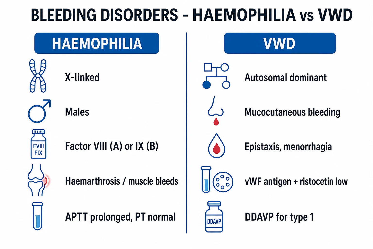
Haemophilia A / B
- X-linked recessive — males affected, females carriers
- Factor VIII (A, 80%) or IX (B, 20%) deficiency
- DEEP bleeds: haemarthrosis (knees/elbows/ankles), muscle, ICH
- APTT prolonged; PT and platelets normal; corrects on mixing
- Severe under 1%; moderate 1 to 5%; mild over 5%
- Treat: factor VIII 25 to 50 IU/kg; emicizumab 1.5 mg/kg SC monthly; gene therapy
Von Willebrand disease
- Autosomal dominant (type 3 recessive)
- vWF quantitative (type 1/3) or qualitative (type 2) defect
- MUCOCUTANEOUS bleeds: epistaxis, menorrhagia, gum, dental
- vWF antigen and ristocetin cofactor low; factor VIII may be low
- Type 1 mild/commonest; type 2B has thrombocytopenia; type 3 severe
- Treat: DDAVP 0.3 mcg/kg (type 1); vWF concentrate (types 2, 3); tranexamic acid 1 g TDS
Acquired haemophilia
- Autoantibodies (inhibitors) neutralise factor VIII
- Elderly, pregnancy/postpartum, autoimmune, malignancy
- No childhood/family history; bleeding disproportionate to factor level
- APTT prolonged, does NOT correct on mixing (inhibitor)
- Bethesda assay quantifies inhibitor titre
- Treat: bypassing agents (rFVIIa, FEIBA); immunosuppression (steroids, rituximab)
Haemophilia severity — the single most important prognostic variable — is graded by the endogenous factor activity, not by bleeding history:[1]
- Severe — factor level under 1 IU/dL (under 1 percent): spontaneous bleeds (joints, muscles, intracranial) beginning in infancy; 2 to 5 spontaneous bleeds per month without prophylaxis.
- Moderate — factor level 1 to 5 IU/dL (1 to 5 percent): bleeding after minor trauma or surgery; occasional spontaneous bleeds.
- Mild — factor level 5 to 40 IU/dL (5 to 40 percent): bleeding only after significant trauma, surgery or dental work; may escape diagnosis until adulthood. [1]
Epidemiology & Risk Factors
Haemophilia A affects approximately 1 in 5000 male live births and haemophilia B approximately 1 in 30 000 — together roughly 400 000 people worldwide. All ethnicities are equally affected because the defect is X-linked; males are affected, females are obligate carriers (rare symptomatic carriers occur with skewed X-inactivation, Turner syndrome, or homozygosity). About 30 percent of cases arise from a de novo mutation with no family history — so a negative family history does not exclude haemophilia in a boy with a suggestive bleeding pattern.[1]
Von Willebrand disease is the commonest inherited bleeding disorder, with a symptomatic prevalence of up to 1 in 100 (much higher prevalence estimates from screening reflect mostly asymptomatic low-vWF individuals). Type 1 accounts for about 75 percent, type 2 for 20 to 25 percent, and type 3 is rare (1 in 500 000 to 1 in 3 million). Blood group O lowers vWF by about 25 percent and is a common reason for borderline-low vWF in someone with mild bleeding.[2]
Risk factors for an acquired coagulopathy include advanced age (acquired haemophilia autoantibodies), pregnancy and the postpartum period, autoimmune disease (systemic lupus erythematosus, rheumatoid arthritis), lymphoproliferative and solid malignancy, sepsis and trauma (DIC), chronic liver disease, malabsorption, biliary obstruction, prolonged antibiotic use and parenteral nutrition without vitamin K (vitamin K deficiency), and anticoagulant therapy (warfarin, direct oral anticoagulants).[6]
Pathophysiology
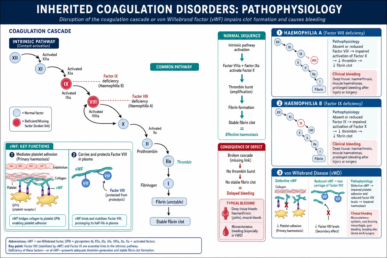
The coagulation cascade is organised into a thrombin-generation engine. The intrinsic tenase complex — factor IXa together with its cofactor factor VIIIa on the activated platelet phospholipid surface — activates factor X to Xa, which (with Va) drives the thrombin burst that converts fibrinogen to a stable, cross-linked fibrin clot. Factor VIII and factor IX are therefore the rate-limiting cofactors of secondary haemostasis. Deficiency of either cripples thrombin generation: a weak, delayed fibrin clot forms, with poor activation of factor XIII (so the clot is not properly cross-linked) and defective activation of the thrombin-activatable fibrinolysis inhibitor (so the clot is unusually susceptible to premature lysis). The net result is the clinical hallmark of haemophilia — delayed, deep-tissue rebleeding into joints and muscles.[1]
Von Willebrand factor has two distinct jobs. First, the high-molecular-weight multimers of vWF bridge exposed subendothelial collagen to the platelet glycoprotein GPIb-IX-V receptor under high shear stress — this is the initiating step of primary haemostasis (platelet adhesion). Second, vWF is the plasma carrier and stabiliser of factor VIII, protecting it from premature clearance and proteolysis and extending its half-life from minutes to roughly 12 hours. VWD is therefore a double hit: defective platelet adhesion produces the mucocutaneous bleeding phenotype, and loss of the carrier function produces a secondary factor VIII deficiency (low vWF leads to unstable, rapidly cleared factor VIII, which lowers factor VIII levels and prolongs the APTT). This explains why VWD can mimic haemophilia and why both conditions can show a prolonged APTT with a low factor VIII.[2]
The four VWD subtypes reflect distinct molecular lesions. In type 1 there is a partial quantitative reduction in normally functioning vWF. In type 2 the vWF is present but dysfunctional: 2A (loss of high-molecular-weight multimers, the commonest type 2), 2B (gain-of-function mutation causing spontaneous binding to platelets and thrombocytopenia, worsened by desmopressin), 2M (normal multimer distribution but reduced function), and 2N (a defective factor VIII binding site on vWF causing isolated factor VIII reduction that mimics mild haemophilia A). In type 3, homozygous or compound-heterozygous loss produces virtual absence of vWF, the severest form.[2]
Clinical Presentation
Haemophilia A and B
The bleeding phenotype of haemophilia A and haemophilia B is clinically indistinguishable; they differ only in the deficient factor. Severity dictates the tempo. Severe haemophilia (factor under 1 percent) presents in infancy with spontaneous bleeding; moderate bleeds after minor trauma; mild may not declare itself until adult life after surgery or dental extraction.[1]
Haemarthrosis is the hallmark — bleeding into the knees, elbows and ankles (the "target joints"). The joint becomes swollen, warm, painful and held in flexion; recurrent bleeds drive haemophilic arthropathy (synovial hypertrophy, cartilage destruction, fixed deformity) — historically the leading cause of long-term disability. Intramuscular bleeds favour the iliopsoas (groin pain, hip held flexed, femoral nerve compression with quadriceps weakness), the calf and forearm (compartment syndrome risk), and the gluteal region. Mucocutaneous and visceral bleeds — gastrointestinal (often from peptic ulcer or vascular malformation), haematuria (common, usually painless) and epistaxis — occur but are less dominant than in VWD. [1]
Intracranial haemorrhage is the most feared bleed and the leading cause of death in severe haemophilia, especially in neonates after instrumental delivery and in infants and toddlers after minor head trauma. Any haemophilia patient with a headache, vomiting, altered consciousness, seizure or a head injury must be treated empirically with factor to 100 percent before imaging.[1]
Von Willebrand disease
VWD causes mucocutaneous bleeding: epistaxis, gum bleeding, easy bruising, prolonged bleeding after dental work or minor wounds, and — characteristically in women — heavy menstrual bleeding (menorrhagia) and postpartum haemorrhage. Type 3, with virtual absence of vWF (and consequently a secondary severe factor VIII deficiency), produces a severe, haemophilia-like phenotype with haemarthrosis and intramuscular bleeds. Type 2B presents with thrombocytopenia that may worsen in pregnancy or after desmopressin.[2]
Atypical presentations
Examiners test the corners deliberately. Mild haemophilia may declare only after surgery or a dental extraction with prolonged oozing. Symptomatic female carriers of haemophilia can present with menorrhagia or excessive postpartum bleeding (skewed X-inactivation). A neonate may bleed after heel-prick, circumcision, vacuum extraction or intramuscular vitamin K. Acquired haemophilia appears in an elderly patient with no childhood or family history presenting with dramatic soft-tissue and subcutaneous bleeding. Acquired VWD accompanies lymphoproliferative disease, aortic stenosis, hypothyroidism or myeloproliferative neoplasms. Type 2N VWD masquerades as mild haemophilia A in a female (autosomal, so both sexes equally affected).[6]
Differential Diagnosis
A prolonged APTT with bleeding is not always haemophilia, and mucocutaneous bleeding is not always VWD. Distinguish the following — each with the feature that separates it:[1][2]
- Platelet disorders (immune thrombocytopenia, Glanzmann thrombasthenia, Bernard-Soulier syndrome) — petechiae and mucocutaneous bleeding with a normal coagulation screen (normal PT and APTT). Glanzmann (GPIIb/IIIa defect) and Bernard-Soulier (GPIb defect) show abnormal platelet function studies.
- Disseminated intravascular coagulation — global consumption of factors and platelets; both PT and APTT prolonged, low fibrinogen, raised D-dimer, thrombocytopenia, with an underlying trigger (sepsis, trauma, obstetric catastrophe, malignancy).
- Liver disease — global factor deficiency (PT prolonged first, then APTT) with thrombocytopenia from hypersplenism; abnormal liver function tests and a long bleeding history consistent with chronic liver disease.
- Vitamin K deficiency — deficiency of factors II, VII, IX and X (and proteins C and S); PT prolonged first (factor VII has the shortest half-life), APTT later; responds to vitamin K within 6 to 12 hours.
- Anticoagulant-related bleeding — warfarin raises the PT (and later APTT); direct oral anticoagulants produce variable abnormalities depending on the agent; the drug history is diagnostic.
- Acquired haemophilia A — autoantibodies neutralising factor VIII in the elderly, in pregnancy or with autoimmune/malignant disease; APTT prolonged that does NOT correct on mixing, low factor VIII, positive Bethesda assay, no childhood or family history.[6]
- Rare factor deficiencies — factor XI (post-surgical bleeding disproportionate to APTT), factor XIII (normal PT and APTT with umbilical stump bleeding and recurrent miscarriage), afibrinogenaemia (prolonged PT, APTT and thrombin time).
PT / APTT pattern — what it tells you
- APTT only prolonged, corrects on mixing → factor VIII or IX deficiency (haemophilia), or VWD
- APTT prolonged, does NOT correct → inhibitor (acquired haemophilia, lupus anticoagulant)
- PT and APTT both prolonged, low fibrinogen, high D-dimer → DIC
- PT prolonged first, APTT later, normal platelets → vitamin K deficiency or early liver disease
- PT, APTT and thrombin time all prolonged, low fibrinogen → afibrinogenaemia / dysfibrinogenaemia
- Normal PT, APTT and platelets, abnormal bleeding → factor XIII, alpha2-antiplasmin, platelet function defect
Clinical & Bedside Assessment
The bleeding assessment tool (BAT) endorsed by the ISTH-SSC quantifies the lifetime bleeding history by scoring each symptom type and severity. A high score (typically 4 or more in adult males, 6 or more in adult females) suggests an underlying bleeding disorder and, in a person with mucocutaneous bleeding, favours VWD or a platelet disorder over haemophilia. A low or absent bleeding score in someone with an abnormal APTT should prompt consideration of an incidental (non-bleeding) abnormality such as a lupus anticoagulant or factor XII deficiency.[2]
The focused examination has three goals. First, characterise active bleeding: a swollen, warm, flexed joint (haemarthrosis); a tender, tense compartment (intramuscular bleed with compartment-syndrome risk); groin pain with hip held flexed and femoral nerve signs (iliopsoas haematoma). Second, document chronic damage: target joints, fixed flexion deformities, muscle wasting, and the synovial thickening of established haemophilic arthropathy. Third, perform a distal neurovascular examination for any limb bleed — the dangers are compartment syndrome and nerve compression (femoral, median, posterior tibial). Fundoscopy and a neurological examination are mandatory if intracranial bleeding is suspected.[1]
Named bedside phenomena worth knowing: the target joint (three or more bleeds into the same joint within six months — a marker for prophylaxis escalation), iliopsoas haemorrhage (inguinal mass, hip held flexed, femoral nerve palsy — easily mistaken for appendicitis or a psoas abscess), and the airway-threatening parapharyngeal or retropharyngeal haematoma in haemophilia (a true emergency). [1]
Investigations
First-line coagulation screen is the entry point and the discriminator:[1][2]
- APTT prolonged in haemophilia (when factor VIII or IX falls below roughly 30 percent) and variably in VWD; PT, thrombin time and platelet count NORMAL.
- Mixing study (APTT correction with pooled normal plasma) — corrects in a factor deficiency (haemophilia, VWD, rare factor deficiencies) and does NOT correct with an inhibitor (acquired haemophilia, lupus anticoagulant). [1]
Specific factor assays confirm and grade severity: factor VIII for haemophilia A and factor IX for haemophilia B, with severity defined by the factor level (severe under 1 percent, moderate 1 to 5 percent, mild over 5 percent). Both assays must be sent before any factor replacement blurs the picture. [1]
Von Willebrand panel — the four tests that define VWD and its subtype:[2]
- vWF antigen (quantity of vWF protein).
- vWF activity / ristocetin cofactor activity (vWF:RCo) or the newer GP1bM assay (functional activity).
- Factor VIII (often low because vWF normally stabilises it).
- vWF multimer analysis to subtype type 2 variants (loss of high-molecular-weight multimers in 2A; abnormally large multimers in 2B). [1]
Interpret the panel in light of blood group (group O lowers vWF antigen by about 25 percent — a common cause of borderline-low vWF in someone with trivial bleeding) and of the acute-phase response (vWF rises with inflammation, surgery, pregnancy and exercise — a normal result during an acute illness can mask VWD; repeat when the patient is well). [1]
Inhibitor screen (Bethesda assay) is mandatory in all newly diagnosed haemophilia and whenever bleeding escalates despite adequate factor replacement. It quantifies neutralising antibodies to factor VIII or IX; a titre of 0.6 Bethesda units (BU) per mL or higher defines a positive inhibitor. Low-responding inhibitors (under 5 BU/mL) may be transient; high-responding inhibitors (5 BU/mL or higher) persist and anamnestically rise on re-exposure.[1]
Genotyping of the F8 (intron 22 inversion accounts for about half of severe haemophilia A) and F9 genes is now standard for family counselling, carrier detection (essential for female relatives and prenatal diagnosis), and selection of gene-therapy candidates (who must be adults with no neutralising anti-AAV antibodies).[4]
Coagulation disorders — key numbers
Management — Resuscitation
An acute bleed in known haemophilia is treated first and investigated after. The cardinal error is to wait for a factor level or imaging before treating.[1]
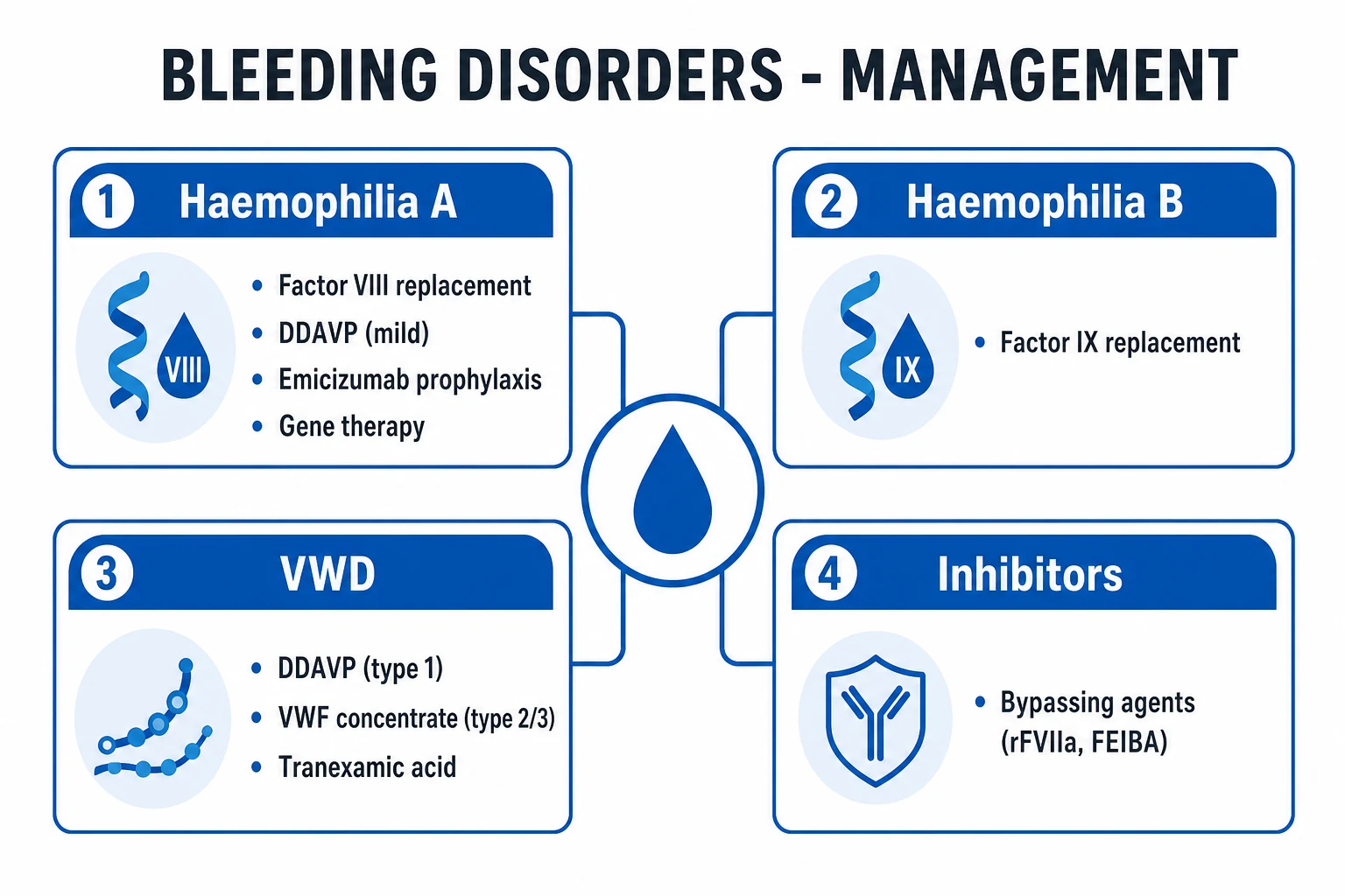
Factor replacement immediately — give the missing factor on suspicion. The dosing rule for factor VIII is one unit per kilogram raises the plasma level by approximately 2 percent (IU/dL); for factor IX one unit per kilogram raises the level by approximately 1 percent with standard (non-extended-half-life) product. Practical targets: [1]
- Joint or muscle bleed — raise factor to 50 to 80 percent; for haemophilia A give factor VIII 25 to 50 IU/kg, repeat at 12-hour intervals (half-life about 12 hours); for haemophilia B give factor IX 40 to 80 IU/kg (longer half-life, less frequent dosing).
- Intracranial haemorrhage, major trauma or major surgery — raise factor to 100 percent immediately and sustain above 50 percent for 7 to 14 days; suspect ICH on clinical grounds and give factor before the CT scan.
- Life-threatening oropharyngeal/airway bleeding — treat as for ICH (100 percent) and secure the airway early.
- Mild haemophilia A with a minor bleed and a documented desmopressin response — desmopressin 0.3 microgram per kilogram intravenously or subcutaneously over 30 minutes may suffice (it releases factor VIII and vWF from endothelial Weibel-Palade stores, roughly tripling the factor VIII level), but a baseline response must have been demonstrated. [1]
Adjuncts and supportive care: RICE (rest, ice, compression, elevation) for joint bleeds; analgesia with paracetamol or opioids — avoid intramuscular injections, aspirin and non-steroidal anti-inflammatory drugs (they worsen bleeding); tranexamic acid (an antifibrinolytic, 15 mg/kg intravenously or 1 g orally three times daily) for mucosal and dental bleeding — but avoid in haematuria (risk of clot obstructing the ureter). Do not attempt arterial or central venous puncture without factor cover; take venous samples from a single peripheral stab.[1]
Management — Definitive & Stepwise
Haemophilia A prophylaxis
The principle of prophylaxis is to maintain the factor trough above 1 to 3 percent so that spontaneous bleeds — and the arthropathy they cause — are prevented. The WFH and modern guidelines now position prophylaxis as the standard of care from early childhood in severe haemophilia.[1]
- Emicizumab (Hemlibra) — a subcutaneous bispecific antibody that simultaneously binds activated factor IX and factor X, functionally replacing factor VIIIa. It is now first-line prophylaxis for severe haemophilia A, with or without inhibitors. Dosing: 3 mg/kg subcutaneously once weekly for the first four weeks, then 1.5 mg/kg once weekly (or 3 mg/kg every two weeks, or 6 mg/kg every four weeks). It reduced the annualised bleed rate by roughly 80 percent in the HAVEN trials and freed patients from frequent intravenous access.[3]
- Recombinant factor VIII prophylaxis — 25 to 40 IU/kg intravenously three times weekly (every other day), or every third day in young children. Extended-half-life FVIII products allow twice-weekly dosing.
- Desmopressin (DDAVP) — for mild haemophilia A only (factor VIII over 5 percent), a documented responder; 0.3 microgram per kilogram subcutaneously or intravenously over 30 minutes, effective for minor bleeds, dental work or minor surgery. Limit to three consecutive days to avoid hyponatraemia (tachyphylaxis also develops).
Haemophilia B prophylaxis
- Extended-half-life recombinant factor IX — 40 to 80 IU/kg intravenously once or twice weekly (FIX half-life roughly 18 to 24 hours, longer with Fc-, albumin- or PEG-fusion products, allowing every-7-to-14-day dosing).
- Fitusiran — an N-acetyl-galactosamine-conjugated small interfering RNA (siRNA) targeting hepatic antithrombin, given subcutaneously weekly; it rebalances haemostasis by lowering antithrombin and is in late-phase development for both haemophilia A and B with and without inhibitors. [1]
Gene therapy — potential functional cure
Two AAV-vector gene therapies are now approved and represent a potential functional cure for selected adults:[4][5][7]
- Valoctocogene roxaparvovec (Roctavian) for haemophilia A — a single intravenous infusion of an AAV5 vector carrying a B-domain-deleted factor VIII gene under a hepatocyte-specific promoter. The GENEr8-1 trial showed mean factor VIII activity of about 40 IU/dL at one year and sustained expression at two years, with the majority of patients stopping routine prophylaxis. Durability gradually declines over several years.[4][5]
- Etranacogene dezaparvovec (Hemgenix) for haemophilia B — an AAV5 vector delivering a factor IX Padua variant (R338L, with intrinsically roughly eightfold higher specific activity). The HOPE-B trial showed stable factor IX activity around 37 IU/dL at 18 months, allowing patients to discontinue prophylaxis. The Padua variant, naturally discovered in a family with thrombosis, is the engine that delivers therapeutically useful FIX levels at modest vector doses.[7]
Candidate selection is strict: adult men only, no pre-existing neutralising anti-AAV antibodies, no active or chronic liver disease, no hepatocellular carcinoma risk factors that preclude a transient transaminitis. Transient corticosteroids are routinely used to manage the immune-mediated transaminitis that accompanies vector infusion.[4]
Von Willebrand disease management
Management is subtype-driven:[2]
- Type 1 (and selected type 2) — desmopressin 0.3 microgram per kilogram subcutaneously or intravenously over 30 minutes, releasing vWF and factor VIII from endothelial stores (test the response first with a DDAVP trial; a 2- to 3-fold rise at 1 to 2 hours defines a responder). Effective for menorrhagia, epistaxis, dental work and minor surgery. Caution in type 2B (can worsen thrombocytopenia) and avoid in pregnancy (risk of maternal hyponatraemia and thrombosis).
- Types 2 and 3 — vWF-containing concentrate (e.g. Haemate-P / Humate-P, Wilate), which delivers both vWF and factor VIII; dosing 20 to 60 IU of ristocetin cofactor activity per kilogram per dose for bleeding or surgery.
- Tranexamic acid — 1 g orally three times daily (or 10 mg/kg intravenously three times daily) or as a mouthwash for dental work, adjunctive across all subtypes. Avoid in haematuria.
- Hormonal management of menorrhagia — combined oral contraceptive pill or a levonorgestrel-releasing intrauterine system. [1]
Desmopressin (DDAVP) — agent, dose, route, timing, rationale
Inhibitors — the biggest complication
Inhibitors (neutralising IgG antibodies to factor VIII, less often factor IX) develop in 20 to 30 percent of children with severe haemophilia A (and in roughly 1 to 5 percent of haemophilia B). Risk is concentrated in the first 50 exposure days. They render standard factor replacement ineffective and are the single most important determinant of morbidity and mortality after severity itself.[1][6]
Acute bleeding with an inhibitor is treated with bypassing agents: [1]
- Recombinant activated factor VII (rFVIIa, novoseven) — 90 microgram per kilogram intravenously every 2 hours until bleeding stops.
- Activated prothrombin complex concentrate (FEIBA) — 50 to 100 units per kilogram intravenously every 8 to 12 hours (cumulative daily ceiling to limit thrombosis).
- Emicizumab provides effective prophylaxis in patients with inhibitors and is now the backbone of long-term care. [1]
Eradication by immune tolerance induction (ITI) — daily or alternate-day factor VIII combined with immunosuppression — achieves tolerance in roughly two-thirds. Rituximab (anti-CD20) is used in refractory cases. Acquired haemophilia uses bypassing agents plus immunosuppression (prednisolone 1 mg/kg/day with or without cyclophosphamide, or rituximab) to eliminate the autoantibody.[6]
Other inherited factor deficiencies
- Factor XI deficiency (haemophilia C) — common in Ashkenazi Jews (carrier frequency up to 8 percent); bleeding is disproportionate to the factor level (some with very low levels bleed little) and is provoked mainly by surgery or trauma at sites of high fibrinolytic activity (mouth, tonsils, urinary tract). Treat with tranexamic acid first-line, fresh frozen plasma or factor XI concentrate for major bleeding. The APTT is prolonged and the PT is normal.[1]
- Factor VII deficiency — the most common of the rare factor deficiencies; PT prolonged, APTT normal. Treat with recombinant activated factor VII 15 to 30 microgram/kg.
- Factor XIII deficiency — PT and APTT are both normal (the clot forms but is not cross-linked and is fragile). Hallmarks: umbilical stump bleeding in neonates, delayed bleeding, recurrent miscarriage and intracranial bleeding. Treat with factor XIII concentrate monthly prophylaxis.
- Fibrinogen disorders — afibrinogenaemia (absent fibrinogen, autosomal recessive; prolonged PT, APTT and thrombin time) and dysfibrinogenaemia (dysfunctional fibrinogen; some variants cause thrombosis). Treat with cryoprecipitate or fibrinogen concentrate.
Acquired coagulopathies
- Acquired haemophilia A — autoantibodies to factor VIII in the elderly, in pregnancy/postpartum, or with autoimmune disease or malignancy. The APTT is prolonged and does not correct on mixing; the Bethesda assay quantifies the inhibitor. Treat bleeds with bypassing agents (rFVIIa, FEIBA) and eradicate the antibody with prednisolone 1 mg/kg/day plus cyclophosphamide or rituximab.[6]
- Disseminated intravascular coagulation — global consumption; treat the underlying cause (sepsis, trauma, obstetric emergency), support with platelets, fresh frozen plasma and cryoprecipitate to keep platelets over 50, fibrinogen over 1.5 g/L and PT/APTT near-normal.
- Liver disease — global factor deficiency; vitamin K 10 mg intravenously if there is a reversible component, fresh frozen plasma and prothrombin complex concentrate for active bleeding.
- Vitamin K deficiency — vitamin K 10 mg intravenously slowly (the PT corrects within 6 to 12 hours; full correction in 24 to 48 hours).
- Anticoagulant-related bleeding — see the dedicated anticoagulation-therapy and anticoagulation-reversal topics; specific reversal agents (idarucizumab for dabigatran, andexanet alfa for factor Xa inhibitors, prothrombin complex concentrate for warfarin).
Specific Subtypes & Scenarios
Severe haemophilia A in a child — the prototypical management scenario. Start prophylaxis before age 2 years (primary prophylaxis) to prevent arthropathy: emicizumab subcutaneously avoids central venous access and is now first-line; those on factor VIII prophylaxis need a port-a-cath for venous access in young children. Vaccines (including hepatitis A and B) must be given subcutaneously, never intramuscularly. [1]
Haemophilia B (Christmas disease) is clinically identical to haemophilia A; the distinction is only by the factor IX assay. Prophylaxis uses extended-half-life factor IX (once or twice weekly) and fitusiran. Gene therapy with etranacogene dezaparvovec delivers the high-activity factor IX Padua variant.[7]
Type 3 VWD behaves like severe haemophilia (haemarthrosis, intramuscular bleeds) with added severe mucocutaneous bleeding; it needs regular vWF concentrate prophylaxis (50 to 100 IU/kg twice weekly). Type 2N VWD is the great mimic of mild haemophilia A — the clue is autosomal inheritance with both sexes affected and a normal vWF antigen but a very low factor VIII. [1]
Acquired haemophilia in the elderly — dramatic subcutaneous and soft-tissue bleeding (haematomas, compartment bleeds) with no childhood history, a prolonged APTT that does not correct on mixing, and a low factor VIII; the underlying trigger (autoimmune disease, lymphoproliferative or solid malignancy, pregnancy) must be sought.[6]
Pregnancy and the peripartum is covered under Special Populations below. [1]
Complications & Pitfalls
The complications fall into three groups: disease-related, treatment-related, and management errors.[1]
Disease-related:
- Haemophilic arthropathy — recurrent haemarthrosis drives synovial hypertrophy, neovascularisation, cartilage degradation and eventually bone destruction, producing the crippling "target joint" deformity of knees, elbows and ankles. It is the leading cause of long-term morbidity and is prevented by primary prophylaxis.
- Intracranial haemorrhage — the leading cause of haemophilia-related death; any head injury, headache or altered consciousness in a haemophilia patient is treated empirically with factor to 100 percent before imaging.
- Compartment syndrome from intramuscular bleeds (calf, forearm, iliopsoas); femoral nerve compression from iliopsoas haematoma.
- Airway compromise from parapharyngeal, retropharyngeal or subglottic haematomas. [1]
Treatment-related:
- Inhibitor formation in 20 to 30 percent of severe haemophilia A — the single most important complication; rendered manageable by emicizumab and bypassing agents.
- Transfusion-transmitted infection — historically devastating: in the 1970s and 1980s plasma-derived factor concentrates transmitted hepatitis C and HIV to a majority of severe haemophilia patients. Modern recombinant and viral-inactivated products are now considered safe, but the historical cohort still bears the consequences (cirrhosis, hepatocellular carcinoma).
- Iron overload in heavily transfused patients (now rare in the recombinant era).
- Thrombosis — paradoxically, with repeated bypassing agents (FEIBA, rFVIIa) or with emicizumab plus activated prothrombin complex concentrate (a dangerous combination that must be avoided).
- Allergic and anamnestic reactions to gene therapy (transient transaminitis; anti-AAV antibody formation precludes re-dosing). [1]
Management errors (classic pitfalls):
- Delaying factor replacement to await levels or imaging in a suspected ICH.
- Intramuscular injections, aspirin or NSAIDs in a haemophilia patient.
- Giving desmopressin in type 2B VWD (worsens thrombocytopenia) or without a prior DDAVP trial.
- Relying on the APTT alone — factor XIII deficiency, mild haemophilia and type 2N VWD can present with a normal APTT.
- Failing to screen for an inhibitor when bleeding escalates on adequate factor.
- Missing acquired haemophilia in an elderly patient with new bleeding (no childhood history) — the mixing study is the key discriminator. [1]
Prognosis & Disposition
With modern prophylaxis — emicizumab, recombinant factor VIII/IX and gene therapy — life expectancy in severe haemophilia is now near-normal in high-income countries (median survival into the seventh and eighth decades), an extraordinary change from the 1970s when most severe haemophilia patients died before age 20. The determinants of outcome are severity (the baseline factor level), the presence and titre of inhibitors, access to comprehensive haemophilia care, and adherence to prophylaxis. Without prophylaxis, severe haemophilia causes crippling arthropathy by adolescence, recurrent life-threatening bleeds and substantially reduced life expectancy.[1]
Disposition from the emergency department depends on the bleed. A joint or muscle bleed is managed with a single factor dose, RICE, analgesia and next-day review. An ICH, airway bleed, iliopsoas or compartment bleed mandates admission, sustained factor cover above 50 to 100 percent for 7 to 14 days, and multidisciplinary input. A patient with a new inhibitor or acquired haemophilia needs haematology admission for bypassing therapy, eradication and investigation of the trigger. All patients benefit from enrolment in a comprehensive haemophilia treatment centre (the WFH-endorsed model of multidisciplinary care).[6]
Special Populations
Neonates
Suspect haemophilia in a male neonate with prolonged bleeding after heel-prick, circumcision, vacuum extraction or intramuscular vitamin K, or with intracranial haemorrhage after birth trauma. Send a cord-blood or peripheral factor VIII and IX assay if there is a known family history; otherwise diagnose after presentation. Do not withhold vitamin K — give it subcutaneously or intravenously, never intramuscularly, in any infant at risk. Avoid instrumental delivery where haemophilia is known in the family.[1]
Pregnancy and the peripartum
Pregnancy is the single most dangerous time for a woman with VWD or for a haemophilia carrier. vWF rises two- to threefold through pregnancy, normalising the bleeding risk in most type 1 VWD carriers — but it crashes within hours of delivery, so postpartum haemorrhage (which can be severe and delayed, up to six weeks) is the dominant risk. Type 3 VWD needs vWF concentrate throughout labour and the puerperium; plan delivery jointly with obstetrics and haemophilia. Avoid neuraxial anaesthesia (epidural/spinal) unless vWF activity and factor VIII are above 50 IU/dL — an intracranial or spinal epidural haematoma is a catastrophic complication.[2]
For a known haemophilia carrier, determine the fetal sex antenatally (the male fetus has a 50 percent chance of being affected); avoid fetal scalp electrodes and instrumental delivery. Cord blood factor levels at birth confirm the diagnosis in the neonate. [1]
The elderly
Acquired haemophilia presents in older patients with no childhood or family history, dramatic subcutaneous bleeding, an APTT that does not correct on mixing, and an underlying autoimmune or neoplastic trigger. The mixing study and Bethesda assay are diagnostic; management combines bypassing agents for acute bleeding with immunosuppression (steroids, cyclophosphamide, rituximab) to eradicate the antibody. Acquired VWD accompanies lymphoproliferative disease, aortic stenosis and hypothyroidism in this age group.[6]
Surgery and dentistry
Plan any elective surgery or dental extraction with haematology in advance. Pre-treatment factor cover (raise factor VIII or IX to 80 to 100 percent) and tranexamic acid mouthwash (for dental work) are essential; sustained factor cover for 7 to 14 days is needed for major surgery. Avoid intramuscular injections and NSAIDs. A patient on emicizumab still needs additional factor replacement for surgery (emicizumab provides prophylaxis, not surgical cover), but the doses can be lower. [1]
Anticoagulated and immunocompromised patients
See the anticoagulation-therapy and anticoagulation-reversal topics for warfarin and direct oral anticoagulant bleeding. The immunocompromised patient with sepsis-triggered DIC is managed by treating the underlying cause plus supportive blood-product replacement. [1]
Evidence, Guidelines & Regional Differences
Landmark evidence and modern guidelines:[1][2]
- Berntorp 2021 (Nature Reviews Disease Primers) — the authoritative modern primer on haemophilia, covering pathophysiology, diagnosis and the full treatment ladder including gene therapy.[1]
- ASH-ISTH-NHF-WFH 2021 VWD guidelines (Connell, Blood Advances) — the consensus international guideline for VWD diagnosis, subtype classification and subtype-specific management.[2]
- Emicizumab HAVEN programme (Mahlangu 2018, NEJM, and the broader HAVEN 1 to 5 trials) — subcutaneous emicizumab reduced the annualised bleed rate by roughly 80 percent in severe haemophilia A with and without inhibitors, transforming prophylaxis.[3]
- Valoctocogene roxaparvovec GENEr8-1 (Ozelo 2022; Mahlangu 2023 two-year follow-up, NEJM) — single-infusion AAV5 gene therapy for haemophilia A producing sustained factor VIII expression out to years; approved by FDA and EMA.[4][5]
- Etranacogene dezaparvovec HOPE-B and the factor IX Padua story (George 2022, Lancet Haematology) — AAV5 gene therapy delivering the high-activity factor IX Padua variant for haemophilia B; approved by FDA and EMA.[7]
- Kruse-Jarres 2017 (American Journal of Hematology) — international guidance on acquired haemophilia A diagnosis and treatment.[6]
- World Federation of Haemophilia (WFH) Treatment Guidelines — the global standard of care, freely available, and the basis of care in most low- and middle-income countries.
Controversies. The durability of gene therapy (factor VIII levels decline gradually over several years; the need for, and ethics of, re-dosing given pre-formed anti-AAV antibodies remain unresolved). Whether emicizumab monotherapy suffices for surgical cover (it does not — additional factor is required). Whether non-factor rebalancing therapies (fitusiran, anti-TFPI agents) will displace factor replacement. How to prioritise the very high cost of curative gene therapy against the established safety and efficacy of emicizumab. [1]
Exam Pearls
- Haemophilia A = factor VIII (X-linked recessive, F8, 80%); haemophilia B = factor IX (X-linked recessive, F9, 20%, Christmas disease).
- VWD = the commonest inherited bleeding disorder (autosomal dominant, up to 1 in 100); mucocutaneous bleeding.
- Haemophilia: deep bleeds (haemarthrosis of knees/elbows/ankles, muscle, ICH). VWD: mucocutaneous (epistaxis, menorrhagia, gums, dental).
- Coagulation screen: APTT prolonged, PT and platelets NORMAL; mixing test corrects = factor deficiency, does not correct = inhibitor.
- Severity: severe under 1%, moderate 1 to 5%, mild 5 to 40%.
- Treat: factor VIII 25 to 50 IU/kg (joint/muscle), to 100% (ICH/surgery); factor IX 40 to 80 IU/kg. DDAVP for mild A and type 1 VWD. vWF concentrate for types 2 and 3. Tranexamic acid 1 g TDS.
- Emicizumab 1.5 mg/kg subcutaneously monthly — first-line prophylaxis for severe haemophilia A.
- Inhibitors (10 to 30% severe A): bypassing agents (rFVIIa 90 mcg/kg, FEIBA 50 to 100 U/kg); emicizumab prophylaxis; ITI eradication.
- Factor XI deficiency — Ashkenazi Jews; bleeding disproportionate to level; treat with tranexamic acid.
- Factor XIII deficiency — normal PT and APTT; umbilical stump bleeding, recurrent miscarriage, delayed bleeding.
- Acquired haemophilia — elderly, no childhood history, APTT does NOT correct on mixing; bypassing agents plus immunosuppression.
- Gene therapy: valoctocogene roxaparvovec (A), etranacogene dezaparvovec (B, factor IX Padua) — single IV infusion, approved.
- DDAVP must NOT be given in type 2B VWD (worsens thrombocytopenia). [1]
Haemophilia — the F.A.C.T.O.R. essentials
FACTOR
the deficient clotting factor; X-linked recessive in males; F8 and F9 genes
PT and platelets NORMAL; corrects on mixing (factor deficiency); does not correct = inhibitor
recurrent haemarthrosis destroys joints; the leading morbidity; prevented by prophylaxis
raise VIII to 50 to 80% (joint) or 100% (ICH); DDAVP for mild A and type 1 VWD
subcutaneous emicizumab 1.5 mg/kg monthly for prophylaxis; valoctocogene/etranacogene as cures
neutralising antibodies (10 to 30% severe A) -> bypassing agents (rFVIIa, FEIBA); Bethesda cut-off 0.6 BU/mL
Exam application bank (NEET-PG / INICET)
One-line answer
Haemophilia A (factor VIII deficiency, X-linked recessive, F8 gene, 1 in 5000 males) and haemophilia B (factor IX deficiency / Christmas disease, F9 gene, 1 in 30 000 males) are the classic inherited coagulopathies, presenting with deep-tissue bleeds — haemarthrosis of knees/elbows/ankles, intramuscular bleeds, and (in severe disease) spontaneous intracranial haemorrhage. Von Willebrand disease (VWD) is the commonest inherited bleeding disorder (up to 1 in 100, autosomal dominant), causing mucocutaneous bleeding (menorrhagia, epistaxis, gum, dental). Diagnosis rests on a prolonged APTT with normal PT and platelets that corrects on mixing, confirmed by factor VIII/IX assays (haemophilia) and vWF antigen plus ristocetin cofactor activity (VWD). Treatment: factor VIII replacement 25 to 50 IU/kg for bleeding and 25 to 40 IU/kg three times weekly for prophylaxis; factor IX 40 to 80 IU/kg [1]
Worked stems (answer without another resource)
Stem 1 — Classic presentation. Map symptoms to mechanism; name the first investigation and first treatment step with dose/route if drug therapy is standard. [1]
Stem 2 — Unstable / complicated. List red flags that force immediate resuscitation, theatre, ICU, antidote, or reperfusion — and what you do in the first 15 minutes. [1]
Stem 3 — Atypical group. Elderly, pregnancy, child, or immunocompromised: how presentation and thresholds change. [1]
Stem 4 — Differential trap. Name the three closest mimics and one discriminator for each. [1]
Stem 5 — Disposition. Who goes home with safety-netting, who is admitted, who needs HDU/ICU/theatre, and what follow-up is mandatory. [1]
Rapid viva checklist
- Definition + classification
- Pathophysiology chain
- Bedside signs / criteria
- Score with exact components (if any)
- Emergency bundle
- Definitive therapy with doses
- Complications of disease and of treatment
- Special populations
- Guideline/trial name if classic
- Three exam traps
Coverage self-check
If you cannot answer any stem above from this page alone, re-read the matching section — the page is intended to be self-sufficient for final-prof and NEET-PG/INICET questions on Coagulation Disorders (Haemophilia & von Willebrand).
References
- [1]Berntorp E, Fischer K, Hart DP, et al. Haemophilia Nat Rev Dis Primers, 2021.PMID 34168126
- [2]Connell NT, Flood VH, Brignardello-Petersen R, et al. ASH ISTH NHF WFH 2021 guidelines on the management of von Willebrand disease Blood Adv, 2021.PMID 33570647
- [3]Mahlangu J, Oldenburg J, Paz-Priel I, et al. Emicizumab Prophylaxis in Patients Who Have Hemophilia A without Inhibitors N Engl J Med, 2018.PMID 30157389
- [4]Ozelo MC, Mahlangu J, Pasi KJ, et al. Valoctocogene Roxaparvovec Gene Therapy for Hemophilia A N Engl J Med, 2022.PMID 35294811
- [5]Mahlangu J, Ozelo MC, Pasi KJ, et al. Two-Year Outcomes of Valoctocogene Roxaparvovec Therapy for Hemophilia A N Engl J Med, 2023.PMID 36812433
- [6]Kruse-Jarres R, Kempton CL, Baudo F, et al. Acquired hemophilia A: Updated review of evidence and treatment guidance Am J Hematol, 2017.PMID 28470674
- [7]George LA, Samelson-Jones BJ, Nathwani AC. Factor IX Padua for haemophilia B gene addition: universal adaptation and repeated success Lancet Haematol, 2022.PMID 35772422