Haematology · haematology
Febrile Neutropenia
Also known as Neutropenic fever · Febrile neutropaenic episode · Neutropenic sepsis · FN
Febrile neutropenia is an oncologic emergency defined as a single oral temperature at least 38.3 degrees Celsius, or 38.0 degrees sustained for one hour, in a patient with an absolute neutrophil count under 0.5 x10^9/L (or under 1.0 with predicted decline). Empiric broad-spectrum antipseudomonal beta-lactam therapy must be given within 60 minutes of recognition, after urgent blood cultures, with MASCC risk stratification guiding oral step-down versus inpatient IV care.
On this page & tools
Your progress
Saved locally on this device.
Exam tags
Red flags
Overview & Definition
Febrile neutropenia is a clinical syndrome rather than a single diagnosis, and it is among the most common and most dangerous complications of cytotoxic chemotherapy, radiation, and haematopoietic stem-cell transplantation (HSCT). It is the paradigm of a time-critical oncologic emergency: the neutropenic host cannot mount the inflammatory response that ordinarily localises and signals infection, so fever may be the only manifestation of bacteraemia or early sepsis. Untreated FN carries a mortality exceeding 50%; with prompt empiric therapy in-hospital mortality falls to roughly 5–12%.[3][1]
The definition adopted jointly by the Infectious Diseases Society of America (IDSA) and the Multinational Association for Supportive Care in Cancer (MASCC) has two components, both required:[1][3]
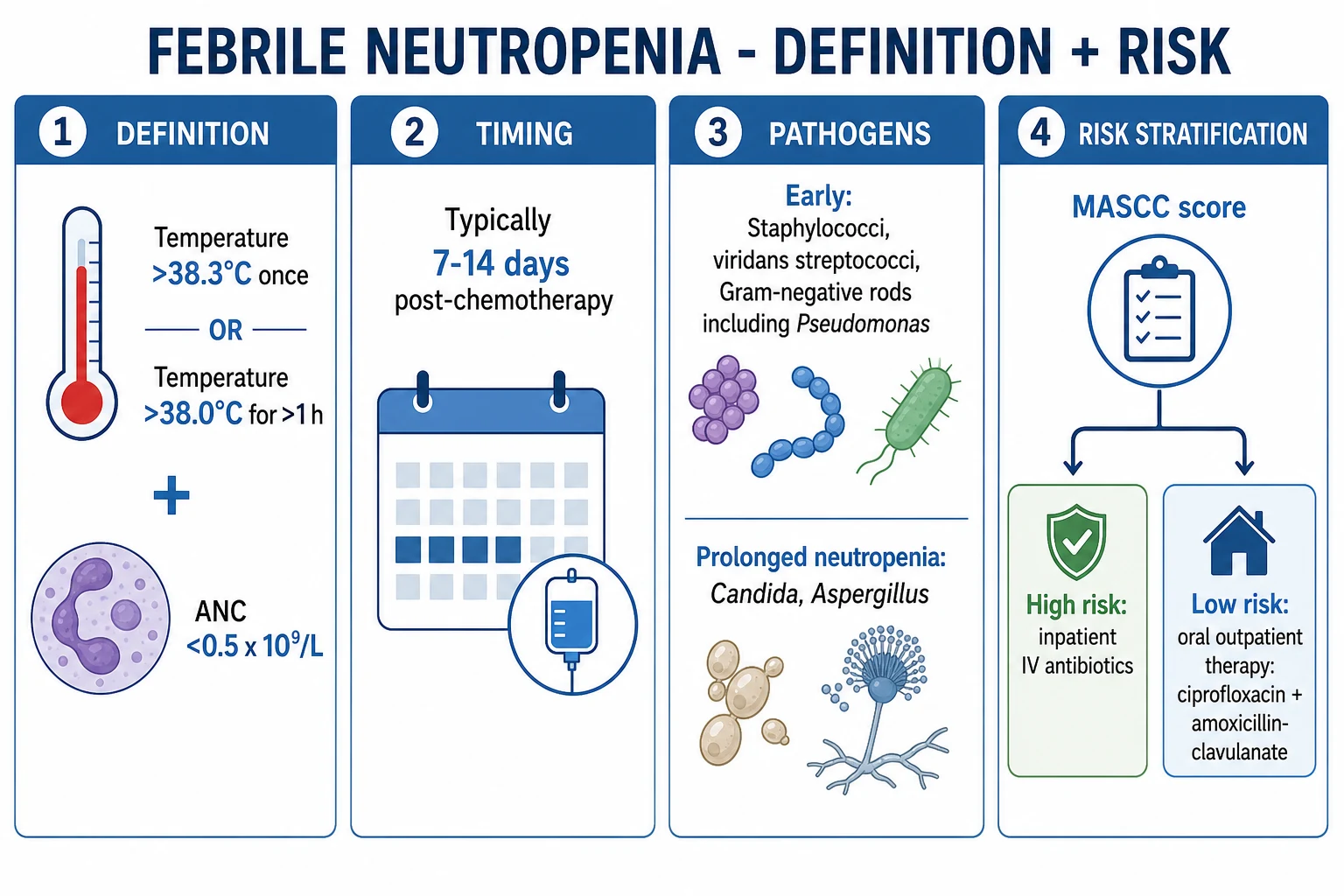
- A fever defined as a single oral temperature 38.3 °C (101 °F) or higher, OR a temperature of 38.0 °C (100.4 °F) or higher sustained for at least one hour; AND
- Neutropenia defined as an absolute neutrophil count (ANC) under 0.5 ×10⁹/L, OR an ANC under 1.0 ×10⁹/L that is expected to fall below 0.5 ×10⁹/L over the next 24 to 48 hours. [1]
Two further terms are essential vocabulary. Profound neutropenia (ANC under 0.1 ×10⁹/L) carries the highest infection risk and is the defining feature of "high-risk" patients. Functional neutropenia describes neutrophils that are present in normal numbers but rendered dysfunctional by chemotherapy, glucocorticoids, or the underlying haematological disease — as in acute myeloid leukaemia — so that classical signs of infection (pus, induration, radiographic infiltrate) are muted or absent. This is the biological reason behind the central maxim of the field: fever in a neutropenic patient is infection until proven otherwise, and the absence of pus does not exclude a life-threatening bacteraemia. [1]
Classification
Febrile neutropenia is classified along three complementary axes that together drive every management decision: risk of medical complication (guides inpatient IV versus oral outpatient care), microbiological certainty of the source (guides duration and escalation), and causative organism (guides antibiotic choice and de-escalation).[1][2]
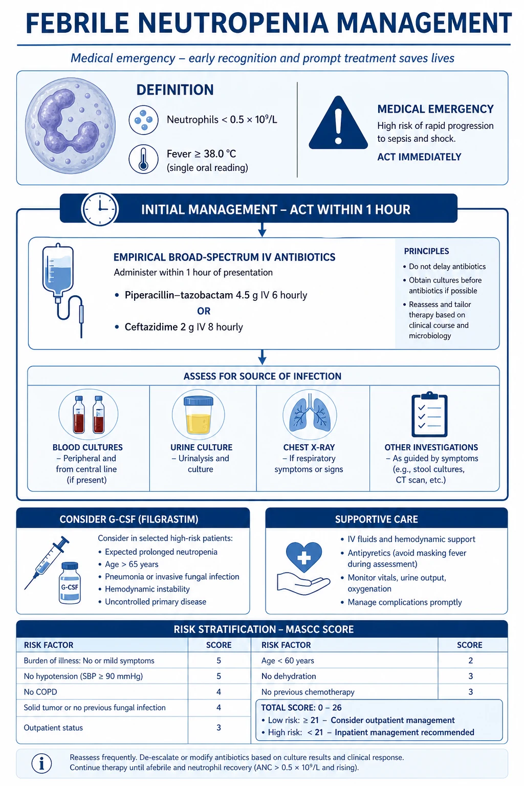
MASCC risk index — the bedside stratification tool
The MASCC risk index, derived by Klastersky and colleagues and validated across thousands of episodes, is the most widely used bedside tool for identifying low-risk patients. It sums eight weighted variables to a maximum of 26 points. A score of 21 or higher identifies low-risk patients (positive predictive value about 91%, sensitivity about 71%) who are candidates for oral step-down or carefully selected outpatient therapy.[1][2]
| Characteristic | Points |
|---|---|
| Burden of illness: no or mild symptoms | 5 |
| Burden of illness: moderate symptoms | 3 |
| No hypotension (systolic BP above 90 mmHg) | 5 |
| No chronic obstructive pulmonary disease | 4 |
| Solid tumour or no previous fungal infection | 4 |
| Outpatient at onset of fever | 3 |
| No dehydration requiring parenteral fluids | 3 |
| Age under 60 years | 2 |
MASCC low risk (score 21+)
- Oral step-down or outpatient IV feasible
- Ciprofloxacin 500 mg BD + co-amoxiclav 625 mg TDS
- Daily review, 24/7 telephone access to treating centre
- Day-1 reassessment; blood cultures every 24–48 h
MASCC high risk (under 21)
- Mandatory inpatient IV antipseudomonal beta-lactam
- Piperacillin-tazobactam 4.5 g IV q6h first-line
- Daily review; escalate for haemodynamic instability
- Add vancomycin/teicoplanin if line infection, cellulitis, or pneumonia
IDSA risk categories — host and depth of neutropenia
The IDSA uses a complementary, organism- and host-oriented split. High-risk patients have profound neutropenia (ANC under 0.1 ×10⁹/L) expected to last more than 7 days, or any significant comorbidity — haemodynamic instability, pneumonia, new abdominal pain, neurological change, renal or hepatic failure. They require inpatient IV antipseudomonal β-lactam. Low-risk patients have solid tumours on standard chemotherapy, a short expected neutropenia (7 days or fewer), clinical stability, no comorbidity, and a MASCC score of 21 or higher; they are candidates for oral step-down or outpatient care.[2][3]
Microbiological classification — drives escalation
- Microbiologically documented infection (MDI): a pathogen isolated from a sterile site (blood, urine, CSF, joint) — about 20–30% of episodes.
- Clinically documented infection (CDI): a focal source identified (pneumonia, cellulitis, typhlitis, line-site infection) without a positive culture — about 20–30%.
- Unexplained fever (UF): no source identified after standard workup — the largest single group, around 50%. [1]
MASCC must-haves — Low Risk
MASCC21
Burden-of-illness none/mild = 5 pts
2 pts; older patients score worse
4 pts; leukaemia scores lower
4 pts; chronic lung disease raises risk
SBP above 90 mmHg = 5 pts
3 pts
3 pts
Oral step-down possible
Epidemiology & Risk Factors
Febrile neutropenia complicates roughly 10–50% of solid-tumour chemotherapy cycles and a far higher proportion — 80% or more — of induction cycles for acute myeloid leukaemia and of myeloablative conditioning for HSCT. The overall in-hospital mortality of treated FN is 5–10% in contemporary cohorts, but it climbs steeply to 30–50% in patients who develop septic shock or invasive fungal infection. Mortality is concentrated in the first 48 hours, which is why the door-to-antibiotic interval is the single most modifiable outcome.[3][1]
The risk of developing FN after any given cycle is governed by an interplay of regimen intensity, host factors, and the integrity of mucosal barriers. The principal risk determinants are: [1]
- Chemotherapy intensity and regimen: high-dose cytarabine, anthracyclines, platinum agents, taxanes, and alkylating agents produce deeper and more prolonged nadirs than weekly fluoropyrimidines or low-dose oral agents. Acute leukaemia induction and HSCT conditioning carry the highest per-cycle risk.
- Expected nadir depth and duration: risk rises sharply once the ANC falls below 0.5 ×10⁹/L and increases further when profound neutropenia (under 0.1 ×10⁹/L) is expected to last more than 7 days.
- Tumour type: haematological malignancies carry the highest risk; among solid tumours, small-cell lung cancer, germ-cell tumours, and aggressive lymphomas are particularly dangerous.
- Patient factors: age over 65, poor performance status (ECOG 2 or higher), hypoalbuminaemia, baseline renal or hepatic dysfunction, advanced-stage disease.
- Prior febrile neutropenia: a previous episode roughly doubles the risk of recurrence on subsequent cycles and is one of the strongest predictors for primary prophylaxis with G-CSF.
- Oral mucositis and mucosal barrier injury: severe mucositis roughly doubles the rate of Gram-negative and viridans-streptococcal bacteraemia.
- Indwelling devices: central venous catheters (tunnelled, PICC, port), urinary catheters, and prosthetic material are niches for coagulase-negative staphylococci, Staphylococcus aureus, and Candida biofilms.
- Corticosteroids and immunosuppression: prolonged steroids impair neutrophil function and T-cell immunity, raising the risk of Pneumocystis, viral reactivation, and mould infection.
- Prior colonisation with resistant organisms (MRSA, VRE, ESBL, carbapenem-resistant Pseudomonas or Acinetobacter) and prior invasive fungal infection. [1]
Key FN epidemiology numbers
Pathophysiology
Neutrophils are the principal effector of innate defence against bacteria and fungi. When the ANC falls below 0.5 ×10⁹/L — and especially below 0.1 ×10⁹/L — five overlapping mechanisms converge to produce clinical infection. [1]
The dominant cascade is mucosal barrier injury driving bacterial translocation. Cytotoxic chemotherapy is taken up by rapidly dividing cells, which include not only the haematopoietic marrow but also the oral, oesophageal, and intestinal epithelium. The mucosal lining ulcerates, loses its tight-junction integrity, and permits luminal organisms to cross into the lamina propria and lymphatics. With the nadir of neutropenia typically falling 7 to 14 days after a cytotoxic cycle — and recovering over the following week — the patient is most vulnerable precisely when both neutrophil number and mucosal integrity are at their lowest. The gastrointestinal tract is therefore the single largest portal of entry, and Gram-negative bacteraemia from Escherichia coli, Klebsiella, and Pseudomonas aeruginosa classically peaks in this nadir window.[3]
The five mechanisms are: [1]
- Quantitative neutrophil deficiency: too few circulating neutrophils to contain the inoculum at any portal of entry — skin, oropharynx, lung alveoli, gut lumen, urinary tract.
- Qualitative neutrophil dysfunction: chemotherapy, glucocorticoids, and the underlying haematological disease impair chemotaxis, phagocytosis, and the oxidative burst, so even the neutrophils that remain are blunted.
- Mucosal barrier injury: chemotherapy-induced ulceration of the oral and intestinal mucosa is the principal route of Gram-negative bacteraemia and Candida translocation.
- Indwelling central venous catheters: biofilm on the catheter hub and lumen is a persistent reservoir for coagulase-negative staphylococci, S. aureus, Candida, and Corynebacterium jeikeium.
- Impaired adaptive surveillance: chemotherapy-induced lymphopenia blunts T-cell and humoral responses, permitting reactivation of herpesviruses (HSV, VZV, CMV, EBV) and invasive mould infection. [1]
The organisms
The microbiology of FN has shifted markedly over thirty years but a recognisable pattern remains. Gram-positive organisms are now the most frequent cause of bacteraemia, accounting for over half of documented bloodstream infections: coagulase-negative staphylococci (line-related), Staphylococcus aureus (including MRSA), viridans streptococci (classically after high-dose cytarabine), enterococci (including VRE), and Streptococcus pneumoniae. Gram-negative organisms carry the highest mortality and include Escherichia coli, Klebsiella pneumoniae, Pseudomonas aeruginosa (the classical high-mortality pathogen and the reason every empiric regimen must be antipseudomonal), enterobacter, and the increasingly important carbapenem-resistant Enterobacteriaceae. Fungal infection — Candida species and Aspergillus species — should be suspected when fever persists beyond 4 to 7 days of broad-spectrum antibacterials in a high-risk host, particularly one with prolonged profound neutropenia.[3][5]
Clinical Presentation
Fever is usually the first and only sign. Because the inflammatory response is blunted, a single rigour in a chemotherapy patient on day 10 post-cycle should trigger the full febrile-neutropenia pathway regardless of the measured temperature. Up to half of episodes never yield an identified source even after exhaustive investigation. [1]
- Fever meeting the definition: single oral temperature 38.3 °C or higher, or 38.0 °C sustained for one hour.
- May be the only symptom: roughly half of episodes have no identifiable localising source.
- Localising symptoms when present: cough, dyspnoea or pleuritic pain (pneumonia); sore throat and odynophagia (mucositis); dysuria (urinary tract infection); perianal pain (perianal abscess, fissure); right-lower-quadrant abdominal pain and diarrhoea (typhlitis, C. difficile colitis).
- Sepsis physiology: hypotension, tachycardia, tachypnoea, new confusion, oliguria, mottled or cool peripheries — mandates resuscitation alongside the first antibiotic dose.
- Central-line site: erythema, tenderness, purulent exudate at the exit site, or tenderness along the tunnel.
- Skin: cellulitis, ecthyma gangrenosum (the classic haemorrhagic necrotic lesion of Pseudomonas septicaemia), petechiae, scattered pustules suggesting disseminated fungal infection, vesicles of HSV or VZV reactivation. [1]
Atypical presentations are deliberately tested at viva. Elderly and diabetic patients may be afebrile or hypothermic in the face of overwhelming sepsis — a normal temperature in a sick-appearing neutropenic patient is itself a red flag. Patients on chronic steroids may not mount a fever. The profoundly neutropenic patient with pneumonia may have a normal chest examination and a near-normal chest X-ray early on, because there are no neutrophils to generate the infiltrate. [1]
Differential Diagnosis
Fever in a neutropenic patient is treated as infection until proven otherwise, but a structured list of non-infectious mimics is essential because each alters the escalation pathway. The key principle: non-infectious causes are diagnoses of exclusion in the neutropenic host — antibiotics come first, the differential is revisited if fever persists despite appropriate therapy.[1]
- Tumour fever: pyrexia driven by cytokine release from active malignancy (Hodgkin lymphoma, renal cell, hepatocellular, leukaemia); classically responds to a naproxen challenge (500 mg BD), now used cautiously.
- Drug fever: from chemotherapy (especially cytarabine, bleomycin), antibiotics, antifungals, cytokines (G-CSF itself causes fever), checkpoint inhibitors, and CAR-T cytokine release syndrome.
- Transfusion reaction: febrile non-haemolytic reaction within four hours of a blood product.
- Venous thromboembolism: catheter-related or limb DVT; pulmonary embolism may present with fever and pleuritic pain.
- Deep seated fungal infection: aspergillosis, candidiasis, mucormycosis — the cause of "persistent fever despite antibiotics".
- Cytokine release / immune-related: CAR-T, bispecific antibodies, engraftment syndrome, graft-versus-host disease. [1]
Infectious
- Bacteraemia, pneumonia, UTI, line infection
- Typhlitis, perianal abscess, severe mucositis
- Invasive aspergillosis or candidaemia
- Viral reactivation — HSV, VZV, CMV
Non-infectious
- Tumour fever (responds to naproxen)
- Drug fever (chemo, antibiotics, biologics)
- Transfusion (febrile non-haemolytic) reaction
- Venous thromboembolism — DVT or PE
- GvHD or cytokine release (CAR-T, bispecifics)
Clinical & Bedside Assessment
The first hour is structured. Examine systematically and document a baseline so that subsequent change is unambiguous — in FN the trend in vital signs, not the absolute value, drives escalation. [1]
- Vital signs and perfusion: temperature, heart rate, blood pressure, respiratory rate, oxygen saturation, urine output, capillary refill, and mental state. Any organ dysfunction automatically up-classifies the patient to high risk.
- Central venous catheter: inspect the exit site, palpate along the tunnel and over the port pocket for tenderness, check the connections.
- Oral cavity: mucosal erythema, ulceration, thrush, herpes vesicles, dental abscess.
- Skin head to toe: petechiae, cellulitis, ecthyma gangrenosum, pustules, VZV lesions, catheter-site infection.
- Perianal region: inspect for fissure, ulceration, or abscess — but avoid digital rectal examination, which risks precipitating bacteraemia in the neutropenic patient.
- Respiratory: auscultate for crackles, consolidation, effusion — but the examination may be entirely silent.
- Abdomen: distension, right-lower-quadrant tenderness (typhlitis), hepatosplenomegaly, rebound.
- Neurological and fundoscopy: mental state, and — if visual or neurological symptoms are present — fundoscopy for CMV retinitis, candida endophthalmitis, or Roth spots. [1]
Investigations
The initial workup must be fast, paired, and pre-antibiotic — but antibiotics are never delayed for investigations. The guiding principle is: draw what you can in the first 15 minutes, then give the antibiotic by the 60-minute mark.[3][1]
- Full blood count and differential: confirm the ANC, the degree of cytopenia (often pancytopenia), and review the trend over the preceding week.
- Urea, electrolytes, creatinine: baseline renal function for drug dosing; acute kidney injury flags sepsis or nephrotoxic drugs.
- Liver function tests, albumin, bilirubin: drug toxicity, sepsis, graft-versus-host disease, fungal disease.
- CRP and procalcitonin: baseline and trend; procalcitonin has higher specificity for bacterial infection and may help de-escalation decisions.
- Lactate: a value over 2 mmol/L is a sepsis red flag and predicts progression to shock.
- Coagulation screen: PT/INR and APTT for baseline before central lines and to detect disseminated intravascular coagulation.
- Blood glucose: hypoglycaemia in sepsis, hyperglycaemia on steroids.
- Blood cultures × 2 sets: one peripheral venepuncture plus one drawn from each lumen of any central venous catheter, labelled with site and time. Inoculate paired aerobic and anaerobic bottles. Volume matters — underfilling reduces yield more than any other technical factor.
- Urine microscopy and culture: clean-catch or catheter specimen.
- Chest X-ray for all patients — pulmonary infection can be clinically and radiographically silent early on.
- Sputum and respiratory viral PCR (influenza, RSV, SARS-CoV-2) if respiratory symptoms.
- Stool culture, C. difficile toxin or PCR, viral PCR if diarrhoea.
- Galactomannan and beta-D-glucan if high-risk for invasive fungal infection (prolonged neutropenia, haematological malignancy, HSCT, prior mould infection).
- CT chest, sinuses, abdomen and pelvis if symptoms localise or if fever persists beyond 4 to 7 days despite empirical antibacterials.
- Viral PCR panel (HSV, VZV, CMV, EBV, respiratory viruses) as clinically indicated.
- Skin biopsy of suspicious lesions for histology and culture; lumbar puncture only if clinical signs of meningitis (rare and not routine).
- Line-tip culture in semi-quantitative (Maki) roll technique if a central line is removed. [1]
Mandatory first-hour workup
Management — Resuscitation
The door-to-needle target is 60 minutes from recognition. Resuscitation, cultures, and the first antibiotic dose run in parallel rather than in sequence.[3][1]
- Airway, Breathing, Circulation assessment; supplemental oxygen to keep saturation at 94% or above; two large-bore intravenous cannulae.
- Blood cultures drawn — peripheral plus each lumen of any central line — but do not delay antibiotics if cultures cannot be obtained promptly; if venous access is difficult, give the antibiotic first.
- Empirical broad-spectrum antipseudomonal β-lactam IV within 1 hour — see the definitive section for the full ladder.
- IV crystalloid resuscitation — 30 mL/kg of balanced crystalloid or 0.9% saline within the first three hours if hypotensive or lactate over 2 mmol/L.
- Vasopressors — noradrenaline first-line, targeting MAP 65 mmHg or above — if shock persists after fluid resuscitation.
- Empirical Gram-positive cover (vancomycin or teicoplanin) added only for specific indications — see below.
- Source control — drain collections, remove infected lines, urgent surgical review for typhlitis or intra-abdominal sepsis. [1]
Management — Definitive & Stepwise
After the first empirical dose, daily re-evaluation focuses on three questions: (1) is there a documented organism, (2) is the patient clinically improving, and (3) is the source controlled? The antibiotics chosen on day 0 are then escalated, de-escalated, or continued based on the answers.[3][1]
First-line empirical antibiotic regimens (high-risk inpatient IV)
The IDSA, ECIL, and ASCO/IDSA guidelines converge on antipseudomonal β-lactam monotherapy as first-line for stable high-risk patients, with escalation to combination therapy only for specific indications.[3][5]
- Piperacillin-tazobactam 4.5 g IV every 6 to 8 hours — the preferred first-line agent in most centres; broad Gram-negative cover including Pseudomonas, anaerobic activity, and reasonable Gram-positive cover.
- Cefepime 2 g IV every 8 hours — an alternative fourth-generation cephalosporin with antipseudomonal activity; recommended by IDSA and ECIL as equivalent first-line.
- Ceftazidime 2 g IV every 8 hours — a third-generation antipseudomonal cephalosporin; alternative where piperacillin-tazobactam is unavailable, but weaker Gram-positive cover.
- Meropenem 1 g IV every 8 hours — a carbapenem reserved for known or suspected ESBL-producing organisms, prior resistant colonisation, severe β-lactam allergy, or as escalation in unstable patients.
- Gentamicin (aminoglycoside) is not recommended for routine empirical use as monotherapy or combination; it adds nephrotoxicity without proven survival benefit in stable patients, and is reserved for short-course synergy in septic shock with a Pseudomonas or resistant Gram-negative focus. [1]
When to add empirical Gram-positive cover
Vancomycin 25–30 mg/kg loading dose, then 15–20 mg/kg every 12 hours (or teicoplanin 6 mg/kg every 12 hours for three doses, then daily) is added to the baseline regimen only for:[3]
- Haemodynamic instability or septic shock.
- Clinically suspected catheter-related infection, cellulitis, or skin/soft-tissue source.
- Severe mucositis (high viridans-streptococcal risk, especially if on fluoroquinolone prophylaxis).
- Pneumonia radiographically confirmed.
- Known prior MRSA colonisation or institutional MRSA prevalence above 20%.
- Positive blood culture for Gram-positive cocci before identification. [1]
Routine empirical vancomycin for every FN patient is not recommended — it does not improve outcomes in unselected patients and adds nephrotoxicity. [1]
Anaerobic and abdominal focus
For perianal abscess, suspected typhlitis (neutropenic enterocolitis), intra-abdominal source, or severe mucositis, add metronidazole 500 mg IV every 8 hours or rely on the inherent anaerobic cover of piperacillin-tazobactam or a carbapenem. [1]
Escalation rules
- Persistent fever at 48 to 72 hours without a source: repeat blood cultures, re-image with CT chest/abdomen/pelvis and sinuses; add empirical antifungal if high-risk for mould or candida (see below).
- New haemodynamic instability at any time: add vancomycin loading dose, broaden to meropenem 1 g IV every 8 hours, and arrange ICU admission.
- Abdominal pain with rising CRP, ileus, or pneumatosis intestinalis → typhlitis: add metronidazole 500 mg IV every 8 hours, keep the patient nil by mouth, nasogastric decompression, urgent surgical consultation.
- Diarrhoea during or after antibiotic exposure → test for Clostridioides difficile; treat with oral vancomycin 125 mg four times daily or fidaxomicin 200 mg twice daily. [1]
Antifungal escalation
For persistent fever on day 4 to 7 of broad-spectrum antibacterials in a high-risk host (prolonged profound neutropenia, allogeneic HSCT, relapsed leukaemia, prior mould), add empirical antifungal therapy:[3][1]
- Caspofungin 70 mg IV loading dose on day 1, then 50 mg IV daily — echinocandin, first-line for suspected candidaemia and favoured by ECIL/IDSA for empirical use.
- Liposomal amphotericin B 3 mg/kg IV daily — broadest mould cover including mucormycosis; preferred if mould is suspected and aspergillosis not confirmed.
- Voriconazole 6 mg/kg IV every 12 hours for two doses, then 4 mg/kg every 12 hours — first-line for proven or probable invasive aspergillosis; therapeutic drug monitoring required. [1]
De-escalation rules
- If a single susceptible organism is identified and the source is controlled, narrow to the smallest-spectrum agent that covers it.
- Afebrile and clinically stable for 48 hours or more with negative cultures at 48 to 72 hours: continue empirical IV until ANC recovery, reassess daily.
- Low-risk (MASCC 21 or higher), afebrile, cultures negative, no comorbidity, tolerating oral intake: step down to oral therapy. [1]
Duration
- Documented bacterial infection: at least 7 to 14 days of appropriate therapy; longer for deep-seated infection (endocarditis, abscess, osteomyelitis).
- Unexplained fever (UF): continue empirical IV until afebrile for 48 hours or more and ANC at or above 0.5 ×10⁹/L, then stop or step down.
- Invasive fungal infection: weeks to months — voriconazole for 6 to 12 weeks for invasive aspergillosis; echinocandin for 14 days after first negative candida blood culture and source control. [1]
Low-risk oral step-down
For the carefully selected MASCC-low-risk patient (score 21 or higher, solid tumour, no comorbidity, clinically stable, normal renal and hepatic function, oral intake preserved, reliable carer and 24/7 access to the treating centre), oral step-down or even primary outpatient therapy is supported by ASCO/IDSA guidance:[2][4]
- Ciprofloxacin 500 mg PO twice daily PLUS co-amoxiclav (amoxicillin-clavulanate) 625 mg PO three times daily — the standard oral regimen.
- Daily telephone or clinic review, with mandatory same-day return for any new symptom, persistent fever, or haemodynamic change. [1]
Source control
- Remove the central line for Staphylococcus aureus, Pseudomonas aeruginosa, Candida, or mycobacterial bacteraemia; persistent bacteraemia after 72 hours of appropriate therapy; clinical tunnel infection; or septic emboli.
- Drainage of abscesses, empyema, septic joints; perianal abscess may need incision and drainage with platelet support.
- Surgical consultation for typhlitis with perforation, ischaemia, or uncontrolled bleeding. [1]
G-CSF (granulocyte colony-stimulating factor) criteria
Therapeutic G-CSF is not routine in established FN — meta-analyses show a reduction in hospitalisation duration but no consistent mortality benefit in unselected patients. ASCO supports reserving therapeutic G-CSF for specific high-risk situations:[7]
- Sepsis, septic shock, or invasive fungal infection.
- Pneumonia.
- Expected prolonged profound neutropenia (more than 7 days) or ANC under 0.1 ×10⁹/L.
- Filgrastim 5 micrograms/kg subcutaneously daily until ANC recovery (typically to 1.0 ×10⁹/L after nadir).
- Pegfilgrastim 6 mg subcutaneously as a single dose is an alternative in selected ambulatory patients.
- Prophylactic G-CSF (given after each cycle) is a separate, evidence-based indication for regimens carrying an FN risk above 20% — distinct from therapeutic use in established FN. [1]
Stepwise Management
A practical MASCC-driven decision pathway:[1][2][4]
| Step | Action | Target / threshold |
|---|---|---|
| 1. Recognise | Single temp 38.3 °C + ANC under 0.5 | Immediate |
| 2. Draw cultures | Peripheral plus each lumen | Within 15 min |
| 3. Stratify | Compute MASCC score | Within 30 min |
| 4. First-dose antibiotic | Piperacillin-tazobactam 4.5 g IV | Within 60 min |
| 5. Reassess at 48 h | Cultures, imaging, source | Twice-daily review |
| 6. Escalate or de-escalate | Add vancomycin or antifungal; narrow if organism known | Daily |
| 7. Step down | MASCC 21+ and afebrile and cultures negative → oral | Day 3–5 |
| 8. Discontinue | Afebrile 48 h + ANC at least 0.5 + no source | Day 7–14 |
Specific Subtypes & Scenarios
- Unexplained fever (UF): the most common category. Treat empirically until ANC recovery; if fever persists, intensify the search for fungal and viral sources.
- Clinically documented infection (CDI): a focal source is identified (pneumonia, cellulitis, typhlitis, perianal abscess, line-site) but no organism is isolated. Treat empirically and add source-specific measures.
- Microbiologically documented infection (MDI): positive culture from a sterile site. Narrow when possible after source control.
- Persistent fever: four or more days of fever despite broad-spectrum antibacterials. Re-image (CT chest, abdomen, pelvis, sinuses); add empirical antifungal in the high-risk host.
- Recurrent fever after defervescence: re-evaluate for a new source, resistant organism, line infection, C. difficile, tumour fever, or drug fever.
- Breakthrough bacteraemia on therapy: consider resistant Gram-negative (ESBL, CRE), MRSA, VRE, or Candida; broaden cover and reassess source control.
- Catheter-related bloodstream infection: coagulase-negative staphylococci may be managed with line retention plus systemic antibiotics and an antibiotic lock; remove the line for S. aureus, Pseudomonas, Candida, mycobacteria, persistent bacteraemia beyond 72 hours, tunnel infection, or septic emboli.
- Typhlitis (neutropenic enterocolitis): the classic triad of fever, right-lower-quadrant pain, and neutropenia, typically after cytarabine or anthracycline induction. CT shows bowel-wall thickening, often with pneumatosis. Manage with piperacillin-tazobactam plus metronidazole 500 mg IV every 8 hours, bowel rest, nasogastric decompression, and urgent surgical review for perforation or uncontrolled bleeding.
- Mucositis: graded oral care protocol, adequate analgesia (often patient-controlled morphine), topical antifungal and HSV prophylaxis as needed.
- Invasive fungal infection: Candida — remove central line, start an echinocandin (caspofungin 70 mg IV day 1 then 50 mg daily), step down to fluconazole when susceptible and stable. Aspergillus — voriconazole; alternative is liposomal amphotericin B 3–5 mg/kg IV daily. Mucormycosis requires liposomal amphotericin B and aggressive surgical debridement.
- Pneumonia: add atypical cover (azithromycin 500 mg IV daily) if features suggest atypical pathogen; consider Pneumocystis jirovecii if prolonged steroids without prophylaxis (add co-trimoxazole once stabilising).
- Paediatric febrile neutropenia: MASCC is validated in adults; children use paediatric-specific rules (e.g. absence of high-risk comorbidity, no hypotension, normal chest X-ray, no focal source). Empirical monotherapy with piperacillin-tazobactam or cefepime is standard; vancomycin reserved for a clear indication.[6]
- HSCT recipients: higher risk of CMV, EBV-PTLD, and mould; use prophylaxis with aciclovir or valganciclovir, letermovir for CMV (seropositive allogeneic), and posaconazole or voriconazole for mould.
- Solid tumours: shorter neutropenia, usually lower MASCC risk, often eligible for oral step-down.
- Haematological malignancies: deeper and longer neutropenia, higher rates of fungal and resistant infection; default to inpatient IV with low threshold for antifungal escalation.
Complications & Pitfalls
- Septic shock and multiorgan dysfunction: the leading cause of early death; mandates ICU admission and full Surviving Sepsis resuscitation.
- Acute respiratory distress syndrome (ARDS): from pneumonia, sepsis, transfusion, or immune reconstitution.
- Acute kidney injury: pre-renal from sepsis, intrinsic from nephrotoxic drugs (vancomycin, aminoglycosides, amphotericin), or post-renal from clot or tumour.
- Invasive fungal superinfection: candidaemia, pulmonary and disseminated aspergillosis, mucormycosis — high mortality even with appropriate therapy.
- Clostridioides difficile colitis: antibiotic exposure plus immunosuppression.
- Line loss: necessitating re-insertion in a thrombocytopenic patient.
- Drug toxicity: vancomycin nephrotoxicity (especially with piperacillin-tazobactam — the "vat" combination), linezolid myelosuppression and serotonin syndrome, voriconazole hepatotoxicity and visual disturbance, amphotericin nephrotoxicity and electrolyte wasting.
- Death: overall in-hospital mortality 5–10%, rising to 30–50% with septic shock or invasive mould disease. [1]
The classic pitfalls are: (1) delaying antibiotics while waiting for the "perfect" culture; (2) omitting central-line cultures; (3) performing a digital rectal examination and precipitating bacteraemia; (4) adding routine vancomycin to every patient; (5) failing to escalate to antifungal at day 4 to 7 of persistent fever; (6) premature outpatient discharge of a borderline MASCC patient; (7) missing typhlitis as the cause of right-lower-quadrant pain; (8) forgetting that G-CSF itself causes fever and bone pain. [1]
Prognosis & Disposition
Prognosis is driven by three variables: the depth and duration of neutropenia, the patient's haemodynamic reserve, and the time-to-antibiotic. [1]
- Favourable: MASCC low risk, solid tumour, ANC recovery expected within 7 days, no comorbidity, prompt response to first-dose antibiotic. Low-risk mortality is under 5%.
- Unfavourable: prolonged profound neutropenia, allogeneic HSCT, refractory or relapsed leukaemia, septic shock, invasive mould disease, multi-drug-resistant bacteraemia. High-risk mortality approaches 10%, and exceeds 50% with septic shock. [1]
Disposition: [1]
- High-risk (MASCC under 21): inpatient IV until afebrile for 48 hours or more and ANC at or above 0.5 ×10⁹/L.
- Low-risk (MASCC 21 or higher): oral step-down or outpatient IV after 24 to 48 hours of inpatient observation if stable and meeting strict criteria.
- ICU: any organ dysfunction — shock, hypoxia, acute kidney injury, encephalopathy. [1]
Prognostic anchors
Special Populations
- Paediatric: MASCC has limited applicability in children; use paediatric-specific FN rules. Empirical monotherapy with piperacillin-tazobactam or cefepime is standard; vancomycin is reserved for a clear clinical indication.[6]
- Elderly (over 65): higher comorbidity, higher mortality, more drug toxicity, and reduced GFR. Dose-adjust aminoglycosides, glycopeptides, and antifungals; the elderly patient may be afebrile or hypothermic in sepsis.
- Palliative or poor-prognosis malignancy: shared decision-making about ICU admission, line insertion, and escalation. Comfort-focused FN management is appropriate when cure is no longer the goal.
- Pregnancy: physiological leukocytosis raises the "normal" ANC; coordinate with obstetrics. Avoid teratogens — fluoroquinolones and tetracyclines in pregnancy, voriconazole in the first trimester. Piperacillin-tazobactam and cephalosporins are generally regarded as safe.
- ICU patients: higher mortality, more resistant organisms, source control critical; consider empirical MRSA, ESBL, and antifungal cover from the outset.
- Patients with severe β-lactam allergy: aztreonam 2 g IV every 8 hours plus vancomycin (where indicated) is the IDSA-endorsed alternative; note that ceftazidime shares a side chain and may cross-react.
Evidence, Guidelines & Regional Differences
Where regional guidance diverges — on first-line agent, the role of aminoglycosides, the threshold for outpatient therapy, and prophylaxis — the principle is universal: antipseudomonal β-lactam within 60 minutes, paired cultures, MASCC-driven stratification, and source control. The disease is global; the drugs and thresholds are regional.[1][1]
Landmark evidence
- Klastersky et al. (2000): established and validated the MASCC risk index, defining the score of 21 as the low-risk threshold and enabling safe outpatient management.[1]
- Freifeld et al. (IDSA 2010/2011): foundational guideline on initial evaluation, empirical antibacterial therapy, and management of FN.[3]
- Taplitz et al. (ASCO/IDSA 2018): outpatient management — defines which low-risk patients can be managed outside hospital with oral antibiotics.[2]
- Averbuch et al. (ECIL 2013): European guidance on empirical therapy in the era of growing resistance.[5]
- Talcott et al. (1994): the original pilot showing home therapy was feasible for low-risk patients — the foundation of modern outpatient pathways.[4]
- Smith/ASCO (2015): G-CSF guidance — defines when primary prophylaxis is indicated and when therapeutic use is reasonable.[7]
- Lehrnbecher et al. (2017): paediatric FN guideline update.[6]
Prevention
Prevention is the highest-yield intervention in FN and is more effective than treatment. The strategy is layered: G-CSF prophylaxis, antimicrobial prophylaxis, and non-pharmacological measures. [1]
G-CSF primary prophylaxis
For chemotherapy regimens carrying an FN risk above 20%, or above 10% with aggravating patient factors (age over 65, prior FN, poor performance status, extensive disease), primary prophylactic G-CSF is recommended:[7]
- Pegfilgrastim 6 mg subcutaneously once per cycle, the day after chemotherapy (or as an on-body autoinjector).
- Filgrastim 5 micrograms/kg subcutaneously daily from the day after chemotherapy until the ANC recovers. [1]
Antimicrobial prophylaxis
- Fluoroquinolone prophylaxis — ciprofloxacin 500 mg orally twice daily or levofloxacin 500 mg daily — for patients expected to have profound neutropenia (ANC under 0.1 ×10⁹/L) for more than 7 days. Reduces Gram-negative bacteraemia but increases resistance and viridans-streptococcal sepsis.
- Antifungal prophylaxis — posaconazole 200 mg three times daily (or 300 mg once daily delayed-release) for prolonged neutropenia or graft-versus-host disease; fluconazole 400 mg daily for lower-risk patients; micafungin as an alternative in HSCT.
- Antiviral prophylaxis — aciclovir 400 mg twice daily or valaciclovir 500 mg twice daily for HSV/VZV-seropositive patients; letermovir 480 mg daily for CMV-seropositive allogeneic HSCT recipients.
- Pneumocystis prophylaxis — co-trimoxazole 960 mg daily or three times weekly for patients on prolonged steroids (prednisolone 20 mg for more than four weeks) or after HSCT. [1]
Non-pharmacological
- Hand hygiene — the single most effective infection-control measure.
- Avoid fresh flowers and plants (soil-borne Pseudomonas, Aspergillus).
- Avoid crowds and sick contacts; protective isolation with high-efficiency particulate (HEPA) filtration for the highest-risk patients.
- A neutropenic diet (avoiding raw foods, soft cheeses, unpasteurised dairy) is traditional but controversial — modern evidence does not show a clear reduction in infection, and most centres now recommend standard food-safety advice rather than a strict "neutropenic" diet.
- Vaccination of household contacts (influenza, COVID-19) and of the patient once recovered; live vaccines are contraindicated during active chemotherapy. [1]
Exam Pearls
- Definition: single oral temperature 38.3 °C or higher OR 38.0 °C sustained for one hour or more, with ANC under 0.5 ×10⁹/L (or under 1.0 with predicted decline).
- Door-to-needle target: 60 minutes. Piperacillin-tazobactam 4.5 g IV every 6 to 8 hours is the workhorse.
- Blood cultures: peripheral plus each lumen of every central line; never delay antibiotics for cultures.
- MASCC score of 21 or higher = low risk → oral step-down (ciprofloxacin plus co-amoxiclav) is possible.
- Vancomycin is NOT routine; add only for line infection, cellulitis, pneumonia, septic shock, severe mucositis, or MRSA risk.
- Gentamicin is NOT routine; reserve for short-course synergy in Pseudomonas septic shock.
- Antifungal escalation at day 4 to 7 of persistent fever in a high-risk host — caspofungin, liposomal amphotericin B, or voriconazole.
- Typhlitis: fever plus right-lower-quadrant pain plus neutropenia → piperacillin-tazobactam plus metronidazole, nil by mouth, surgical review.
- Remove the central line for S. aureus, Pseudomonas, Candida, mycobacteria, persistent bacteraemia beyond 72 hours, or tunnel infection.
- G-CSF is not routine in established FN; reserve for sepsis, pneumonia, or expected ANC under 0.1 for more than 7 days. Primary prophylactic G-CSF is for regimens with over 20% FN risk.
- Discontinue empirical antibiotics when afebrile for 48 hours or more, ANC at or above 0.5, cultures negative, and no source.
- Most common organisms: coagulase-negative staphylococci (line), E. coli, Klebsiella, Pseudomonas aeruginosa, viridans streptococci (post-cytarabine), Candida, Aspergillus. Gram-positives are #1 in frequency; Pseudomonas is #1 in mortality.
- Nadir window: days 7 to 14 post-chemotherapy is the classic FN window.
- Avoid digital rectal examination in the neutropenic patient — risk of precipitating bacteraemia.
- A normal chest X-ray does not exclude pneumonia early in profound neutropenia — there are no neutrophils to form the infiltrate. [1]
Exam application bank (NEET-PG / INICET)
One-line answer
Febrile neutropenia is an oncologic emergency defined as a single oral temperature at least 38.3 degrees Celsius, or 38.0 degrees sustained for one hour, in a patient with an absolute neutrophil count under 0.5 x10^9/L (or under 1.0 with predicted decline). Empiric broad-spectrum antipseudomonal beta-lactam therapy must be given within 60 minutes of recognition, after urgent blood cultures, with MASCC risk stratification guiding oral step-down versus inpatient IV care.
Worked stems (answer without another resource)
Stem 1 — Classic presentation. Map symptoms to mechanism; name the first investigation and first treatment step with dose/route if drug therapy is standard. [1]
Stem 2 — Unstable / complicated. List red flags that force immediate resuscitation, theatre, ICU, antidote, or reperfusion — and what you do in the first 15 minutes. [1]
Stem 3 — Atypical group. Elderly, pregnancy, child, or immunocompromised: how presentation and thresholds change. [1]
Stem 4 — Differential trap. Name the three closest mimics and one discriminator for each. [1]
Stem 5 — Disposition. Who goes home with safety-netting, who is admitted, who needs HDU/ICU/theatre, and what follow-up is mandatory. [1]
Rapid viva checklist
- Definition + classification
- Pathophysiology chain
- Bedside signs / criteria
- Score with exact components (if any)
- Emergency bundle
- Definitive therapy with doses
- Complications of disease and of treatment
- Special populations
- Guideline/trial name if classic
- Three exam traps
Coverage self-check
If you cannot answer any stem above from this page alone, re-read the matching section — the page is intended to be self-sufficient for final-prof and NEET-PG/INICET questions on Febrile Neutropenia.
[1]References
- [1]Klastersky J, Paesmans M, Rubenstein EB, et al. The Multinational Association for Supportive Care in Cancer risk index: A multinational scoring system for identifying low-risk febrile neutropenic cancer patients J Clin Oncol, 2000.PMID 10944139
- [2]Taplitz RA, Kennedy EB, Bow EJ, et al. Outpatient Management of Fever and Neutropenia in Adults Treated for Malignancy: American Society of Clinical Oncology and Infectious Diseases Society of America Clinical Practice Guideline Update J Clin Oncol, 2018.PMID 29461916
- [3]Freifeld AG, Bow EJ, Sepkowitz KA, et al. Clinical practice guideline for the use of antimicrobial agents in neutropenic patients with cancer: 2010 update by the infectious diseases society of america Clin Infect Dis, 2011.PMID 21258094
- [4]Talcott JA, Whalen A, Clark J, Rieker PD, Finberg R Home antibiotic therapy for low-risk cancer patients with fever and neutropenia: a pilot study of 30 patients based on a validated prediction rule J Clin Oncol, 1994.PMID 8270967
- [5]Averbuch D, Orasch C, Cordonnier C, et al. European guidelines for empirical antibacterial therapy for febrile neutropenic patients in the era of growing resistance: summary of the 2011 4th European Conference on Infections in Leukemia Haematologica, 2013.PMID 24323983
- [6]Lehrnbecher T, Robinson P, Fisher B, et al. Guideline for the Management of Fever and Neutropenia in Children With Cancer and Hematopoietic Stem-Cell Transplantation Recipients: 2017 Update J Clin Oncol, 2017.PMID 28459614
- [7]Smith TJ, Bohlke K, Lyman GH, et al. Recommendations for the Use of WBC Growth Factors: American Society of Clinical Oncology Clinical Practice Guideline Update J Clin Oncol, 2015.PMID 26169616