haematology · haematology
Hereditary Haemochromatosis
Also known as Hereditary haemochromatosis · HH · Bronze diabetes · Genetic iron overload · HFE haemochromatosis
Hereditary haemochromatosis (HH) is an autosomal recessive disorder of iron homeostasis causing inappropriate, unregulated intestinal iron absorption that progressively deposits in parenchymal organs — liver (cirrhosis, hepatocellular carcinoma), pancreas (diabetes), heart (cardiomyopathy, arrhythmia), skin (bronzing), joints (arthropathy), pituitary and gonads (hypogonadism). The commonest cause is homozygous C282Y mutation in the HFE gene on chromosome 6p (Type 1). Screen with transferrin saturation over 45 percent and serum ferritin; confirm with HFE genetic testing. Cornerstone treatment is therapeutic venesection (phlebotomy) to target ferritin 50 to 100 micrograms per litre, with iron chelation reserved for those who cannot be venesected. Treated before cirrhosis, life expectancy is normal.
On this page & tools
Your progress
Saved locally on this device.
Exam tags
Red flags
Overview & Definition
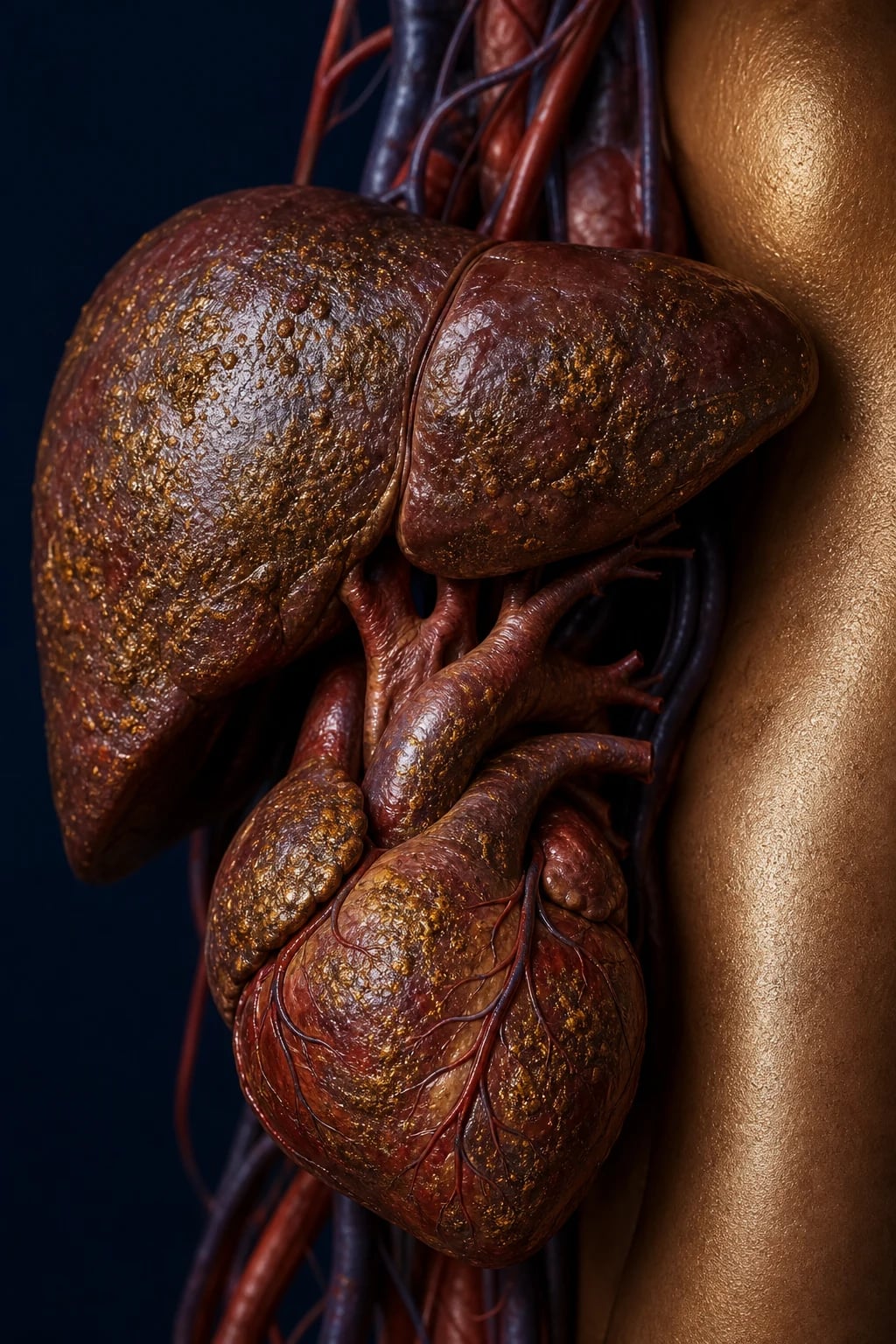
Haemochromatosis is a syndrome of excessive systemic iron accumulation that, untreated, deposits in and ultimately destroys parenchymal organs. The term spans two broad categories:[3][7]
- Hereditary (genetic) haemochromatosis (HH) — inherited defects that decrease hepcidin production or activity, removing the brake on intestinal iron absorption and macrophage iron release. Most cases are autosomal recessive; Type 4 (ferroportin) is autosomal dominant.
- Secondary iron overload — iron enters from outside (transfusions, dietary, parenteral) or accumulates from ineffective erythropoiesis (thalassaemia, sideroblastic anaemia, MDS). Macrophage iron is the dominant pool here, and the molecular defect is not in the hepcidin–ferroportin axis (although secondary hepcidin suppression occurs). [1]
In day-to-day practice and in NEET-PG/INICET, "haemochromatosis" almost always means HFE Type 1 hereditary haemochromatosis. The clinical skill is recognising iron overload early (when fatigue and arthralgia are the only symptoms) and treating with venesection before cirrhosis develops, because once cirrhosis is established the risk of hepatocellular carcinoma persists even after complete iron depletion.[1]
The disease is highly penetrant biochemically but weakly penetrant clinically: most C282Y homozygotes develop raised transferrin saturation and ferritin, but only a minority develop the full bronze-diabetes-cirrhosis picture. This explains why screening of first-degree relatives is so rewarding.[5][6]
Classification
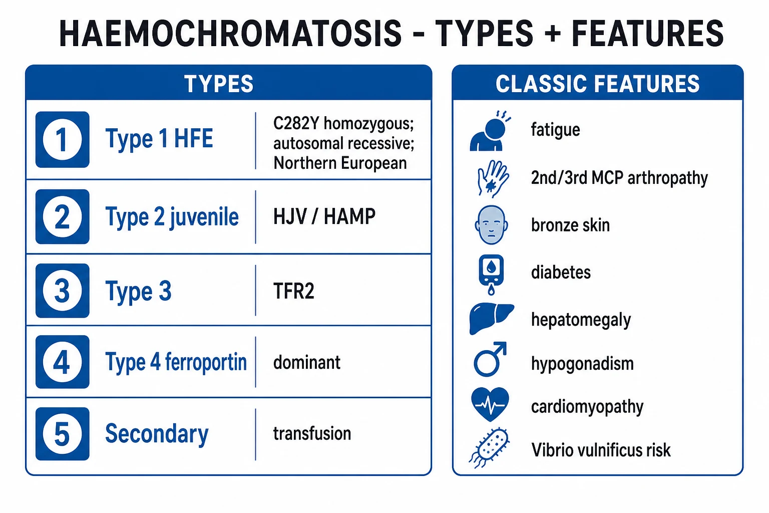
Hereditary haemochromatosis is classified by the mutated gene, the inheritance, the age of onset, and the distribution of iron deposition (parenchymal versus reticuloendothelial/macrophage):[2][3]
Type 1 — HFE
- Gene: HFE on chromosome 6p21.3 (C282Y homozygous commonest; H63D and S65C weak)
- Inheritance: autosomal recessive
- Onset: adult (40–60 years men; post-menopause women)
- Iron pattern: parenchymal (hepatocytes, heart, pancreas)
- Commonest type (>80 percent of HH in Northern Europeans)
Type 2 — Juvenile
- Type 2A: HJV (hemojuvelin); Type 2B: HAMP (hepcidin)
- Inheritance: autosomal recessive
- Onset: before age 30 (2nd–3rd decade) — severe, rapid
- Iron pattern: parenchymal; severe cardiac and endocrine
- Leading cause of death: cardiomyopathy and hypogonadism
Type 3 — TFR2
- Gene: TFR2 (transferrin receptor 2)
- Inheritance: autosomal recessive
- Onset: young adult (intermediate severity)
- Iron pattern: parenchymal, similar to Type 1
- Rare, more common in Southern Italy
Type 4 — Ferroportin
- Gene: SLC40A1 (ferroportin)
- Inheritance: autosomal dominant (unique among HH)
- Onset: adult
- Iron pattern: RETICULOENDOTHELIAL (Kupffer cells, macrophages) — spared parenchyma early
- Tolerates venesection poorly (early anaemia); lower-volume phlebotomy
Non-HFE genetic iron overload (rarer, but examinable):[2]
- Aceruloplasminemia (CP gene, AR) — the triad of diabetes, retinal degeneration and basal-ganglia neurological disease (parkinsonism, ataxia) with low serum iron and absent caeruloplasmin; iron trapped in brain and pancreas.
- Atransferrinemia (TF gene) — severe iron-loading anaemia with low transferrin.
- DMT1 (SLC11A2) mutation — microcytic anaemia with hepatic iron loading. [1]
Secondary iron overload (must be distinguished from HH before venesection): transfusional iron overload (thalassaemia major, sickle-cell, MDS), chronic ineffective erythropoiesis (sideroblastic anaemia), chronic haemolysis, dietary iron overload (African/Bantu siderosis), excess parenteral iron, and iron overload of chronic liver disease (alcohol, hepatitis C, NAFLD/NASH, porphyria cutanea tarda).[3]
Epidemiology & Risk Factors
- HFE C282Y homozygosity is the commonest monogenic disorder in people of Northern European (Celtic, Nordic) descent — gene frequency about 1 in 200 to 1 in 250, with a carrier rate of about 1 in 8 to 1 in 10 in this population.[5]
- Clinical penetrance is low: only about 1 to 2 percent of C282Y homozygotes develop the full bronze-diabetes-cirrhosis picture; biochemical penetrance (raised iron indices) is about 50 to 75 percent in men and lower in women.[6]
- Sex: clinical disease is 5 to 10 times more common in men. Women lose iron through menstruation and pregnancy, which delays onset until post-menopause (or after hysterectomy); men present at 40 to 60 years.[3]
- Ethnicity: rare in African, Asian, Hispanic and Polynesian populations (HFE C282Y essentially absent; African iron overload is a separate, non-HFE entity).
- Alcohol: a major co-factor — alcohol increases iron absorption and adds direct hepatotoxicity, accelerating cirrhosis.
- Co-factors that accelerate expression: hepatitis B or C, NAFLD, porphyria cutanea tarda (50 to 70 percent of PCT patients carry HFE mutations), and exogenous iron or vitamin C supplementation.[3]
Pathophysiology
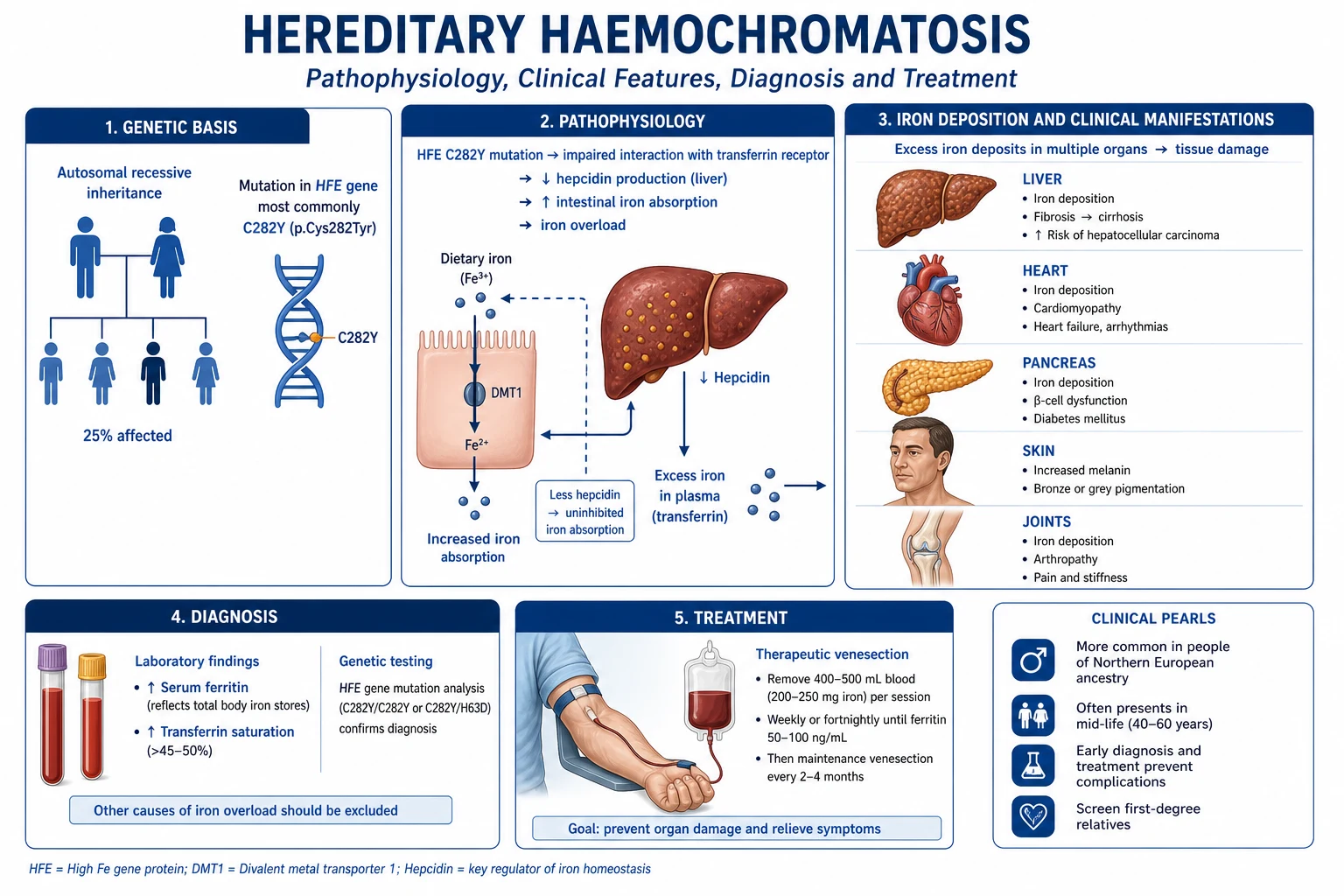
The hepcidin–ferroportin axis is the master regulator of systemic iron. Understanding it explains every clinical feature of HH.[2][7]
Normal iron physiology: [1]
- Total body iron is about 3 to 4 g in adults; about 2 to 2.5 g in haemoglobin, about 1 g in storage (ferritin, haemosiderin), the rest in myoglobin and enzymes. There is no regulated iron excretion — iron balance is controlled solely by absorption.
- Daily loss is only about 1 mg (desquamated skin, gut mucosa, menstruation adds about 0.5 mg/day). Duodenal absorption matches loss at 1 to 2 mg/day.
- Hepcidin (25-amino-acid peptide, made by hepatocytes) is the iron-regulatory hormone. It binds ferroportin — the only known cellular iron exporter — on the basolateral membrane of duodenal enterocytes, macrophages, hepatocytes and placental cells, causing ferroportin internalisation and degradation. The result: iron is trapped inside cells and plasma iron falls.
- Hepcidin is induced by high body iron (a negative-feedback loop) and suppressed by erythropoietic drive, hypoxia and inflammation (anaemia needs iron). [1]
What goes wrong in HFE haemochromatosis: [1]
- Mutant HFE protein fails to signal hepcidin up-regulation in response to iron loading. C282Y disrupts a disulphide bond so HFE cannot bind beta-2-microglobulin and reach the cell surface; the liver behaves as if iron-deficient.
- Hepcidin is inappropriately low (or inappropriately normal) for the degree of iron load.
- Ferroportin remains active — duodenal enterocytes and macrophages keep pumping iron into plasma unregulated.
- Daily iron absorption rises from 1 to 2 mg to 3 to 6 mg; with no excretion mechanism, iron accumulates at about 1 g per year of active disease.
- Over decades, body iron reaches 20 to 50 g (10 to 20 times normal) and deposits as haemosiderin in parenchymal cells (hepatocytes, cardiac myocytes, pancreatic beta-cells, pituitary, gonads, synovium, skin). Macrophage iron is relatively spared in HFE disease (in contrast to Type 4 ferroportin and transfusional overload).
- Free (non-transferrin-bound) iron catalyses Fenton chemistry — Fe²⁺ + H₂O₂ → Fe³⁺ + hydroxyl radical + hydroxide ion — generating reactive oxygen species that peroxidate membrane lipids, damage mitochondrial and nuclear DNA, and trigger apoptosis and fibrogenesis. [1]
At the molecular level, C282Y is a cysteine-to-tyrosine substitution at position 282 of the HFE protein. HFE is structurally MHC class I-like; to function it must fold correctly, bind beta-2-microglobulin, and travel to the basolateral hepatocyte membrane where it interacts with transferrin receptor 1. The C282Y mutation abolishes the critical disulphide bond, so the misfolded protein is retained in the endoplasmic reticulum and never reaches the cell surface — the liver literally cannot "see" its own iron stores and under-produces hepcidin. The milder H63D mutation (aspartate-to-histidine at position 63) distorts the ligand-binding pocket but still allows surface trafficking, which is why H63D homozygotes and C282Y/H63D compound heterozygotes load iron far less often and rarely develop end-organ disease.[2][7]
Organ-specific consequences of parenchymal iron:[3][7]
- Liver — hepatocyte necrosis activates Kupffer cells, which release TGF-beta and activate hepatic stellate cells, driving fibrosis → cirrhosis. Cirrhosis carries a 200-fold increased risk of hepatocellular carcinoma (the leading cause of death in treated adult HH).
- Pancreas — beta-cell iron deposition impairs insulin secretion, producing diabetes mellitus (often insulin-requiring).
- Heart — myocyte iron causes dilated cardiomyopathy and conduction disease/arrhythmia (atrial fibrillation, ventricular tachycardia). Cardiac failure is the leading cause of death in juvenile (Type 2) disease.
- Skin — increased melanin plus dermal iron produces the characteristic bronze/slate-grey pigmentation (so the disease is "bronze" rather than purely "iron-grey").
- Pituitary and gonads — iron in gonadotrophs causes hypogonadotropic hypogonadism: impotence and loss of libido in men, amenorrhoea and infertility in women, testicular atrophy, gynaecomastia.
- Joints — iron in synoviocytes promotes calcium pyrophosphate deposition (chondrocalcinosis, pseudogout) and a characteristic osteoarthritis of the 2nd and 3rd metacarpophalangeal joints with hook-like osteophytes.
- Infection susceptibility — iron is an essential growth factor for Vibrio vulnificus (raw shellfish, fatal septicaemia), Listeria monocytogenes, Yersinia enterocolitica and some Candida; iron overload impairs neutrophil and macrophage function. [1]
Clinical Presentation
The classic tetrad (only seen in late, untreated disease) is cirrhosis, diabetes mellitus, skin pigmentation and hypogonadism — historically called bronze diabetes.[3] Most modern patients present earlier and more subtly, and many are detected by screening iron studies before any symptom.
Untreated HFE haemochromatosis — the silent decades
Early / non-specific presentation (the commonest modern picture): [1]
- Fatigue, lethargy, weakness (80 percent of symptomatic cases) — vague and often misattributed.
- Arthralgia (40 to 60 percent) — often the first specific symptom, typically 2nd and 3rd MCP ("handshake pain"), wrists, knees, ankles; may precede organ damage by years.
- Right-upper-quadrant discomfort, abdominal pain.
- Loss of libido, erectile dysfunction, amenorrhoea. [1]
Established / late presentation — by organ: [1]
- Skin — bronze or slate-grey pigmentation, most marked in sun-exposed areas, flexural creases, scars and oral mucosa; due to increased melanin plus dermal iron.
- Liver — hepatomegaly early; later, stigmata of chronic liver disease (palmar erythema, spider naevi, jaundice, ascites, encephalopathy, splenomegaly from portal hypertension).
- Pancreas — diabetes mellitus; frequently insulin-requiring; ketoacidosis uncommon.
- Heart — dilated cardiomyopathy (exertional dyspnoea, oedema, raised JVP, S3 gallop), arrhythmias (atrial fibrillation, ventricular ectopy, sudden death), conduction blocks.
- Endocrine — hypogonadotropic hypogonadism (impotence, testicular atrophy, gynaecomastia, amenorrhoea, loss of axillary/pubic hair); hypothyroidism and adrenal insufficiency less common.
- Joints — chronic arthropathy (2nd and 3rd MCP with hook-like osteophytes, wrists, knees, ankles), chondrocalcinosis (knee menisci, pubic symphysis), pseudogout attacks; spine involvement rare. [1]
Atypical presentations to recognise (a recurring examiner theme): [1]
- Women — disease delayed to post-menopause by menstrual and pregnancy iron losses; may present after hysterectomy or with early menopause.
- Asymptomatic — abnormal iron indices on routine bloods or during evaluation of fatigue, arthritis, raised ALT, or incidentally found hepatomegaly.
- Elderly — may present with arthropathy alone (mistaken for osteoarthritis), new atrial fibrillation, or heart failure with preserved ejection fraction.
- Juvenile (Type 2, HJV/HAMP) — severe cardiomyopathy and hypogonadism before age 30, often with milder liver disease than HFE; cardiac death if untreated.
- Porphyria cutanea tarda — photosensitive bullous lesions on sun-exposed skin, fragile skin, hypertrichosis; often co-inherited with HFE mutations.
- Acute severe presentation — acute liver failure (rare), or severe sepsis with raw-shellfish exposure (Vibrio vulnificus) or Yersinia bacteraemia in unrecognised iron overload. [1]
Differential Diagnosis
A complete differential, with distinguishing features, follows:[2][3]
- Transfusional iron overload (thalassaemia major, sickle-cell, MDS, aplastic anaemia) — clear transfusion history (each unit delivers about 200 mg iron); iron loads macrophages/reticuloendothelial system first, then spills into parenchyma; anaemic patient; marrow shows ringed sideroblasts; HFE negative.
- Alcoholic liver disease — alcohol history, AST:ALT ratio over 2, macrocytosis, gamma-GT markedly raised; ferritin elevated as an acute-phase reactant but transferrin saturation usually under 45 percent; liver biopsy macrovesicular steatosis and Mallory bodies, mild iron in Kupffer cells.
- Non-alcoholic fatty liver disease (NAFLD/NASH) — metabolic syndrome, normal or low transferrin saturation, raised ferritin from inflammation; biopsy shows steatosis with mild iron in Kupffer cells, not hepatocytes.
- Chronic viral hepatitis (HCV/HBV) — positive viral serology; ferritin modestly elevated; iron not the dominant histological feature.
- African (Bantu) iron overload — sub-Saharan diet brewed in iron pots, plus a non-HFE genetic factor; affects both hepatocytes and macrophages.
- Aceruloplasminemia — diabetes, retinal degeneration and basal-ganglia parkinsonism/ataxia; low serum iron and undetectable caeruloplasmin; brain MRI shows iron in basal ganglia.
- Wilson disease (a copper disorder, in the differential of cirrhosis plus neuropsychiatric disease in young adults) — low caeruloplasmin, Kayser–Fleischer rings, neurological movement disorder, low serum copper.
- Porphyria cutanea tarda — photosensitive bullae on sun-exposed skin, fragile skin, hypertrichosis, dark urine (pink fluorescence under Wood's lamp); often coexists with HFE mutations.
- Osteoarthritis / rheumatoid arthritis / CPPD pseudogout (differential of the arthropathy alone) — osteoarthritis affects DIP/PIP not MCP; rheumatoid arthritis gives symmetrical MCP synovitis, morning stiffness over 1 hour, RF/anti-CCP positive; calcium pyrophosphate (CPPD/pseudogout) affects knee and wrist with rhomboid weakly positive birefringent crystals.
- Addison's disease / drug pigmentation (differential of skin pigmentation alone) — Addison's gives mucocutaneous pigmentation, weight loss, hypotension, hyponatraemia, hyperkalaemia, low cortisol/high ACTH; drug pigmentation from amiodarone, minocycline, heavy metals.
- Adult-onset Still's disease / haemophagocytic lymphohistiocytosis — extremely high ferritin as acute-phase marker, but transferrin saturation low (anaemia of chronic disease). [1]
Clinical & Bedside Assessment
A focused bedside examination looks for organ-specific iron damage:[1]
- Skin — bronze/slate-grey pigmentation, especially sun-exposed areas, scars, palmar creases, oral mucosa, external genitalia; look for stigmata of chronic liver disease.
- Hands — 2nd and 3rd MCP bony swelling, "handshake sign" (pain on dorsiflexion of the index/MCP joints), hook-like osteophytes; chondrocalcinosis of wrist/knee.
- Abdomen — hepatomegaly (early), later splenomegaly, ascites, caput medusae of portal hypertension; test for hepatic encephalopathy (asterixis).
- Cardiovascular — signs of dilated cardiomyopathy (raised JVP, displaced apex, S3 gallop, basal crackles), irregularly irregular pulse of atrial fibrillation.
- Endocrine / genital — testicular atrophy, gynaecomastia, loss of body hair in men; signs of hypothyroidism; clinical diabetes (random glucose).
- Neurological — peripheral neuropathy, and (in aceruloplasminemia) cerebellar/extrapyramidal signs. [1]
Named signs: "Handshake sign" (pain on dorsiflexion of the second and third MCP joints) is characteristic; bronze diabetes is the classic eponymous phenotype; Kayser–Fleischer rings are not present in haemochromatosis (they belong to Wilson disease). [1]
Investigations
A two-step strategy: screening iron indices, then confirmatory HFE genotyping, then staging of organ damage.[1][4]
Step 1 — Screening iron studies
- Fasting transferrin saturation (TSAT) = serum iron divided by total iron-binding capacity (TIBC) multiplied by 100. The earliest and most sensitive abnormality. Thresholds: over 45 percent (some guidelines say over 50 percent in men, over 45 percent in women) warrants HFE testing. A TSAT over 60 percent in men or over 50 percent in women is highly suggestive.[1]
- Serum ferritin — proportional to body iron stores but is an acute-phase reactant (rises with inflammation, infection, liver disease, alcohol). Investigate if over 300 micrograms per litre (men) or over 200 micrograms per litre (women); over 1000 micrograms per litre strongly suggests significant iron loading and is the trigger for liver fibrosis assessment.
- Repeat abnormal iron indices fasting, on two separate occasions, when the patient is well before pursuing the diagnosis (false positives from recent alcohol, hepatitis, inflammation).
Hereditary haemochromatosis
- Transferrin saturation HIGH (over 45 percent) — the single best discriminator
- Ferritin high and tracks with body iron; falls predictably with each venesection
- C282Y homozygous on HFE genotyping confirms Type 1
- Parenchymal iron on biopsy (Perls' Prussian blue in hepatocytes); hepatic iron index over 1.9
Inflammation / infection / malignancy
- Transferrin saturation LOW or normal — ferritin is behaving as an acute-phase reactant
- Ferritin high but disproportionate to true iron stores; CRP raised
- Underlying sepsis, autoimmune flare or occult cancer drives the rise
- Normalises when the inflammatory stimulus resolves; do NOT venesect
Alcohol / NAFLD / chronic liver disease
- Transferrin saturation usually under 45 percent
- Ferritin mildly to moderately raised from hepatocyte injury and the acute-phase response
- AST:ALT pattern (over 2 in alcohol), steatosis on imaging, metabolic syndrome features
- Iron sits in Kupffer cells, not hepatocytes, on biopsy
Adult Still's / HLH
- Ferritin grossly elevated (often over 10,000 micrograms per litre) as a marker of macrophage activation
- Transferrin saturation LOW (anaemia of chronic disease)
- Fever, evanescent salmon-pink rash, arthralgia, hepatosplenomegaly, high soluble IL-2 receptor
- Glycosylated ferritin fraction under 20 percent is characteristic of adult Still's disease
Step 2 — Confirmatory genetic testing
- HFE genotyping for C282Y and H63D (and S65C). C282Y homozygosity with raised iron indices confirms Type 1 HH and obviates the need for liver biopsy purely to confirm the diagnosis.[1]
- C282Y/H63D compound heterozygotes and H63D homozygotes have low penetrance; treat only if iron loading is confirmed biochemically.
- Non-C282Y cases with confirmed iron loading — request HJV, HAMP, TFR2, SLC40A1 sequencing (juvenile, ferroportin).
Step 3 — Quantify and stage
- Liver biopsy with Perls' Prussian blue stain — historically the gold standard. Quantifies hepatic iron concentration (HIC); hepatic iron index (HII) = HIC divided by age; over 1.9 micromoles per gram dry weight per year supports HH (vs secondary overload). Biopsy is now reserved for staging fibrosis when ferritin over 1000 micrograms per litre, age over 40, elevated AST/ALT, or non-C282Y homozygotes.[1][4]
- Non-invasive fibrosis — FibroScan (transient elastography), FIB-4, APRI; MRI-based methods.
- MRI T2-star — non-invasive quantification of hepatic and cardiac iron; FerriScan (R2 MRI) is FDA-cleared for liver iron concentration. T2-star under 20 ms indicates significant cardiac iron loading.
- Organ function tests — LFTs, glucose/HbA1c, fasting lipid, FSH/LH/testosterone (or oestradiol), TSH, morning cortisol, alpha-fetoprotein (AFP).
- Cardiac assessment — 12-lead ECG (arrhythmia, conduction), echocardiogram (systolic/diastolic function), cardiac MRI T2-star.
- Liver ultrasound plus AFP every 6 months — hepatocellular carcinoma surveillance in cirrhotics.
- Exclusion of co-factors — hepatitis B and C serology, alcohol history, lipids, glucose; screen for porphyria cutanea tarda (urinary porphyrins) if skin lesions.
Family screening — all first-degree relatives (siblings and children) should have HFE genotyping plus fasting transferrin saturation and ferritin. C282Y homozygous relatives are followed even if biochemically normal.[1][5]
Management — Resuscitation
HH is a chronic disease and rarely presents acutely, but two scenarios need urgent management:[3]
- Decompensated cirrhosis — treat as for any acute-on-chronic liver failure: ABCDE, correct coagulopathy/hypoglycaemia, treat ascites (spironolactone 100 mg oral daily plus furosemide 40 mg oral daily, dose-titrated), encephalopathy (lactulose 15 to 30 mL oral two to three times daily to achieve two to three soft stools), and refer to a hepatology/transplant centre.
- Cardiac decompensation — standard heart-failure therapy (loop diuretic, ACE inhibitor/ARB, beta-blocker once euvolaemic, mineralocorticoid antagonist), rate/rhythm control for atrial fibrillation; urgent iron chelation (deferoxamine continuous IV infusion plus deferiprone) for life-threatening cardiac iron loading; arrhythmia may need cardiology input and ICU monitoring.
- Sepsis with iron overload (especially Vibrio vulnificus from raw shellfish, Yersinia) — broad-spectrum antibiotics (e.g. doxycycline plus ceftazidime or a fluoroquinolone), ITU support, and do not give iron during the acute episode. [1]
Management — Definitive & Stepwise
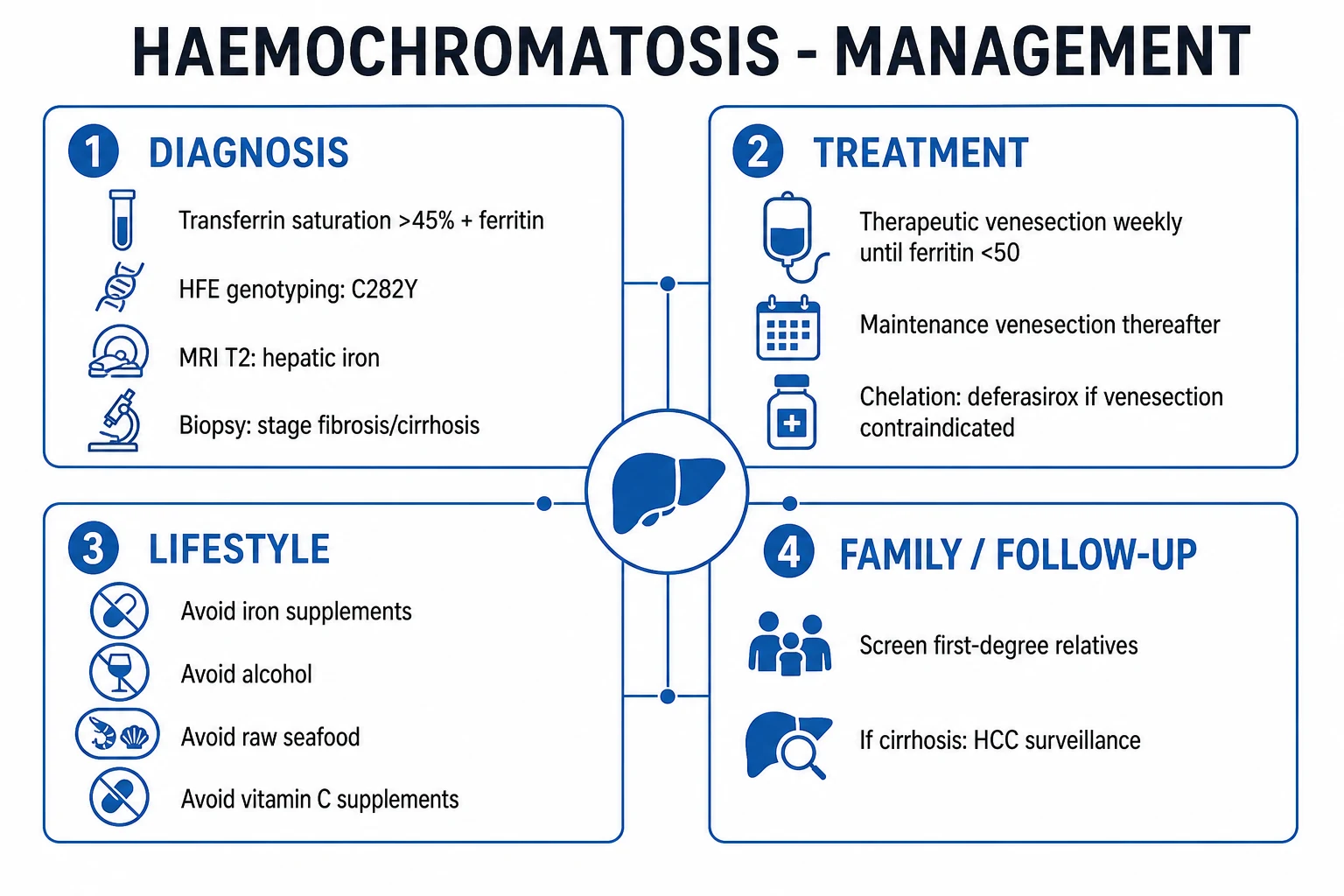
The cornerstone of treatment is iron removal.[1][4]
Therapeutic venesection protocol — induction then lifelong maintenance
Confirm the diagnosis (raised fasting transferrin saturation plus ferritin; C282Y homozygous on HFE genotyping) and exclude anaemia (haemoglobin at least 110 g/L) before the first venesection
INDUCTION — remove one unit (450 to 500 mL) of whole blood weekly or fortnightly; each unit strips about 200 to 250 mg of iron
Check haemoglobin before every session — postpone if haemoglobin under 110 g/L or it falls more than 20 g/L between sessions; check serum ferritin and transferrin saturation every 4 to 8 weeks
Continue induction until serum ferritin reaches 50 to 100 micrograms per litre and transferrin saturation is under 50 percent — typically 1 to 2 years and 20 to 50 venesections
Do NOT drive ferritin under 20 micrograms per litre — over-venesection causes iron-deficiency anaemia, fatigue and restless legs
MAINTENANCE — switch to one venesection every 2 to 4 months (about 3 to 4 per year) for LIFE; recheck ferritin before each session and keep it in target range
Use smaller-volume (250 mL) phlebotomy for the elderly, frail, cardiac patients and Type 4 ferroportin disease to avoid precipitating anaemia
First-line: therapeutic venesection (phlebotomy)
- Rationale — each 450 to 500 mL (one unit) of whole blood removes about 200 to 250 mg of iron. This is the most efficient, cheapest and safest iron-depletion method and is first-line for HFE Types 1, 2 and 3.
- Induction phase — remove one unit (450 to 500 mL) weekly or fortnightly in adults who tolerate it. Monitor haemoglobin before each venesection (postpone if haemoglobin under 110 g/L or drops more than 20 g/L between sessions) and serum ferritin every 4 to 8 weeks (and transferrin saturation).
- Target — serum ferritin 50 to 100 micrograms per litre and transferrin saturation under 50 percent; avoid driving ferritin under 20 micrograms per litre (over-venesection causes iron-deficiency anaemia and fatigue).
- Maintenance phase — once at target, switch to one venesection every 2 to 4 months (typically 3 to 4 per year) to keep ferritin in target; lifelong.
- Volume for special groups — smaller-volume (250 mL) phlebotomy for the elderly, frail, cardiac patients, and Type 4 ferroportin disease (anaemia-prone). [1]
Second-line: iron chelation (when venesection contraindicated)
Indications: anaemia (thalassaemia, MDS, sideroblastic), severe cardiac failure/cardiomyopathy where venesection not tolerated, Type 4 ferroportin disease with early anaemia.[3]
- Deferasirox — oral, once-daily dispersible tablet; 20 to 40 mg/kg/day; monitor renal function (creatinine), LFTs, audiology and ophthalmology (ototoxicity, retinopathy). Convenient but renal adverse effects limit use.
- Deferiprone — oral, 75 mg/kg/day in three divided doses; effective at cardiac iron removal; risk of agranulocytosis (mandatory weekly FBC) and arthropathy.
- Deferoxamine (desferrioxamine) — subcutaneous infusion 40 to 50 mg/kg/day over 8 to 12 hours via pump, 5 to 7 nights/week; the original chelator; reserved for severe disease especially cardiac iron (IV infusion in acute cardiac decompensation). [1]
Erythrocytapheresis
Removes more iron per session than simple phlebotomy (plasma returned, only red cells removed) — useful where available for rapid depletion or anaemia-prone patients. [1]
Lifestyle measures
- Avoid iron supplements (including multivitamins with iron) and high-dose vitamin C (vitamin C enhances iron absorption and, in massive overload, has caused fatal arrhythmia).
- Avoid alcohol (accelerates cirrhosis); counsel abstinence or strict limits.
- Avoid raw shellfish and undercooked seafood (Vibrio vulnificus risk).
- Avoid uncooked marine fish if severely iron loaded; the risk falls to normal once iron depleted. [1]
Management of complications
- Hepatocellular carcinoma surveillance — liver ultrasound plus alpha-fetoprotein every 6 months in all cirrhotic HH patients (HCC risk persists after iron depletion).
- Diabetes — standard therapy; insulin frequently required; metformin if appropriate.
- Hypogonadism — testosterone replacement (men), hormone replacement (women, until usual menopause age); fertility needs gonadotrophin therapy.
- Arthropathy — simple analgesia (paracetamol first-line, NSAIDs with caution in cirrhosis/renal impairment), physiotherapy; joint replacement for end-stage disease.
- Cirrhosis — variceal screening (OGD at diagnosis and every 1 to 3 years), hepatocellular carcinoma surveillance, vaccinations (hepatitis A and B, influenza, pneumococcal).
- Liver transplantation — for decompensated cirrhosis or hepatocellular carcinoma meeting criteria; outcomes are worse than for other indications because of cardiac iron involvement and infectious risk, so optimise iron pre-transplant. [1]
Family screening and genetic counselling
- Test all first-degree relatives of confirmed cases with HFE genotyping and iron studies.
- Asymptomatic C282Y homozygous relatives: lifelong clinical and biochemical surveillance from early adulthood; venesection if iron loading develops.
- Reproductive counselling for juvenile and ferroportin forms (AR vs AD inheritance changes the recurrence risk). [1]
Specific Subtypes & Scenarios
- Juvenile haemochromatosis (Type 2, HJV/HAMP) — severe, early-onset; cardiomyopathy and hypogonadism dominate and are the leading causes of death. Treat aggressively with venesection plus chelation from diagnosis; cardiac transplantation may be needed.
- Ferroportin disease (Type 4, SLC40A1) — autosomal dominant; iron in macrophages/Kupffer cells rather than hepatocytes, so transferrin saturation is normal or low and ferritin is markedly elevated. Aggressive venesection causes early anaemia — use smaller-volume phlebotomy (250 mL weekly to fortnightly) or chelation.
- African iron overload — non-HFE; dietary (iron-pot brewing) plus genetic predisposition; affects both hepatocytes and macrophages; treat with dietary counselling and gentle venesection.
- Porphyria cutanea tarda (PCT) — venesection is also the treatment for PCT; many PCT patients carry HFE mutations, so test for HH in every PCT case. Phlebotomy to ferritin under 50 micrograms per litre plus low-dose hydroxychloroquine (200 mg twice weekly) clears the skin lesions.
- Hereditary haemochromatosis in a woman of reproductive age — defer venesection during pregnancy; resume postpartum. Avoid chelation (deferiprone is teratogenic; deferasirox not recommended in pregnancy).
- Iron overload in a chronic-anaemia patient (thalassaemia, MDS) — never venesect; use chelation from the first 10 to 20 transfusions; cardiac T2-star MRI surveillance. [1]
Complications & Pitfalls
Complications:[3]
- Cirrhosis — present in about 10 to 15 percent of untreated C282Y homozygotes at diagnosis.
- Hepatocellular carcinoma — 200-fold increased risk, only in cirrhotics; the leading cause of death in treated adult HH; risk persists after iron depletion.
- Cardiomyopathy and arrhythmia — dilated cardiomyopathy, atrial fibrillation, sudden cardiac death; dominant in juvenile disease.
- Diabetes mellitus — usually irreversible.
- Hypogonadism, impotence, infertility — often irreversible.
- Arthropathy — usually irreversible despite iron depletion; major quality-of-life burden.
- Osteoporosis — from hypogonadism.
- Infection — Vibrio vulnificus (raw oysters, septicaemia, haemorrhagic bullae), Yersinia enterocolitica (mesenteric adenitis, septicaemia — iron-dependent), Listeria, Candida; iron withdrawal (venesection) reduces risk. [1]
Pitfalls: [1]
- Misdiagnosing elevated ferritin as iron overload — ferritin rises in inflammation, infection, alcohol, NAFLD, hepatitis and malignancy; transferrin saturation distinguishes true iron overload (high) from inflammation (low/normal).
- Forgetting to screen the family — a single diagnosis should trigger testing of all first-degree relatives; pre-symptomatic treatment prevents disease.
- Over-venesecting to very low ferritin — causes iron-deficiency anaemia, fatigue and restless legs; aim for 50 to 100 micrograms per litre, not zero.
- Missing HCC surveillance — cirrhotic HH needs 6-monthly ultrasound plus AFP for life, even after iron normalisation.
- Using aggressive venesection in Type 4 ferroportin disease or anaemia — causes dangerous anaemia; switch to small-volume phlebotomy or chelation.
- Failing to vaccinate cirrhotic patients (hepatitis A and B, influenza, pneumococcal). [1]
Prognosis & Disposition
- Treated before cirrhosis — normal life expectancy. Iron depletion reverses fatigue, skin pigmentation and hepatic inflammation and may partly improve diabetes and cardiac function.[1]
- Once cirrhosis develops — survival reduced; HCC risk persists even after complete iron depletion; lifelong HCC surveillance required.
- Arthropathy, hypogonadism and (often) diabetes are irreversible despite iron depletion; manage symptomatically.
- Cardiac iron overload — potentially reversible with chelation and venesection; the dominant reversible cause of death in juvenile disease.
- Disposition — most patients managed as outpatients; admit for decompensated cirrhosis, severe cardiac failure/arrhythmia, severe sepsis or for intensive chelation. Coordinate care between hepatology, cardiology, endocrinology and haematology; consider specialist iron-overload clinic.
Special Populations
- Women of reproductive age / pregnancy — disease delayed by menstruation and pregnancy; defer venesection during pregnancy; no iron chelation in pregnancy (deferiprone teratogenic; deferasirox avoid); resume venesection postpartum and during lactation.
- Children — HFE Type 1 rarely manifests clinically before adulthood; do not venesect asymptomatic children; juvenile (Type 2) presents in adolescence with severe cardiomyopathy and hypogonadism — urgent specialist management.
- Elderly — lower-volume venesection (250 mL), longer intervals; watch for anaemia and cardiac decompensation; comorbidities (heart failure, chronic kidney disease) shift to chelation.
- Patients with chronic anaemia (thalassaemia, MDS, sideroblastic) — never venesect; chelation is first-line; coordinate with haematology.
- Anticoagulated / bleeding-disorder patients — venesection is generally still feasible (no coagulopathy issue, since you are removing blood intentionally) but discuss with haematology if platelets under 50 or on warfarin/DOAC. [1]
Evidence, Guidelines & Regional Differences
- AASLD 2011 practice guideline (Bacon et al.) — fasting transferrin saturation over 45 percent triggers HFE testing in symptomatic patients or those with family history; HFE genotyping confirms Type 1; venesection first-line to ferritin 50 to 100 micrograms per litre; liver biopsy to stage fibrosis when ferritin over 1000 micrograms per litre, age over 40, or elevated ALT/AST.
- EASL 2010 guideline — broadly concordant; emphasises non-invasive fibrosis assessment and 6-monthly HCC surveillance in cirrhosis.
- BSH (British Society for Haematology) / UK guideline (Dooley & Worwood) — recommends screening of first-degree relatives and the same iron-study thresholds; UK uses ferriScan and MRI T2-star increasingly. [1]
Clinical evidence
Multi-ethnic, cross-sectional screening study of 99,711 North American primary-care adults
Population: Adults across five ethnic groups (white, black, Asian, Hispanic, Native American)
Key finding
C282Y homozygosity in 0.44 percent of white participants; biochemical penetrance high but clinical disease (cirrhosis, HCC, pigmentation, diabetes) found in only about 1 to 2 percent — not significantly above non-carriers
Clinical evidence
Prospective Australian population cohort (Melbourne Collaborative Cohort Study) with long follow-up
Population: C282Y homozygotes and age-matched controls
Key finding
Iron-overload-related disease developed in 28 percent of C282Y homozygous men but only 1 percent of women; penetrance strongly male-predominant and age-dependent
Controversies: [1]
- Population screening for HFE — favoured in Northern-European-origin populations on cost-effectiveness grounds but contested because of low clinical penetrance and the ethical burden of identifying a gene most carriers never express.
- Chelation versus venesection in HFE disease with mild anaemia — venesection remains first-line; chelation is rarely needed in pure HFE Type 1. [1]
Exam Pearls
Organ targets of iron — BRONZE CHARM
CHARM
dilated cardiomyopathy, arrhythmias — leading cause of death in juvenile HH
cirrhosis and hepatocellular carcinoma (200-fold risk in cirrhosis)
2nd and 3rd MCP with hook-like osteophytes, chondrocalcinosis
bronze/slate-grey pigmentation (melanin plus dermal iron)
pancreas (diabetes), pituitary (hypogonadotropic hypogonadism), testes, thyroid
Hereditary haemochromatosis — the numbers examiners ask
- Commonest cause of HH in Northern Europeans = C282Y/C282Y homozygosity of HFE on chromosome 6p21.3 (autosomal recessive). H63D is common but weakly penetrant.[2]
- Bronze diabetes = cirrhosis plus diabetes plus skin pigmentation (plus hypogonadism for the full tetrad).
- Screen with fasting transferrin saturation (over 45 percent) and ferritin; confirm with HFE genotyping (no biopsy needed for diagnosis in C282Y homozygotes).
- Pathophysiology in one sentence: mutant HFE fails to up-regulate hepcidin; ferroportin unchecked; unregulated iron absorption; Fenton-chemistry oxidative injury to parenchymal cells.
- Treatment = venesection first-line (one unit/week, target ferritin 50 to 100 micrograms per litre); chelation (deferasirox, deferiprone, deferoxamine) if venesection contraindicated.
- Mnemonic "ABCDEF" for organs: Arthropathy, Bronze skin, Cardiomyopathy, Diabetes, Endocrine (hypogonadism), Fibrosis (liver).
- Iron deposits in parenchymal cells (hepatocytes) — distinguishes HFE disease from secondary overload (macrophage-predominant).
- Avoid alcohol, iron supplements, vitamin C, raw shellfish (Vibrio vulnificus).
- HCC only in cirrhosis; surveillance 6-monthly ultrasound plus AFP for life.
- Cardiac death is the leading cause in juvenile (Type 2) HH (HJV or HAMP).
- Handshake sign — pain on dorsiflexion of 2nd/3rd MCP.
- Screen all first-degree relatives (siblings and children) with HFE plus iron studies.
- Porphyria cutanea tarda co-inherits HFE mutations — venesection treats both.
- Treated before cirrhosis — normal life expectancy.
Exam application bank (NEET-PG / INICET)
One-line answer
Hereditary haemochromatosis (HH) is an autosomal recessive disorder of iron homeostasis causing inappropriate, unregulated intestinal iron absorption that progressively deposits in parenchymal organs — liver (cirrhosis, hepatocellular carcinoma), pancreas (diabetes), heart (cardiomyopathy, arrhythmia), skin (bronzing), joints (arthropathy), pituitary and gonads (hypogonadism). The commonest cause is homozygous C282Y mutation in the HFE gene on chromosome 6p (Type 1). Screen with transferrin saturation over 45 percent and serum ferritin; confirm with HFE genetic testing. Cornerstone treatment is therapeutic venesection (phlebotomy) to target ferritin 50 to 100 micrograms per litre, with iron chelation reserved for those who cannot be venesected. Treated before cirrhosis, life expectancy is normal.
Worked stems (answer without another resource)
Stem 1 — Classic presentation. Map symptoms to mechanism; name the first investigation and first treatment step with dose/route if drug therapy is standard. [1]
Stem 2 — Unstable / complicated. List red flags that force immediate resuscitation, theatre, ICU, antidote, or reperfusion — and what you do in the first 15 minutes. [1]
Stem 3 — Atypical group. Elderly, pregnancy, child, or immunocompromised: how presentation and thresholds change. [1]
Stem 4 — Differential trap. Name the three closest mimics and one discriminator for each. [1]
Stem 5 — Disposition. Who goes home with safety-netting, who is admitted, who needs HDU/ICU/theatre, and what follow-up is mandatory. [1]
Rapid viva checklist
- Definition + classification
- Pathophysiology chain
- Bedside signs / criteria
- Score with exact components (if any)
- Emergency bundle
- Definitive therapy with doses
- Complications of disease and of treatment
- Special populations
- Guideline/trial name if classic
- Three exam traps
Coverage self-check
If you cannot answer any stem above from this page alone, re-read the matching section — the page is intended to be self-sufficient for final-prof and NEET-PG/INICET questions on Hereditary Haemochromatosis.
References
- [1]Bacon BR, Adams PC, Kowdley KV, Powell LW, Tavill AS. Diagnosis and management of hemochromatosis: 2011 practice guideline by the American Association for the Study of Liver Diseases Hepatology, 2011.PMID 21452290
- [2]Pietrangelo A. Hereditary hemochromatosis--a new look at an old disease N Engl J Med, 2004.PMID 15175440
- [3]Powell LW, Seckington RC, Deugnier Y. Haemochromatosis Lancet, 2016.PMID 26975792
- [4]European Association for the Study of the Liver. EASL clinical practice guidelines for HFE hemochromatosis J Hepatol, 2010.PMID 20471131
- [5]Adams PC, Reboussin DM, Barton JC, et al. Hemochromatosis and iron-overload screening in a racially diverse population N Engl J Med, 2005.PMID 15858186
- [6]Allen KJ, Gurrin LC, Constantine CC, et al. Iron-overload-related disease in HFE hereditary hemochromatosis N Engl J Med, 2008.PMID 18199861
- [7]Brissot P, Pietrangelo A, Adams PC, de Graaff B, McLaren CE, Loréal O. Haemochromatosis Nat Rev Dis Primers, 2018.PMID 29620054