Haematology · Haematology
Haemolytic Anaemia
Also known as Hemolytic anemia · Haemolysis · AIHA · Autoimmune haemolytic anaemia · Extravascular haemolysis
Haemolytic anaemia is anaemia caused by premature destruction of red blood cells (lifespan shortened from the normal 120 days) at a rate that exceeds marrow compensation. Classify by site (intravascular vs extravascular) and by origin (inherited vs acquired). Biochemical signature: raised reticulocytes, raised LDH, raised unconjugated bilirubin, low/absent haptoglobin, with haemoglobinaemia/haemoglobinuria in intravascular forms. The direct antiglobulin (DAT/Coombs) test is the single most important discriminator: positive = immune (warm IgG AIHA, cold IgM agglutinin, paroxysmal cold haemoglobinuria), negative = non-immune (hereditary spherocytosis, G6PD deficiency, PNH, microangiopathic, sickle cell, thalassaemia). Management is cause-specific: warm AIHA — prednisolone 1–1.5 mg/kg; cold agglutinin — avoid cold + rituximab; hereditary spherocytosis — folate +/- splenectomy (vaccinate before); G6PD — avoid triggers; PNH — eculizumab/ravulizumab; MAHA — treat underlying (TTP = plasma exchange + caplacizumab).
On this page & tools
Your progress
Saved locally on this device.
Practise this topic
Exam tags
Red flags
Overview & Definition
Haemolytic anaemia is defined as anaemia that arises when red blood cells are destroyed before their normal lifespan of about 120 days is complete, and at a rate that overwhelms the bone marrow's compensatory capacity (which can raise output six- to eight-fold by increasing erythropoietin-driven reticulocyte release). The clinician's task is not simply to confirm that haemolysis is present — that is the easy part — but to localise the mechanism and identify the specific cause, because management of warm autoimmune haemolytic anaemia (steroids), hereditary spherocytosis (splenectomy), G6PD deficiency (trigger avoidance) and paroxysmal nocturnal haemoglobinuria (complement blockade) could not be more different. The direct antiglobulin (Coombs) test (DAT) is the single most powerful branch point in the diagnostic algorithm.[1][2]
Classification
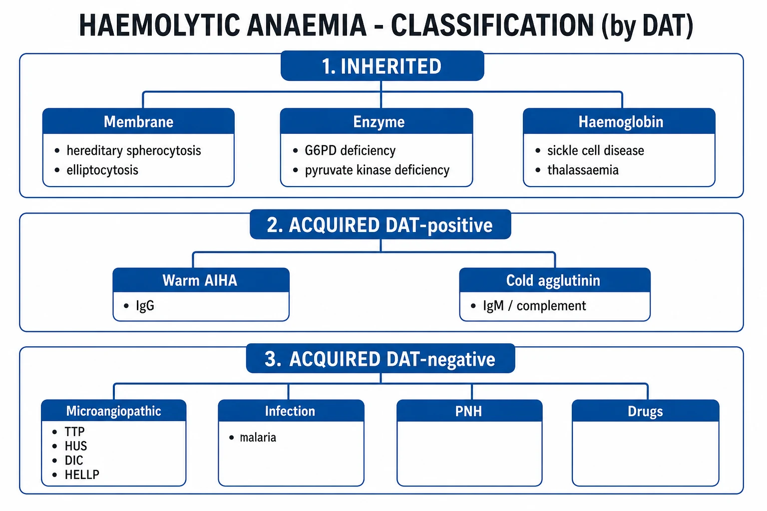
Haemolytic anaemia is classified along two independent axes — by the anatomical site of destruction and by the origin (inherited or acquired). Both axes carry immediate clinical implications: extravascular destruction tends to be gentler and splenic, while intravascular destruction produces the dramatic triad of haemoglobinaemia, haemoglobinuria and acute kidney injury; inherited causes declare themselves in childhood with a positive family history, while acquired causes demand a search for triggers, drugs and systemic disease. [1]
Intravascular haemolysis
- Red cells lyse WITHIN the circulation (free haemoglobin spills into plasma)
- Hallmarks: haemoglobinaemia, haemoglobinuria (dark/cola urine), haemosiderinuria, methaemalbumin
- Haptoglobin ABSENT (saturates and clears within hours)
- LDH markedly raised, often over 1000 U/L
- Causes: ABO mismatch, PNH, cold agglutinin, G6PD crisis, MAHA/TTP, prosthetic valve, march haemoglobinuria, severe burns, malaria, clostridial sepsis, Wilson crisis
Extravascular haemolysis
- Red cells destroyed by SPLENIC/HEPATIC macrophages (Fc and C3b receptors)
- Hallmarks: jaundice (unconjugated bilirubin), splenomegaly, spherocytes (warm AIHA/HS)
- Haptoglobin LOW but may be detectable; LDH moderately raised
- Causes: warm AIHA (IgG), hereditary spherocytosis, sickle cell, thalassaemia, haemolytic disease of newborn
- Commoner than intravascular
Inherited (corpuscular) defects
- MEMBRANE: hereditary spherocytosis (spectrin/ankyrin/pallidin/band 3 — mostly AD), hereditary elliptocytosis
- ENZYME: G6PD deficiency (X-linked recessive, commonest), pyruvate kinase deficiency (AR, chronic non-spherocytic)
- HAEMOGLOBIN: sickle cell disease, thalassaemia, unstable haemoglobins (Hb Koln)
- Clues: family history, lifelong/childhood onset, chronic jaundice, pigment gallstones, DAT NEGATIVE
Acquired (extracorpuscular)
- IMMUNE: warm AIHA (IgG, 37C), cold agglutinin disease (IgM, 4C), paroxysmal cold haemoglobinuria (biphasic IgG, Donath-Landsteiner), drug-induced
- NON-IMMUNE: microangiopathic (TTP/HUS/DIC/HELLP/malignant HTN), PNH, infection (malaria, clostridium, bartonella), mechanical (prosthetic valve), severe burns, Wilson
- Clues: adult onset, drug/infection trigger, autoimmune or lymphoproliferative disease, DAT POSITIVE (if immune)
Epidemiology & Risk Factors
Inherited haemolytic anaemias track along ethnic and geographic lines that mirror the historical distribution of falciparum malaria — the so-called malaria belt. Carriage of the sickle mutation, alpha- and beta-thalassaemia traits, and G6PD deficiency all confer partial protection against severe malaria in heterozygotes, which explains their persistence at high frequency. G6PD deficiency is the commonest human enzyme defect, affecting an estimated 400 million people worldwide, with particularly high prevalence in sub-Saharan Africa, the Mediterranean, the Middle East and South-East Asia. Sickle cell trait is carried by roughly 1 in 12 African Americans and is similarly frequent in parts of India and the Middle East. Hereditary spherocytosis is the commonest inherited haemolytic anaemia in Northern Europeans (incidence about 1 in 2000) and is most often autosomal dominant. The thalassaemias are concentrated across the Mediterranean (Greek for "sea blood"), the Middle East, the Indian subcontinent and South-East Asia.[3][4]
Among the acquired haemolytic anaemias, autoimmune haemolytic anaemia (AIHA) is by far the commonest, with an overall incidence of about 1 to 3 per 100,000 per year. Warm AIHA accounts for roughly 70 to 80 percent of cases (mediated by IgG at body temperature) and may be idiopathic (about half) or secondary to lymphoproliferative disease (chronic lymphocytic leukaemia, lymphoma), systemic autoimmune disease (systemic lupus erythematosus), immunodeficiency, or drugs (methyldopa, penicillin, cephalosporins, fludarabine). Cold agglutinin disease accounts for about 15 to 20 percent and may follow Mycoplasma pneumoniae (anti-I) or infectious mononucleosis (anti-i), or arise from a B-cell lymphoproliferative disorder. Paroxysmal nocturnal haemoglobinuria (PNH) is rare (incidence about 1 to 2 per million) but disproportionately important because it is fatal if untreated and fully transformed by complement inhibition.[2]
[1]Pathophysiology
The unifying lesion is a red-cell lifespan shortened from ~120 days to as little as a few days, after which the bone marrow's ability to compensate (driven by erythropoietin) is exceeded and anaemia supervenes. Three mechanistic questions define the workup: (1) is the destruction intravascular or extravascular? (2) is the cell intrinsically abnormal or is it being attacked extrinsically? and (3) is antibody or complement bound to the cell (immune) or not?[1][2]
Extravascular destruction — the splenic macrophage
The normal spleen filters senescent red cells through a network of macrophages that recognise physiological ageing signals (band 3 clustering, immunoglobulin binding, oxidative membrane damage). In extravascular haemolysis this process is accelerated. The archetype is warm AIHA: IgG antibodies (class 1, optimally reactive at 37 °C, often directed at Rh antigens) coat the red cell, which is then phagocytosed by splenic macrophages via their Fc-gamma receptors. Because macrophages nibble away only parts of the antibody-coated membrane, the cell loses surface area faster than volume and becomes a spherocyte — small, dense, with no central pallor — the morphological signature of warm AIHA and hereditary spherocytosis. Hereditary spherocytosis achieves the same end by a different route: loss of membrane skeletal proteins (spectrin, ankyrin, band 3, pallidin) weakens the lipid bilayer attachment; bits of membrane bud off into the splenic cords, again producing spherocytes that are then trapped and destroyed.[2]
Intravascular destruction — free haemoglobin
When the red cell is lysed within the circulation, free haemoglobin spills into plasma. The body's first-line defence is haptoglobin, a liver-derived alpha-2 globulin that binds free haemoglobin with extremely high affinity; the haptoglobin–haemoglobin complex is cleared within minutes to hours by the monocyte-macrophage system, so haptoglobin falls rapidly and may become undetectable (a level under 25 mg/dL is approximately 95 percent sensitive for haemolysis; a normal haptoglobin effectively excludes significant intravascular haemolysis). Once haptoglobin is saturated, free oxyhaemoglobin circulates (haemoglobinaemia) and is filtered by the kidney, producing haemoglobinuria (classic dark, cola-coloured urine) and, over days, haemosiderinuria (detected by Prussian-blue staining of urinary sediment). Residual haem is bound to albumin to form methaemalbumin (detected by Schumm's test). The iron loss drives iron deficiency in chronic intravascular haemolysis (e.g. PNH).[1]
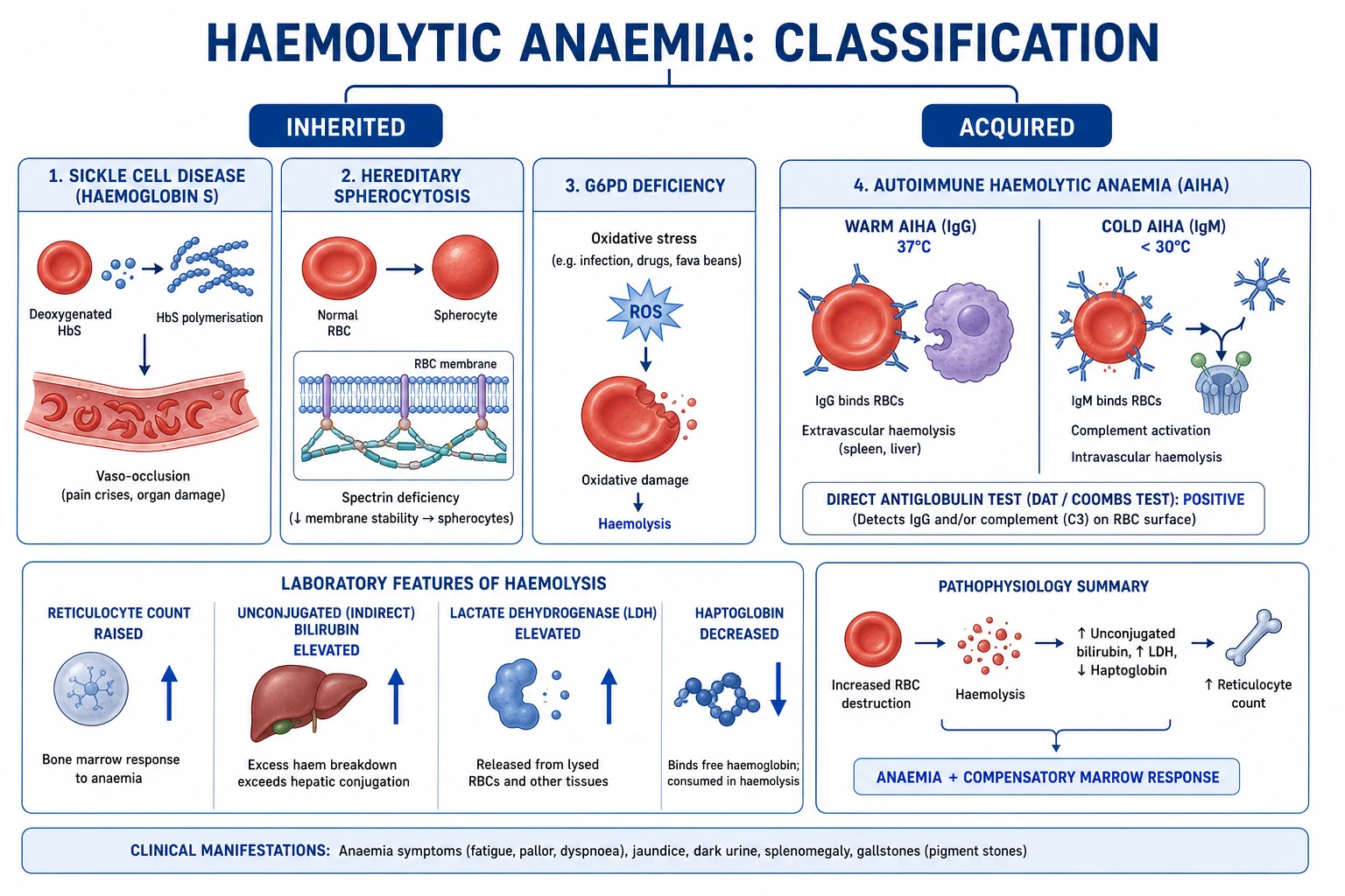
The biochemical signature
Regardless of site, the breakdown of haemoglobin releases the porphyrin ring as unconjugated bilirubin, which is conjugated in the liver and excreted in bile; the load overwhelms conjugation so unconjugated (indirect) bilirubin rises. Intracellular enzymes leak from destroyed red cells — most usefully lactate dehydrogenase (LDH), particularly the LDH-2 isoenzyme, which rises in proportion to the rate of haemolysis. The marrow responds with reticulocytosis (polychromatic, bluish, large cells on the film; the corrected reticulocyte count above 2 percent or a reticulocyte production index above 2 confirms an adequate marrow response and is the single most useful marker of a haemolytic process). [1]
Mechanism by mechanism — the molecular detail
Warm AIHA (IgG, 37 °C)
- IgG1/IgG3 directed at Rh-system antigens coats the red cell
- Splenic macrophage Fc-gamma receptors phagocytose — extravascular
- Spherocytes on film; DAT positive IgG +/- C3d
- Steroids reduce antibody production and Fc-receptor phagocytosis
Cold agglutinin (IgM, 4 °C)
- IgM pentamer (anti-I or anti-i) binds RBC in cold extremities
- Activates classical complement cascade (C1q -> C3b)
- On rewarming IgM dissociates leaving C3b; Kupffer cells in LIVER (C3b receptors) destroy — extravascular
- DAT positive C3d ONLY (IgG negative); cold agglutinin titre raised
Hereditary spherocytosis
- AD loss of spectrin/ankyrin/band 3/pallidin weakens membrane skeleton
- Membrane vesiculates off -> loss of surface-to-volume ratio -> spherocyte
- Spleen traps and destroys rigid spherocytes
- Osmotic fragility increased; EMA-binding flow cytometry reduced
G6PD deficiency
- X-linked recessive; G6PD maintains glutathione in reduced (GSH) form
- Without GSH, oxidant stress (fava bean, sulphonamides, primaquine, infection) denatures Hb
- Denatured Hb precipitates as Heinz bodies; spleen bites them out -> bite cells
- Acute intravascular haemolysis with haemoglobinuria; assay falsely NORMAL during attack
PNH
- Acquired PIG-A mutation in haematopoietic stem cell
- Absent GPI anchor -> absent CD55 (DAF) and CD59 (MIRL) on red cells
- Uncontrolled complement activation on RBC surface -> intravascular lysis
- FLAER flow cytometry on granulocytes diagnostic; eculizumab (anti-C5) is transformative
Microangiopathic (MAHA)
- Red cells fragmented by fibrin strands / platelet-rich microthrombi in small vessels
- Produces schistocytes (helmet cells, triangle cells) on film
- Causes: TTP (ADAMTS13 deficiency), HUS (shiga toxin), DIC, HELLP, malignant HTN
- Treat the cause: TTP = plasma exchange + steroids + caplacizumab
Clinical Presentation
The clinical face of haemolysis combines the generic features of any anaemia with the specific features of accelerated red-cell turnover, and then superimposes the fingerprints of the individual cause. [1]
General features of haemolysis
- Anaemia — fatigue, exertional dyspnoea, pallor (conjunctival, palmar creases, mucous membranes), tachycardia, flow murmurs; in the elderly or those with coronary disease, angina or heart failure may be the presenting complaint.
- Jaundice — a lemon-yellow (lemon tinge) discolouration of sclera and skin from raised unconjugated bilirubin; classically without dark stools and without pruritus (because the bilirubin never reaches the gut in excess conjugated form, though stool may be darker from increased stercobilinogen).
- Splenomegaly — the spleen is the principal site of red-cell clearance and is palpably enlarged in chronic extravascular haemolysis (warm AIHA, hereditary spherocytosis, thalassaemia, sickle cell in children before autosplenectomy).
- Dark urine — cola-coloured / port-wine urine is the hallmark of intravascular haemolysis (haemoglobinuria in PNH, cold agglutinin, G6PD crisis, ABO mismatch, march haemoglobinuria); a positive dipstick for blood with no red cells on microscopy is the clue.
- Pigment gallstones — chronic bilirubin overload leads to calcium bilirubinate (pigment) stones; cholecystitis, biliary colic or pancreatitis may be the first presentation of an inherited haemolysis. [1]
Clinical fingerprints by subtype
- Warm AIHA — subacute onset of anaemia and jaundice in a middle-aged or older adult; look for underlying CLL, lymphoma, SLE, or a recent drug (methyldopa, fludarabine).
- Cold agglutinin disease — older patient with acrocyanosis, Raynaud-type phenomena and livedo on cold exposure, often with chronic low-grade haemolysis that worsens in winter; a post-infectious form in young adults follows Mycoplasma pneumoniae (anti-I, often severe but self-limiting) or infectious mononucleosis (anti-i).[2]
- Hereditary spherocytosis — childhood or young-adult onset of chronic mild haemolysis punctuated by haemolytic crises (worsening with intercurrent infection), aplastic crises (parvovirus B19 — sudden Hb drop with low reticulocytes) and megaloblastic crises (folate depletion); family history often positive; frontal bossing and pigment stones in long-standing disease.
- G6PD deficiency — a male (X-linked) of African, Mediterranean or Asian ancestry with acute intravascular haemolysis (jaundice, dark urine, back pain) within 24 to 72 hours of exposure to fava beans, sulphonamides, primaquine, dapsone, rasburicase, naphthalene or an infection (hepatitis, pneumonia).[1]
- PNH — young adult with classic triad of haemolytic anaemia, pancytopenia and thrombosis at unusual sites (hepatic, portal, cerebral venous sinus); nocturnal haemoglobinuria is often historic rather than observed.
- Microangiopathic (TTP/HUS/DIC) — schistocytic haemolysis (the cell is fragmented in the vessel) with thrombocytopenia; TTP adds a fluctuating neurological deficit and fever; HUS adds acute kidney injury after bloody diarrhoea in children; DIC adds a coagulopathy and prolonged PT/APTT.
- Mechanical — a patient with a prosthetic heart valve and chronic low-grade intravascular haemolysis (raised LDH, low haptoglobin, haemosiderinuria, iron deficiency) — listen for the valve click or regurgitant murmur.
- Donath-Landsteiner / paroxysmal cold haemoglobinuria (PCH) — a child with acute intravascular haemolysis after cold exposure, often after a viral illness or in association with syphilis; the antibody is a biphasic IgG that binds in the cold and fixes complement, then lyses on rewarming.
Atypical and special presentations
- Elderly — may present with cardiac decompensation (heart failure, angina) from anaemia rather than with classical jaundice, or as a worsening of pre-existing CLL/lymphoma.
- Pregnant — physiological haemodilution and increased folate demand can unmask an inherited haemolysis; HELLP syndrome is a microangiopathic haemolysis unique to pregnancy; transfusion thresholds differ.
- Neonate — haemolytic disease of the newborn (Rh or ABO incompatibility) presents with early severe jaundice, anaemia and risk of kernicterus; positive DAT, raised unconjugated bilirubin, may need phototherapy or exchange transfusion.
- Immunocompromised — higher risk of drug-induced and parainfective haemolysis; consider parvovirus B19 aplastic crisis in chronic haemolysis with sudden reticulocytopenia. [1]
Differential Diagnosis
Several non-haemolytic states produce some of the haemolytic signature (raised bilirubin, raised LDH, even low haptoglobin) and must be actively excluded, because mislabelling them as haemolysis leads to immunosuppression or splenectomy that the patient does not need. [1]
Megaloblastic / ineffective erythropoiesis
- B12/folate deficiency, MDS — intramedullary death of precursors releases LDH and bilirubin
- MACROCYTIC anaemia with LOW/normal reticulocytes (NOT raised)
- Hypersegmented neutrophils, macro-ovalocytes; normal haptoglobin
- Treat with B12 (hydroxocobalamin) or folate
Reabsorbing haematoma / internal haemorrhage
- Retroperitoneal bleed, large bruise — resorbed RBC breakdown raises bilirubin and lowers haptoglobin
- NO reticulocytosis initially, NO spherocytes, NO schistocytes
- Falls over days; associated pain, falling Hb, rising then settling LDH
Gilbert syndrome
- Isolated UNCONJUGATED hyperbilirubinaemia; normal Hb, normal reticulocytes, normal LDH, normal haptoglobin
- Family history positive; worsens with fasting, illness, alcohol
- Reduced UDP-glucuronosyltransferase activity; benign, no treatment
Hepatic cirrhosis with anaemia
- Splenomegaly and jaundice but MIXED (conjugated + unconjugated) bilirubin, abnormal LFTs, low platelets
- NO reticulocytosis, NO haemolysis signature
- Stigmata of chronic liver disease; manage underlying cirrhosis
Septic / malarial haemolysis (true haemolysis, but a CAUSE)
- Falciparum malaria — ruptured parasitised RBCs; film shows parasites
- Clostridial sepsis — massive intravascular lysis by lecithinase
- Bartonella bacilliformis; Babesiosis — spleen-independent
- Treat the infection; transfuse for severity
Wilson disease (acute)
- Massive copper release causes acute intravascular haemolysis + acute liver failure
- Low ceruloplasmin, high urinary copper, Kayser-Fleischer rings
- Emergency: chelation, often liver transplant
The decisive discriminator between true haemolysis and its mimics is the reticulocyte response: an adequately marrow-responding haemolytic anaemia shows a corrected reticulocyte count above 2 percent or reticulocyte production index above 2; a low reticulocyte count in the face of raised bilirubin and LDH points to ineffective erythropoiesis (megaloblastic, MDS) or a superimposed aplastic crisis in a chronic haemolytic patient. [1]
Clinical & Bedside Assessment
A focused examination secures the presence of haemolysis and searches for its cause. [1]
- General inspection — pallor (conjunctivae, palmar creases, mucous membranes), lemon-yellow jaundice (sclera, sublingual, skin), frontal bossing and maxillary overgrowth (thalassaemia major, chronic severe haemolysis), leg ulcers (sickle cell disease, venous stasis), jaundice with scratch marks (more biliary than haemolysis).
- Hands — koilonychia suggests iron deficiency (NOT haemolysis, except PNH), clubbing suggests chronic disease; look for splinter haemorrhages if endocarditis or prosthetic valve.
- Abdomen — splenomegaly (grade in cm below costal margin; massive in thalassaemia, CML, malarial; moderate in warm AIHA and HS; autosplenectomy in sickle cell beyond childhood); hepatomegaly (lymphoma, haemolytic neonate); check for gallbladder tenderness (pigment stones).
- Lymph nodes — generalised lymphadenopathy points to a lymphoproliferative cause of warm AIHA (CLL, lymphoma).
- Cardiovascular — tachycardia, flow murmur (anaemia); prosthetic valve click or regurgitant murmur (mechanical haemolysis); features of infective endocarditis.
- Skin — malar rash, oral ulcers, alopecia (SLE causing warm AIHA); acrocyanosis, livedo, Raynaud-type on cold exposure (cold agglutinin); purpura (Evan syndrome — AIHA + ITP; HSP; DIC); Kayser-Fleischer rings (Wilson).
- Urine — colour at the bedside: cola / coca-cola / port-wine = haemoglobinuria (intravascular). Dipstick positive for blood with no red cells on microscopy is the bedside signature of haemoglobinuria.
- Neurology — fluctuating deficit with thrombocytopenia and schistocytes = TTP; cranial nerve palsy or hemiparesis in sickle cell = stroke. [1]
Investigations
The investigation strategy is tiered: first confirm haemolysis and localise the site, then identify the specific cause. [1]
Tier 1 — Confirm and localise haemolysis
- Full blood count — anaemia (severity varies); MCV often raised (reticulocytes are larger); the red cell distribution width (RDW) is raised; MCHC may be raised in hereditary spherocytosis (dense cells).
- Reticulocyte count — raised (the marrow's response); express as a corrected reticulocyte count (retic percentage × patient Hct / normal Hct) — above 2 percent confirms haemolysis; reticulocyte production index (RPI) above 2 likewise. A LOW reticulocyte count in a haemolytic picture = aplastic crisis (parvovirus B19) or megaloblastic crisis (folate deficiency).
- LDH — raised, particularly the LDH-2 isoenzyme; very high in intravascular haemolysis and MAHA (over 1000 U/L).
- Unconjugated (indirect) bilirubin — raised (typically 30 to 100 µmol/L); conjugated is normal.
- Haptoglobin — low or absent; a level under 25 mg/dL is approximately 95 percent sensitive for haemolysis, and a normal haptoglobin effectively excludes significant intravascular haemolysis. Note haptoglobin is an acute-phase reactant and may be misleadingly normal if there is concurrent inflammation; it is also congenitally absent in ~2 percent of the population.
- Peripheral blood film — the morphology often gives the diagnosis:
- Spherocytes — warm AIHA, hereditary spherocytosis
- Schistocytes (helmet / triangle cells) — microangiopathic (TTP/HUS/DIC/HELLP), mechanical valve
- Sickled cells + target cells — sickle cell disease
- Target cells, microcytes, basophilic stippling — thalassaemia
- Bite cells + Heinz bodies (supravital stain) — G6PD deficiency
- Elliptocytes — hereditary elliptocytosis
- Polychromasia — reticulocytosis (macrophage response)
- Howell-Jolly bodies — asplenia (sickle cell) [1]
Tier 2 — The single most important test: the DAT (direct Coombs)
The direct antiglobulin test detects antibody or complement bound to the patient's red cells. It is the master branch point of the haemolytic workup. [1]
DAT POSITIVE (immune haemolysis)
- Warm AIHA: IgG +/- C3d positive (IgG dominant)
- Cold agglutinin disease: C3d only (IgG NEGATIVE); cold agglutinin titre raised
- Paroxysmal cold haemoglobinuria: Donath-Landsteiner test positive (biphasic haemolysin)
- Drug-induced: penicillin (hapten), methyldopa (autoantibody), cephalosporin, fludarabine
- Haemolytic disease of newborn (ABO / Rh), delayed transfusion reaction
DAT NEGATIVE (non-immune haemolysis)
- Hereditary spherocytosis — osmotic fragility / EMA-binding flow cytometry
- G6PD deficiency — enzyme assay (BewarE: normal during acute attack)
- Pyruvate kinase deficiency — enzyme assay
- Sickle cell disease — Hb electrophoresis / HPLC
- Thalassaemia — Hb electrophoresis / HPLC, genetics
- PNH — FLAER / CD55-CD59 flow cytometry
- Microangiopathic — film (schistocytes), coagulation, ADAMTS13
- Mechanical valve — clinical; haemosiderinuria; iron deficiency
Tier 3 — Confirmatory / cause-specific tests
- Cold agglutinin titre — raised in cold agglutinin disease (typically over 1:64 at 4 °C); the antibody specificity is anti-I (Mycoplasma, most cases) or anti-i (infectious mononucleosis).[2]
- Donath-Landsteiner test — biphasic haemolysin for paroxysmal cold haemoglobinuria.
- Osmotic fragility test (classic) / EMA-binding flow cytometry (modern) — for hereditary spherocytosis. EMA binds to band 3; reduced mean fluorescence confirms HS even in cases with few spherocytes.
- G6PD enzyme assay — quantitative; BewarE the assay is falsely normal during an acute attack because the oldest (most deficient) cells are lysed first and only the youngest, reticulocyte-rich cells remain — repeat 2 to 3 months after the episode. A fluorescent spot test is a rapid screening version.[1]
- Haemoglobin electrophoresis / HPLC — for sickle cell (HbS over 50 percent in disease) and thalassaemia (raised HbA2 over 3.5 percent in beta-thal trait; HbF raised in beta-thal major and intermedia).[3][4]
- Flow cytometry for CD55 and CD59 (FLAER test) — for PNH; absence of GPI-anchored proteins on granulocytes confirms the diagnosis.
- ADAMTS13 activity — under 10 percent with inhibitor confirms TTP (distinguishing it from HUS and other MAHA).
- Coagulation screen — PT, APTT, fibrinogen, D-dimer for DIC.
- Bone marrow — rarely needed; excludes marrow failure or infiltration when reticulocytes are inappropriately low.
- Haemoglobinuria — positive dipstick for blood with no red cells on microscopy; haemosiderinuria on Prussian-blue staining of urinary sediment is a marker of chronic intravascular haemolysis (PNH, mechanical valve).
- Underlying cause screen — autoimmune screen (ANA, anti-dsDNA, rheumatoid factor), immunoglobulins and serum electrophoresis (lymphoproliferative disease), viral serology (HIV, hepatitis, EBV, mycoplasma), LDH and urate (high cell turnover), renal and liver function, iron studies (chronic intravascular haemolysis causes iron deficiency).
Haemolytic anaemia — key numbers
Management — Resuscitation
Acute severe haemolysis is a medical emergency. The resuscitation priorities are: secure the airway and circulation, transfuse if haemodynamically compromised or symptomatic, protect the kidneys, and stop the trigger. [1]
- Transfusion — transfuse if Hb under 70 g/L in a healthy adult, under 80 g/L if symptomatic (chest pain, heart failure, presyncope) or in the elderly, or under 100 g/L in severe haemolysis with rapid fall. In AIHA the autoantibody reacts with all donor cells, so a full crossmatch is impossible — request least-incompatible blood (the unit with the weakest incompatibility on crossmatch) and inform the haematology laboratory; transfuse cautiously under observation. In cold agglutinin disease use a blood warmer to avoid worsening haemolysis by the cold-reactive antibody.
- Intravenous fluids — crystalloid to maintain urine output above 1 mL/kg/h; protects against acute kidney injury from haemoglobinuria (free haemoglobin is nephrotoxic in acidotic, volume-contracted states).
- Stop the offending drug — penicillin, methyldopa, quinine, sulphonamides, primaquine, dapsone, rasburicase, naphthalene, fludarabine, cephalosporins.
- Folate — folic acid 5 mg orally daily to support the compensatory erythropoiesis (haemolysis consumes folate; deficiency causes a superimposed megaloblastic crisis).
- Treat the trigger — antibiotics for infection, oxygen and hydration for a sickle crisis, plasma exchange for TTP, eculizumab for PNH.
- Vaccinate before splenectomy — see Special Populations. [1]
Management — Definitive & Stepwise
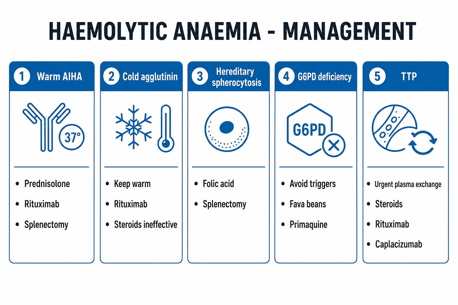
Definitive treatment is entirely cause-specific. The general principle is: extrinsic causes are reversible (withdraw drug, treat infection, plasma exchange for TTP), intrinsic defects require lifelong measures or surgery (folate, splenectomy, transfusion, chelation), and immune causes require immunomodulation (steroids, rituximab, splenectomy). [1]
Warm autoimmune haemolytic anaemia (IgG, 37 °C)
First-line
- Prednisolone 1 to 1.5 mg/kg/day orally (maximum 80 mg/day), usually 60 to 100 mg
- Most respond within 1 to 3 weeks (rising Hb, falling reticulocytes)
- Taper SLOWLY over months once stable — alternate-day regime as dose falls
- Add gastroprotection (PPI), bone protection (calcium/vitamin D +/- bisphosphonate), glycaemic monitoring
- Folic acid 5 mg/day
Second-line
- Rituximab 375 mg/m2 IV weekly for 4 weeks (anti-CD20) — now often first-line in older/frail patients
- Splenectomy (after vaccination) — removes the principal site of IgG-mediated clearance; response ~two-thirds
- Immunosuppressants — azathioprine, mycophenolate mofetil, ciclosporin, cyclophosphamide
- Fostamatinib (SYK inhibitor) — approved for refractory warm AIHA
Refractory / severe
- High-dose steroids (methylprednisolone 1 g IV for 3 days)
- IV immunoglobulin 0.4 g/kg/day for 5 days (short-lived effect)
- Plasma exchange, rituximab + bendamustine, immunosuppression combination
- Investigate relentlessly for an occult lymphoproliferative cause (CT, marrow)
Cold agglutinin disease (IgM, 4 °C)
Steroids and splenectomy are ineffective in cold agglutinin disease because the destruction is C3b-mediated in the liver (not IgG-Fc-mediated in the spleen). Management is built around avoidance of cold and rituximab.[2]
- Keep the patient warm — including extremities, nose, and during transfusion (use a blood warmer); avoid cold drinks and cold exposure.
- Rituximab 375 mg/m2 IV weekly for 4 weeks — first-line; ~50 percent response; rituximab + bendamustine for lymphoma-associated disease.
- Sutimlimab (anti-C1s, complement inhibitor) — approved 2022; rapidly raises Hb by blocking classical complement activation; does NOT treat underlying clonal disease.
- Plasma exchange — for life-threatening acute haemolysis (removes the IgM).
- Avoid cold-stored fluids and surgery-induced hypothermia — even cold intravenous fluids can trigger haemolysis. [1]
Hereditary spherocytosis
- Folic acid 5 mg orally daily — lifelong, to prevent megaloblastic crisis.
- Splenectomy — indicated for moderate-to-severe disease (chronic transfusion need, severe symptomatic anaemia, recurrent haemolytic crises, symptomatic gallstones); cures the haemolysis (the spherocytes remain but are no longer destroyed). In children, partial splenectomy is preferred in some centres to preserve immune function. Mandatory pre-splenectomy vaccination and lifelong penicillin V prophylaxis (250 to 500 mg twice daily) — see Special Populations.
- Cholecystectomy — for symptomatic pigment gallstones.
- Transfusion — in aplastic crisis (parvovirus B19) or severe haemolytic crisis. [1]
G6PD deficiency
The cornerstone is trigger avoidance; the disease is otherwise benign.[1]
- Avoid: fava (broad) beans; sulphonamides (co-trimoxazole, sulfasalazine, dapsone — note dapsone is a particular problem in dermatitis herpetiformis and leprosy); primaquine, tafenoquine (antimalarials); rasburicase (tumour lysis); naphthalene (mothballs); methylene blue (paradoxically oxidant in G6PD-deficient patients); analine dyes; certain analgesics in large doses (aspirin, phenacetin); and avoid infections (oxidative burst of neutrophils contributes).
- Supportive — fluids to protect the kidneys; transfusion for severe acute haemolysis.
- Counsel family members (X-linked recessive — males affected, females carriers). [1]
Paroxysmal nocturnal haemoglobinuria (PNH)
- Complement inhibition is transformative:
- Eculizumab — anti-C5 monoclonal antibody, 600 mg IV every 7 days for 4 weeks, then 900 mg every 14 days; meningococcal vaccination and prophylactic penicillin mandatory (encapsulated infection risk).
- Ravulizumab — long-acting anti-C5, weight-based loading then maintenance every 8 weeks (more convenient).
- Crovalimab — recently approved long-acting anti-C5.
- Anticoagulation — for thrombosis (a leading cause of death in PNH); prophylaxis for high-risk patients on complement blockade is debated.
- Bone marrow transplant — the only curative option; reserved for severe disease, marrow failure, or in regions where complement blockade is unavailable.
- Iron supplementation — for the iron deficiency of chronic intravascular haemolysis (give cautiously; transferrin saturation target). [1]
Microangiopathic haemolytic anaemia (MAHA)
Treat the underlying cause — the haemolysis itself is not directly treated. [1]
- TTP (thrombotic thrombocytopenic purpura) — emergency plasma exchange (removes anti-ADAMTS13 antibody and replenishes enzyme), prednisolone 1 mg/kg/day, caplacizumab (anti-vWF nanobody, blocks platelet aggregation) — mortality reduced from ~90 percent to under 10 percent. Folate; avoid platelet transfusion (fuels microthrombi) unless life-threatening bleeding.
- HUS (haemolytic uraemic syndrome) — supportive; eculizumab for atypical (complement-mediated) HUS; dialysis for AKI; fluid and electrolyte management.
- DIC — treat the underlying cause (sepsis, malignancy, obstetric); supportive component therapy (FFP, cryoprecipitate, platelets) for bleeding; heparin in thrombotic-predominant DIC.
- HELLP — delivery of the baby. [1]
Drug-induced immune haemolytic anaemia
- Stop the offending drug (penicillin, cephalosporin, methyldopa, fludarabine, quinine, NSAIDs).
- Mechanisms: hapten (penicillin — drug coats the cell, antibody to drug), autoantibody (methyldopa — true autoantibody against Rh), immune complex (quinine — drug-antibody complex deposits on cell).
- Steroids for severe haemolysis; usually self-limiting once drug stopped. [1]
Sickle cell disease and thalassaemia
See the dedicated topics — sickle-cell-disease (hydroxycarbamide, transfusion, transplant, gene therapy) and thalassaemia (transfusion, iron chelation with deferasirox, transplant).[3][4]
Specific Subtypes & Scenarios
Hereditary spherocytosis in depth
The commonest inherited haemolysis in Northern Europeans, autosomal dominant in ~75 percent (recessive forms exist). Mutations in ANK1 (ankyrin, commonest), SPTB (beta-spectrin), SPTA1 (alpha-spectrin), EPB42 (pallidin), SLC4A1 (band 3) weaken the membrane skeleton, so membrane is lost in the splenic cords and the cell becomes a spherocyte. Diagnosis: spherocytes on film, raised osmotic fragility (spherocytes lyse in less hypotonic saline than normal cells), and EMA-binding flow cytometry (reduced fluorescence, modern gold standard). Severity ranges from asymptomatic carrier (compensated), through mild/moderate chronic haemolysis with pigment gallstones, to severe transfusion-dependent disease. Crises: haemolytic (infection), aplastic (parvovirus B19 — sudden Hb drop, low reticulocytes), megaloblastic (folate depletion in pregnancy or growth spurts). [1]
Donath-Landsteiner (paroxysmal cold haemoglobinuria, PCH)
Rare but examinable. A biphasic IgG haemolysin (anti-P specificity) binds to the red cell in the cold (extremities), fixes complement, and then lyses on rewarming at 37 °C. Classically in children after a viral illness (now the commonest cause) or in syphilis (congenital or tertiary). Diagnosis: Donath-Landsteiner test (biphasic haemolysis in vitro). Usually self-limiting; supportive care, transfuse P-negative blood if available, keep warm. [1]
Pyruvate kinase deficiency
Autosomal recessive chronic non-spherocytic haemolytic anaemia (the commonest inherited non-spherocytic haemolysis after G6PD). Reduced ATP causes rigid echinocytes. Splenectomy partially helps; mitapirdine (PK activator) is a newer therapy. [1]
Mechanical (microangiopathic and prosthetic) haemolysis
Red cells are fragmented mechanically — in the fibrin-rich microvasculature (TTP, HUS, DIC, HELLP, malignant hypertension) or across a prosthetic heart valve (especially regurgitant mitral valves). Film shows schistocytes (helmet, triangle, fragmented cells); LDH very high; haptoglobin absent; haemosiderinuria and iron deficiency mark chronicity. Treat the cause; iron supplementation for valve-related iron loss; valve revision if severe. [1]
Wilson disease — acute haemolysis
A rare but recognisable presentation of acute Wilson crisis: massive copper release causes acute intravascular haemolysis with acute liver failure and Coombs-negative haemolysis. Diagnose with low ceruloplasmin, high urinary copper, Kayser-Fleischer rings; treat with chelation and liver transplant. [1]
Aplastic crisis — the exception to reticulocytosis
In any patient with chronic haemolysis (HS, sickle cell, thalassaemia) who presents with a sudden fall in haemoglobin and a LOW reticulocyte count, suspect parvovirus B19 infection of erythroid progenitors. The marrow stops producing reticulocytes, so the underlying haemolysis runs unchecked with no compensation. Self-limiting (1 to 2 weeks); transfuse to bridge; IgG serology shows recent infection. [1]
Complications & Pitfalls
- Pigment (calcium bilirubinate) gallstones — the classical long-term complication of any chronic haemolysis; cholecystitis, biliary colic, pancreatitis; consider prophylactic cholecystectomy at splenectomy in HS.
- Folate deficiency (megaloblastic crisis) — compensatory erythropoiesis consumes folate; prevent with folic acid 5 mg daily.
- Aplastic crisis (parvovirus B19) — sudden reticulocytopenia; the only setting in which a haemolytic anaemia shows a LOW reticulocyte count.
- Iron overload — from chronic transfusion (sickle, thalassaemia, refractory AIHA); cardiomyopathy, cirrhosis, endocrine failure; chelate with deferasirox.
- Iron deficiency — chronic intravascular haemolysis (PNH, mechanical valve) causes urinary iron loss; check ferritin; supplement cautiously.
- Acute kidney injury — from haemoglobinuria; prevent with hydration.
- Thrombosis — particularly PNH (hepatic, portal, cerebral venous sinus) and sickle cell; lifelong risk.
- Hypersplenism — in chronic extravascular haemolysis; worsens cytopenias.
- OPSI (overwhelming post-splenectomy infection) — encapsulated organisms (Streptococcus pneumoniae, Haemophilus influenzae, Neisseria meningitidis); mortality 50 to 70 percent even with treatment; prevented by vaccination and prophylactic penicillin.
- Immunosuppression complications — steroids (osteoporosis, diabetes, infection, hypertension), rituximab (hepatitis B reactivation, PML rarely), splenectomy (infection).
- Pitfalls — (1) interpreting a falsely normal G6PD assay during an acute attack as excluding the diagnosis — repeat at 2 to 3 months; (2) treating cold agglutinin disease with steroids/splenectomy (ineffective); (3) under-transfusing AIHA because of crossmatch incompatibility (transfuse the least-incompatible unit; the risk of under-transfusion exceeds the risk of a minor incompatibility); (4) missing a superimposed parvovirus B19 aplastic crisis because the reticulocyte count is low; (5) giving primaquine (or dapsone, rasburicase, methylene blue) to a patient with unrecognised G6PD deficiency.[1]
Prognosis & Disposition
- Warm AIHA — most respond to steroids within 1 to 3 weeks; ~one-third achieve long-term remission, ~one-third remain steroid-dependent, and ~one-third are refractory and need second-line therapy (rituximab, splenectomy, immunosuppression). Overall mortality 8 to 11 percent at 1 year, driven by underlying disease and thromboembolism.
- Cold agglutinin disease — chronic relapsing course; rituximab responses in ~50 percent; newer complement inhibitors (sutimlimab) improve Hb but do not cure.
- Hereditary spherocytosis — normal lifespan after splenectomy in severe cases; mild forms well compensated.
- G6PD deficiency — excellent with trigger avoidance; haemolysis is self-limiting.
- PNH — transformed by complement inhibitors (eculizumab, ravulizumab); previously median survival 10 to 15 years (thrombosis, marrow failure), now near-normal in treated patients.
- TTP — untreated mortality ~90 percent; with plasma exchange and caplacizumab under 10 percent.
- HUS — most children recover; atypical (complement) HUS has poorer prognosis without eculizumab. [1]
Disposition — severe acute haemolysis requires hospital admission (transfusion, IV fluids, monitoring of Hb, renal function, electrolytes); stable chronic haemolysis is managed as an outpatient with folate, surveillance for gallstones and iron overload, and pre-splenectomy vaccination planning. All splenectomised patients need lifelong follow-up for infection prophylaxis. [1]
Special Populations
Children
- Weight-based dosing — prednisolone 2 mg/kg/day (max 60 to 80 mg) for warm AIHA; folic acid by weight.
- Screening — neonatal sickle screening; G6PD assay in high-risk groups before prescribing oxidant drugs.
- Parvovirus B19 aplastic crisis — commoner in children with chronic haemolysis; transfuse.
- Hereditary spherocytosis — partial splenectomy preferred in some paediatric centres to preserve immune function.
- PCH — commoner in children; usually self-limiting.
- HUS — typical (post-diarrhoeal, shiga-toxin) HUS in children under 5. [1]
Pregnancy
- Physiological haemodilution lowers Hb; folate demand rises (give 5 mg daily); transfusion thresholds differ.
- HELLP syndrome is a microangiopathic haemolysis unique to pregnancy (haemolysis, elevated liver enzymes, low platelets) — deliver the baby.
- Warm AIHA in pregnancy — steroids first-line; rituximab avoided if possible; multidisciplinary haematology-obstetric care.
- Hereditary spherocytosis — most tolerate pregnancy well with folate; splenectomy deferred. [1]
Elderly
- Lower threshold to transfuse (cardiac comorbidity); watch for cardiac decompensation as the presenting feature.
- Always investigate for an underlying lymphoproliferative cause of warm AIHA (CLL, lymphoma) — blood film, immunoglobulins, marrow, CT. [1]
Immunocompromised
- Higher risk of drug-induced and parainfective haemolysis; check G6PD before oxidant drugs.
- Rituximab patients — recheck hepatitis B; PML rarely. [1]
Post-splenectomy — the inviolable bundle
The post-splenectomy bundle is the same regardless of why the spleen was removed (haemolysis, ITP, trauma): [1]
Vaccinate (ideally 2 to 4 weeks BEFORE)
- Pneumococcal: PCV13 then PPSV23 (or PCV20 alone); booster of PPSV23 at 5 years
- Haemophilus influenzae type b (Hib) conjugate
- Meningococcal ACWY conjugate (MenACWY) + booster every 5 years; MenB (Bexsero/Trumenba)
- Annual influenza vaccine
- If already splenectomised, vaccinate at least 2 weeks post-op; ideally 2 to 4 weeks pre-op
Antibiotic prophylaxis (lifelong in UK)
- Penicillin V 250 to 500 mg orally twice daily (phenoxymethylpenicillin)
- Erythromycin if penicillin-allergic
- Lifelong in UK; debate exists in US about duration but most experts lifelong if under 16 or over 50, or impaired immunity
- Standby rescue dose: amoxicillin 1 g (patient carries; take if fever and seek emergency care)
Patient education
- Spleen alert card / medical ID at all times
- Any fever is a medical emergency — present within 1 hour, empirical IV ceftriaxone 2 g
- Animal bites — capnocytophaga (cover with amoxicillin-clavulanate)
- Malaria prophylaxis if travelling
- Avoid asplenia-associated travel risks
Evidence, Guidelines & Regional Differences
Landmark trials and therapies
- MSH trial (Charache et al., NEJM 1995) — hydroxycarbamide reduced sickle crises by ~50 percent in adults; the foundation of modern sickle management (now recommended from 9 months).
- Hillmen et al. (NEJM 2006, 2013) — eculizumab (anti-C5) transformed PNH; reduced haemolysis, transfusion need, thrombosis and improved survival.
- HERCULES (NEJM 2019) — caplacizumab (anti-vWF nanobody) accelerated recovery and reduced mortality in acquired TTP when added to plasma exchange.
- CARLILNE / Cardinal (NEJM 2022) — sutimlimab (anti-C1s) raised Hb in cold agglutinin disease; first complement inhibitor approved for this condition (2022).[2]
- STOP trial (NEJM 1998) — chronic transfusion guided by transcranial Doppler reduced stroke by ~90 percent in sickle cell children.
Guidelines
- British Society for Haematology (BSH) guidelines on the diagnosis and management of autoimmune haemolytic anaemia (2016/2020) and hereditary spherocytosis.
- American Society of Hematology (ASH) guidelines on warm AIHA, TTP/HUS, and PNH.
- UK Green Book (DH) on immunisation of the asplenic/hyposplenic patient.
- NICE guidance on eculizumab/ravulizumab and sutimlimab for PNH and cold agglutinin disease. [1]
Regional differences
- Penicillin prophylaxis duration after splenectomy — lifelong in UK (Green Book) for all ages; variable in US (lifelong if under 16, over 50, or immunocompromised; otherwise individualised).
- Vaccine choice — PCV13/PPSV23 vs PCV20 alone; MenACWY vs MenACWY + MenB; boosters every 5 years for meningococcal in the UK.
- Splenectomy approach in children — partial splenectomy preferred in some European paediatric centres to preserve immune function; less common in the US.
- G6PD screening — routine in many African, Mediterranean and Asian countries before prescribing oxidant drugs; opportunistic in the UK/US.
- PNH management — eculizumab universally funded in some healthcare systems (UK NHS) but rationed by disease severity in others. [1]
Exam Pearls
Haemolytic anaemia — DAT split
COOMBS
THE master discriminator — positive = immune (AIHA, HDN, drug), negative = non-immune (HS, G6PD, PNH, MAHA, sickle, thalassaemia)
inherited (membrane/enzyme/Hb) vs acquired (immune/non-immune) — family history + age of onset
extravascular (spleen, spherocytes, haptoglobin low) vs intravascular (haemoglobinaemia, haemoglobinuria, haptoglobin absent)
reticulocytes RAISED (corrected over 2 percent); LOW reticulocytes = parvovirus B19 aplastic crisis or megaloblastic crisis
both raised; bilirubin UNCONJUGATED; LDH-2 isoenzyme; haptoglobin under 25 mg/dL ~95 percent sensitive
vaccinate BEFORE (pneumococcal, Hib, meningococcal, influenza); lifelong penicillin V; OPSI risk
- Pigment (calcium bilirubinate) gallstones = chronic haemolysis (HS, sickle, thalassaemia, hereditary elliptocytosis) — NOT cholesterol stones.
- Cola-coloured urine with a positive dipstick for blood but NO red cells on microscopy = haemoglobinuria (intravascular haemolysis — PNH, cold agglutinin, G6PD crisis, ABO mismatch, march haemoglobinuria, mechanical valve).
- MCHC raised on the FBC → think hereditary spherocytosis (dense, dehydrated spherocytes).
- A LOW reticulocyte count in a haemolytic patient = parvovirus B19 aplastic crisis (the single most-tested exception).
- Haptoglobin under 25 mg/dL is ~95 percent sensitive for haemolysis; a normal haptoglobin effectively excludes significant intravascular haemolysis (but it is an acute-phase reactant, so may be misleadingly normal with inflammation).
- Evan syndrome = warm AIHA + immune thrombocytopenia (ITP) — look for underlying autoimmune/lymphoproliferative disease.
- Schistocytes on film with thrombocytopenia = MAHA — TTP (ADAMTS13 under 10 percent, neurology), HUS (renal, post-diarrhoeal), DIC (coagulopathy), HELLP (pregnancy).
- Mechanical valve haemolysis — chronic intravascular, iron deficiency, haemosiderinuria; treat iron, consider valve revision.
- Splenectomy before vaccination = OPSI risk — encapsulated organisms, 50 to 70 percent mortality; vaccinate 2 to 4 weeks before; lifelong penicillin V.
- Methylene blue for methaemoglobinaemia is contraindicated in G6PD deficiency (it is itself an oxidant and ineffective in deficient patients — use ascorbic acid or exchange transfusion).
- Rasburicase and dapsone are absolutely contraindicated in G6PD deficiency. [1]
Exam application bank (NEET-PG / INICET)
One-line answer
Haemolytic anaemia is anaemia caused by premature destruction of red blood cells (lifespan shortened from the normal 120 days) at a rate that exceeds marrow compensation. Classify by site (intravascular vs extravascular) and by origin (inherited vs acquired). Biochemical signature: raised reticulocytes, raised LDH, raised unconjugated bilirubin, low/absent haptoglobin, with haemoglobinaemia/haemoglobinuria in intravascular forms. The direct antiglobulin (DAT/Coombs) test is the single most important discriminator: positive = immune (warm IgG AIHA, cold IgM agglutinin, paroxysmal cold haemoglobinuria), negative = non-immune (hereditary spherocytosis, G6PD deficiency, PNH, microangiopathic, sickle cell, thalassaemia). Management is cause-specific: warm AIHA — prednisolone 1–1.5 mg/kg; cold agglutinin — avoid cold + rituximab; hereditary spherocytosis — folate +/- splenectomy (vaccinate bef [1]
Worked stems (answer without another resource)
Stem 1 — Classic presentation. Map symptoms to mechanism; name the first investigation and first treatment step with dose/route if drug therapy is standard. [1]
Stem 2 — Unstable / complicated. List red flags that force immediate resuscitation, theatre, ICU, antidote, or reperfusion — and what you do in the first 15 minutes. [1]
Stem 3 — Atypical group. Elderly, pregnancy, child, or immunocompromised: how presentation and thresholds change. [1]
Stem 4 — Differential trap. Name the three closest mimics and one discriminator for each. [1]
Stem 5 — Disposition. Who goes home with safety-netting, who is admitted, who needs HDU/ICU/theatre, and what follow-up is mandatory. [1]
Rapid viva checklist
- Definition + classification
- Pathophysiology chain
- Bedside signs / criteria
- Score with exact components (if any)
- Emergency bundle
- Definitive therapy with doses
- Complications of disease and of treatment
- Special populations
- Guideline/trial name if classic
- Three exam traps
Coverage self-check
If you cannot answer any stem above from this page alone, re-read the matching section — the page is intended to be self-sufficient for final-prof and NEET-PG/INICET questions on Haemolytic Anaemia.
[1]References
- [1]Cappellini MD, Fiorelli G. Glucose-6-phosphate dehydrogenase deficiency Lancet, 2008.PMID 18177777
- [2]Berentsen S. New Insights in the Pathogenesis and Therapy of Cold Agglutinin-Mediated Autoimmune Hemolytic Anemia Front Immunol, 2020.PMID 32318071
- [3]Muncie HL Jr, Campbell J. Alpha and beta thalassemia Am Fam Physician, 2009.PMID 19678601
- [4]Kattamis A, Kwiatkowski JL, Aydinok Y, et al. Thalassaemia Lancet, 2022.PMID 35691301