Haematology · General Medicine
Inherited Thrombophilia (Factor V Leiden & Protein C/S Deficiency)
Also known as Inherited thrombophilia · Hereditary thrombophilia · Factor V Leiden · Protein C deficiency · Protein S deficiency · Antithrombin deficiency · Prothrombin G20210A · Hyperhomocysteinaemia
Inherited thrombophilias are genetic disorders increasing the risk of venous thromboembolism (VTE). The commonest are factor V Leiden (activated protein C resistance; the single commonest inherited thrombophilia in Europeans, autosomal dominant, carrier rate 3 to 8 percent) and the prothrombin G20210A mutation (raised prothrombin; 2 percent of Europeans). Less common but higher-risk are deficiencies of the natural anticoagulants — antithrombin, protein C, protein S — which cause more severe, younger-onset, unusual-site thrombosis and warfarin-induced skin necrosis (protein C/S). Hyperhomocysteinaemia (MTHFR mutations) raises both venous and arterial thrombosis risk. Indications to test: VTE under 50, unusual site (cerebral, mesenteric, portal, hepatic), recurrent unprovoked VTE, family history, VTE in pregnancy/OCP, warfarin-induced skin necrosis. Testing is selective, not universal; the result does NOT change anticoagulation duration for low-risk defects alone. Treat with anticoagulation; high-risk defects (AT deficiency, homozygous FVL) warrant extended/lifelong therapy. Pregnancy uses LMWH (warfarin teratogenic).
On this page & tools
Your progress
Saved locally on this device.
Exam tags
Red flags
Overview & Definition
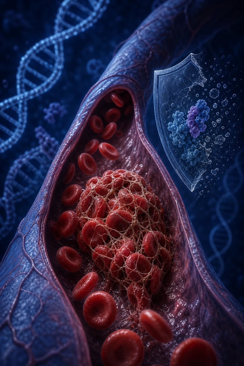
Inherited thrombophilias are a heterogeneous group of genetic defects that shift the haemostatic balance toward venous thrombosis — the hypercoagulability arm of Virchow's triad. They work by either removing a natural anticoagulant brake (antithrombin, protein C, protein S) or strengthening a procoagulant drive (factor V Leiden resists degradation; prothrombin G20210A increases factor II levels). The net cellular consequence is unopposed thrombin generation in the venous circulation.[1]
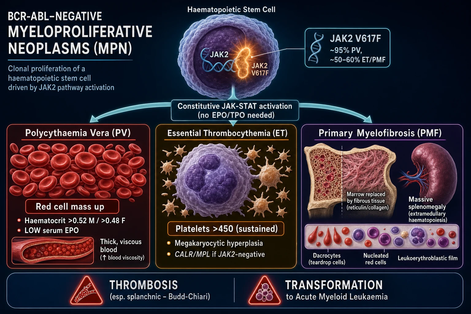
These disorders must be distinguished from acquired thrombophilia, which an examiner will deliberately contrast: antiphospholipid syndrome (antibody-mediated), malignancy (Trousseau syndrome), heparin-induced thrombocytopenia, pregnancy, oestrogen therapy, nephrotic syndrome, myeloproliferative neoplasms (JAK2), and paroxysmal nocturnal haemoglobinuria. The clinical significance of inherited thrombophilia is risk-stratified by the defect: factor V Leiden and the prothrombin mutation are common but confer only a modest VTE increase, whereas the natural-anticoagulant deficiencies are rare but high-risk.[1][5]
The critical clinical skill — and the single most examined point — is not memorising every defect. It is knowing whom to test (selectively), when and how to test (timing matters profoundly; acute illness, pregnancy, and anticoagulants distort functional assays), and how the result changes management — which for most low-risk defects is not at all. All major inherited thrombophilias are autosomal dominant with incomplete penetrance: most heterozygous carriers never thrombose.[1][2]
Classification
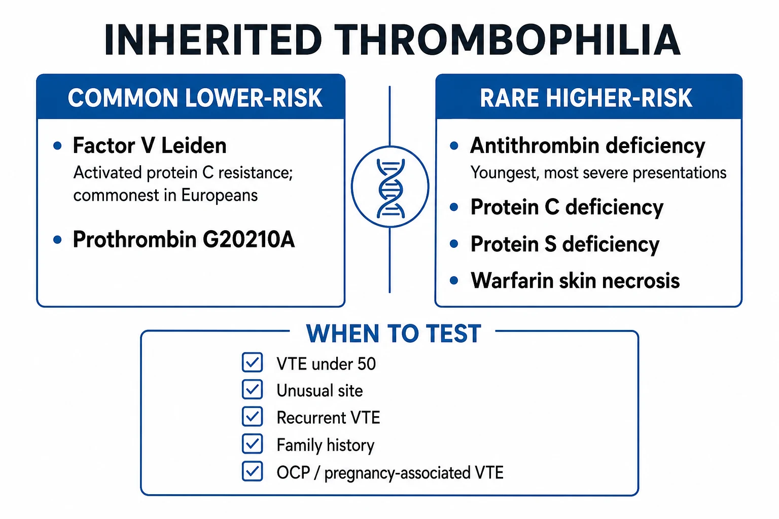
The six clinically significant inherited thrombophilias divide cleanly into two risk tiers by frequency, mechanism, and clinical impact. The common, lower-risk defects work by amplifying procoagulant activity; the rare, higher-risk defects work by removing the body's natural anticoagulant brakes. This distinction governs everything downstream — testing strategy, anticoagulation duration, pregnancy prophylaxis, and family counselling.[1][5]
Common, lower-risk (procoagulant gain)
- Factor V Leiden — APC resistance; heterozygous 3 to 8-fold VTE risk, homozygous ~80-fold; 3 to 8 percent of Europeans
- Prothrombin G20210A — raised prothrombin (factor II); 2 to 5-fold VTE risk; 2 percent of Europeans
- Hyperhomocysteinaemia (MTHFR) — venous AND arterial thrombosis; treatable with folate/B6/B12
- Testing rarely changes anticoagulation duration
- Often need a second hit (OCP, pregnancy, surgery) to thrombose
Rare, higher-risk (anticoagulant loss)
- Antithrombin deficiency — youngest, most severe; 10 to 25-fold VTE; heparin resistance; under 1 percent prevalence
- Protein C deficiency — warfarin-induced skin necrosis; neonatal purpura fulminans (homozygous)
- Protein S deficiency — cofactor for APC; warfarin skin necrosis; under 1 percent
- Each warrants extended or lifelong anticoagulation after a first event
- Functional assays needed (not just antigen); repeat abnormal results off therapy
Epidemiology & Risk Factors
The population prevalence of each defect varies enormously, and the population genetics of factor V Leiden is a favourite examiner question. Carrier frequencies differ by ethnicity because these mutations arose from founder effects in specific populations.[1][7]
- Factor V Leiden is the single commonest inherited thrombophilia in Europeans: carrier frequency roughly 3 to 8 percent in Caucasian populations. It is uncommon in African, East Asian, Australasian, and Native American populations (under 1 percent), a population-genetics fact that reflects its origin as a European founder mutation. The mutation is a single G-to-A substitution at nucleotide 1691 (G1691A) in the F5 gene on chromosome 1q24, producing an Arg506Gln (R506Q) amino acid change in the factor V molecule.[7]
- Prothrombin G20210A is the second commonest inherited thrombophilia in Europeans (carrier frequency approximately 2 percent). It is a G-to-A transition at position 20210 in the 3-prime untranslated region of the F2 gene on chromosome 11, which raises plasma prothrombin levels by roughly 25 to 30 percent.[1]
- Antithrombin, protein C, and protein S deficiencies are each rare (prevalence under 1 percent in the general population) but are disproportionately over-represented in cohorts of young patients with unprovoked, recurrent, or unusual-site VTE. Antithrombin deficiency is the rarest (approximately 1 in 2000 to 5000) but carries the highest VTE risk of any single inherited defect.[4]
- Hyperhomocysteinaemia due to MTHFR C677T is polymorphic: the homozygous TT genotype occurs in roughly 10 to 15 percent of European populations, though only a fraction develop clinically significant hyperhomocysteinaemia (those with low folate intake).[1]
Inheritance is autosomal dominant for all six defects, but with incomplete penetrance. Most heterozygous carriers of factor V Leiden or the prothrombin mutation never develop VTE (lifetime risk approximately 5 to 10 percent). Homozygous factor V Leiden or compound/double defects (factor V Leiden plus prothrombin mutation) carry an additive, substantially higher VTE risk and are treated as high-risk. The concept of the second hit is central: a carrier's modest genetic risk is amplified by acquired or transient provoking factors — oestrogen exposure (combined oral contraceptive pill, pregnancy, hormone replacement therapy), surgery, trauma, immobility, malignancy, nephrotic syndrome, infection, and the puerperium. A factor V Leiden carrier on the combined pill has roughly a 35-fold increased VTE risk versus a non-carrier not on the pill — the classic Vandenbroucke Lancet figure.[3][7]
Pathophysiology
The coagulation balance
Haemostasis is a dynamic equilibrium between procoagulant drivers (tissue factor → factor VIIa → factor Xa → thrombin → fibrin) and natural anticoagulant brakes (antithrombin, the protein C and protein S system, tissue factor pathway inhibitor). Every inherited thrombophilia works by either removing a brake or strengthening a procoagulant — the net effect is unopposed thrombin generation and venous clot formation.[1]
Mechanism by defect
Factor V Leiden (Arg506Gln, G1691A): The factor V molecule is normally activated by thrombin to factor Va, which then serves as a cofactor for the prothrombinase complex (converting prothrombin to thrombin). Activated protein C (APC) downregulates this by cleaving factor Va at three sites — Arg306, Arg506, and Arg679. The factor V Leiden mutation changes Arg506 to Gln506, which is the primary APC cleavage site. Without this cleavage, factor Va is inactivated approximately 10 times more slowly, so the procoagulant cofactor persists in the circulation, generating more thrombin. This produces the laboratory phenotype of activated protein C (APC) resistance: a functional assay ratio of less than 2.0 (normal is above 2.0), later confirmed by genetic PCR.[7]
Prothrombin G20210A: The mutation sits in the 3-prime untranslated region of the prothrombin gene (F2). It does not change the protein sequence but increases messenger RNA stability and translation efficiency, raising plasma prothrombin (factor II) concentrations by roughly 25 to 30 percent. More prothrombin means more substrate for the prothrombinase complex, more thrombin generation, and a modestly elevated VTE risk. The defect is not detectable by APC-resistance testing — it requires specific genetic PCR.[1]
Antithrombin deficiency: Antithrombin (formerly antithrombin III) is a serine protease inhibitor (serpin) produced by the liver. It neutralises thrombin (factor IIa), factor Xa, factor IXa, factor XIa, and factor XIIa by forming irreversible 1:1 complexes. Heparin potentiates this inactivation several thousand-fold by causing a conformational change in antithrombin — this is the molecular basis of heparin and LMWH therapy. In antithrombin deficiency, this brake is lost, so thrombin and factor Xa persist. Type I deficiency = low antigen and low activity (quantitative defect); Type II = normal antigen but dysfunctional protein (qualitative, with variants affecting the reactive site, heparin-binding site, or pleiotropic). The heparin-binding-site variants are the ones that cause heparin resistance: the APTT fails to rise despite escalating heparin doses because there is insufficient functional antithrombin for heparin to potentiate.[4]
Protein C deficiency: Protein C is a vitamin K-dependent glycoprotein synthesised in the liver. When thrombin binds to thrombomodulin on healthy endothelial cells (not at sites of injury), it changes its substrate specificity from procoagulant (fibrinogen, factor V) to anticoagulant: it activates protein C. Activated protein C (APC), with its essential cofactor protein S, then proteolytically cleaves and inactivates factors Va and VIIIa, shutting down further thrombin generation. In protein C deficiency, this brake is lost. Heterozygous deficiency causes VTE; homozygous deficiency (usually Type I, severe) presents in the neonatal period with purpura fulminans — widespread cutaneous microvascular thrombosis causing full-thickness skin necrosis, disseminated intravascular coagulation, and death if untreated.[1]
Protein S deficiency: Protein S is also vitamin K-dependent and serves as the essential cofactor for activated protein C in the inactivation of factors Va and VIIIa. It also has APC-independent anticoagulant activity by directly inhibiting the prothrombinase and tenase complexes. Approximately 60 percent of circulating protein S is bound to C4b-binding protein (and is functionally inactive); only the free protein S (approximately 40 percent) is the active cofactor. Deficiency can be Type I (low total and free), Type II (low activity, normal antigen), or Type III (normal total but low free protein S). As with protein C deficiency, homozygous deficiency causes neonatal purpura fulminans, and heterozygous deficiency predisposes to warfarin-induced skin necrosis.[8]
Hyperhomocysteinaemia (MTHFR C677T): Homocysteine is a sulphur-containing amino acid intermediate in the methionine cycle. The MTHFR C677T polymorphism (Ala222Val) reduces MTHFR enzyme activity (thermolabile variant), impairing the remethylation of homocysteine to methionine — particularly when folate intake is low. Elevated homocysteine promotes thrombosis through endothelial dysfunction, oxidative stress, reduced thrombomodulin activity, increased tissue factor expression, and impaired fibrinolysis (via increased lipoprotein(a)). The MTHFR A1298C variant is less commonly associated with hyperhomocysteinaemia. Unlike the other defects, hyperhomocysteinaemia raises both venous AND arterial thrombosis risk (myocardial infarction, stroke), and is treatable with vitamin supplementation (folate, vitamin B6, vitamin B12).[1]
The warfarin-induced skin necrosis mechanism
This is the highest-yield pathophysiology fact in inherited thrombophilia and a guaranteed viva question. The four vitamin K-dependent procoagulant factors are II (prothrombin), VII, IX, and X. There are also two vitamin K-dependent anticoagulant proteins: protein C and protein S. The key pharmacological fact is that protein C has the shortest half-life (approximately 8 hours) of all the vitamin K-dependent factors, followed by factor VII (6 hours), then factors IX (24 hours) and II/prothrombin (60 to 72 hours).[8]
When warfarin is started, it blocks vitamin K epoxide reductase, halting gamma-carboxylation of all vitamin K-dependent factors. Because protein C has the shortest half-life, its levels fall first — within 24 to 48 hours — creating a transient but profound procoagulant window: the anticoagulant protein C is gone while the long-lived procoagulant factors II, IX, and X are still circulating at near-normal levels. In a patient who is already genetically deficient in protein C (or S), this window is catastrophic: the already-low anticoagulant reserve is driven to near-zero, producing cutaneous microvascular thrombosis and full-thickness skin necrosis — classically on the breasts, buttocks, thighs, and penis — within 3 to 10 days of starting warfarin. This is precisely why warfarin must always be started with bridging heparin (UFH or LMWH) and continued until the INR is therapeutic for two consecutive days, and why warfarin-induced skin necrosis is a red flag for protein C or S deficiency.[8]
Clinical Presentation
Inherited thrombophilia is a laboratory diagnosis masquerading as a clinical one. There is no pathognomonic bedside sign, symptom, or physical finding that distinguishes a thrombophilic patient from a non-thrombophilic one. The presentation is simply venous thromboembolism, and the underlying defect is inferred from the pattern — age, site, recurrence, and family history. The specific clinical features that should prompt testing are:[2][5]
- Unprovoked VTE at a young age — DVT or pulmonary embolism in a patient under 50 years without a surgical, immobility, or malignancy trigger. This is the single most important indication.
- Unusual-site venous thrombosis — thrombosis in atypical locations: cerebral venous sinus (headache, papilloedema, seizures), mesenteric (abdominal pain, bowel ischaemia), portal vein (portal hypertension, ascites), hepatic vein (Budd-Chiari syndrome: hepatomegaly, ascites, abdominal pain), renal vein (flank pain, haematuria, nephrotic-range proteinuria), retinal vein (painless visual loss), and axillary or subclavian veins (often effort-related, Paget-Schroetter syndrome).
- Recurrent VTE despite adequate anticoagulation therapy.
- VTE on the combined oral contraceptive pill or in pregnancy or the puerperium.
- Strong family history of VTE — two or more affected first-degree relatives, especially at a young age.
- Warfarin-induced skin necrosis within the first 3 to 10 days of warfarin therapy — pathognomonic for protein C or S deficiency.
- Recurrent fetal loss or late pregnancy complications — recurrent first-trimester miscarriage (though antiphospholipid syndrome is the more common cause), second or third trimester fetal loss, pre-eclampsia, placental abruption, intrauterine growth restriction, or stillbirth. The prothrombin G20210A mutation has been associated with recurrent pregnancy loss in meta-analytic data.[6]
- Neonatal purpura fulminans — disseminated cutaneous microvascular thrombosis in a neonate is the presenting feature of homozygous protein C or S deficiency.[1]
Atypical and important presentations: most heterozygous factor V Leiden carriers are completely asymptomatic throughout their lives (penetrance approximately 5 to 10 percent lifetime VTE risk). Clinical presentation therefore usually requires a second hit — the OCP, pregnancy, surgery, or immobility amplifying the baseline genetic risk. Antithrombin deficiency tends to present at the youngest age (often first VTE under 30, sometimes in adolescence) and may cause heparin resistance during treatment — the APTT fails to rise despite escalating unfractionated heparin doses, signalling insufficient functional antithrombin for heparin to potentiate.[4]
Differential Diagnosis
The essential distinction at the bedside and in the laboratory is inherited versus acquired hypercoagulability. Before labelling a patient as genetically thrombophilic, the clinician must identify and exclude the secondary, potentially treatable mimics. Acquired thrombophilic states are often more common, more dangerous, and more amenable to specific therapy than inherited defects.[1][2]
Antiphospholipid syndrome
- Acquired, antibody-mediated (lupus anticoagulant, anticardiolipin, anti-beta-2-GPI)
- Arterial AND venous thrombosis plus recurrent pregnancy loss
- APTT prolonged, does not correct on mixing study
- Primary or secondary to SLE; check all three antibodies
- Lifelong anticoagulation; DOACs avoided in triple-positive APS
Malignancy (Trousseau)
- Migratory superficial thrombophlebitis; classically pancreatic or mucinous cancer
- Acquired, not genetic; exclude with age-appropriate cancer screen
- Often unusual-site or migratory thrombosis
- May cause heparin resistance via consumption
Myeloproliferative neoplasm
- JAK2 V617F mutation; polycythaemia vera, essential thrombocythaemia, myelofibrosis
- Strong link to splanchnic vein thrombosis (Budd-Chiari, portal vein)
- Acquired clonal disorder; check full blood count and JAK2
- Can coexist with inherited thrombophilia
Heparin-induced thrombocytopenia
- Falling platelet count 5 to 14 days into heparin therapy
- Paradoxical thrombosis (both venous and arterial)
- Diagnose with 4Ts score and anti-PF4 antibody assay
- Stop ALL heparin; switch to argatroban, bivalirudin, or danaparoid
Other causes to exclude before labelling a patient as having an inherited defect include nephrotic syndrome (urinary antithrombin loss causes acquired antithrombin deficiency), paroxysmal nocturnal haemoglobinuria (splanchnic and cerebral thrombosis with Coombs-negative haemolytic anaemia and pancytopenia), sickle cell disease, inflammatory bowel disease, homocystinuria (a severe, distinct inborn error versus the common MTHFR polymorphisms), and the physiological hypercoagulability of pregnancy, sepsis, and severe liver disease — all of which can lower natural-anticoagulant levels and mimic inherited deficiency on functional assays.[1]
Clinical & Bedside Assessment
There is no pathognomonic bedside sign of an inherited thrombophilia — the focused clinical assessment is that of the thrombotic event itself plus a structured hunt for clues, complications, and the family history that will determine whether testing is warranted.[2]
- Deep vein thrombosis: calf or thigh swelling, tenderness along the deep venous system, erythema, warmth, and superficial venous dilation. Measure the difference in calf circumference at a fixed point (more than 3 cm difference is significant). Apply the Wells score for DVT to stratify pretest probability before imaging. Distinguish proximal (popliteal, femoral, iliac) from distal (calf) DVT — proximal carries higher PE risk.
- Pulmonary embolism: tachycardia, tachypnoea, hypoxia, pleuritic chest pain, haemoptysis, and signs of right-heart strain (elevated JVP, right ventricular heave, loud P2) in massive PE. Apply the Wells or PERC score for PE pretest probability; calculate Pulmonary Embolism Severity Index (PESI) to determine outpatient versus inpatient management.
- Unusual-site thrombosis patterns: progressive headache, papilloedema, and focal seizures suggest cerebral venous sinus thrombosis; abdominal pain, ascites, and diarrhoea suggest mesenteric or portal vein thrombosis; hepatomegaly, right upper quadrant pain, and refractory ascites suggest Budd-Chiari (hepatic vein) thrombosis; sudden painless visual loss suggests retinal vein occlusion.
- Skin inspection: look for warfarin-induced skin necrosis — well-demarcated erythema progressing through purpura to full-thickness necrosis, typically on breasts, buttocks, thighs, and penis, appearing within 3 to 10 days of starting warfarin. This is a clinical emergency and pathognomonic for protein C or S deficiency.
- Family history: take a three-generation pedigree focusing on VTE events (DVT, PE, thrombophlebitis), pregnancy complications, and known thrombophilia diagnoses. A family history of two or more affected first-degree relatives is one of the strongest single discriminants for an inherited cause.[5]
Investigations
Principles of thrombophilia testing
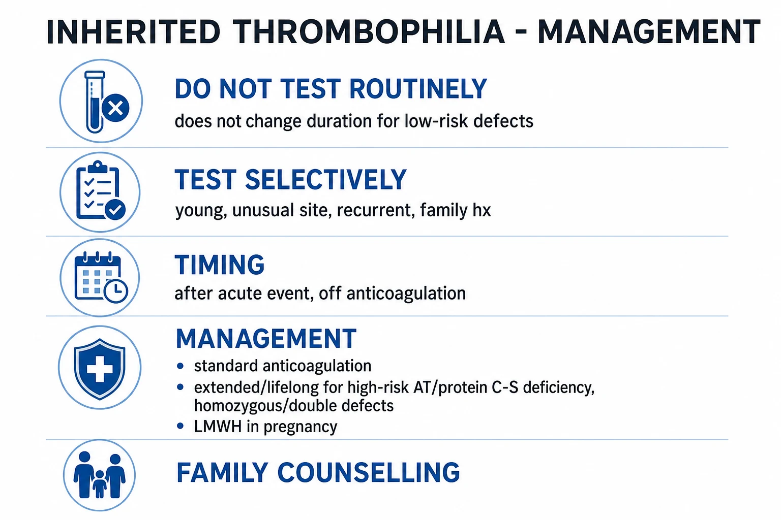
Thrombophilia testing is selective, not universal. The American Society of Hematology (ASH), the British Society for Haematology (BSH), and the International Society on Thrombosis and Haemostasis (ISTH) all endorse a selective approach: routine testing of all VTE patients is not cost-effective and rarely changes management, because most common defects (factor V Leiden, prothrombin mutation) do not alter anticoagulation duration. The decision to test should be guided by the probability that the result will change management — not by curiosity.[1][5]
When to test (indications): unprovoked VTE under 50; unusual-site VTE (cerebral, mesenteric, portal, hepatic, renal, retinal); recurrent unprovoked VTE; VTE on the combined OCP or in pregnancy; strong family history (two or more first-degree relatives with VTE); warfarin-induced skin necrosis; recurrent fetal loss or severe pregnancy complications; heparin resistance (suggests antithrombin deficiency); and first-degree relatives of confirmed probands (cascade screening).[5]
When NOT to test: most provoked VTE in older patients; VTE where the result would not change anticoagulation duration; critically ill patients on multiple confounders; and during the acute thrombotic event itself (when acute-phase reactants distort assays).[5]
Inherited thrombophilia — key numbers
The thrombophilia panel
| Test | What it detects | Interpretation pitfalls |
|---|---|---|
| APC-resistance assay (functional) | Factor V Leiden phenotype — APC sensitivity ratio less than 2.0 | False-positive in pregnancy, OCP, lupus anticoagulant; confirm with PCR |
| Factor V Leiden PCR (genetic) | G1691A / Arg506Gln genotype — heterozygous or homozygous | The confirmatory genetic test; unaffected by anticoagulants or acute illness — can test anytime |
| Prothrombin G20210A PCR (genetic) | The F2 3-prime UTR mutation | Raised prothrombin level is a clue but non-specific; PCR is diagnostic; unaffected by anticoagulants |
| Functional antithrombin (activity assay) | Antithrombin activity — heparin cofactor assay | Lowered by heparin therapy, nephrotic syndrome, liver disease, acute thrombosis, pregnancy, asparaginase; repeat off therapy |
| Functional protein C (activity assay) | Protein C activity — chromogenic or clotting | Lowered by warfarin — test off warfarin for 2-plus weeks or use genetic testing; also lowered in liver disease, pregnancy, sepsis, DIC |
| Free protein S antigen | Free (active) protein S level | Lowered by warfarin, pregnancy, OCP, inflammation, liver disease, nephrotic syndrome; C4b-binding protein rises in inflammation and binds more protein S, lowering free levels |
| Fasting homocysteine | Elevated homocysteine level | Raised by folate, B12, or B6 deficiency; renal impairment; hypothyroidism; MTHFR mutations; test fasting |
| MTHFR C677T / A1298C PCR | Common MTHFR polymorphisms | Controversial clinical utility — homocysteine level is more clinically relevant than genotype in most guidelines |
| Lupus anticoagulant + anticardiolipin + anti-beta-2-GPI | Antiphospholipid antibodies (acquired) | Always test for APS alongside inherited defects; must be positive twice, 12 weeks apart, to diagnose APS |
Timing and interpretation
Timing is everything. The commonest laboratory error in thrombophilia is testing at the wrong time. The rules are:[1][5]
- Do NOT test during the acute thrombotic event — consumption of clotting factors and acute-phase responses distort functional assays. Wait at least 4 to 6 weeks after the acute event.
- Do NOT test on anticoagulation — warfarin lowers protein C and protein S (they are vitamin K-dependent); heparin lowers antithrombin. Either stop anticoagulation for a sufficient washout (2-plus weeks for warfarin, though this is rarely practical or safe) or use genetic tests (factor V Leiden PCR, prothrombin PCR), which are unaffected by anticoagulants and can be done at any time.
- Do NOT test during pregnancy or on the OCP — both lower free protein S and antithrombin levels; defer until postpartum or after stopping the OCP. The genetic tests remain valid.
- Always exclude acquired causes first — antiphospholipid antibodies, malignancy screen (age-appropriate), full blood count and blood film for myeloproliferative disease (check JAK2 if splanchnic thrombosis), and liver and renal function.
- Repeat any abnormal functional result before labelling a patient as having an inherited deficiency — transient acquired deficiency from sepsis, pregnancy, liver disease, or nephrotic syndrome is common and can masquerade as inherited deficiency on a single assay.[1]
Management — Resuscitation
An inherited thrombophilia is not an emergency in itself — the emergency is the thrombotic event it has caused, which is managed identically to any VTE, with one specific pharmacological trap related to antithrombin deficiency.[2]
- Acute DVT or PE: weight-based low-molecular-weight heparin (e.g. enoxaparin 1 mg per kg subcutaneously every 12 hours, or 1.5 mg per kg once daily) or fondaparinux (5 to 10 mg subcutaneously once daily by body weight), then transition to oral anticoagulation (DOAC or warfarin). For massive PE with haemodynamic instability (hypotension, shock), give systemic thrombolysis (alteplase 50 to 100 mg IV over 2 hours) or consider catheter-directed thrombolysis or surgical embolectomy. In antithrombin deficiency, unfractionated or low-molecular-weight heparin may fail to achieve therapeutic levels because there is insufficient functional antithrombin to potentiate — monitor with anti-Xa levels (not APTT) and administer antithrombin concentrate if heparin-resistant.[2][4]
- Warfarin-induced skin necrosis: STOP warfarin immediately, administer therapeutic-dose heparin (LMWH or intravenous unfractionated), give vitamin K (5 to 10 mg IV or oral) to reverse warfarin, and administer protein C concentrate (or fresh frozen plasma if concentrate unavailable) to replace the deficient anticoagulant. Surgical debridement may be required for necrotic skin. The complication is entirely preventable by always bridging warfarin with heparin until the INR is therapeutic for two consecutive days, especially in any patient with suspected protein C or S deficiency.[8]
- Neonatal purpura fulminans (homozygous protein C/S deficiency): a dermatological and haematological emergency — give protein C concentrate (or FFP), initiate therapeutic anticoagulation with heparin, and plan for lifelong protein C replacement or anticoagulation. Liver transplantation is curative for severe protein C deficiency.[1]
Management — Definitive & Stepwise
The thrombotic event is treated with standard anticoagulation; the defect type mainly determines the DURATION of therapy. The commonest error in examinations — and in clinical practice — is over-treating a low-risk defect. A heterozygous factor V Leiden carrier with a single provoked DVT does not need lifelong anticoagulation simply because of the genetic result.[1][5]
Duration by defect tier
| Defect tier | Anticoagulation duration | Rationale |
|---|---|---|
| Low-risk (heterozygous FVL, prothrombin mutation), single provoked event | Standard duration — provoked VTE approximately 3 months | The defect rarely changes the recurrence-prevention calculus |
| Low-risk, unprovoked or recurrent VTE | Extended (at least 6 to 12 months; reassess) | Driven by the nature of the event (unprovoked), not by the defect genotype |
| High-risk (antithrombin deficiency, homozygous FVL, double defect) | Extended or lifelong | High recurrence rate; the defect does change duration |
| VTE with antiphospholipid syndrome overlap | Lifelong (warfarin preferred; DOACs avoided in triple-positive APS) | Highest recurrence; warfarin remains first-line |
Key principle: for most patients with a first VTE, the decision to extend anticoagulation is driven by whether the event was provoked or unprovoked, the bleeding risk, and patient preference — not by the thrombophilia genotype alone. The ASH, BSH, and ISTH guidelines all caution against extending therapy solely on the basis of a low-risk thrombophilia result.[5]
Anticoagulant choice
A DOAC (apixaban, rivaroxaban, dabigatran, or edoxaban) or warfarin is first-line for most VTE. LMWH is preferred in pregnancy and active cancer (per Khorana score and guideline recommendations). In antithrombin deficiency with heparin resistance, the APTT is unreliable — monitor with anti-Xa levels and consider antithrombin concentrate perioperatively, peripartum, or during acute thrombosis. The role of DOACs in antithrombin deficiency is less established and warrants specialist input; however, DOACs act independently of antithrombin and may be effective. In protein C or S deficiency, always bridge warfarin with heparin for at least 5 days and until the INR is therapeutic for two consecutive days to prevent warfarin-induced skin necrosis.[1][4]
Hyperhomocysteinaemia management
Unlike the other inherited thrombophilias, hyperhomocysteinaemia is directly treatable with vitamin supplementation regardless of the thrombotic event: folate 0.4 to 5 mg daily, vitamin B6 (pyridoxine) 25 to 100 mg daily, and vitamin B12 (cyanocobalamin) 0.4 to 1 mg daily lower homocysteine levels. However, while vitamin supplementation normalises homocysteine, the evidence that it reduces recurrent VTE risk is weak — multiple randomised trials have shown homocysteine-lowering therapy does not significantly reduce VTE recurrence. Therefore, anticoagulation duration is still determined by the thrombotic event itself, and vitamin supplementation is given as adjunctive therapy.[1]
Specific Subtypes & Scenarios
Factor V Leiden (FVL)
- Mutation: G1691A in exon 10 of F5 gene on chromosome 1q24; Arg506Gln (R506Q). The cleavage site for activated protein C is lost, causing APC resistance.[7]
- Inheritance: autosomal dominant with incomplete penetrance.
- VTE risk: heterozygous approximately 3 to 8-fold increased; homozygous approximately 80-fold increased; compound heterozygous (FVL plus prothrombin G20210A) approximately 20-fold.
- Clinical: DVT and PE; the commonest inherited cause of unprovoked VTE in young Caucasians. Most heterozygotes never thrombose.
- Management: heterozygous with a single provoked event — standard anticoagulation duration (3 months). Heterozygous with unprovoked VTE — extended anticoagulation (at least 6 to 12 months), driven by the event not the genotype. Homozygous or compound defect — extended or lifelong anticoagulation. Counsel on OCP avoidance (35-fold synergistic risk).[3][7]
Prothrombin G20210A
- Mutation: G-to-A transition at nucleotide 20210 in the 3-prime untranslated region of the F2 gene on chromosome 11p11. Raises plasma prothrombin levels by approximately 25 to 30 percent via increased mRNA stability and translation.[1]
- Inheritance: autosomal dominant with incomplete penetrance.
- VTE risk: heterozygous approximately 2 to 5-fold increased. Homozygous or compound with FVL is higher-risk.
- Clinical: DVT and PE; also associated with recurrent pregnancy loss in systematic review and meta-analysis data.[6]
- Management: same as factor V Leiden — heterozygous single provoked event gets standard duration; the genotype alone does not extend anticoagulation. Counsel on OCP avoidance.
Protein C deficiency
- Gene: PROC on chromosome 2q13 to q14; autosomal dominant.
- Types: Type I (low antigen and activity — quantitative); Type II (normal antigen, low activity — qualitative).
- VTE risk: heterozygous approximately 3 to 15-fold increased, depending on subtype. Presents with unprovoked VTE, often at a young age, and warfarin-induced skin necrosis.[8]
- Homozygous: neonatal purpura fulminans — widespread cutaneous microvascular thrombosis, DIC, and death if untreated. Treat with protein C concentrate, FFP, and lifelong anticoagulation. Liver transplantation is curative.[1]
- Management: always bridge warfarin with heparin to prevent skin necrosis. Extended anticoagulation after a first event is generally recommended.
Protein S deficiency
- Gene: PROS1 on chromosome 3p11.1 to 3q11.2; autosomal dominant.
- Types: Type I (low total and free antigen); Type II (low activity, normal antigen); Type III (normal total, low free protein S). Measure free protein S antigen (the active fraction).
- VTE risk: heterozygous approximately 2 to 10-fold increased. Presents similarly to protein C deficiency with young, unprovoked VTE and warfarin-induced skin necrosis.[8]
- Homozygous: neonatal purpura fulminans (rare).
- Management: same as protein C deficiency — always bridge warfarin with heparin; extended anticoagulation after a first event.
Antithrombin deficiency
- Gene: SERPINC1 on chromosome 1q25; autosomal dominant.
- Types: Type I (low antigen and activity — quantitative); Type II (normal antigen, low activity — qualitative). Type II heparin-binding-site variants are the most thrombogenic.[4]
- VTE risk: the strongest single inherited thrombophilia — approximately 10 to 25-fold increased VTE risk. Presents at the youngest age (often first VTE under 30), with unusual-site thrombosis (mesenteric, cerebral), and may cause heparin resistance during acute treatment (APTT fails to rise despite escalating UFH doses).[4]
- Acquired deficiency: nephrotic syndrome (urinary antithrombin loss), liver disease (reduced synthesis), asparaginase chemotherapy, DIC, severe sepsis — exclude before diagnosing inherited deficiency.
- Management: extended or lifelong anticoagulation after a first VTE event. During acute treatment, monitor with anti-Xa levels (APTT is unreliable in heparin resistance) and administer antithrombin concentrate to overcome resistance. Perioperative and peripartum antithrombin concentrate may be needed.[4]
Hyperhomocysteinaemia (MTHFR)
- Mutation: MTHFR C677T (Ala222Val, thermolabile variant) and MTHFR A1298C (Glu429Ala) on chromosome 1p36.3. Homozygous C677T (TT genotype) occurs in approximately 10 to 15 percent of Europeans.
- Mechanism: impaired remethylation of homocysteine to methionine, especially with low folate intake, leading to elevated plasma homocysteine. Endothelial dysfunction, oxidative stress, reduced thrombomodulin, and impaired fibrinolysis.[1]
- Thrombosis risk: venous AND arterial — DVT, PE, MI, stroke. Unlike other defects which are predominantly venous, hyperhomocysteinaemia is a risk factor for arterial thrombosis too.
- Diagnosis: fasting total homocysteine level (greater than 15 micromol per litre is elevated); MTHFR genotyping is secondary (the phenotype matters more than the genotype for most guidelines).
- Treatment: folate 0.4 to 5 mg daily, vitamin B6 25 to 100 mg daily, vitamin B12 0.4 to 1 mg daily normalises homocysteine. However, homocysteine-lowering therapy has NOT been shown to reduce VTE recurrence in randomised trials — anticoagulation duration is still determined by the thrombotic event.[1]
Combined (double) defects
Approximately 1 to 2 percent of thrombophilia patients carry two defects — most commonly factor V Leiden plus prothrombin G20210A. The VTE risks are additive or synergistic (combined risk approximately 20 to 60-fold). These patients are treated as high-risk with extended or lifelong anticoagulation, and their relatives should be offered cascade screening for both defects.[1]
Complications & Pitfalls
| Complication or pitfall | Notes |
|---|---|
| Recurrent VTE | Higher recurrence than in non-carriers, especially with high-risk defects (AT, protein C/S). Anticoagulation adherence is critical. |
| Post-thrombotic syndrome | Chronic calf swelling, pain, heaviness, hyperpigmentation, and venous ulceration after DVT. Graduated compression stockings reduce severity. |
| Chronic thromboembolic pulmonary hypertension (CTEPH) | Occurs in approximately 0.4 to 4 percent of PE survivors — progressive dyspnoea, right-heart failure. Diagnose with V/Q scan; treat with pulmonary endarterectomy or riociguat. |
| Warfarin-induced skin necrosis | Protein C or S deficiency; preventable by heparin bridging. Requires immediate warfarin cessation, heparin, vitamin K, and protein C concentrate. |
| Cerebral venous sinus thrombosis | Unusual-site thrombosis; presents with headache, seizures, focal deficits. Higher risk with high-risk defects or combined defects. |
| Pregnancy complications | Recurrent miscarriage, pre-eclampsia, IUGR, placental abruption, stillbirth. APS is a more common cause than inherited thrombophilia. |
| Pitfall: false results from wrong timing | Testing during acute thrombosis, on heparin or warfarin, in pregnancy, or during sepsis gives falsely low antithrombin, protein C, or protein S — leading to misdiagnosis. Use genetic tests or repeat off therapy. |
| Pitfall: over-testing low-risk provoked VTE | Testing where the result would not change duration wastes resources, causes anxiety, and may lead to inappropriate extended anticoagulation. |
| Pitfall: omitting acquired causes | APS, malignancy, nephrotic syndrome, myeloproliferative disease, PNH, and HIT must be excluded before attributing thrombophilia to an inherited cause. |
| Pitfall: heparin resistance mismanaged | Escalating heparin doses without recognising antithrombin deficiency delays effective anticoagulation. Check anti-Xa and give antithrombin concentrate. |
Prognosis & Disposition
Penetrance is incomplete for all inherited thrombophilias, and this is the single most important prognostic concept for counselling. Most heterozygous factor V Leiden carriers never develop VTE — the lifetime risk is approximately 5 to 10 percent for heterozygous FVL and 2 to 4 percent for heterozygous prothrombin G20210A. In contrast, homozygous factor V Leiden and antithrombin deficiency carry much higher lifetime risks (often over 50 percent), and protein C or S deficiency heterozygotes have an intermediate risk. The annual incidence of VTE in untreated antithrombin-deficient relatives is approximately 1 to 3 percent per year — substantially higher than the baseline population rate of approximately 0.1 percent.[1][4]
Recurrence after a first VTE event is higher in thrombophilic patients than in non-carriers, and the magnitude depends on the defect: low-risk defects have a modest increase (1.5 to 2-fold above non-carriers), whereas high-risk defects (antithrombin, homozygous FVL, double defects) have a 2 to 4-fold higher recurrence. This is why high-risk defects justify extended or lifelong anticoagulation.[5]
Disposition is outpatient anticoagulation once the acute event is stable, with lifelong surveillance for high-risk defects and counselling of first-degree relatives through cascade screening (test parents, siblings, and adult children; counsel regarding OCP avoidance, pregnancy prophylaxis, and the incomplete penetrance so they are not needlessly alarmed).[1][2]
Special Populations
Pregnancy and thrombophilia
Pregnancy is a physiological hypercoagulable state (increased factors II, VII, VIII, X, and fibrinogen; decreased protein S; venous stasis; uterine compression of the IVC). A thrombophilic defect amplifies this risk. Management is guided by the type of defect, the personal VTE history, and the family history, using RCOG Green-top Guideline No. 37 (UK) or ASH guidance (US).[2][5]
- Anticoagulant choice in pregnancy: LMWH is the mainstay (e.g. enoxaparin 40 mg subcutaneously daily for prophylaxis, or weight-based therapeutic dosing for treatment). Warfarin is teratogenic — it crosses the placenta and causes embryopathy (nasal hypoplasia, stippled epiphyses, limb hypoplasia), particularly between weeks 6 and 12 of gestation, and causes fetal and placental haemorrhage in later pregnancy. DOACs are avoided in pregnancy and breastfeeding (insufficient safety data). Transition from warfarin to LMWH before 6 weeks gestation (ideally pre-conception in known thrombophilia).
- Postpartum: continue LMWH (or switch to warfarin, which is safe in breastfeeding) for at least 6 weeks postpartum, the period of highest VTE risk. Warfarin is safe in lactation — it does not appear in breast milk in clinically significant amounts (unlike in pregnancy).[2]
- Risk stratification: asymptomatic low-risk carriers (heterozygous FVL or prothrombin mutation) with no prior VTE often need postpartum prophylaxis only. High-risk defects (antithrombin deficiency, homozygous FVL) or any defect with a prior VTE warrant antenatal and postpartum prophylactic or therapeutic LMWH. A very strong family history (first-degree relative with VTE) may tip a low-risk carrier toward antenatal prophylaxis.[5]
Combined oral contraceptive pill (OCP)
The combined OCP is contraindicated in women with high-risk defects (antithrombin, protein C, protein S deficiency, homozygous FVL) due to synergistic VTE risk. Factor V Leiden carriers on the combined pill have approximately a 35-fold increased VTE risk versus non-carriers not on the pill.[3] Counsel to switch to a progestogen-only pill (desogestrel), a levonorgestrel intrauterine system (Mirena), or non-hormonal contraception (copper IUD, barrier methods). The progestogen-only pill does not significantly increase VTE risk.
Neonates
Homozygous protein C or S deficiency presents with neonatal purpura fulminans — a dermatological and haematological emergency characterised by widespread, rapidly progressive cutaneous microvascular thrombosis, DIC, and skin necrosis in the first hours to days of life. Treatment is protein C concentrate (or fresh frozen plasma), therapeutic anticoagulation with heparin, and lifelong protein C replacement or anticoagulation. Liver transplantation is curative for severe protein C deficiency.[1]
Asymptomatic relatives (cascade screening)
Targeted cascade screening of first-degree relatives of a confirmed proband can guide OCP avoidance, pregnancy prophylaxis, and situational prophylaxis (surgery, immobilisation). It is not emergency testing and should be accompanied by genetic counselling explaining incomplete penetrance — most carriers never thrombose, and the result should empower informed decisions, not cause anxiety.[5]
Anticoagulated patient needing surgery
For patients on anticoagulation needing elective surgery, bridge with LMWH based on the thrombotic risk. In antithrombin deficiency, plan for antithrombin concentrate perioperatively to prevent both thrombosis and heparin resistance. In protein C or S deficiency, restart warfarin carefully with heparin bridging. For DOACs, stop 24 to 48 hours preoperatively based on bleeding risk and renal function; no bridging is usually needed given the short half-life.[2]
Evidence, Guidelines & Regional Differences
Selective testing is the global consensus, endorsed by the ASH (American Society of Hematology) 2020 Choosing Wisely campaign and guidelines, the BSH (British Society for Haematology), and the ISTH (International Society on Thrombosis and Haemostasis). The rationale: routine thrombophilia testing of all VTE patients is not cost-effective, rarely changes anticoagulation duration for low-risk defects, and can cause harm through unnecessary extended anticoagulation, patient anxiety, and insurance implications.[5]
Pregnancy prophylaxis is stratified by RCOG Green-top Guideline No. 37 (UK), which provides a risk table combining defect type with personal and family VTE history to determine whether antenatal prophylaxis, postpartum prophylaxis, or both are warranted. The ASH 2018 guidelines on VTE in pregnancy similarly recommend risk-stratified prophylaxis with LMWH.[5]
DOACs (apixaban, rivaroxaban, dabigatran, edoxaban) are first-line for most VTE globally, including in patients with inherited thrombophilia. However, in antiphospholipid syndrome (triple-positive), DOACs are avoided in favour of warfarin (RE-CIRCULATE, TRAPS trials). In antithrombin deficiency with heparin resistance, the role of DOACs is less established — they act independently of antithrombin and may be effective, but specialist input is advised. The classic Trousseau association (migratory thrombophlebitis with malignancy) is an acquired state, not inherited, and must be separated from inherited thrombophilia in the differential.[2][5]
Controversy — MTHFR testing: most guidelines (ASH, BSH, ACMG) now recommend against routine MTHFR genotyping, because the C677T and A1298C polymorphisms are extremely common, their association with VTE is weak, and the clinical action (homocysteine-lowering vitamins) does not reduce VTE recurrence. Measuring fasting homocysteine may be more useful if clinically indicated (e.g. young arterial thrombosis with a family history), but even this is controversial. The topic remains in curricula because of its historical importance and exam frequency.[1]
Exam Pearls
Whom to TEST for inherited thrombophilia
TEST
unprovoked VTE under 50
cerebral, mesenteric, portal, hepatic, renal, retinal veins
two or more affected first-degree relatives; warfarin-induced skin necrosis
recurrent VTE; VTE on OCP or in pregnancy
The natural anticoagulants — three BRAKES
CAPS
cleaves Va and VIIIa; warfarin-induced skin necrosis if deficient
neutralises thrombin and Xa; heparin resistance if deficient; strongest single defect
cofactor for APC; warfarin-induced skin necrosis if deficient
all three act at specific steps of the coagulation cascade; deficiency removes the brake
Exam application bank (NEET-PG / INICET)
One-line answer
Inherited thrombophilias are genetic disorders increasing the risk of venous thromboembolism (VTE). The commonest are factor V Leiden (activated protein C resistance; the single commonest inherited thrombophilia in Europeans, autosomal dominant, carrier rate 3 to 8 percent) and the prothrombin G20210A mutation (raised prothrombin; 2 percent of Europeans). Less common but higher-risk are deficiencies of the natural anticoagulants — antithrombin, protein C, protein S — which cause more severe, younger-onset, unusual-site thrombosis and warfarin-induced skin necrosis (protein C/S). Hyperhomocysteinaemia (MTHFR mutations) raises both venous and arterial thrombosis risk. Indications to test: VTE under 50, unusual site (cerebral, mesenteric, portal, hepatic), recurrent unprovoked VTE, family history, VTE in pregnancy/OCP, warfarin-induced skin necrosis. Testing is selective, not universal; the
Worked stems (answer without another resource)
Stem 1 — Classic presentation. Map symptoms to mechanism; name the first investigation and first treatment step with dose/route if drug therapy is standard. [1]
Stem 2 — Unstable / complicated. List red flags that force immediate resuscitation, theatre, ICU, antidote, or reperfusion — and what you do in the first 15 minutes. [1]
Stem 3 — Atypical group. Elderly, pregnancy, child, or immunocompromised: how presentation and thresholds change. [1]
Stem 4 — Differential trap. Name the three closest mimics and one discriminator for each. [1]
Stem 5 — Disposition. Who goes home with safety-netting, who is admitted, who needs HDU/ICU/theatre, and what follow-up is mandatory. [1]
Rapid viva checklist
- Definition + classification
- Pathophysiology chain
- Bedside signs / criteria
- Score with exact components (if any)
- Emergency bundle
- Definitive therapy with doses
- Complications of disease and of treatment
- Special populations
- Guideline/trial name if classic
- Three exam traps
Coverage self-check
If you cannot answer any stem above from this page alone, re-read the matching section — the page is intended to be self-sufficient for final-prof and NEET-PG/INICET questions on Inherited Thrombophilia (Factor V Leiden & Protein C/S Deficiency).
References
- [1]Phillippe HM, Hornsby LB, Treadway S, Farrell J, Finks S. Inherited Thrombophilia J Pharm Pract, 2014.PMID 24739277
- [2]Chopard R, Albertsen IE, Piazza G,_SPIEFF,Goldshtein NM,Witt DM. Diagnosis and Treatment of Lower Extremity Venous Thromboembolism: A Review JAMA, 2020.PMID 33141212
- [3]Vandenbroucke JP, Koster T, Briet E, Reitsma PH, Bertina RM, Rosendaal FR. Increased risk of venous thrombosis in oral-contraceptive users who are carriers of factor V Leiden mutation Lancet, 1994.PMID 7968118
- [4]Patnaik MM, Moll S. Inherited antithrombin deficiency: a review Haemophilia, 2008.PMID 19141163
- [5]Stevens SM, Woller SC, Bauer KA, Kasthuri R, Cushman M, Streiff M, Lim W, Douketis JD, Gould MK, Lopes RD, Dager WE, Lansberg MG, Kahn SR, Bhatt DL, Smythe MA, Freeman MW, Kern H, Thomson L, Ortel TL, Kahn SR, Crowther M, Smythe MA. Guidance for the evaluation and treatment of hereditary and acquired thrombophilia J Thromb Thrombolysis, 2016.PMID 26780744
- [6]Gao H, Huang Z, Wang L, Chen H, Liu Y, Chen Q, Wang H. Prothrombin G20210A mutation is associated with recurrent pregnancy loss: a systematic review and meta-analysis update Thromb Res, 2015.PMID 25528068
- [7]Kujovich JL. Factor V Leiden thrombophilia Genet Med, 2011.PMID 21116184
- [8]Sallah S, Thomas DP, Roberts HR. Recurrent warfarin-induced skin necrosis in kindreds with protein S deficiency Haemostasis, 1998.PMID 9885367