Haematology · General Medicine
Multiple Myeloma
Also known as Multiple myeloma · Myeloma · Plasma cell myeloma · Kahler disease
Multiple myeloma is a malignant clonal neoplasm of terminally differentiated B-cells (plasma cells) that accumulates in the bone marrow and secretes a monoclonal protein (paraprotein / M-band) and/or free light chains. End-organ damage is summarised by CRAB — hyperCalcaemia, Renal impairment (cast nephropathy), Anaemia, Bone lesions (lytic). Diagnosis (IMWG 2014) requires clonal marrow plasma cells at least 10 percent (or biopsy-proven plasmacytoma) plus a myeloma-defining event: CRAB features or a biomarker of malignancy (serum involved-to-uninvolved free light chain ratio at least 100, more than one focal lesion on MRI, or 60 percent or more clonal marrow plasma cells). Workup: SPEP + immunofixation, serum free light chains, urine Bence Jones, marrow biopsy with FISH, whole-body low-dose CT, beta-2 microglobulin, LDH, albumin (R-ISS). Treatment: VRd (bortezomib + lenalidomide + dexamethasone) induction then autologous stem-cell transplant for fit patients then lenalidomide maintenance; relapsed disease — daratumumab (anti-CD38), carfilzomib, pomalidomide, bispecific antibodies, anti-BCMA CAR-T. Supportive: zoledronic acid or denosumab for bone. Median survival now 7 to 10 years.
On this page & tools
Your progress
Saved locally on this device.
Practise this topic
Exam tags
Red flags
Overview & Definition
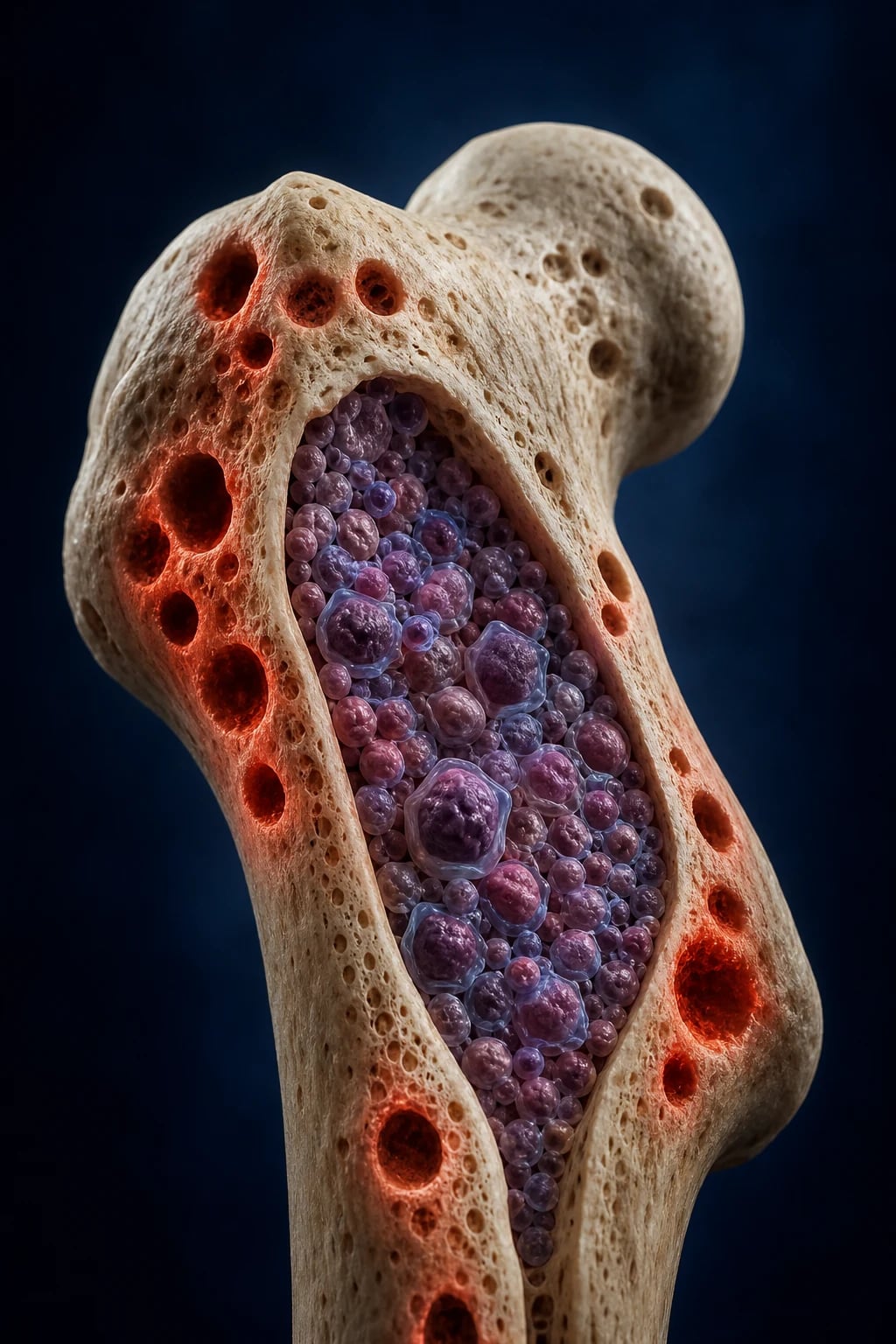
Multiple myeloma is a malignant clonal neoplasm of terminally differentiated B-cells (plasma cells) that accumulates in the bone marrow and secretes a monoclonal immunoglobulin (paraprotein / M-protein) and/or free light chains. The clone is a post-germinal-centre B-cell that has undergone somatic hypermutation and class-switch recombination but has acquired transforming genetic lesions that drive uncontrolled proliferation and survival in the marrow. The disease causes harm through three linked mechanisms: (1) infiltration of the bone marrow by plasma cells (anaemia, cytopenias), (2) secretion of monoclonal protein and cytokines (renal cast nephropathy, hyperviscosity, bone destruction, immune paresis), and (3) cytokine-driven uncoupling of bone remodelling (RANKL-mediated osteoclast activation producing lytic lesions and hypercalcaemia).[1]
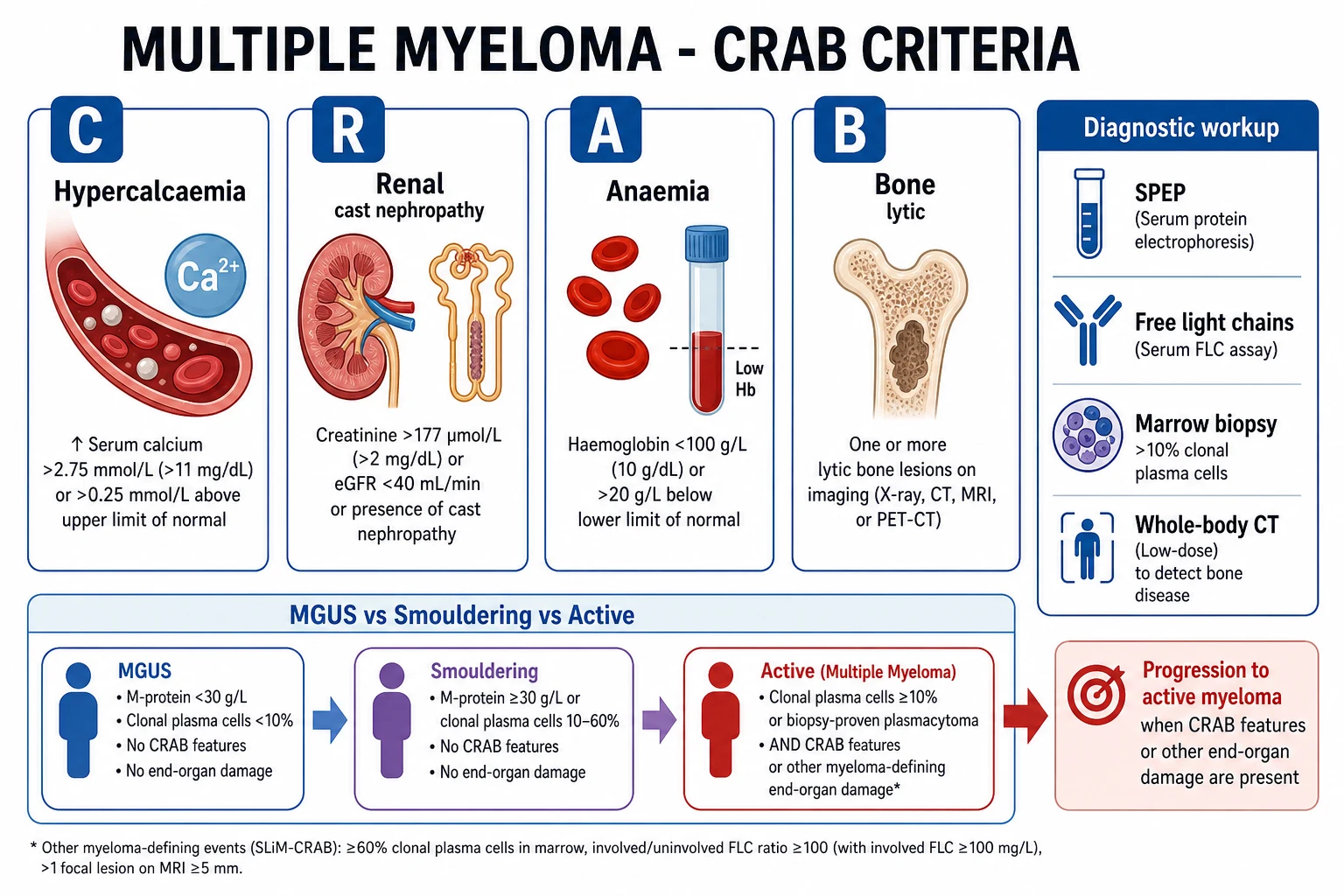
End-organ damage is summarised by the CRAB mnemonic — hyperCalcaemia, Renal impairment, Anaemia, Bone lesions — and myeloma is distinguished from its precursor conditions (MGUS, smouldering myeloma) by the presence of end-organ damage or a myeloma-defining biomarker. Myeloma is the second commonest haematological malignancy after non-Hodgkin lymphoma, predominantly affecting older adults (median age about 70). The arrival of proteasome inhibitors (bortezomib, carfilzomib, ixazomib), immunomodulatory drugs (thalidomide, lenalidomide, pomalidomide) and monoclonal antibodies (daratumumab) — now joined by bispecific antibodies and anti-BCMA CAR-T cell therapy — has transformed a previously uniformly fatal disease into a chronic, manageable condition with median survival of 7 to 10 years.[1][2]
Multiple myeloma — the headline numbers
Classification
Myeloma sits at the symptomatic end of a plasma-cell dyscrasia spectrum distinguished by three axes — the paraprotein load, the marrow clonal plasma-cell percentage, and the presence or absence of end-organ damage or a biomarker of malignancy.[1][3]
MGUS
- Serum M-protein under 30 g/L AND marrow clonal plasma cells under 10 percent
- No CRAB end-organ damage, no myeloma-defining biomarkers
- Normal free light chain ratio (abnormal ratio less than 100)
- No evidence of amyloidosis or other B-cell lymphoproliferative disorder
- Monitor annually; progression risk about 1 percent per year
Smouldering (asymptomatic) myeloma
- Serum M-protein at least 30 g/L (IgG or IgA) and/or urinary monoclonal protein at least 500 mg per 24 h, AND/OR marrow clonal plasma cells 10 to 60 percent
- No CRAB end-organ damage, no myeloma-defining biomarker
- No amyloidosis
- Risk stratify: high-risk features (FLC ratio at least 100 but under the myeloma threshold, more than one focal MRI lesion, marrow plasma cells at least 50 percent, M-protein at least 30 g/L) carry higher progression risk
- Close observation; early therapy for high-risk smouldering disease (the frente/quebec score)
Symptomatic (active) multiple myeloma
- Clonal marrow plasma cells at least 10 percent (or biopsy-proven plasmacytoma)
- PLUS a myeloma-defining event — CRAB attributable to the plasma-cell disorder OR a biomarker of malignancy
- Biomarkers: involved-to-uninvolved serum FLC ratio at least 100 (involved FLC at least 100 mg/L); more than one focal lesion on MRI or CT; marrow clonal plasma cells at least 60 percent
- Requires systemic treatment
- Sub-classify by secreted paraprotein type and cytogenetic risk
By secreted paraprotein type (the immunoglobulin the clone produces): IgG myeloma is the commonest (about 50 percent), followed by IgA (about 20 percent). Light-chain only (Bence Jones) myeloma accounts for 15 to 20 percent — there is no intact M-band on serum electrophoresis, so the diagnosis is easily missed if only a serum SPEP is sent. The rare IgD and IgE myelomas and the non-secretory forms (about 1 to 2 percent) are diagnosed by marrow biopsy and imaging and monitored with serum free light chains, since even "non-secretory" clones usually produce measurable free light chains. [1]
Related plasma-cell disorders (examiners test these by their distinguishing features): [1]
- Solitary plasmacytoma of bone — a single clonal plasma-cell tumour of the skeleton with no systemic disease (normal marrow elsewhere, no CRAB, no biomarker). Treated with radiotherapy with curative intent; about half progress to systemic myeloma within 10 years.
- Extramedullary plasmacytoma — a plasma-cell tumour of soft tissue (commonly upper respiratory tract); treated with radiotherapy, better prognosis than solitary plasmacytoma of bone.
- POEMS syndrome — Polyneuropathy, Organomegaly, Endocrinopathy, Monoclonal protein (almost always lambda), Skin changes; plus osteosclerotic bone lesions and a markedly raised serum VEGF. Distinguished from classic myeloma by the osteosclerotic (not lytic) bone lesion and the disabling neuropathy.
- AL amyloidosis — a plasma-cell clone producing light chains that misfold as amyloid fibrils deposited in tissues (heart, kidney, liver, nerves); features include macroglossia, periorbital purpura, nephrotic-range proteinuria, restrictive cardiomyopathy. May overlap with myeloma or stand alone with a tiny paraprotein.
- Heavy-chain disease — rare lymphoplasmacytic neoplasms producing truncated heavy chains (alpha, gamma, mu). [1]
Epidemiology & Risk Factors
Multiple myeloma accounts for about 1 to 2 percent of all cancers and roughly 10 to 15 percent of haematological malignancies, making it the second commonest haematological malignancy after non-Hodgkin lymphoma. The median age at diagnosis is about 69 to 70 years, with under 2 percent of cases occurring before 40. There is a slight male predominance and, notably, roughly twice the incidence in people of African ancestry (Black populations), who also present at a younger median age — the reasons include a higher prevalence of MGUS and possible germline variants. Incidence is rising globally, partly from ageing populations and better detection.[1]
Recognised risk factors: [1]
- Increasing age — fundamentally a disease of older adults.
- Male sex and African ancestry (Black populations — two- to threefold higher incidence; in contrast, lower in Asian populations).
- Family history of myeloma or MGUS — roughly twofold increased risk in first-degree relatives.
- Obesity — consistently associated with an increased risk of myeloma and of progression from MGUS.
- Prior MGUS — the obligate precursor in essentially all cases. MGUS is present in over 3 percent of people over 50 and in over 5 percent of those over 70, and progresses to myeloma (or a related lymphoproliferative disorder) at about 1 percent per year.
- Possible environmental factors: ionising radiation, and chronic agricultural, pesticide and petroleum-solvent exposures; chronic immune stimulation (e.g. some chronic infections) has a weak association. [1]
In India and other low- and middle-income countries the apparent incidence is lower (partly from under-diagnosis and shorter life expectancy), but the clinical profile is younger presentation and more advanced disease at diagnosis. Access to lenalidomide, bortezomib, autologous transplant and especially daratumumab and CAR-T is limited by cost, and thalidomide + melphalan + prednisolone (MPT) or bortezomib + melphalan + prednisolone (VMP) remain widely used regimens. Blood Cancer India and ICMR registries are improving epidemiology capture.[1]
Pathophysiology
The malignant clone is a post-germinal-centre, terminally differentiated B-cell (a plasma cell) that has escaped normal apoptotic regulation and proliferates in the bone marrow. It is supported by the marrow microenvironment: bone-marrow stromal cells secrete interleukin-6 (IL-6), VEGF and other cytokines, and bind the myeloma cells via adhesion molecules (VLA-4, CD44). This niche contact activates NF-kB, STAT3 and PI3K/AKT survival pathways that promote plasma-cell proliferation and drug resistance. The four CRAB features each have a distinct, mechanistic basis that an examiner will probe.[1]
The mechanism of each CRAB feature
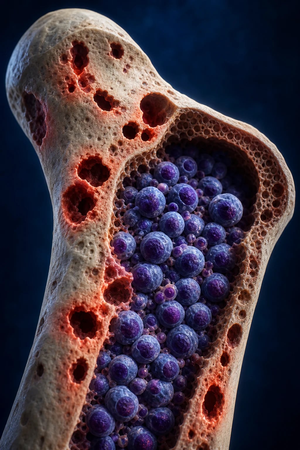
- Bone destruction (B and the hypercalcaemia of C) — malignant plasma cells and marrow stromal cells secrete RANKL (receptor activator of NF-kB ligand), which binds the RANK receptor on osteoclast precursors and activates osteoclasts. Simultaneously the plasma cells release DKK1 (Dickkopf-1) and other Wnt-pathway antagonists that suppress osteoblasts. The result is uncoupled bone resorption without matching formation: punched-out lytic lesions, pathological fractures, and hypercalcaemia as calcium is released into the circulation from resorbed bone. The characteristic "raindrop" or "punched-out" skull appearance reflects this focal destructive process.
- Renal damage (R) — excess monoclonal free light chains (Bence Jones protein) are filtered by the glomerulus, taken up by proximal tubular cells, and precipitate as obstructive tubular casts with a giant-cell reaction (myeloma cast nephropathy) — the commonest myeloma kidney lesion. Other mechanisms compound this: AL amyloidosis and light-chain deposition disease (light chains depositing in glomeruli and tubules), hypercalcaemic nephropathy (volume depletion and nephrocalcinosis), uric-acid nephropathy from tumour lysis, dehydration, and exposure to nephrotoxic drugs (NSAIDs) and iodinated contrast. The combination of light-chain toxicity and a second insult (contrast, dehydration, hypercalcaemia) is what typically precipitates acute kidney injury.
- Anaemia (A) — a normocytic, normochromic anaemia from a combination of marrow infiltration by plasma cells (crowding out normal haematopoiesis), renal anaemia (the damaged kidney produces less erythropoietin), cytokine-mediated suppression (hepcidin), and sometimes folate or iron deficiency. Anaemia is the commonest cytopenia; thrombocytopenia and leucopenia occur in advanced disease.
- Immune paresis and recurrent infection — the heavy paraprotein load and suppression of normal B-cell function reduce normal polyclonal immunoglobulin production, while neutropenia (marrow infiltration or treatment) compounds the risk. Infections are typically with encapsulated respiratory organisms (Streptococcus pneumoniae, Haemophilus influenzae) and Gram-negatives, and herpes zoster reactivation is common on proteasome inhibitors. [1]
Cytogenetics and molecular risk
Myeloma is divided into two broad cytogenetic groups. Hyperdiploid myeloma (odd-numbered trisomies — 3, 5, 7, 9, 11, 15, 19, 21) is generally standard-risk and tends to present with bone disease. Non-hyperdiploid myeloma includes the IgH translocations at 14q32, the most important being t(4;14) (FGFR3/MMSET), t(14;16) (c-MAF) and t(14;20) (MAFB). The high-risk lesions — del(17p)/TP53 loss, t(4;14), t(14;16), and gain(1q) — are detected by FISH on purified (CD138-selected) plasma cells, because conventional karyotyping misses most lesions (plasma cells divide poorly in culture). The Revised ISS incorporates these high-risk lesions into staging. TP53 loss (del(17p)) carries the worst prognosis and is the dominant driver of extramedullary and plasma-cell leukaemia biology.[2]
Clinical Presentation
The typical presentation is an older adult with persistent bone pain (back, ribs, long bones — often worse on movement and at night), fatigue (anaemia), recurrent infections, and symptoms of hypercalcaemia or renal failure (thirst, polyuria, constipation, confusion, nausea). Bone pain is the single commonest presenting symptom, arising in the spine, ribs, skull, pelvis and proximal long bones. Up to a third of patients are diagnosed after a pathological fracture, and a meaningful minority are picked up incidentally through a raised ESR or an M-band found on routine bloods.[1]
The CRAB features in detail
- C — hyperCalcaemia — from bone resorption. Symptoms: thirst, polyuria, polydipsia, dehydration, constipation, abdominal pain, anorexia, nausea, confusion, drowsiness (the "stones, bones, groans, moans" tetrad). Corrected calcium is typically over 2.75 mmol/L (11 mg/dL); severe hypercalcaemia is a medical emergency.
- R — Renal impairment — usually from cast nephropathy; presents as AKI or progressive CKD, often precipitated by dehydration, infection, iodinated contrast or NSAIDs. The urine is negative for albumin on dipstick despite proteinuria because dipsticks detect albumin, not light chains.
- A — Anaemia — normocytic, normochromic; causes fatigue, dyspnoea, pallor. May be the only feature at diagnosis.
- B — Bone lesions — punched-out lytic lesions (skull "raindrop", spine, pelvis, ribs, proximal long bones), diffuse osteopenia, and pathological fractures (vertebral compression, rib, femoral neck). Intractable bone pain and fractures dominate the quality-of-life burden. [1]
Presenting oncological emergencies
Hyperviscosity and amyloidosis
Hyperviscosity syndrome is more typical of Waldenstrom macroglobulinaemia (IgM is a large pentamer) but can occur with very high IgG or IgA; features are visual disturbance (retinopathy with "sausage-link" veins), headache, mucosal bleeding, vertigo and chest pain. [1]
AL amyloidosis features that may accompany myeloma (or signal amyloidosis alone): macroglossia (an enlarged, firm tongue), periorbital purpura ("raccoon eyes"), nephrotic-range proteinuria, restrictive cardiomyopathy (cardiomegaly, raised JVP, orthopnoea, low-voltage ECG, increased septal thickness on echo with "sparkling" texture), hepatomegaly, and a sensory and autonomic neuropathy (orthostatic hypotension, diarrhoea, erectile dysfunction). [1]
Atypical presentations
In the elderly, classic features may be subtle: frailty, recurrent falls, delirium, fatigue alone, or unexplained weight loss. A lower threshold to screen with SPEP and serum free light chains is warranted in any older adult with unexplained anaemia, renal impairment, hypercalcaemia, or bone pain. [1]
Recurrent infection (pneumonia, pyelonephritis, septicaemia) may be the first clue, from immune paresis. Bleeding (epistaxis, gum bleeding) may reflect acquired von Willebrand disease, thrombocytopenia, or amyloidosis. Peripheral neuropathy may reflect amyloidosis, POEMS, or (paradoxically) be a side-effect of bortezomib once treatment starts. [1]
Differential Diagnosis
A monoclonal protein on serum or urine electrophoresis is not diagnostic of myeloma on its own — most monoclonal gammopathies are MGUS. The differential separates into the plasma-cell/lymphoplasmacytic disorders, the mimics of bone lesions, and the mimics of hypercalcaemia and renal failure.[1][3]
MGUS
- M-protein under 30 g/L, marrow under 10 percent, no CRAB, no biomarker
- Normal FLC ratio, no amyloidosis
- Stable on repeat testing
- ~1 percent per year progression risk; observe
Smouldering myeloma
- M-protein at least 30 g/L and/or marrow at least 10 percent (under 60 percent)
- No CRAB, no biomarker
- Distinguish by absence of end-organ damage and FLC ratio under 100
- Risk-stratify; some progress within months
Waldenstrom macroglobulinaemia
- Lymphoplasmacytic lymphoma producing IgM paraprotein
- Marrow infiltration by lymphoplasmacytic cells (not pure plasma cells)
- Hyperviscosity, anaemia, lymphadenopathy, neuropathy
- No lytic bone lesions; MYD88 L265P mutation
Reactive polyclonal hypergammaglobulinaemia
- Broad-based, polyclonal (not sharp) band on SPEP
- Chronic infection (HIV, HBV, HCV), liver disease, autoimmune disease
- Both kappa and lambda raised proportionally
- No clonal plasma cells in marrow
Lytic bone lesions must be distinguished from metastatic carcinoma (breast, prostate, lung, kidney, thyroid — though prostate and breast metastases are usually mixed or sclerotic, while renal and thyroid are classically lytic and can mimic myeloma), osteoporotic compression fractures (diffuse, age-appropriate, not discrete punched-out lesions), brown tumours of hyperparathyroidism, and Paget disease (sclerotic, thickened cortex). A single destructive lesion with a paraprotein suggests solitary plasmacytoma; multiple lesions need full myeloma workup. [1]
Hypercalcaemia may alternatively reflect primary hyperparathyroidism (high or inappropriately normal PTH), solid-tumour humoral hypercalcaemia (PTHrP-mediated), vitamin-D toxicity, or skeletal metastases — myeloma hypercalcaemia has a suppressed PTH and a paraprotein, with the calcium driven by bone resorption (RANKL). [1]
Renal failure of uncertain cause in an older adult must prompt SPEP and free light chains — cast nephropathy is easily missed and labelled "hypertensive kidney disease". Anaemia with a very high ESR (over 100 mm/h) and rouleaux on the blood film should always trigger a paraprotein screen. [1]
Clinical & Bedside Assessment
The focused bedside examination has three jobs: detect the CRAB features, screen for amyloidosis, and exclude the oncological emergencies (cord compression, hyperviscosity).[1]
- General — pallor (anaemia), cachexia, bruising (thrombocytopenia, amyloidosis), signs of dehydration or confusion (hypercalcaemia). Document performance status (ECOG or Karnofsky) — central to fitness for autologous transplant.
- Skeleton — palpate for bony tenderness over the spine, skull, sternum, ribs, pelvis and long bones; look for spinal deformity (kyphosis from vertebral collapse) and a pathological-fracture deformity.
- Neurology — examine for spinal cord compression (motor weakness, a sensory level, sphincter disturbance) — a myeloma emergency; assess for peripheral neuropathy (amyloidosis, treatment toxicity, POEMS) and monoclonal-protein-related neuropathy.
- Cardiorespiratory — signs of anaemia and cardiac failure/restriction (amyloid cardiomyopathy — raised JVP, bibasal crackles, hepatomegaly, low-voltage ECG). Hyperviscosity may cause retinopathy.
- Abdomen — hepatosplenomegaly (suggests amyloidosis, Waldenstrom, or overlap lymphoma); assess volume status and check for a distended bladder (cord compression).
- Amyloidosis screen — macroglossia, periorbital purpura ("raccoon eyes"), shoulder-pad sign (amyloid in periarticular tissue), nephrotic-range proteinuria on dipstick (if albuminuric) and signs of restrictive cardiomyopathy.
- Eyes and fundi — hyperviscosity retinopathy (dilated, tortuous "sausage-link" veins, haemorrhages). [1]
Investigations
The myeloma workup has four pillars: bloods and urine (anaemia, renal function, calcium, the paraprotein, prognostic markers), bone marrow biopsy (the clonal plasma-cell burden and cytogenetics), imaging (the bone lesions), and the diagnostic and staging criteria that pull it together.[1][3]
First-line bloods and urine
Full blood count and film: normocytic, normochromic anaemia is common; the film shows rouleaux (red cells stacked like coins) from the high serum immunoglobulin, and the ESR is markedly raised (often over 100 mm/h). Leucopenia and thrombocytopenia occur in advanced disease. A blood film with rouleaux and a high ESR is a strong clue. [1]
Biochemistry: urea, electrolytes, creatinine and eGFR (renal impairment), corrected calcium (hypercalcaemia), albumin (low — part of ISS staging), LDH (raised — proliferative marker, part of R-ISS), C-reactive protein (prognostic), urate (raised). Beta-2 microglobulin is essential — it is the single most powerful prognostic marker and a core component of the ISS and R-ISS. [1]
The paraprotein workup (do not omit any of these)
- Serum protein electrophoresis (SPEP) with immunofixation — detects the sharp, narrow M-band (M-protein) and identifies the heavy- and light-chain type (IgG kappa, IgA lambda, etc.). A broad-based band suggests polyclonal hypergammaglobulinaemia, not myeloma.
- Serum free light chain (FLC) assay with kappa-to-lambda ratio — essential. A normal SPEP does not exclude light-chain or non-secretory myeloma; only the FLC assay detects these. The assay measures free (unbound) kappa and lambda, which are cleared by the kidney and rise in renal failure (so the ratio, not absolute values, matters).
- Urine electrophoresis (UPEP) and immunofixation on a 24-hour collection — detects Bence Jones protein (monoclonal free light chains in the urine). A routine urine dipstick is negative in Bence Jones proteinuria because dipsticks detect albumin, not light chains — a classic exam point. [1]
Bone marrow aspirate and trephine biopsy
A unilateral posterior iliac-crest aspirate and trephine gives the clonal plasma-cell percentage (morphology — multinucleate, nucleolated, perinuclear-hof plasma cells), immunophenotype by immunohistochemistry or flow cytometry (CD138+, CD38+, aberrant CD56+, monoclonal light-chain restriction), and — critically — FISH cytogenetics for the high-risk lesions. Plasma-cell percentage is reported on the aspirate; the trephine assesses architecture and infiltration pattern. [1]
Imaging
Whole-body low-dose CT (WBLDCT)
- The modern IMWG standard for lytic lesions
- Much more sensitive than conventional plain skeletal survey
- Detects lytic lesions, fractures, extramedullary disease
- Replaces the older skull, spine, pelvis, long-bone X-ray series
MRI spine / whole-body MRI
- Most sensitive for focal lesions and bone-marrow infiltration
- Essential for suspected spinal cord compression (urgent)
- Detects more than one focal lesion (a myeloma-defining biomarker)
- Useful in smouldering myeloma risk stratification
FDG PET-CT
- Staging, active-disease assessment, and extramedullary disease
- Combines metabolic activity with anatomy
- Useful for response assessment and relapse detection
- Alternative to WBLDCT in many centres
Plain skeletal survey (historic)
- Conventional X-rays of skull, spine, pelvis, humeri, femurs
- Detects only lesions that have destroyed 30 to 50 percent of cortical bone
- Now superseded by WBLDCT — too insensitive
- Still used where CT/MRI not available
Diagnostic criteria — IMWG 2014 (reproduced verbatim)
Symptomatic (active) multiple myeloma requires clonal bone-marrow plasma cells at least 10 percent (or biopsy-proven plasmacytoma) PLUS one or more myeloma-defining events attributable to the plasma-cell disorder:[3]
CRAB end-organ damage: [1]
- C — hyperCalcaemia — corrected serum calcium more than 0.25 mmol/L (1 mg/dL) above the upper limit of normal, or over 2.75 mmol/L (11 mg/dL).
- R — Renal insufficiency — creatinine clearance under 40 mL/min or serum creatinine over 177 micromol/L (2 mg/dL).
- A — Anaemia — haemoglobin more than 2 g/dL (20 g/L) below the lower limit of normal, or under 100 g/L.
- B — Bone lesions — one or more osteolytic lesions on whole-body CT, PET-CT, or MRI (or, where imaging is unavailable, on conventional radiography). [1]
OR a biomarker of malignancy (myeloma-defining event even without CRAB): [1]
- Serum involved-to-uninvolved free light chain ratio at least 100 (with the involved FLC level at least 100 mg/L).
- More than one focal lesion on MRI (each at least 5 mm).
- Clonal bone-marrow plasma cells at least 60 percent on aspirate or biopsy. [1]
CRAB + 3 biomarkers — what makes a myeloma-defining event
CRAB-60
calcium over 2.75 mmol/L (or 0.25 above normal)
CrCl under 40 mL/min or creatinine over 177 micromol/L
Hb under 100 g/L or more than 20 g/L below normal
at least one lytic lesion on CT/PET-CT/MRI
a biomarker — even without CRAB
involved-to-uninvolved FLC ratio at least 100 (involved FLC at least 100 mg/L)
Staging — the International Staging System (ISS) and the Revised ISS (R-ISS)
The ISS uses serum beta-2 microglobulin and albumin alone.[4]
- ISS Stage I — beta-2 microglobulin under 3.5 mg/L AND albumin at least 35 g/L.
- ISS Stage II — neither stage I nor stage III (either beta-2 microglobulin under 3.5 with albumin under 35, or beta-2 microglobulin 3.5 to 5.5).
- ISS Stage III — beta-2 microglobulin at least 5.5 mg/L. [1]
The Revised ISS (R-ISS) adds LDH and high-risk cytogenetics to better separate outcomes:[4]
R-ISS staging (median overall survival, modern therapy)
Stage III
Beta-2 microglobulin at least 5.5, OR high-risk cytogenetics with high LDH — median OS ~29 months
The high-risk cytogenetic lesions in R-ISS are del(17p)/TP53, t(4;14), and t(14;16) (by FISH). A raised LDH (above the upper limit of normal) is the third adverse factor. [1]
Investigations — the must-not-miss set
Management — Resuscitation
Treat the presenting emergencies first, then start systemic anti-myeloma therapy (which itself treats the underlying driver of the crisis). Four emergencies dominate.[1]
Hypercalcaemia of myeloma
Manage as for any severe hypercalcaemia: aggressive IV isotonic saline (3 to 6 L over the first 24 hours, titrated to volume status), an IV bisphosphonate (zoledronic acid 4 mg IV over 15 minutes) or denosumab (120 mg subcutaneously) to inhibit osteoclast-mediated bone resorption, and treat the underlying myeloma. Avoid thiazides (they retain calcium); consider calcitonin (4 IU/kg subcutaneously every 12 hours) for rapid but transient calcium lowering in severe symptomatic hypercalcaemia. [1]
Myeloma cast nephropathy / acute kidney injury
The aim is to reduce the filtered light-chain load and remove the precipitant. Give aggressive IV hydration (isotonic saline, 3 L/day, monitored), strictly avoid NSAIDs and iodinated contrast, correct hypercalcaemia, treat infection, and start bortezomib-based anti-myeloma therapy promptly (bortezomib is hepatically metabolised and safe in renal failure, including dialysis-dependent patients). Consider high-cutoff haemodialysis and plasma exchange in selected severe cases (evidence is mixed, so these are not routine). Even dialysis-dependent patients can recover renal function if the light-chain burden is controlled quickly. [1]
Spinal cord compression
An oncological emergency. Give IV dexamethasone 10 to 16 mg immediately, organise urgent whole-spine MRI within 24 hours, and obtain urgent clinical review (clinical oncology, neurosurgery, spinal surgery) for local therapy — radiotherapy for radiosensitive disease, or surgical decompression for spinal instability, an unknown primary, or neurological deterioration despite steroids. Time is cord: the sooner treatment starts, the better the neurological outcome. [1]
Hyperviscosity syndrome
Treat with urgent plasmapheresis (plasma exchange) to remove the excess immunoglobulin, followed immediately by systemic anti-myeloma therapy to suppress further production. Aspirate for plasmapheresis may be via a central line; exchange 1 to 1.5 plasma volumes daily until symptoms resolve. [1]
Infection
Begin empirical broad-spectrum antibiotics promptly (cover encapsulated respiratory organisms and Gram-negatives; add antifungal cover per protocol for febrile neutropenia), ensure prophylaxis as the regimen requires (aciclovir 400 mg orally daily on bortezomib for herpes zoster; co-trimoxazole for pneumocystis on high-dose steroid regimens; antifungal per policy), and rehydrate. [1]
The first 24 hours in suspected myeloma with AKI and hypercalcaemia
Aggressive IV isotonic saline (3 to 6 L/24 h, monitor volume); insert a urinary catheter and consider central access
Stop all nephrotoxins — NSAIDs, iodinated contrast, ACE inhibitors if hypotensive; correct hypercalcaemia with zoledronic acid 4 mg IV
Send the diagnostic bundle: SPEP + immunofixation, serum free light chains, urine for Bence Jones, beta-2 microglobulin, LDH, albumin, calcium, FBC, FISH-ready marrow biopsy
Image with whole-body low-dose CT (avoid contrast unless essential)
Start bortezomib-based anti-myeloma therapy (e.g. VCd) promptly; bortezomib is hepatically cleared and safe in renal failure
Monitor urine output, creatinine, calcium, fluid balance; consider dialysis support or high-cutoff haemodialysis in severe AKI
Management — Definitive & Stepwise
Myeloma therapy is risk-adapted and transplant-status-driven. The disease remains incurable but highly treatable, and is managed as a chronic relapsing-remitting condition with sequential lines of therapy. Fit, younger patients receive induction then autologous stem-cell transplant then maintenance; older or frail patients receive continuous combination therapy. Every patient gets supportive care.[1][2][5]
Induction for transplant-eligible patients
The standard induction is VRd — bortezomib, lenalidomide, dexamethasone — for 4 to 6 cycles, aiming for a deep response before stem-cell harvest and autologous transplant.[5]
VRd induction (transplant-eligible)
Alternative inductions depend on cytogenetics, renal function and access: VTD (bortezomib + thalidomide + dexamethasone), KCD (carfilzomib + cyclophosphamide + dexamethasone), Dara-VRd (adding daratumumab, supported by the GRIFFIN and CASSIOPEIA data). For high-risk cytogenetics, bortezomib-containing regimens are essential (bortezomib overcomes the adverse effect of t(4;14)).[2][5]
Autologous stem-cell transplant (ASCT)
For fit patients (generally under 70 to 75 with good performance status, ECOG 0 to 1, without severe comorbidity — selected by physiological, not chronological, age), ASCT after high-dose melphalan conditioning prolongs progression-free and overall survival. The procedure: mobilise peripheral-blood stem cells (with cyclophosphamide + G-CSF, plus plerixafor if needed), apherese CD34+ cells (aim for at least 2 to 4 million CD34+ cells/kg), then condition with high-dose melphalan 200 mg/m2 IV over two days, and re-infuse the autologous stem cells to rescue haematopoiesis. Engraftment takes 10 to 14 days. A second (tandem) ASCT is considered for patients not achieving a complete response after the first. Lenalidomide maintenance follows. [1]
Maintenance
Lenalidomide 10 to 15 mg orally daily, days 1 to 21 of a 28-day cycle, until progression — prolongs progression-free survival and is standard after ASCT. For high-risk cytogenetics, bortezomib-based maintenance may be preferred. [1]
Induction for transplant-INeligible (older or frail) patients
The standard is DRd — daratumumab + lenalidomide + dexamethasone — given continuously until progression (the MAIA regimen). Alternatives include VRd-lite (reduced-dose bortezomib, lenalidomide, dexamethasone), Dara-VMP (daratumumab + bortezomib + melphalan + prednisolone — the ALCYONE regimen), and VMP alone.[1]
DRd (transplant-ineligible, continuous)
Relapsed and refractory myeloma
Myeloma relapses in virtually every patient. The relapsed/refractory armamentarium is deep, with therapy chosen by prior exposure, response, tolerance, and access:[2]
Anti-CD38 monoclonal antibodies
- Daratumumab — IV or subcutaneous; can be re-used at relapse
- Isatuximab — alternative anti-CD38
- Infusion reactions, blood-typing interference, neutropenia
Proteasome inhibitors
- Carfilzomib — IV, more potent than bortezomib; cardiac toxicity (hypertension, heart failure) and dyspnoea
- Ixazomib — oral, weekly; convenient, mild neuropathy
IMiDs (next-generation)
- Pomalidomide — oral, active in lenalidomide-refractory disease
- Thalidomide — older, neuropathy, somnolence, constipation, teratogenic
Bispecific antibodies
- Teclistamab — BCMA x CD3 bispecific, subcutaneous
- Elranatamab, Talquetamab (GPRC5D x CD3)
- Cytokine-release and infections (especially reactivated viruses) — need antiviral prophylaxis
Anti-BCMA CAR-T cell therapy
- Idecabtagene vicleucel (ide-cel), ciltacabtagene autoleucel (cilta-cel)
- For heavily pretreated relapsed/refractory disease
- Cytokine-release syndrome, neurotoxicity (ICANS), cytopenias, prolonged B-cell aplasia with infection risk
Other
- Selinexor (XPO1 inhibitor) — oral; thrombocytopenia, GI toxicity
- Venetoclax — effective in t(11;14) myeloma specifically
- Melflufen, belantamab mafodotin (anti-BCMA antibody-drug conjugate)
Supportive care (every patient, every line of therapy)
The supportive-care bundle — given to every myeloma patient
Bone: zoledronic acid 4 mg IV monthly (or every 3 months) OR denosumab 120 mg subcutaneously monthly — reduces skeletal-related events, fractures and hypercalcaemia. Dental review before bisphosphonates (osteonecrosis of the jaw risk).
Anaemia: erythropoiesis-stimulating agents (darbepoetin, epoetin) for chemotherapy-associated anaemia; transfusion for symptomatic anaemia.
Infection: aciclovir 400 mg orally daily on bortezomib (VZV); pneumocystis and antifungal prophylaxis per regimen; **vaccination** (influenza, COVID, pneumococcal, recombinant zoster) — avoid live vaccines.
VTE prophylaxis: aspirin 75 to 100 mg orally daily OR low-molecular-weight heparin with lenalidomide/thalidomide-based regimens.
Renal: avoid nephrotoxins (NSAIDs, contrast); manage cast nephropathy with hydration and bortezomib.
Hypercalcaemia: bisphosphonates/denosumab; hydration.
Symptom control: analgesia (avoid NSAIDs), laxatives, antiemetics; vertebroplasty or kyphoplasty for painful vertebral fractures; radiotherapy for painful localised bony lesions or solitary plasmacytoma.
Psychosocial: counselling, financial and transplant-education support, advance care planning.
Specific Subtypes & Scenarios
- Smouldering (asymptomatic) myeloma — by definition no CRAB and no myeloma-defining biomarker. Risk-stratify using the Spanish (PETHEMA) and Mayo models (combining marrow plasma-cell percentage, M-protein size, FLC ratio, and MRI focal lesions). The myeloma-defining biomarkers (FLC ratio at least 100, more than one focal MRI lesion, marrow plasma cells at least 60 percent) automatically reclassify the patient as having symptomatic myeloma needing treatment. High-risk smouldering disease may be treated early with lenalidomide ± dexamethasone (the QuiRedex trial showed delayed progression), though this remains a controversy.
- Non-secretory and light-chain (Bence Jones) myeloma — no measurable intact M-band on SPEP; monitor with serum free light chains and imaging (PET-CT or WBLDCT). Non-secretory disease is rare and usually produces measurable free light chains despite the absent intact immunoglobulin.
- Solitary plasmacytoma of bone — a single clonal plasma-cell tumour of the skeleton with no systemic disease; treat with radiotherapy with curative intent (40 Gy in 20 fractions). Monitor with serial SPEP, free light chains and imaging — about half progress to systemic myeloma within 10 years.
- Extramedullary plasmacytoma — soft-tissue plasma-cell tumour (commonly sinonasal, upper respiratory tract); radiotherapy, excellent prognosis, lower rate of progression to systemic myeloma.
- POEMS syndrome — polyneuropathy (predominantly motor, ascending, disabling), organomegaly, endocrinopathy (hypothyroidism, diabetes, adrenal failure, hypogonadism), monoclonal protein (almost always lambda light chain), skin changes (hyperpigmentation, thickening, haemangiomas, white nails); plus osteosclerotic bone lesions and a markedly raised serum VEGF. Treat with radiotherapy for a dominant sclerotic lesion or lenalidomide-based systemic therapy for disseminated disease.
- AL amyloidosis with a low-level paraprotein — multi-organ involvement (cardiac, renal, hepatic, neural, soft tissue); diagnose with Congo-red staining (apple-green birefringence) of a fat-pad or marrow biopsy, and a serum amyloid P scan or cardiac MRI for organ staging. Treat with proteasome-inhibitor or daratumumab-based therapy (daratumumab is now first-line in the ANDROMEDA regimen for AL amyloidosis).
- Plasma-cell leukaemia — primary (de novo, aggressive) or secondary (leukaemic transformation of refractory myeloma); defined by more than 2,000 circulating plasma cells per microlitre or 20 percent plasma cells on the blood film. Aggressive, high-risk cytogenetics, treated with VRd/Dara-VRd and ASCT in fit patients; prognosis poor. [1]
Complications & Pitfalls
Disease complications — pathological fractures (vertebral collapse, long-bone fracture) and spinal cord compression, hypercalcaemic crisis, renal failure (cast nephropathy, amyloidosis, light-chain deposition disease), recurrent infection (immune paresis, encapsulated organisms, herpes zoster), hyperviscosity syndrome, AL amyloidosis (cardiomyopathy, nephrotic syndrome, neuropathy, macroglossia), cytopenias (anaemia, thrombocytopenia, leucopenia), and extramedullary disease in advanced or plasma-cell leukaemia forms.[1]
Treatment complications — examiners test these by drug: [1]
Drug-specific toxicities you must be able to name
Classic diagnostic pitfalls (each is a high-yield exam point):[1]
- Missing a small IgD, IgE or light-chain myeloma by sending only a SPEP — always add serum free light chains.
- Urine dipstick is negative in Bence Jones proteinuria — use UPEP, not dipstick, to detect light chains.
- Attributing renal failure or anaemia to "old age" in a 70-year-old — send a paraprotein screen (SPEP + FLC).
- Performing a plain skeletal survey instead of whole-body low-dose CT (plain X-rays miss lesions that have not yet destroyed 30 to 50 percent of cortex).
- Forgetting FISH cytogenetics on the marrow — conventional karyotyping misses the high-risk lesions.
- Giving iodinated contrast or NSAIDs to a patient with cast nephropathy (precipitates or worsens AKI).
- Forgetting aciclovir prophylaxis on bortezomib (herpes zoster reactivation is common and preventable).
- Forgetting VTE prophylaxis with lenalidomide/thalidomide regimens. [1]
Prognosis & Disposition
The Revised International Staging System (R-ISS) stratifies outcome and is the bedside prognostic tool:[4]
- Stage I — beta-2 microglobulin under 3.5 mg/L, albumin at least 35 g/L, normal LDH, no high-risk cytogenetics — median survival about 62 to 83 months and improving with novel therapy.
- Stage II — intermediate — median survival roughly 40 to 50 months.
- Stage III — beta-2 microglobulin at least 5.5 mg/L OR high-risk cytogenetics with high LDH — median survival about 29 months. [1]
Overall median survival is now 7 to 10 years with modern therapy, and over 10 years for standard-risk transplant-eligible patients who achieve a complete response. Adverse factors include high-risk cytogenetics [del(17p)/TP53, t(4;14), t(14;16), gain(1q)], a high LDH, renal failure at diagnosis, extramedullary disease, plasma-cell leukaemia, frailty, and high R-ISS stage. [1]
Myeloma remains incurable but treatable: the depth of response (complete response, MRD-negativity by next-generation flow or sequencing) correlates with longer survival. Follow-up is by serial M-protein or free-light-chain monitoring every 1 to 3 months, imaging for new bone disease, surveillance for treatment toxicity (peripheral neuropathy, secondary malignancy with melphalan, ONJ), and vigilance for second primaries. Relapse is expected; the relapsed/refractory armamentarium is deep but finite. [1]
CASSIOPEIA (Moreau, Lancet 2019)
Population: Newly diagnosed transplant-eligible myeloma
Key finding
Adding daratumumab to VTD induction and consolidation around autologous stem-cell transplant in transplant-eligible newly diagnosed myeloma significantly deepened response (higher stringent complete response and MRD-negativity) and improved progression-free survival — establishing Dara-VTD as a new standard for fit patients.
Special Populations
- Elderly and frail — dose-reduced regimens (VRd-lite, DRd, Dara-VMP), continuous therapy until progression or intolerance; ASCT selected by physiological, not chronological, age (fit 75-year-olds can be transplanted; frail 60-year-olds cannot). Use a geriatric assessment to guide intensity.
- Renal impairment — bortezomib-based therapy is the backbone (bortezomib is hepatically metabolised and safe in renal failure, including dialysis); lenalidomide is renally cleared and must be dose-adjusted to eGFR (10 mg for CrCl 30 to 50, 15 mg every other day for CrCl under 30, 7.5 mg three times weekly on dialysis); avoid NSAIDs and iodinated contrast; even dialysis-dependent patients can receive ASCT. Cast nephropathy can be reversible if treated promptly.
- Resource-limited settings — thalidomide + melphalan + prednisolone (MPT) or bortezomib + melphalan + prednisolone (VMP) as lower-cost alternatives where lenalidomide, ASCT, daratumumab and CAR-T are unavailable or unaffordable. Access, not biology, is the main determinant of treatment choice in LMICs.
- Pregnancy — extremely rare (myeloma is a disease of older adults); management requires a multidisciplinary team. Avoid ionising radiation where possible (use MRI), avoid teratogenic drugs (thalidomide and lenalidomide are absolutely contraindicated — strict contraception and risk-management programme), and balance the timing of therapy against fetal wellbeing.
- Immunocompromised / post-transplant — higher infection risk; aggressive prophylaxis (antiviral, antifungal, pneumocystis), vaccination (non-live vaccines), and close monitoring for reactivated viral infections (especially with bispecific antibodies and CAR-T). [1]
Evidence, Guidelines & Regional Differences
IMWG 2014 (Rajkumar, Lancet Oncol) updated the diagnostic criteria by adding myeloma-defining biomarkers — FLC ratio at least 100, more than one focal MRI lesion, marrow plasma cells at least 60 percent — so that patients at high risk of progression can be treated before end-organ damage (CRAB) develops. This was a paradigm shift from "treat only when CRAB" to "treat when the biology says it will cause harm."[3]
Landmark trials and what they changed
Regional guideline deltas: [1]
International Myeloma Working Group (IMWG) criteria and recommendations are the global spine for diagnosis, staging and treatment selection. NCCN (US) and ESMO (Europe) guidelines broadly align with IMWG but differ in drug sequencing and access. NICE (UK) restricts some expensive agents (e.g. daratumumab in certain lines) by cost-effectiveness. In India and LMICs, cost and transplant access dominate: thalidomide/melphalan-prednisolone and VMP remain workhorses, lenalidomide is increasingly generic, and ASCT is available in major centres; daratumumab, bispecifics and CAR-T are limited by cost and infrastructure.[1]
Controversies the exam may probe: early treatment of high-risk smouldering myeloma versus watchful waiting; continuous versus fixed-duration therapy; MRD-guided treatment discontinuation; the role of tandem (double) ASCT in high-risk disease; the duration of maintenance (indefinite versus fixed); and the place of bispecifics and CAR-T in earlier lines. The move towards MRD-negativity as a treatment endpoint, and four-drug (Dara-VRd) induction for fit patients, are current trends. [1]
Exam Pearls
CRAB — the myeloma-defining end-organ damage
CRAB
from bone resorption (RANKL); thirst, confusion, constipation; Ca over 2.75 mmol/L
free light chains precipitate as tubular casts (cast nephropathy); CrCl under 40 or creatinine over 177
normocytic, from marrow infiltration plus low erythropoietin; Hb under 100 or more than 20 below normal
at least one punched-out lytic lesion on imaging; pathological fractures
Quick self-test — name the three myeloma-defining biomarkers
(1) Serum involved-to-uninvolved free light chain ratio at least 100 (with involved FLC at least 100 mg/L); (2) more than one focal lesion on MRI/CT; (3) marrow clonal plasma cells at least 60 percent. Any one of these, plus clonal plasma cells at least 10 percent in the marrow, makes symptomatic myeloma even without CRAB features.[3]
Frequently-misremembered facts, stated correctly: [1]
- MGUS (under 30 g/L, under 10 percent, no CRAB) versus smouldering (at least 30 g/L or at least 10 percent, no CRAB/biomarker) versus symptomatic (CRAB or biomarker).
- Normal SPEP does not exclude myeloma — always add serum free light chains (light-chain and non-secretory disease).
- Urine dipstick is negative in Bence Jones proteinuria — use UPEP; rouleaux and a high ESR are the bedside clues.
- Bortezomib is a proteasome inhibitor; key toxicities — peripheral neuropathy and herpes zoster (give aciclovir prophylaxis). It is hepatically metabolised and safe in renal failure.
- Lenalidomide is an IMiD; VTE risk (give aspirin or LMWH prophylaxis); renally cleared — dose-adjust to eGFR; teratogenic.
- Daratumumab is anti-CD38; interferes with blood-typing (CD38 on red cells — notify the transfusion lab).
- Bone: zoledronic acid or denosumab reduce skeletal events and hypercalcaemia; beware osteonecrosis of the jaw (do a dental review) and hypocalcaemia (worse with denosumab).
- Cast nephropathy AKI — hydrate aggressively, avoid NSAIDs and contrast, start bortezomib-based therapy.
- Spinal cord compression is an emergency — IV dexamethasone and urgent MRI within 24 hours, then radiotherapy or surgical decompression.
- Hypercalcaemia thresholds: calcium over 2.75 mmol/L (11 mg/dL), or 0.25 mmol/L above the upper limit of normal.
- R-ISS = beta-2 microglobulin + albumin + LDH + high-risk cytogenetics; the ISS uses only beta-2 microglobulin and albumin.
- Plasma-cell leukaemia — more than 2,000 circulating plasma cells per microlitre or 20 percent plasma cells on the blood film.
- POEMS — lambda paraprotein, osteosclerotic (not lytic) lesion, raised VEGF. [1]
Exam application bank (NEET-PG / INICET)
One-line answer
Multiple myeloma is a malignant clonal neoplasm of terminally differentiated B-cells (plasma cells) that accumulates in the bone marrow and secretes a monoclonal protein (paraprotein / M-band) and/or free light chains. End-organ damage is summarised by CRAB — hyperCalcaemia, Renal impairment (cast nephropathy), Anaemia, Bone lesions (lytic). Diagnosis (IMWG 2014) requires clonal marrow plasma cells at least 10 percent (or biopsy-proven plasmacytoma) plus a myeloma-defining event: CRAB features or a biomarker of malignancy (serum involved-to-uninvolved free light chain ratio at least 100, more than one focal lesion on MRI, or 60 percent or more clonal marrow plasma cells). Workup: SPEP + immunofixation, serum free light chains, urine Bence Jones, marrow biopsy with FISH, whole-body low-dose CT, beta-2 microglobulin, LDH, albumin (R-ISS). Treatment: VRd (bortezomib + lenalidomide + dexamet
Worked stems (answer without another resource)
Stem 1 — Classic presentation. Map symptoms to mechanism; name the first investigation and first treatment step with dose/route if drug therapy is standard. [1]
Stem 2 — Unstable / complicated. List red flags that force immediate resuscitation, theatre, ICU, antidote, or reperfusion — and what you do in the first 15 minutes. [1]
Stem 3 — Atypical group. Elderly, pregnancy, child, or immunocompromised: how presentation and thresholds change. [1]
Stem 4 — Differential trap. Name the three closest mimics and one discriminator for each. [1]
Stem 5 — Disposition. Who goes home with safety-netting, who is admitted, who needs HDU/ICU/theatre, and what follow-up is mandatory. [1]
Rapid viva checklist
- Definition + classification
- Pathophysiology chain
- Bedside signs / criteria
- Score with exact components (if any)
- Emergency bundle
- Definitive therapy with doses
- Complications of disease and of treatment
- Special populations
- Guideline/trial name if classic
- Three exam traps
Coverage self-check
If you cannot answer any stem above from this page alone, re-read the matching section — the page is intended to be self-sufficient for final-prof and NEET-PG/INICET questions on Multiple Myeloma.
References
- [1]Kumar SK, Rajkumar V, Kyle RA, et al. Multiple myeloma Nat Rev Dis Primers, 2017.PMID 28726797
- [2]Sonneveld P, Avet-Loiseau H, Lonial S, et al. Treatment of multiple myeloma with high-risk cytogenetics: a consensus of the International Myeloma Working Group Blood, 2016.PMID 27002115
- [3]Rajkumar SV, Dimopoulos MA, Palumbo A, et al. International Myeloma Working Group updated criteria for the diagnosis of multiple myeloma Lancet Oncol, 2014.PMID 25439696
- [4]Palumbo A, Avet-Loiseau H, Oliva S, et al. Revised International Staging System for Multiple Myeloma: A Report From International Myeloma Working Group J Clin Oncol, 2015.PMID 26240224
- [5]Moreau P, Attal M, Hulin C, et al. Bortezomib, thalidomide, and dexamethasone with or without daratumumab before and after autologous stem-cell transplantation for newly diagnosed multiple myeloma (CASSIOPEIA): a randomised, open-label, phase 3 study Lancet, 2019.PMID 31171419