Haematology · General Medicine
Myelodysplastic Syndromes
Also known as Myelodysplastic syndrome · MDS · Myelodysplasia · Preleukaemia · Smouldering leukaemia
Myelodysplastic syndromes (MDS) are a heterogeneous group of clonal haematopoietic stem-cell neoplasms defined by dysplastic, ineffective haematopoiesis, peripheral cytopenia(s) and a variable risk of transformation to acute myeloid leukaemia. Diagnosis requires dysplasia of at least 10 percent in one or more myeloid lineages (or an MDS-defining cytogenetic lesion) plus a persistent unexplained cytopenia after excluding secondary causes (B12, folate, copper deficiency, alcohol, infection). The 20 percent blast threshold separates MDS from AML. Risk is stratified by the IPSS-R and, increasingly, the molecular IPSS-M (blast percentage, cytogenetics, haemoglobin, platelets, plus TP53, SF3B1, FLT3 mutations). Lower-risk disease is managed with supportive care, erythropoiesis-stimulating agents, lenalidomide for del(5q) and luspatercept for ringed sideroblasts; higher-risk disease gets a hypomethylating agent (azacitidine or decitabine), often with venetoclax and the only curative modality — allogeneic stem-cell transplant. Median survival spans from over eight years in very-low-risk disease to under a year in very-high-risk or multi-hit TP53 disease.
On this page & tools
Your progress
Saved locally on this device.
Exam tags
Red flags
Overview & Definition
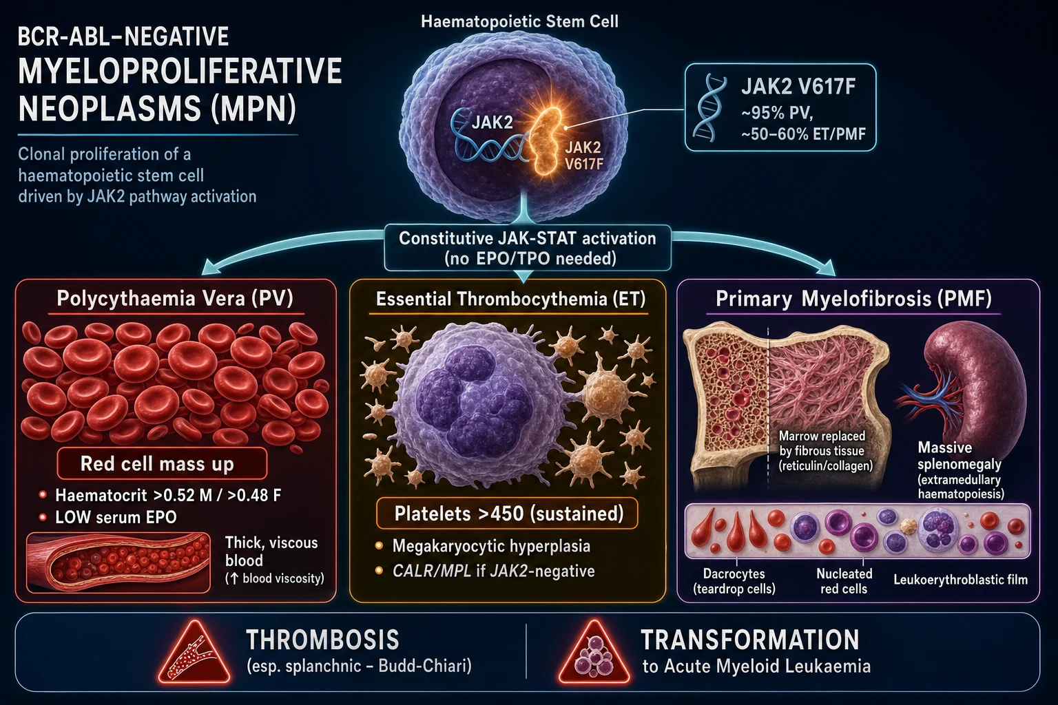
The myelodysplastic syndromes are a heterogeneous group of clonal haematopoietic stem-cell neoplasms in which disordered, ineffective haematopoiesis produces one or more persistent peripheral cytopenias — anaemia most often, but also neutropenia and thrombocytopenia — with a variable propensity to transform into acute myeloid leukaemia. They are the commonest myeloid malignancy of older adults and are increasingly recognised as the overt-malignancy end of a spectrum that begins with age-related clonal haematopoiesis.[1]
The diagnosis rests on a triad that an examiner will always probe: [1]
- Dysplasia of at least 10 percent in one or more myeloid lineages (erythroid, granulocytic, megakaryocytic), or the presence of an MDS-defining cytogenetic abnormality such as isolated del(5q).
- A persistent unexplained cytopenia (a cytopenia that persists for at least four to six months and is not attributable to a secondary cause).
- Exclusion of secondary and reactive causes — vitamin B12 and folate deficiency, copper deficiency, alcohol excess, viral infection (HIV, parvovirus B19), and drug effect.[1]
The central functional lesion is ineffective haematopoiesis: increased apoptosis (programmed cell death) of maturing progenitors within the marrow. The clone proliferates but its offspring die before they reach the blood. The result is the paradox that defines MDS — a hyper- or normocellular marrow producing peripheral cytopenia, rather than the empty marrow of aplastic anaemia. A parallel process of blast accumulation and clonal evolution accounts for progression to AML in about a quarter to a third of patients. The 20 percent marrow or peripheral blast threshold separates MDS from AML (the WHO lowered the FAB threshold of 30 percent to 20 percent in 2001).[1][7]
Classification
MDS classification has evolved from morphology alone (the 1982 FAB system) through the 2008 and 2016 WHO editions to the current WHO 5th edition (2022) and the parallel International Consensus Classification (ICC, 2022), both of which reorganise the disease around genetics. The key thresholds a student must reproduce are the 10 percent dysplasia cut-off, the 5 percent blast line that separates low-blast from high-blast MDS, and the 20 percent blast line that defines AML.[7]
WHO 5th edition (2022) — the current standard
The 2022 WHO divides MDS into two groups: entities defined by a genetic abnormality, and entities defined morphologically when no defining lesion is found.[7]
MDS with low blasts and isolated del(5q) — the 5q- syndrome
- Macrocytic anaemia with or without other cytopenia, isolated del(5q), under 5 percent marrow and under 2 percent blood blasts
- Classically favourable prognosis; high response to lenalidomide
- Female predominance; one of the most distinctive MDS subtypes
MDS with low blasts and SF3B1 mutation (MDS-SF3B1)
- Under 5 percent blasts, SF3B1 mutation, 15 percent or more ringed sideroblasts
- Favourable prognosis; superior response to luspatercept
- Replaces the old RARS/RCMD-RS categories
MDS with low blasts (MDS-LB)
- Under 5 percent marrow and under 2 percent blood blasts; dysplasia in one (single-lineage) or more (multilineage) lineages
- Most common morphological category; generally lower risk
- ESA-responsive if serum EPO is low; outcome depends on cytogenetics and mutations
MDS with increased blasts — MDS-IB
- MDS-IB1: 2 to 9 percent blood blasts or 5 to 9 percent marrow blasts
- MDS-IB2: 5 to 19 percent blood or 10 to 19 percent marrow blasts, or Auer rods
- Higher risk of infection, bleeding and AML transformation; disease-modifying therapy (HMA) and transplant consideration
- Replaces the older MDS-EB1 and MDS-EB2 names
MDS with biallelic TP53 inactivation (MDS-biTP53)
- Two or more TP53 hits (mutations, deletion or copy-neutral loss of heterozygosity), often complex karyotype
- Frequent in therapy-related MDS; aggressive course with early AML transformation
- Very poor prognosis; HMA-based therapy and selected transplant
MDS with fibrosis (MDS-f) and MDS, NOS
- MDS-f: marked reticulin fibrosis with dysplasia; often dry tap, diagnose on trephine
- MDS, NOS: morphologically defined MDS when genetics are unavailable
- Hypoplastic MDS (marrow cellularity under 25 percent for age) sits within this group and may respond to immunosuppression
The older FAB classification (still encountered in older texts and exam stems): refractory anaemia (RA), refractory anaemia with ringed sideroblasts (RARS), refractory anaemia with excess blasts (RAEB), RAEB in transformation (RAEB-t, 20 to 30 percent blasts — now reclassified as AML), refractory cytopenia with multilineage dysplasia (RCMD), and the 5q- syndrome. Chronic myelomonocytic leukaemia (CMML) is no longer MDS — it is an MDS/MPN overlap neoplasm defined by persistent peripheral monocytosis of at least 1.0 x 10^9 per litre with dysplasia.[1]
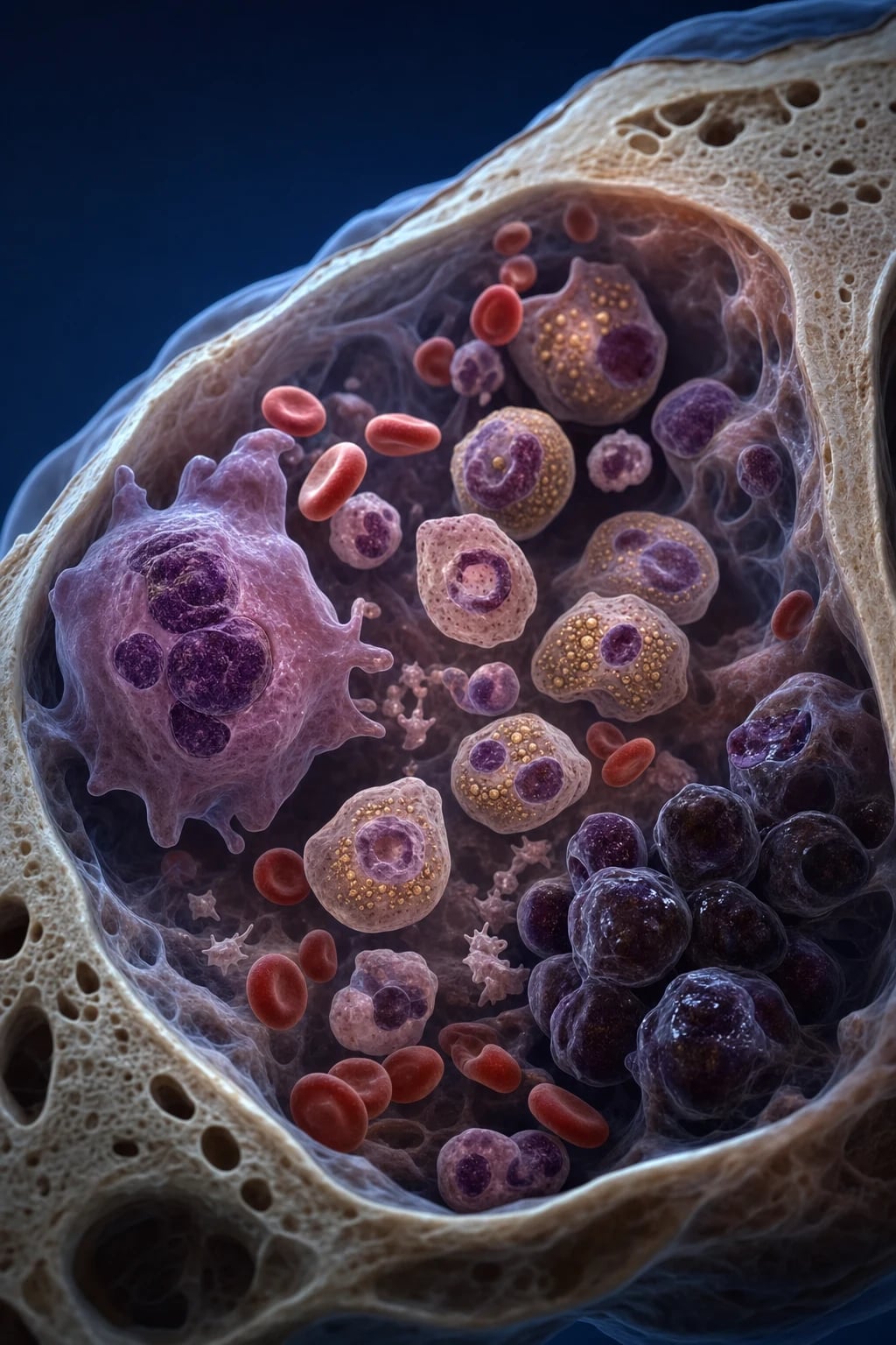
WHO 2022 versus ICC 2022: the two systems agree on most categories but differ at the AML boundary. Both recognise MDS-biTP53 and the SF3B1/del(5q) genetic entities, and both retain the 20 percent blast threshold for AML when no defining AML genetics are present. The ICC, however, sets the AML threshold at 10 percent blasts for cases with a defining AML genetic abnormality (such as t(8;21) or NPM1 mutation), and merges some low-blast categories. In practice, treat the IPSS-R and IPSS-M risk scores — not the classification name — as the primary guide to therapy.[7]
Epidemiology & Risk Factors
MDS has an overall incidence of about 4 per 100,000 per year, rising steeply with age to over 30 per 100,000 in those over 70. The median age at diagnosis is about 76 years (most series report 70 to 75), with a slight male predominance (male-to-female ratio roughly 1.5 to 1). It is among the commonest haematological malignancies of older adults and is substantially more common than AML in the elderly.[1]
Recognised acquired and environmental risk factors: [1]
- Increasing age — the dominant risk factor; clonal haematopoiesis becomes almost universal beyond 70.
- Male sex and tobacco smoking.
- Benzene and petroleum-solvent exposure, and ionising radiation.
- Prior cytotoxic therapy — the single most important modifiable risk factor. [1]
Therapy-related MDS (t-MDS) arises after chemo- or radiotherapy for a prior malignancy and carries two mechanistic subtypes that an examiner may distinguish: an alkylating-agent/radiation-related form (latency 5 to 7 years, chromosomes 5 and 7 abnormalities, complex karyotype, TP53 mutation, poor prognosis) and a topoisomerase-II-inhibitor-related form (latency 2 to 3 years, balanced translocations of 11q23/MLL (KMT2A) or RUNX1 (21q22), often presenting directly as AML). t-MDS is grouped with therapy-related AML and has the worst outcomes in the disease.[1]
Inherited bone-marrow-failure and germline-predisposition syndromes that evolve to MDS include Fanconi anaemia, severe congenital neutropenia, Shwachman-Diamond syndrome, Diamond-Blackfan anaemia, dyskeratosis congenita and other telomere-biology disorders, and germline mutations in GATA2, RUNX1, ETV6, DDX41 and CEBPA. A young patient with MDS should trigger testing for an underlying germline predisposition — this changes donor selection (avoid affected relatives) and the transplant conditioning protocol.[1]
The antecedent spectrum explains most "de novo" MDS in the elderly. Clonal haematopoiesis of indeterminate potential (CHIP) — the presence of a somatic mutation (DNMT3A, TET2, ASXL1, TP53, JAK2) at a variant allele fraction of at least 2 percent in someone with a normal blood count — is detectable in over 10 percent of people over 70. CHIP carries a small annual risk of progression to a haematological malignancy. When CHIP is accompanied by an unexplained cytopenia but insufficient dysplasia to call MDS, the state is clonal cytopenia of undetermined significance (CCUS); without a clone it is idiopathic cytopenia of undetermined significance (ICUS). The same founding mutations (DNMT3A, TET2, ASXL1) recur across the spectrum.[1]
Pathophysiology
An acquired somatic mutation in a multipotent haematopoietic stem or progenitor cell confers a clonal proliferative or survival advantage. The disease then evolves through multiple genetic hits: [1]
- Early founding mutations in DNA-methylation and chromatin genes — TET2, DNMT3A, ASXL1 — are shared with CHIP and establish the clone.
- Spliceosome mutations (SF3B1, SRSF2, U2AF1, ZRSR2) commonly follow; SF3B1 is the hallmark of ringed-sideroblast disease.
- Late, progression-associated mutations — TP53 (multi-hit), RUNX1, EZH2, NRAS/KRAS, FLT3, IDH1/2 — drive blast accumulation and evolution to AML.[1]
The dominant functional consequence is ineffective haematopoiesis: the clone proliferates but its maturing progenitors undergo excessive intramedullary apoptosis (up to three times normal), so the marrow is frequently hyper- or normocellular while the blood shows cytopenia — the paradox that defines MDS. A contributing mechanism is a pro-inflammatory dysfunctional bone-marrow microenvironment (abnormal cytokine signalling, altered mesenchymal stromal cells, T-cell-mediated immune attack on normal residual stem cells) that selectively favours the clone. A parallel process of blast accumulation and clonal evolution accounts for progression to AML in about a quarter to a third of patients.[1]
Recurrent cytogenetic lesions and their prognostic signal: [1]
- Favourable / good: isolated del(5q), del(20q), -Y, normal karyotype (about half of all MDS).
- Intermediate: del(7q), trisomy 8 (+8), i(17q), and single or double abnormalities not classified elsewhere.
- Adverse / poor: monosomy 7 (-7), del(5q) with other abnormalities, inv(3)/t(3;3), complex karyotype (at least 3 unrelated abnormalities) and monosomal karyotype; multi-hit TP53 commonly coexists with a complex karyotype.[2]
Recurrent gene mutations and their prognostic signal: SF3B1 (ringed sideroblasts — favourable); TP53 multi-hit (complex karyotype, therapy-related — very adverse); ASXL1, RUNX1, EZH2, NRAS/KRAS (adverse); TET2, DNMT3A (common, largely intermediate). These mutations — captured by the molecular IPSS-M — refine prognosis well beyond the IPSS-R.[1][5]
Clinical Presentation
The commonest presentation is an incidental finding of cytopenia, macrocytosis or dysplastic morphology on a routine full blood count in an older adult — frequently asymptomatic. When symptoms occur they reflect the affected lineage(s):[1]
- Anaemia (the most common cytopenia, present in over 80 percent at diagnosis) — fatigue, pallor, exertional dyspnoea, and worsening of angina or cardiac failure in those with vascular disease.
- Neutropenia — recurrent bacterial infection (chest, skin, perianal), fever, and occasionally neutropenic sepsis as the first clue.
- Thrombocytopenia — easy bruising, petechiae, purpura, mucosal bleeding, menorrhagia, and, rarely, major haemorrhage. [1]
Bicytopenia or pancytopenia is common; an isolated cytopenia (usually anaemia) is the other pattern. Splenomegaly and lymphadenopathy are uncommon in MDS; their presence should prompt consideration of an MDS/MPN overlap syndrome (CMML), primary myelofibrosis, CML, or a separate lymphoid process.[1]
Atypical and paraneoplastic presentations that examiners test deliberately: acute febrile neutrophilic dermatosis (Sweet syndrome) and cutaneous vasculitis may accompany or even precede MDS, as may autoimmune phenomena (polyarthralgia, uveitis, hypothyroidism). In the very elderly, MDS may present as isolated fatigue, falls, delirium, or exacerbation of cardiac failure from unrecognised anaemia, the cytopenia found only on testing. Acute presentations include neutropenic sepsis, major bleeding from severe thrombocytopenia, and symptomatic severe anaemia with high-output cardiac failure.[1]
Differential Diagnosis
Dysplasia alone is not MDS — reactive and secondary causes must be excluded before diagnosing a clonal disorder. This single principle generates more exam questions than any other in the topic.[1]
Nutritional deficiencies
- Vitamin B12 and folate deficiency — megaloblastic change with hypersegmented neutrophils and macro-ovalocytes; check serum B12 and RBC folate
- Copper deficiency — anaemia plus neutropenia with vacuolated erythroid and myeloid precursors and ringed sideroblasts; a sensory (dorsal-column) neuropathy; classic after gastric or bariatric surgery or zinc excess; a notorious MDS mimic
- Corrects with replacement — the MDS diagnosis is only safe once these are normal
Toxic, drug and infectious mimics
- Alcohol excess — macrocytosis, vacuolated red-cell precursors, thrombocytopenia
- Drug effect — chemotherapy, antibiotics, immunosuppressants, myelotoxic agents
- Viral infection — HIV (cytopenias with mild dysplasia), parvovirus B19 (pure red-cell aplasia), EBV, CMV, hepatitis
- Heavy metals — lead (basophilic stippling), arsenic
Other clonal and marrow-failure disorders
- Aplastic anaemia — hypocellular marrow without dysplasia or a clone
- Large granular lymphocytic leukaemia — pure red-cell aplasia, chronic neutropenia, rheumatoid arthritis
- Paroxysmal nocturnal haemoglobinuria — haemolysis, cytopenia, thrombosis; flow cytometry (FLAER) defines it
- Primary myelofibrosis — teardrop cells, splenomegaly, marked fibrosis
- Overt AML — blasts at least 20 percent
Borderline pre-MDS states
- CHIP — a clone (DNMT3A/TET2/ASXL1/TP53/JAK2 VAF at least 2 percent) but a normal count
- CCUS — a clone plus an unexplained cytopenia but insufficient dysplasia or genetics to call MDS
- ICUS — an unexplained cytopenia without a clone
- Observation and re-biopsy resolve most; do not over-treat
The single most important teaching point: always send B12, folate and a serum copper level, take an alcohol and drug history, and exclude HIV before labelling dysplasia as MDS.[1]
Clinical & Bedside Assessment
The focused assessment documents the consequences of the cytopenia, screens for alternative diagnoses, and gathers the information that will drive intensity of therapy (especially transplant eligibility).[1]
- General — look for pallor, bruising, petechiae and purpura, and signs of infection (fever, focus). Document performance status (ECOG or Karnofsky) and comorbidity burden (HCT-CI) — both gate transplant decisions.
- Abdomen — examine for hepatosplenomegaly (suggests overlap/CML/CMML/myelofibrosis or infiltration) and lymphadenopathy (suggests lymphoma).
- Cardiovascular — assess the consequences of anaemia (tachycardia, flow murmur, signs of cardiac failure, exertional tolerance) and document any transfusion-related iron overload (no early signs, so measure ferritin).
- Frailty, cognition and social support — central to transplant decisions and therapy intensity in the predominantly elderly population.
- History — a focused drug, occupational (benzene, solvents, radiation) and prior-cancer-treatment exposure history, and a family history of haematological malignancy (germline predisposition: GATA2, RUNX1, ETV6, DDX41). [1]
There are no pathognomonic bedside signs of MDS — the diagnosis is made in the laboratory. [1]
Investigations
First-line bloods: full blood count and film (macrocytosis — MCV often 100 to 120 fL, hypogranular or hyposegmented pseudo-Pelger-Huet neutrophils, giant hypogranular platelets, inappropriately low reticulocytes, peripheral blast count), LDH (raised in higher-risk disease), and a screen to exclude secondary causes (vitamin B12, folate, copper, ferritin, HIV and viral serology, and an alcohol history). The MCV is typically raised but MDS with isolated del(5q) can be normocytic; a low reticulocyte count in the face of anaemia points to a production defect.[1]
Bone-marrow aspirate and trephine biopsy is the diagnostic cornerstone and every patient needs one: [1]
- Cellularity — hyper-, normo- or hypocellular (hypoplastic MDS mimics aplastic anaemia).
- Dysplasia of at least 10 percent in one or more lineages — erythroid (nuclear budding, multinucleation, ringed sideroblasts), granulocytic (hypogranular, hyposegmented Pelgeroid forms), megakaryocytic (micromegakaryocytes — small mononuclear or binucleate forms).
- Blast percentage — counted precisely (the 5, 10 and 20 percent lines all matter).
- Prussian-blue (Perls) iron stain — for ringed sideroblasts (erythroid precursors with at least 5 siderotic granules encircling at least one-third of the nucleus); 15 percent or more with an SF3B1 mutation defines MDS-SF3B1.
- Reticulin fibrosis — grades MDS-f.[1]
Cytogenetics: conventional G-banding karyotype plus FISH for the common lesions (-7/del(7q), del(5q), del(20q), trisomy 8, complex karyotype). A normal karyotype (about half) and isolated del(5q) are favourable; -7 and complex karyotype are adverse. Molecular next-generation sequencing for TP53, SF3B1, ASXL1, RUNX1, TET2, EZH2, NRAS, FLT3, IDH1/2 refines prognosis and increasingly guides therapy — for example, lenalidomide for del(5q), venetoclax consideration, and TP53-directed trials.[1]
Erythropoietin level — a serum EPO under about 500 mU/mL with a low transfusion burden (under 2 units per month) predicts a good response to erythropoiesis-stimulating agents. [1]
Risk stratification — the Revised IPSS (IPSS-R) and the molecular IPSS-M
Risk stratification drives every management decision. The IPSS-R (Greenberg, Blood 2012) combines four clinical variables into five risk categories; the IPSS-M (Bernard, NEJM Evidence 2022) layers gene-mutation data on top to reclassify about 20 percent of patients — usually upwards.[2][5]
The IPSS-R combines marrow blast percentage, cytogenetic risk group, haemoglobin and platelet count: [1]
IPSS-R cytogenetic risk groups
The blast percentage, cytogenetic group, haemoglobin (cut-points at 9 and 10.5 g/dL) and platelet count (cut-points at 50 and 100 x 10^9 per litre) combine into a score that distributes patients into five categories:[2]
IPSS-R risk categories and median overall survival
Intermediate
Higher blasts, adverse cytogenetics, or two cytopenias — median OS ~3.0 years
The molecular IPSS-M (IPSS-M, Bernard 2022) incorporates the IPSS-R clinical variables plus 31 gene mutations — weighted most heavily for multi-hit TP53 (adverse) and SF3B1 (favourable), with FLT3 also adverse. It produces six categories with the following approximate median overall survivals:[5]
IPSS-M risk categories and median survival
In practice, the IPSS-M reclassifies about one patient in five, most often from lower to higher risk — which is why molecular testing is now standard in fit patients. Iron-overload monitoring: serial serum ferritin, and consider chelation when ferritin exceeds about 1000 micrograms per litre (or after 20 to 25 red-cell units) in chronically transfused patients.[1]
Management — Resuscitation
Treat the acute cytopenic consequences first, then start risk-adapted disease therapy. The immediate threats are severe symptomatic anaemia, major bleeding, and neutropenic sepsis.[1]
- Severe or symptomatic anaemia — red-cell transfusion. Use leucodepleted products, irradiated products if the patient is a transplant candidate or immunosuppressed, and CMV-negative products if CMV-negative and a transplant candidate. Transfuse one unit at a time in older patients and reassess, watching for volume overload; aim for the lowest haemoglobin that relieves symptoms (often 8 to 9 g/dL, higher in cardiac disease).
- Thrombocytopenia — platelet transfusion for active bleeding, before procedures, or as prophylaxis when platelets are under 10 x 10^9 per litre (or under 20 with fever or bleeding risk). The thrombopoietin-receptor agonists (eltrombopag, romiplostim) carry a caution for blast progression and are not routine.
- Febrile neutropenia — a medical emergency: blood cultures and empirical broad-spectrum antipseudomonal beta-lactam (e.g. piperacillin-tazobactam 4.5 g IV every 8 hours or ceftazidime 2 g IV every 8 hours) within one hour; add cover for the local focus and consider antifungal cover if neutropenia is prolonged.
- Iron overload from chronic transfusion — initiate chelation when ferritin exceeds about 1000 micrograms per litre to protect hepatic, cardiac and endocrine function.[1]
Management — Definitive & Stepwise
Therapy is risk-adapted using the IPSS-R (and increasingly IPSS-M) and calibrated to age, fitness and patient goals. Lower-risk disease (IPSS-R very low, low, and many intermediate) is managed with supportive care and growth factors; higher-risk disease (IPSS-R high and very high, and IPSS-M high/very-high) warrants disease-modifying therapy and consideration of transplant.[1][3]
Lower-risk MDS — supportive and growth-factor therapy
The aim in lower-risk disease is to relieve cytopenia, improve quality of life and delay transformation, not to eradicate the clone.[1]
Lower-risk MDS — the stepwise ladder
Supportive care for all — red-cell and platelet transfusion as needed; iron chelation (deferasirox) once ferritin exceeds ~1000 microg/L; antimicrobial prophylaxis in higher-risk neutropenia.
Erythropoiesis-stimulating agents if serum EPO under ~500 mU/mL and low transfusion burden — epoetin alfa 40,000 U subcutaneously weekly (or darbepoetin 150 to 300 mcg weekly); ~40 to 60 percent erythroid response.
Luspatercept 1.0 mg/kg subcutaneously every 3 weeks (titrate to 1.75 mg/kg) for ringed-sideroblast MDS that is ESA-resistant (MEDALIST).
Lenalidomide 10 mg orally daily, days 1 to 21 of a 28-day cycle, for del(5q) MDS — high erythroid and cytogenetic response; also off-label in non-del(5q) ESA failure.
Immunosuppression (antithymocyte globulin plus ciclosporin) for the younger, hypocellular, HLA-DR15-positive lower-risk subset resembling aplastic anaemia.
Re-assess with marrow and cytogenetics at 4 to 6 months; escalate to HMA if blasts rise or transfusion dependence develops.
Lower-risk MDS — erythropoiesis-stimulating agents and targeted therapy
Higher-risk MDS — disease-modifying therapy and transplant
The aim in higher-risk disease is to modify the disease course and pursue the only curative option — allogeneic transplant in fit patients. The standard disease-modifying agent is a hypomethylating agent (HMA) — azacitidine first-line — increasingly combined with the BCL-2 inhibitor venetoclax.[3][8]
Higher-risk MDS — hypomethylating agents and venetoclax
Higher-risk MDS — the curative pathway
Confirm higher-risk disease (IPSS-R high/very high, or IPSS-M high/very-high, or 10 to 19 percent blasts, or adverse cytogenetics).
Assess transplant candidacy — physiological age (generally under 70 to 75, but selected older fit patients eligible), ECOG 0 to 1, low comorbidity index (HCT-CI), adequate organ function.
If transplant-eligible: start HMA (azacitidine 75 mg/m2 x7d every 28 days, plus/minus venetoclax) for cytoreduction while a donor search proceeds (matched sibling first, then matched unrelated, then haploidentical).
Proceed to allogeneic haematopoietic stem-cell transplant with reduced-intensity conditioning (fludarabine plus busulfan or fludarabine plus melphalan) in older adults.
If transplant-ineligible: continue HMA (azacitidine or decitabine) every 28 days for as long as it works, plus best supportive care and clinical-trial enrolment.
For multi-hit TP53 and therapy-related MDS: HMA backbone plus clinical trial; transplant offers limited but real benefit in selected patients.
Allogeneic haematopoietic stem-cell transplant is the only potentially curative therapy. It is offered to fit higher-risk patients, generally with reduced-intensity conditioning in older adults, exploiting a graft-versus-leukaemia effect (the donor immune system recognises and destroys residual malignant cells). Selection balances the high transplant-related mortality (10 to 20 percent at one year) and graft-versus-host disease against the poor natural history of untreated higher-risk disease. TP53-mutated and therapy-related MDS carry the worst post-transplant outcomes — relapse is common — but transplant still offers the best chance of long-term survival in selected patients, and is best paired with clinical-trial enrolment.[1][3]
Specific Subtypes & Scenarios
Isolated del(5q) — the 5q- syndrome
- Macrocytic anaemia with or without thrombocytopenia (often raised platelets), isolated del(5q), under 5 percent blasts
- Female predominance; one of the most favourable MDS subtypes
- High erythroid and cytogenetic response to lenalidomide 10 mg daily (List, NEJM 2006)
MDS with ringed sideroblasts and SF3B1 mutation
- Under 5 percent blasts, SF3B1 spliceosome mutation, 15 percent or more ringed sideroblasts
- Favourable outcome with low transformation risk
- Superior response to luspatercept in ESA-resistant disease (MEDALIST)
MDS with biallelic TP53 inactivation (MDS-biTP53)
- Two or more TP53 hits (mutations and/or 17p deletion), usually complex karyotype
- Frequent in therapy-related MDS; aggressive course
- Very poor prognosis with early AML transformation; HMA plus clinical trial; transplant case-by-case
Therapy-related MDS (t-MDS)
- Arises 2 to 7 years after chemo/radiotherapy; alkylator type has -5/-7, complex karyotype
- Grouped with therapy-related AML; aggressive biology
- Poor response to all therapy; HMA backbone and selected transplant
Hypoplastic MDS
- Hypocellular marrow (under 25 percent for age) mimicking aplastic anaemia
- A subset (younger, HLA-DR15-positive) responds to immunosuppression (antithymocyte globulin plus ciclosporin)
- Distinguished from aplastic anaemia by dysplasia and a cytogenetic/molecular clone
Chronic myelomonocytic leukaemia (CMML)
- The prototypic MDS/MPN overlap — persistent peripheral monocytosis at least 1.0 x 10^9/L with dysplasia
- Splenomegaly common (unlike typical MDS)
- Manage with hydroxycarbamide 500 to 1500 mg daily for the proliferative phase, HMA, and transplant in selected patients
Complications & Pitfalls
Disease complications — progressive cytopenia (infection, bleeding, transfusion dependence), transfusional iron overload with end-organ damage (cirrhosis, cardiac failure, diabetes, hypogonadism, hypothyroidism), and transformation to AML in about 25 to 30 percent of patients overall (higher in high-blast and TP53-mutated disease). Infection is the leading cause of death in MDS.[1]
Treatment complications that examiners test by drug: [1]
Drug-specific toxicities in MDS therapy
Classic diagnostic pitfalls: mistaking B12, folate or copper deficiency (or alcohol) for MDS; overcalling reactive dysplasia (post-infection, growth-factor effect); failing to send cytogenetics and molecular testing on the marrow; not excluding HIV and other viruses; and diagnosing MDS from dysplasia alone without a persistent cytopenia or a clone. Treatment pitfalls: over-treating lower-risk disease like leukaemia; failing to chelate iron in chronically transfused patients; delaying transplant referral for fit higher-risk patients; and using thrombopoietin-receptor agonists without monitoring blast progression (a real concern for AML evolution).[1]
Prognosis & Disposition
Prognosis is highly heterogeneous and is driven by the risk score. The IPSS-R median survivals range from about 8.8 years (very low) down to about 0.8 years (very high); overall median survival is roughly 2 to 5 years. The IPSS-M refines this further, with median survivals from about 8.8 years (very low) down to about 0.9 years (very high).[2][5]
Adverse prognostic factors: higher marrow blast percentage, adverse or complex cytogenetics (especially -7, inv(3) and multi-hit TP53), low haemoglobin, low platelet count, high LDH, older age and poor performance status. Favourable factors include isolated del(5q), an SF3B1 mutation, a normal karyotype, and a low blast count with limited transfusion need.[2]
Allogeneic stem-cell transplant is the only modality with curative potential, selected by age, comorbidity (HCT-CI), performance status and donor availability; the graft-versus-leukaemia effect is central to its efficacy. Follow-up: serial full blood counts and transfusion requirement, periodic marrow with blast and cytogenetic reassessment, ferritin and iron monitoring, and surveillance for infection and treatment toxicity.[3]
Special Populations
- Younger patients — prioritise donor search, germline (inherited-predisposition) testing (GATA2, RUNX1, ETV6, DDX41), and early allogeneic transplant referral, since age and fitness confer the greatest transplant benefit; a younger patient with apparent "de novo" MDS always warrants a germline work-up before selecting a donor.
- Elderly and frail patients — emphasise supportive care, transfusion and iron chelation, with HMA and reduced-intensity transplant reserved for fit higher-risk individuals selected by physiological rather than chronological age; a fit 72-year-old may be transplanted, a frail 60-year-old may not.
- Therapy-related MDS — poor-risk biology; discuss goals of care, HMA-based therapy and clinical trials; transplant selected case-by-case.
- Inherited bone-marrow-failure syndromes (e.g. Fanconi anaemia) — require a modified, fludarabine-based reduced-intensity transplant protocol avoiding radiation, and donor/relative genetic screening to avoid using an affected relative as donor.
- Resource-limited settings — manage with transfusion, affordable HMA where available, and supportive antimicrobial prophylaxis; transplant access and novel-agent cost are major barriers, so access — not biology — often drives treatment choice.
- Pregnancy (very rare) — supportive care with transfusion; defer disease-modifying therapy where possible; lenalidomide is absolutely contraindicated (teratogenic).[1]
Evidence, Guidelines & Regional Differences
The WHO 5th edition (2022) and the International Consensus Classification (ICC, 2022) both reorganise MDS around genetics, recognise MDS-biTP53 and the SF3B1/del(5q) entities, and retain the 20 percent blast threshold for AML (the ICC defining AML by genetics at the 10 percent blast count for defining recurrent genetic abnormalities).[7]
The Revised IPSS (2012, Greenberg) remains the standard bedside risk tool, while the molecular IPSS-M (2022, Bernard) incorporates mutations (multi-hit TP53, SF3B1, FLT3 and others) to refine risk and reclassify about one patient in five.[2][5]
Landmark trials and what they changed in MDS
Regional practice differs sharply by access. In high-income settings (NCCN — US; ESMO/ELN — Europe) there is routine availability of HMA, luspatercept, lenalidomide, iron chelation and allogeneic transplant, with molecular NGS guiding therapy. In India and other low/middle-income countries (ICMR context), care is largely transfusion- and HMA-based supportive therapy, with limited transplant and novel-agent access — cost, not biology, is the main determinant. Controversies include the optimal use of HMA plus venetoclax in higher-risk MDS (versus AML alone), the safety of thrombopoietin-receptor agonists (blast-progression concern), transplant timing and upper-age limits, luspatercept in non-sideroblastic disease, and MRD-guided therapy after transplant.[1]
Exam Pearls
[1]The MDS diagnostic triad
DYC
at least 10 percent in one or more myeloid lineages (or an MDS-defining cytogenetic lesion such as isolated del(5q))
persistent unexplained cytopenia (anaemia, neutropenia and/or thrombocytopenia) for at least four to six months
rule out B12, folate, copper deficiency, alcohol, HIV and drugs before calling it MDS
What the IPSS-M adds
MDS-RISK
IPSS-M layers 31 gene mutations onto the IPSS-R clinical variables
founding hits TET2, DNMT3A, ASXL1 (shared with CHIP)
SF3B1 — ringed sideroblasts, favourable
about one patient in five moves risk category, usually upward
multi-hit TP53 — the worst mutation, complex karyotype
categories — Very Low, Low, Moderate-Low, Moderate-High, High, Very High
cytogenetics still weighted; -7 and complex are adverse
- 20 percent blasts is the MDS/AML boundary (WHO; FAB was 30 percent); the ICC uses 10 percent for AML with defining genetics.
- Ineffective haematopoiesis = hypercellular marrow with peripheral cytopenia (the defining paradox).
- MDS-IB1 = 5 to 9 percent marrow blasts; MDS-IB2 = 10 to 19 percent marrow blasts (WHO 2022 renamed "excess blasts" to "increased blasts").
- 5q- syndrome — macrocytic anaemia, isolated del(5q), low blasts, favourable, lenalidomide-responsive.
- SF3B1 mutation + ringed sideroblasts — favourable, luspatercept-responsive.
- Multi-hit TP53 — worst prognosis, complex karyotype, therapy-related setting.
- Copper deficiency is the classic mimic (vacuolated precursors, neuropathy, after gastric surgery or zinc).
- Infection is the leading cause of death in MDS.
- Iron chelation (deferasirox 20 to 40 mg/kg daily) when ferritin exceeds 1000 microg/L. [1]
Exam application bank (NEET-PG / INICET)
One-line answer
Myelodysplastic syndromes (MDS) are a heterogeneous group of clonal haematopoietic stem-cell neoplasms defined by dysplastic, ineffective haematopoiesis, peripheral cytopenia(s) and a variable risk of transformation to acute myeloid leukaemia. Diagnosis requires dysplasia of at least 10 percent in one or more myeloid lineages (or an MDS-defining cytogenetic lesion) plus a persistent unexplained cytopenia after excluding secondary causes (B12, folate, copper deficiency, alcohol, infection). The 20 percent blast threshold separates MDS from AML. Risk is stratified by the IPSS-R and, increasingly, the molecular IPSS-M (blast percentage, cytogenetics, haemoglobin, platelets, plus TP53, SF3B1, FLT3 mutations). Lower-risk disease is managed with supportive care, erythropoiesis-stimulating agents, lenalidomide for del(5q) and luspatercept for ringed sideroblasts; higher-risk disease gets a hypo
Worked stems (answer without another resource)
Stem 1 — Classic presentation. Map symptoms to mechanism; name the first investigation and first treatment step with dose/route if drug therapy is standard. [1]
Stem 2 — Unstable / complicated. List red flags that force immediate resuscitation, theatre, ICU, antidote, or reperfusion — and what you do in the first 15 minutes. [1]
Stem 3 — Atypical group. Elderly, pregnancy, child, or immunocompromised: how presentation and thresholds change. [1]
Stem 4 — Differential trap. Name the three closest mimics and one discriminator for each. [1]
Stem 5 — Disposition. Who goes home with safety-netting, who is admitted, who needs HDU/ICU/theatre, and what follow-up is mandatory. [1]
Rapid viva checklist
- Definition + classification
- Pathophysiology chain
- Bedside signs / criteria
- Score with exact components (if any)
- Emergency bundle
- Definitive therapy with doses
- Complications of disease and of treatment
- Special populations
- Guideline/trial name if classic
- Three exam traps
Coverage self-check
If you cannot answer any stem above from this page alone, re-read the matching section — the page is intended to be self-sufficient for final-prof and NEET-PG/INICET questions on Myelodysplastic Syndromes.
References
- [1]Cazzola M Myelodysplastic Syndromes N Engl J Med, 2020.PMID 32997910
- [2]Greenberg PL, Tuechler H, Schanz J, et al. Revised international prognostic scoring system for myelodysplastic syndromes Blood, 2012.PMID 22740453
- [3]Fenaux P, Mufti GJ, Hellstrom-Lindberg E, et al. Efficacy of azacitidine compared with that of conventional care regimens in the treatment of higher-risk myelodysplastic syndromes: a randomised, open-label, phase III study Lancet Oncol, 2009.PMID 19230772
- [4]Fenaux P, Platzbecker U, Mufti GJ, et al. Luspatercept in Patients with Lower-Risk Myelodysplastic Syndromes N Engl J Med, 2020.PMID 31914241
- [5]Bernard E, Tuechler H, Greenberg PL, et al. Molecular International Prognostic Scoring System for Myelodysplastic Syndromes NEJM Evid, 2022.PMID 38319256
- [6]List A, Dewald G, Bennett J, et al. Lenalidomide in the myelodysplastic syndrome with chromosome 5q deletion N Engl J Med, 2006.PMID 17021321
- [7]Khoury JD, Solary E, Abla O, et al. The 5th edition of the World Health Organization Classification of Haematolymphoid Tumours: Myeloid and Histiocytic/Dendritic Neoplasms Leukemia, 2022.PMID 35732831
- [8]DiNardo CD, Jonas BA, Pullarkat V, et al. Azacitidine and Venetoclax in Previously Untreated Acute Myeloid Leukemia N Engl J Med, 2020.PMID 32786187