Haematology · General Medicine
Myeloproliferative Disorders (PV, ET, MF & CML)
Also known as Myeloproliferative neoplasms · MPN · Polycythaemia vera · Essential thrombocythemia · Primary myelofibrosis · Chronic myeloid leukaemia · PV ET MF CML
Myeloproliferative neoplasms (MPN) are clonal disorders of haematopoietic stem cells causing overproduction of mature myeloid lineages. Polycythaemia vera (PV) = raised haematocrit (over 0.52 M / over 0.48 F) plus JAK2 V617F (~95%) and low serum erythropoietin; presents with hyperviscosity (headache, visual disturbance, thrombosis, aquagenic pruritus), splenomegaly, gout; treat with venesection to haematocrit under 0.45 plus low-dose aspirin plus cytoreduction (hydroxycarbamide 15 to 35 mg/kg/day). Essential thrombocythemia (ET) = sustained platelet count over 450 with megakaryocytic hyperplasia (JAK2 50 to 60 percent, CALR 25 percent, MPL 5 percent); risk of thrombosis and bleeding; treat with aspirin 75 mg and cytoreduction if high-risk. Primary myelofibrosis (PMF) = marrow fibrosis, massive splenomegaly, tear-drop cells, leukoerythroblastic film; treat with ruxolitinib (JAK1/2 inhibitor); allogeneic stem cell transplant is the only cure. Chronic myeloid leukaemia (CML) is driven by the BCR-ABL1 fusion (Philadelphia chromosome, t(9;22)) and treated with tyrosine-kinase inhibitors (imatinib 400 mg, dasatinib 100 mg, nilotinib 300 mg twice daily) with RT-PCR BCR-ABL1 transcript monitoring; allogeneic stem cell transplant is reserved for blast phase or T315I mutation. Splanchnic vein thrombosis (Budd-Chiari) warrants JAK2 testing even with normal counts. Thrombosis is the leading cause of death in PV and ET.
On this page & tools
Your progress
Saved locally on this device.
Exam tags
Red flags
Overview & Definition
The myeloproliferative neoplasms (MPN) are a family of clonal disorders of the haematopoietic stem cell in which the malignant clone retains the capacity to differentiate into mature, functional blood cells, but produces too many of them. William Dameshek recognised this shared biology in 1951, grouping polycythaemia vera (PV), essential thrombocythemia (ET), primary myelofibrosis (PMF) and chronic myeloid leukaemia (CML) as "myeloproliferative disorders". Modern WHO classification splits them by molecular driver: the BCR-ABL1-positive CML stands apart on the Philadelphia chromosome, while PV, ET and PMF share activation of the JAK-STAT pathway through the JAK2, CALR and MPL driver mutations.[1]
Unlike acute leukaemia — where the marrow is flooded by immature, non-functional blasts and the patient suffers marrow failure — the MPN produce mature, granular, working cells. The damage therefore comes from excess: hyperviscosity, thrombosis, bleeding, organomegaly, and — over years — marrow fibrosis or transformation to acute myeloid leukaemia (AML). Recognising each entity hinges on which lineage is overproduced (red cells in PV, platelets in ET, none effectively in PMF because marrow is scarred, granulocytes in CML), and on characteristic blood-film, molecular and cytogenetic findings. The clinical art is to separate a true clonal MPN from the far commoner reactive causes of a high haematocrit, platelet or white-cell count, and to risk-stratify each patient to prevent the dominant complication — thrombosis.[1][2]
Classification
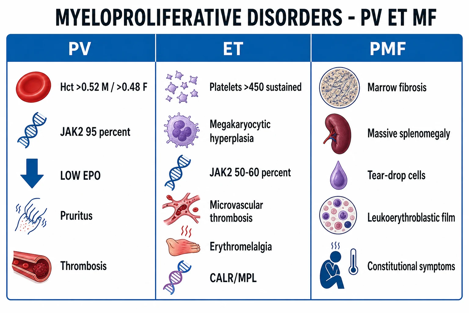
The four classic MPN are distinguished by the dominant overproduced lineage, the marrow morphology, and above all the molecular driver. JAK2 V617F is the unifying lesion of the BCR-ABL1-negative trio; BCR-ABL1 defines CML and must be excluded before labelling a case ET, PV or PMF.[1]
Polycythaemia vera (PV)
- Red-cell lineage: raised haematocrit (over 0.52 M / over 0.48 F) or raised red-cell mass
- JAK2 V617F in ~95 percent (or JAK2 exon 12 in ~3 to 5 percent)
- Serum EPO LOW (autonomous clone suppresses it)
- Plethoric facies, aquagenic pruritus, thrombosis, splenomegaly, gout
- Hypercellular marrow with panmyelosis and pleomorphic megakaryocytes
Essential thrombocythemia (ET)
- Platelet lineage: platelets over 450 x10^9/L sustained
- Megakaryocytic hyperplasia with large mature megakaryocytes
- JAK2 50 to 60 percent; CALR ~25 percent; MPL ~5 percent (triple-negative ~15 percent)
- Microvascular thrombosis (erythromelalgia) and bleeding; acquired vWD over 1500
- Normal haematocrit and essentially normal marrow cellularity
Primary myelofibrosis (PMF)
- Fibrosis replaces marrow — ineffective extramedullary haematopoiesis
- Reticulin/collagen fibrosis on trephine; atypical megakaryocyte clusters
- JAK2 ~60 percent, CALR ~25 percent, MPL ~5 to 10 percent
- Massive splenomegaly, tear-drop cells, leukoerythroblastic film, high LDH
- Worst prognosis of the three; highest leukaemic transformation (~10 to 20 percent)
Chronic myeloid leukaemia (CML)
- Granulocyte lineage: high WBC with left shift, **basophilia**, thrombocytosis
- BCR-ABL1 fusion from t(9;22)(q34;q11.2) — Philadelphia chromosome — is **defining**
- Low leucocyte alkaline phosphatase (historical); driver = p210 BCR-ABL1 tyrosine kinase
- Massive splenomegaly, fatigue, sweats, gout; three phases (chronic, accelerated, blast)
- Treated with tyrosine-kinase inhibitors (imatinib/dasatinib/nilotinib) — near-normal survival
WHO 2016 and the 2022 ICC/WHO revisions integrate morphology, genetics and clinical features. A key subtype is pre-fibrotic PMF (pre-PMF), which mimics ET on counts but carries a higher leukaemic-transformation risk and is distinguished by atypical megakaryocyte clusters, mild reticulin fibrosis (grade 0 to 1), a raised LDH, anaemia and constitutional symptoms. Chronic myeloid leukaemia is defined by BCR-ABL1 and is excluded from the JAK2-driven trio by a negative test for the fusion. The clinical lesson: a sustained high count is never labelled an MPN until reactive causes are excluded and the relevant driver mutation is confirmed.[1]
Epidemiology & Risk Factors
The MPN are diseases of middle and older age (median presentation 50 to 70 years), though CML has a younger peak and ET occurs in young women — relevant because pregnancy planning shapes management. Incidence is roughly 0.7 to 2.6 per 100,000 per year for PV, 0.6 to 2.5 for ET, 0.2 to 1.0 for PMF (the least common but most aggressive), and 1 to 2 for CML. ET has a slight female predominance; PV and CML a slight male predominance. Secondary (reactive) causes of a raised haematocrit, platelet or white-cell count are far commoner than any MPN and must always be excluded first.[1][2][3]
Recognised risk factors include increasing age, male sex (for PV and CML), rare familial predisposition to clonal haematopoiesis, and ionising-radiation or benzene/solvent exposure (classically linked to CML and AML). Almost all PV is JAK2 V617F positive (or exon 12); CALR and MPL account for most JAK2-negative ET/PMF. In CML the BCR-ABL1 fusion is acquired somatic (not inherited) and is present in virtually 100 percent of cases. [1]
Pathophysiology
The molecular spine of the BCR-ABL1-negative MPN is the JAK2 V617F mutation — a valine-to-phenylalanine substitution at position 617 in the JAK2 pseudokinase (JH2) domain that releases the kinase from autoinhibition, rendering it constitutively active. JAK2 is the intracellular signalling partner of the erythropoietin (EPO) receptor, thrombopoietin receptor (MPL) and G-CSF receptor, so the clone proliferates without the normal cytokine stimulus: PV red-cell precursors form endogenous erythroid colonies (EEC) in culture without added EPO, and serum EPO is suppressed by negative feedback from the autonomous red-cell mass. This is the basis of the low serum EPO test, the single best discriminator of PV from secondary polycythaemia.[1]
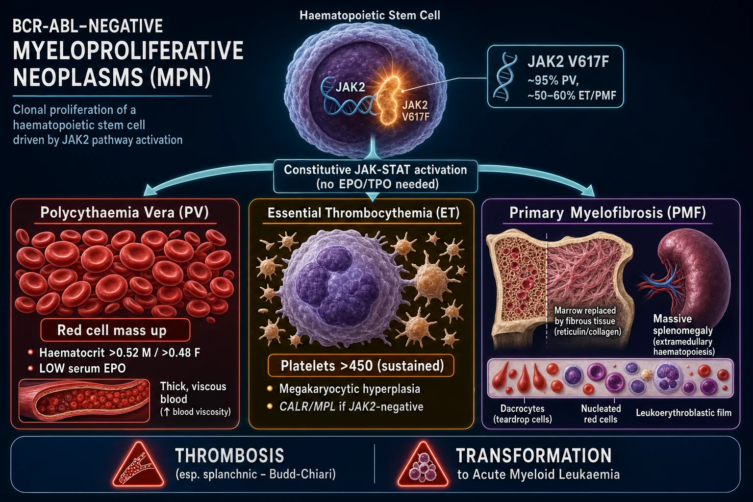
The downstream mechanisms are lineage-specific: [1]
- PV — hyperviscosity: the raised red-cell mass increases blood viscosity, producing sluggish flow, endothelial activation and thrombosis (stroke, MI, Budd-Chiari). High cell turnover causes hyperuricaemia and gout, and histamine release from basophils contributes to aquagenic pruritus (itch triggered by a warm bath or shower).
- ET — platelet dysfunction: the clone makes large, dysregulated platelets that aggregate in arterioles, producing microvascular thrombosis (erythromelalgia, transient visual disturbance, atypical chest pain). Paradoxically, at platelet counts over 1500 the platelets adsorb high-molecular-weight von Willebrand factor, producing acquired von Willebrand disease and bleeding — the counterintuitive "high count, bleed not clot" trap.
- PMF — fibrosis and extramedullary haematopoiesis: abnormal megakaryocytes and monocytes release cytokines — TGF-beta, PDGF and bFGF — that stimulate marrow fibroblasts to lay down reticulin and collagen, effacing the marrow space. Haematopoiesis shifts to the spleen and liver (extramedullary haematopoiesis), producing massive organomegaly; mechanically distorted cells leave the film with tear-drop (dacryocyte) poikilocytes and nucleated red cells and immature granulocytes — the leukoerythroblastic picture.
- CML — BCR-ABL1 kinase: the Philadelphia chromosome t(9;22) fuses the ABL1 gene (chromosome 9) with the BCR breakpoint on chromosome 22, generating the p210 BCR-ABL1 fusion protein — a constitutively active tyrosine kinase that drives RAS/MAPK, JAK/STAT and PI3K/AKT signalling and uncontrolled granulocyte proliferation with characteristic basophilia.[3][4]
The alternative driver mutations are CALR (calreticulin) and MPL in JAK2-negative ET/PMF; CALR-mutated disease tends to a higher platelet count, lower thrombotic risk and better survival than JAK2-mutated disease, and is the commonest driver in JAK2-negative ET and PMF. Triple-negative MPN (JAK2, CALR and MPL all wild-type) carries a distinct, often less favourable, prognosis and demands meticulous exclusion of mimics.[1][7][8]
Clinical Presentation
The MPN present either as an incidental abnormal full blood count or with symptoms of hyperviscosity, thrombosis, bleeding, organomegaly or hypermetabolism. The tempo differs by entity. [1]
Polycythaemia vera classically presents with a plethoric (ruddy) complexion, headache, dizziness, tinnitus, blurred vision (hyperviscosity), aquagenic pruritus (intense itch after a warm bath or shower — virtually pathognomonic), splenomegaly with early satiety or left-upper-quadrant fullness, gout from hyperuricaemia, and thrombosis (stroke, myocardial infarction, peripheral arterial occlusion, or Budd-Chiari / splanchnic vein thrombosis). Erythromelalgia — warm, red, painful, swollen hands and feet relieved by aspirin — is shared with ET.[2][9]
Essential thrombocythemia is most often an incidental thrombocytosis on a routine count, but symptomatic disease produces microvascular thrombosis — erythromelalgia, atypical chest pain, headache, visual disturbance and transient ischaemic attacks — and, less often, major arterial or venous thrombosis or bleeding (especially mucocutaneous bleeding at extreme thrombocytosis from acquired von Willebrand disease). Splenomegaly is mild and present in roughly half. [1]
Primary myelofibrosis presents with marked constitutional symptoms (fatigue, weight loss, night sweats, low-grade fever), massive splenomegaly (early satiety, abdominal discomfort, splenic infarction pain, portal hypertension and ascites), hepatomegaly, bone pain, gout, pruritus, and symptoms of cytopenias (infection, bleeding, exertional dyspnoea from anaemia). The spleen can reach the pelvis.[1]
Chronic myeloid leukaemia in chronic phase is frequently discovered on a routine blood count showing marked leucocytosis with a left shift, basophilia, eosinophilia and thrombocytosis. Symptomatic patients describe fatigue, weight loss, early satiety, left-upper-quadrant fullness from splenomegaly (often massive), night sweats and gout. Accelerated or blast phase presents as an acute leukaemic illness with infection, bleeding, bone pain and rising blasts.[3][4]
Presenting emergencies: Budd-Chiari or splanchnic vein thrombosis (abdominal pain, ascites, tender hepatomegaly), stroke or MI from hyperviscosity in PV/ET, splenic infarction and hyperuricaemic acute kidney injury in PMF, leucostasis (pulmonary or cerebral) in extreme hyperleucocytosis of CML blast phase, priapism (CML), and — in any MPN — rising blasts signalling transformation to AML.[1]
Atypical presentation: an incidental abnormal blood count, non-specific fatigue, or a JAK2-positive splanchnic vein thrombosis with a NORMAL peripheral count (occult MPN) — a favourite exam trap. A young woman with recurrent pregnancy loss and a high platelet count may have ET with antiphospholipid overlap. [1]
Differential Diagnosis
A raised haematocrit, platelet count, white-cell count or spleen has a wide differential, and reactive causes are far commoner than MPN. The single best discriminator of primary from secondary polycythaemia is serum EPO.[2]
Primary (PV)
- Clonal — JAK2 V617F (or exon 12) positive
- Serum EPO LOW
- Raised absolute red-cell mass; arterial oxygen saturation normal
- Plethora, aquagenic pruritus, splenomegaly, thrombosis, gout
- Hypercellular marrow with panmyelosis; EEC positive
Secondary — appropriate (HIGH EPO)
- Chronic hypoxia: COPD, pulmonary fibrosis, high-altitude living
- Cyanotic / right-to-left congenital heart disease
- Smoking, obstructive sleep apnoea
- EPO appropriately HIGH; treat the underlying hypoxia
- No JAK2 mutation; normal spleen; normal oxygen saturation excludes most
Secondary — inappropriate (HIGH EPO)
- EPO-secreting tumour: renal cell carcinoma, hepatocellular carcinoma, uterine fibroid, cerebellar haemangioblastoma, pheochromocytoma
- Renal cysts, post-renal-transplant erythrocytosis
- EPO HIGH but no hypoxia; image kidneys/liver/cerebellum
- No JAK2 mutation
Apparent (relative) polycythaemia
- Dehydration, diuretics, smoking, hypertension, obesity — Gaisbock syndrome
- Normal red-cell MASS but reduced plasma volume
- EPO normal; resolves with rehydration / lifestyle change
- No splenomegaly, no JAK2
Thrombocytosis is distinguished from ET by excluding reactive causes — infection, inflammation (CRP high), iron deficiency (check ferritin), post-splenectomy, post-bleed, malignancy, rebound after marrow recovery — using CRP, ferritin, history and persistence over time; a clonal JAK2, CALR or MPL mutation or typical marrow morphology confirms ET. Reactive thrombocytosis typically settles as the trigger resolves and rarely exceeds 1000 x10^9/L.[2]
Leucocytosis with a left shift and splenomegaly (the CML picture) is mimicked by leukaemoid reaction (massive reactive leucocytosis from sepsis, often with toxic granulation and a high leucocyte alkaline phosphatase score, in contrast to CML's low score), chronic neutrophilic leukaemia, other myeloid neoplasms, and severe infection — distinguished by BCR-ABL1 testing, blood film and the clinical context. [1]
Massive splenomegaly with a leukoerythroblastic film (the PMF picture) is also seen with metastatic carcinoma, miliary tuberculosis, visceral leishmaniasis, storage disorders (Gaucher), hairy cell leukaemia and other marrow-infiltrative processes — distinguished by marrow biopsy and the relevant infection/typing screens. [1]
Splanchnic (Budd-Chiari, portal or mesenteric) vein thrombosis should always prompt JAK2 (and CALR/MPL) testing even with a normal blood count, because occult MPN is a leading cause and the count may not yet be abnormal.[1]
Clinical & Bedside Assessment
A focused examination at the bedside can suggest the MPN entity and reveal its complications: [1]
- General: plethoric (ruddy) complexion and conjunctival suffusion point to PV; cachexia and sweating to PMF; pallor and bruising to cytopenias (PMF, blast phase).
- Hands and skin: erythromelalgia (warm red swollen digits relieved by aspirin) in PV/ET; gouty tophi; scratch marks from pruritus; bruising from acquired vWD or cytopenias.
- Abdomen: palpate for splenomegaly (mild in PV/ET, moderate in CML, massive in PMF) and hepatomegaly; assess for ascites (portal hypertension in PMF or Budd-Chiari).
- Cardiovascular and neurological: signs of thrombosis (focal neurology, ischaemic limb, hypertension) and a hepatic bruit/ascites (Budd-Chiari).
- Lymph nodes: generally not enlarged in MPN; prominent lymphadenopathy points to lymphoma or blast-phase transformation. [1]
Named clinical signs worth knowing: plethora (PV), aquagenic pruritus (PV, after a warm bath), erythromelalgia (PV/ET), splenomegaly grading by centimetres below the costal margin, and the leukoerythroblastic film (PMF and marrow infiltration). [1]
Investigations
First-line bloods in any suspected MPN: full blood count with film, JAK2 V617F mutation screen, serum erythropoietin, urea and electrolytes, liver function tests, urate, LDH, ferritin and CRP; add CALR and MPL screens if JAK2 is negative and PV is excluded. The film is diagnostic-steering: in PV it is densely packed with red cells; in ET large platelets dominate; in PMF it shows tear-drop poikilocytes, nucleated red cells and a leukoerythroblastic picture; in CML it shows a full granulocytic left shift with basophilia and eosinophilia and a low LAP score (now rarely used).[1][2]
Bone-marrow aspirate and trephine biopsy with reticulin staining is central for all four. PV shows a hypercellular marrow with panmyelosis (trilineage proliferation) and pleomorphic megakaryocytes, with absent iron stores; ET shows megakaryocytic hyperplasia with large, mature megakaryocytes and essentially normal cellularity; PMF shows atypical megakaryocyte clusters and reticulin (grade 2 to 3) or collagen fibrosis, often with osteosclerosis and a "dry tap" on aspiration; CML shows marked granulocytic hyperplasia with small dwarf megakaryocytes and a low lipid-laden sea-blue histiocyte count. Cytogenetics exclude CML (BCR-ABL1 negative) in PV/ET/PMF and define high-risk lesions (complex karyotype, del(5q), monosomy 7/-7, i(17q), +8, 12p-) in PMF.[1][3]
In suspected CML, the definitive tests are karyotyping (t(9;22) Philadelphia chromosome), FISH for BCR-ABL1, and quantitative RT-PCR for the BCR-ABL1 transcript (p210) on peripheral blood or marrow — the same RT-PCR is then used to monitor response to tyrosine-kinase therapy on the International Scale (IS).[3][4]
Diagnostic criteria (reproduced)
WHO 2016 — Polycythaemia vera (diagnosis requires either all 3 major, or the first 2 major plus 1 minor):
- Major 1 — Haemoglobin over 18.5 g/dL in men / over 16.5 g/dL in women, OR haematocrit over 0.52 in men / over 0.48 in women, OR raised red-cell mass over 25 percent above predicted.
- Major 2 — JAK2 V617F or JAK2 exon 12 mutation.
- Minor — Bone marrow trilineage myeloproliferation; subnormal serum EPO; or endogenous erythroid colony (EEC) growth.[2]
WHO 2016 — Essential thrombocythemia (all 4 major required):
- Platelets over 450 x10^9/L sustained.
- Bone marrow proliferation mainly megakaryocytic, with enlarged mature megakaryocytes; no significant granulocytic or erythroid left-shift.
- Does not meet criteria for CML (BCR-ABL1 negative), PV, PMF or MDS.
- JAK2, CALR or MPL clonal marker present, OR reactive thrombocytosis excluded.[2]
WHO 2016 — Primary myelofibrosis distinguishes overt (fibrotic) PMF from pre-fibrotic PMF by reticulin grade; both require the clonal markers, megakaryocytic atypia, and exclusion of CML/MDS/reactive fibrosis. Unfavourable PMF cytogenetics include complex karyotype, del(5q), -7/del(7q), i(17q), +8 and 12p-.[1]
Risk scores (reproduced)
DIPSS — Dynamic International Prognostic Scoring System for PMF (Passamonti). Scored at any point in the disease; each factor carries the points shown: [1]
DIPSS — PMF risk score
DIPSS categories and median survival: Low (0 points) ~11.3 years; Intermediate-1 (1 to 2) ~7.9 years; Intermediate-2 (3 to 4) ~4 years; High (5 to 6) ~2.3 years. Intermediate-2 and High risk in a transplant-eligible patient trigger consideration of allogeneic stem cell transplant.[1]
IPSET-thrombosis — ET (International Prognostic Score for Thrombosis in WHO-essential thrombocythemia) assigns points for independent predictors of thrombosis: age over 60 (1 point), prior thrombosis (1 point), cardiovascular risk factors (1 point), and JAK2 V617F (2 points). Risk groups: Low (0 points), Intermediate (1 to 2), High (3 points or more) — but in routine practice most clinicians use the simpler low-risk (under 60, no thrombosis) vs high-risk (over 60 or prior thrombosis) split, with JAK2 positivity nudging borderline patients toward treatment.[2]
CML — phases are defined by blood and marrow: chronic phase (blasts under 10 percent), accelerated phase (blasts 10 to 19 percent, basophilia over 20 percent, platelets under 100 or over 1000 x10^9/L unrelated to therapy, clonal cytogenetic evolution, or progressive splenomegaly), and blast phase (blasts 20 percent or more — about 30 percent lymphoid, 70 percent myeloid). Phase determines prognosis and whether an allogeneic transplant is on the table.[3][4]
Management — Resuscitation
Treat the acute event first, then initiate disease control.[1][2]
- Acute thrombosis (stroke, MI, Budd-Chiari) — standard anticoagulation or reperfusion as indicated plus urgent cytoreduction: venesection in PV (to drive the haematocrit down toward 0.45), and hydroxycarbamide and/or plateletpheresis in ET with extreme thrombocytosis.
- Budd-Chiari / splanchnic vein thrombosis — therapeutic-dose low-molecular-weight heparin then warfarin or a direct oral anticoagulant, lifelong if an underlying MPN is confirmed; screen for JAK2/CALR/MPL regardless of count.
- Leucostasis (CML blast phase with very high WBC) — hydration, hydroxycarbamide, leucapheresis, urgent tyrosine-kinase inhibitor, allopurinol and tumour-lysis prophylaxis.
- Hyperuricaemic acute kidney injury — aggressive hydration, allopurinol 300 mg oral daily (or rasburicase), and treat the underlying cell turnover with cytoreduction.
- Acquired von Willebrand bleeding at extreme thrombocytosis — stop aspirin, cytoreduce urgently; von Willebrand factor–containing concentrate if bleeding is severe. [1]
Management — Definitive & Stepwise
Management is risk-adapted: the goal in PV and ET is thrombosis prevention, in PMF symptom, spleen and cytopenia control (and cure for the transplant-eligible minority), and in CML deep molecular response on a tyrosine-kinase inhibitor with the option of treatment-free remission.[1][2][3]
Polycythaemia vera
- Venesection to a target haematocrit under 0.45 — the ECLAP / Marchioli target. The randomised CYTO-PV study showed that tight control (under 0.45) versus liberal control (0.45 to 0.50) roughly halves cardiovascular death and major thrombosis. Start with 400 to 500 mL removed every few days (smaller volumes, 250 to 300 mL, in older/cardiac patients), tapering to monthly once target is held; iron deficiency will develop and actually helps suppress erythropoiesis.[9]
- Low-dose aspirin 75 to 100 mg oral daily — the ECLAP trial (Landolfi, NEJM 2004) showed reduced cardiovascular death, myocardial infarction, stroke and major thrombosis without excess major bleeding; avoid in active bleeding or acquired vWD.[6]
- Cytoreduction when high-risk (age over 60, prior thrombosis, platelet-driven symptoms, intolerance of venesection): hydroxycarbamide 15 to 35 mg/kg oral daily, dose-adjusted every two to four weeks to maintain target counts and platelets under 400 x10^9/L. Alternatives in younger patients or pregnancy: pegylated interferon alpha-2a 90 micrograms weekly (or interferon-alpha 3 million units three times weekly); in resistant or intolerant disease, ruxolitinib 10 mg oral twice daily (RESPONSE trials).[2]
- Supportive: allopurinol 300 mg oral daily for hyperuricaemia/gout; antihistamines and SSRIs for pruritus; statin and antihypertensive cardiovascular-risk control; thrombosis prophylaxis perioperatively and in hospital.
Essential thrombocythemia
- All patients: control cardiovascular risk factors (smoking, hypertension, diabetes, lipids).
- Low-risk (under 60, no thrombosis): observation, or low-dose aspirin 75 mg oral daily if JAK2-positive, microvascular symptoms, or cardiovascular risk factors; cytoreduce only for symptoms or extreme thrombocytosis.
- High-risk (over 60 or prior thrombosis): low-dose aspirin 75 mg oral daily (unless contraindicated) plus cytoreduction. First-line: hydroxycarbamide 15 mg/kg oral daily, dose-adjusted to a platelet count under 400 to 600 x10^9/L. The PT-1 / MRC-PT1 trial showed hydroxycarbamide superior to anagrelide for the composite of arterial thrombosis, major bleeding and transformation. Anagrelide 0.5 mg twice daily titrated (usual 1 to 2 mg twice daily) is second-line and selective for platelets but causes headache, palpitations, fluid retention and arrhythmia. Pegylated interferon alpha-2a is an alternative, especially in younger patients.[2]
- Extreme thrombocytosis over 1500 with bleeding (acquired vWD): hold aspirin, urgent cytoreduction (hydroxycarbamide); plateletpheresis if bleeding is life-threatening.
- Refractory/intolerant: switch to interferon-alpha or anagrelide; busulfan in selected older patients.
Primary myelofibrosis
- Ruxolitinib (oral JAK1/2 inhibitor) for splenomegaly and constitutional symptoms — validated by the COMFORT-I and COMFORT-II (Harrison, NEJM 2012) trials with durable spleen reduction and symptom benefit. Dose by platelet count: 20 mg twice daily if platelets over 200, 15 mg twice daily if 100 to 200; monitor for cytopenias, infection (tuberculosis, herpes zoster) and a withdrawal syndrome on abrupt cessation (taper). Newer agents: fedratinib, pacritinib (for thrombocytopenic PMF) and momelotinib (also improves anaemia).[5]
- Anaemia: transfusion to symptomatic target; danazol 200 mg three times daily, erythropoiesis-stimulating agents (if EPO low), lenalidomide or thalidomide (especially with del(5q)), luspatercept.
- Splenomegaly refractory to ruxolitinib: consider radiotherapy for painful splenomegaly; splenectomy is controversial (high morbidity — thrombosis, bleeding, infection, and post-splenectomy leucocytosis/thrombocytosis).
- Allogeneic stem cell transplant — the only curative therapy — for Intermediate-2 / High DIPSS and fit, younger patients with a donor; myeloablative or reduced-intensity conditioning. Decision balances transplant-related mortality (~10 to 30 percent) against the disease's natural history.[1]
- Supportive and palliative: transfusion support (iron chelation with deferasirox if heavy), antimicrobial prophylaxis, symptom control, and management of portal hypertension.
Chronic myeloid leukaemia
Tyrosine-kinase inhibitors (TKIs) have transformed CML from a near-fatal disease (median survival 3 to 5 years in the pre-imatinib era) to a near-normal life expectancy. Treatment is started immediately on a confirmed BCR-ABL1-positive chronic phase; the choice of TKI and lifelong molecular monitoring are guided by the European LeukemiaNet (ELN) 2020 recommendations.[3][4]
CML — first-line TKI choice and monitoring (ELN 2020)
Confirm BCR-ABL1 (p210) by RT-PCR and karyotype/FISH; assess phase (chronic / accelerated / blast)
Start a first-line TKI in chronic phase: imatinib 400 mg oral once daily (reference standard; cheapest), OR dasatinib 100 mg oral once daily (faster/deeper responses; pulmonary arterial hypertension risk), OR nilotinib 300 mg oral twice daily (deep responses; QT prolongation, metabolic), OR bosutinib 400 to 500 mg once daily
Monitor BCR-ABL1 transcript by qPCR (International Scale) every 3 months for the first year
Early molecular response at 3 months: BCR-ABL1 IS under 10 percent — target achieved; reassess drug and adherence if not
Major molecular response (MMR) by 12 months: BCR-ABL1 IS under 0.1 percent — optimal
Deep molecular response (MR4 / MR4.5, IS under 0.01 to 0.0032 percent) sustained over 2 years — consider treatment-free remission (TFR) attempt in selected patients
Failure or suboptimal response: BCR-ABL1 kinase-domain mutation testing, switch TKI (dasatinib, nilotinib, bosutinib, ponatinib 45 mg daily then taper, or asciminib 40 mg once or twice daily)
T315I mutation or resistance to two or more TKIs, or accelerated/blast phase: consider ponatinib/asciminib and allogeneic stem cell transplant
Allogeneic stem cell transplant is no longer first-line; it is reserved for T315I mutation, failure of two or more TKIs, accelerated phase not responding to a TKI, or blast phase (ideally after achieving a second chronic phase with a TKI plus chemotherapy).[3][4]
Escalation triggers across the MPN: failure to reach the target haematocrit on venesection; intolerance or resistance to hydroxycarbamide; progressive splenomegaly or worsening symptoms in PMF; rising blasts (transformation to AML); and transplant-eligibility assessment. For CML, loss of a previously achieved molecular response is itself an escalation trigger prompting mutation testing. [1]
Specific Subtypes & Scenarios
- Pre-fibrotic PMF (pre-PMF) — mimics ET but distinguished by atypical megakaryocyte clusters, mild reticulin fibrosis (grade 0 to 1), a raised LDH, anaemia and more constitutional symptoms; carries a higher leukaemic-transformation risk than ET, so is managed with closer surveillance and earlier consideration of transplant.
- Post-PV and post-ET myelofibrosis — PV and ET may each evolve into a myelofibrotic phase (post-PV MF, post-ET MF) over years; suspect when splenomegaly worsens, new cytopenias or tear-drop cells appear, and LDH rises; management parallels PMF.
- JAK2-positive splanchnic vein thrombosis with normal counts — treat as occult MPN: lifelong anticoagulation, cytoreduction if counts later rise, and surveillance.
- CML treatment-free remission (TFR) — patients with stable deep molecular response (MR4.5) for two or more years on a TKI, in a specialist centre, may attempt to discontinue the TKI under close molecular monitoring; roughly 40 to 60 percent maintain remission, the remainder relapse (usually within 6 months) and regain response on restarting.
- CML blast phase — myeloid vs lymphoid; treated with TKI plus chemotherapy appropriate to the phenotype to achieve a second chronic phase, then allogeneic stem cell transplant.
- Pregnancy and the MPN — see Special Populations below. [1]
Disease evolution across the MPN — what to watch for over years
Complications & Pitfalls
Disease complications: arterial and venous thrombosis (stroke, MI, peripheral arterial occlusion, Budd-Chiari — the leading cause of death in PV and ET), major bleeding from acquired von Willebrand disease at extreme thrombocytosis, transformation to post-PV/post-ET myelofibrosis, transformation to acute myeloid leukaemia (risk highest in PMF, ~10 to 20 percent; ~5 to 10 percent in PV; ~2 to 5 percent in ET), and — in PMF — massive splenomegaly (splenic infarction, portal hypertension, cachexia) and transfusion iron overload. In CML, untreated or resistant disease progresses through accelerated to blast phase, with worsening cytopenias, infection and bleeding.[1][3]
Treatment complications: hydroxycarbamide — cytopenias, oral and leg ulcers, mucocutaneous pigmentation (its long-term leukaemogenicity is debated but generally considered low-risk at standard doses); anagrelide — headache, palpitations, fluid retention, tachyarrhythmia, pulmonary hypertension; interferon-alpha — flu-like illness, depression, autoimmune phenomena, thyroid dysfunction; ruxolitinib — cytopenias, immunosuppression (tuberculosis, herpes zoster reactivation) and a withdrawal syndrome on abrupt cessation. TKIs: imatinib — fluid retention, periorbital oedema, cytopenias, nausea; dasatinib — pleural effusion, pulmonary arterial hypertension; nilotinib — QT prolongation, hyperglycaemia, pancreatitis, vascular events; ponatinib — arterial and venous thrombosis (notably cardiovascular); asciminib — pancreatitis.[2][4]
Classic diagnostic pitfalls: [1]
- Diagnosing "secondary" polycythaemia without checking serum EPO and JAK2 (missing PV) — a normal oxygen saturation does not exclude a clonal cause.
- Treating extreme thrombocytosis in ET with more aspirin when the cause of bleeding is acquired von Willebrand disease — aspirin must be stopped, not given.
- Labelelling leucocytosis with splenomegaly as CML without a BCR-ABL1 test (a leukaemoid reaction from sepsis has a high LAP score and settles with treatment).
- Missing pre-fibrotic PMF behind a label of ET — check marrow, LDH and megakaryocyte morphology.
- Forgetting that JAK2-positive splanchnic vein thrombosis can have a normal blood count. [1]
Prognosis & Disposition
PV and ET have a near-normal or modestly reduced life expectancy with good control — median survival roughly 14 years (PV) and 20 years (ET) — but this is reduced by thrombosis (the leading cause of death) and by transformation. PV carries a roughly 5 to 10 percent lifetime risk of AML transformation and a 10 to 15 percent risk of progression to post-PV myelofibrosis; ET transforms to AML in 2 to 5 percent and to post-ET MF in roughly 10 percent. PMF carries the worst prognosis of the BCR-ABL1-negative trio (median survival 4 to 7 years overall, but dictated by DIPSS — from ~11 years in low-risk to ~2 years in high-risk); younger, transplant-eligible patients may be cured by allogeneic stem cell transplant.[1]
CML prognosis has been transformed by TKIs: 10-year overall survival of 85 to 90 percent with first-line imatinib, and even better with second-generation TKIs; the leading causes of death are now cardiovascular disease and second cancers rather than CML itself, and many patients on a TKI have a near-normal life expectancy. Blast-phase CML has a poor prognosis (median survival under a year without transplant).[3][4]
Adverse factors across the MPN include high-risk cytogenetics (complex karyotype, del(5q), monosomy 7 in PMF), high LDH, and circulating blasts. Leukaemic transformation is highest in PMF. Follow-up is by serial full blood counts, maintaining the haematocrit under 0.45 in PV, monitoring symptoms and spleen size in PMF, BCR-ABL1 RT-PCR quarterly in CML, and surveillance for transformation (rising blasts, new cytopenias) and cardiovascular risk factor control. [1]
Median survival by MPN entity
CML (on TKI)
near-normal
Special Populations
- Pregnancy (chiefly ET in young women; PV and PMF are rarer) — low-dose aspirin 75 mg throughout pregnancy, interferon-alpha (safe in pregnancy) if cytoreduction is needed, avoid hydroxycarbamide and anagrelide, and postpartum low-molecular-weight heparin for six weeks (the highest-risk period). JAK2-positive status increases pregnancy loss, and a history of thrombosis or recurrent loss adds prophylactic LMWH. Coordinate a joint haematology–obstetric plan.
- Elderly — favour hydroxycarbamide (PV/ET) and imatinib (CML) for tolerability; consider reduced-intensity conditioning transplant in carefully selected older PMF patients; manage comorbidity and polypharmacy.
- Children and young adults — prefer interferon-alpha (ET/PV, fertility-sparing) and second-generation TKIs in CML aiming for deep response and possible treatment-free remission; transplantation for high-risk PMF.
- Immunocompromised — caution with ruxolitinib (TB/herpes zoster risk) and aggressive TKIs; screen for hepatitis and tuberculosis before immunosuppressive therapy.
- Anticoagulated patients — manage the dual risk of thrombosis and bleeding; avoid unnecessary antiplatelet plus anticoagulant combinations; balance splanchnic-vein thrombosis risk against bleeding from acquired vWD or cytopenias.
- Surgery — venesect PV to under 0.45 preoperatively, hold anagrelide/aspirin per bleeding risk, ensure thromboprophylaxis. [1]
Evidence, Guidelines & Regional Differences
Landmark trials and what they changed: the ECLAP / Landolfi (NEJM 2004) trial established that low-dose aspirin reduces cardiovascular death and major thrombosis in PV; the CYTO-PV / Marchioli study established the haematocrit-under-0.45 venesection target; the COMFORT-I and COMFORT-II (Harrison, NEJM 2012) trials validated ruxolitinib for splenomegaly and symptom control in myelofibrosis; the RESPONSE trials supported ruxolitinib in resistant/intolerant PV; the PT-1 / MRC-PT1 trial showed hydroxycarbamide superior to anagrelide in ET; and the IRIS trial established imatinib as first-line CML therapy, with long-term follow-up confirming durable responses.[5][6][9]
ECLAP — low-dose aspirin in PV
N Engl J Med, 2004
PMID 14711910
Population: 518 patients with PV, no contraindication to aspirin
Key finding
Reduced cardiovascular death, MI, stroke and major thrombosis; no significant excess of major bleeding
Practice change
Low-dose aspirin 75 to 100 mg daily is standard for all PV patients without contraindication
The WHO 2016 and ICC/WHO 2022 revisions redefined MPN diagnosis around driver mutations (JAK2, CALR, MPL) integrated with marrow morphology, and recognised pre-fibrotic PMF as a distinct, higher-risk entity. CALR was discovered in 2013 (Nangalia, NEJM) as the dominant JAK2-negative driver in ET and PMF, and CALR-mutated disease carries a lower thrombotic risk and better survival than JAK2-mutated disease. Large genomic studies (Grinfeld, NEJM 2018) have begun to deliver personalised prognosis from integrated mutation profiles.[7][8]
Guideline bodies: NCCN (US), European LeukemiaNet (ELN 2020 for CML), BCSH/European Society for Medical Oncology, WHO, and in India ICMR / Blood Cancer India epidemiology and the Indian Council of Medical Research guidance. For CML, ELN 2020 is the global reference for TKI selection and molecular milestones.[4]
High-income settings (US, UK, Europe, Australia): routine access to JAK2/CALR/MPL testing, JAK inhibitors (ruxolitinib, fedratinib, pacritinib, momelotinib), pegylated interferon, all generations of CML TKIs (including ponatinib, asciminib), allogeneic transplant, and molecular monitoring on the International Scale; treatment-free remission programmes in specialist CML centres. [1]
India and low- and middle-income countries: management is hydroxycarbamide / aspirin / anticoagulation-led, driven by drug cost and variable access to JAK inhibitors, interferon, transplant and molecular monitoring. Imatinib dominates CML therapy (generic since 2016, widely affordable); second-generation TKIs and BCR-ABL1 PCR monitoring may be rationed by cost. Transplant access is concentrated in specialist centres. Splanchnic vein thrombosis with occult MPN is over-represented in regions with high infectious/inflammatory vascular disease.
Controversies: whether hydroxycarbamide is leukaemogenic (generally considered low-risk at standard doses, but debated in young patients); the role of early ruxolitinib in PV/ET; the milder phenotype of CALR-driven disease; transplant timing and intensity in PMF; JAK-inhibitor withdrawal; and the selection of patients for CML treatment-free remission versus lifelong TKI. [1]
Exam Pearls
RAISE — secondary (high-EPO) polycythaemia causes
RAISE
cyanotic congenital heart disease
chronic high-altitude living
COPD, pulmonary fibrosis, chronic hypoxia
chronic hypoxic drive
renal cell carcinoma, HCC, uterine fibroid, cerebellar haemangioblastoma — inappropriate EPO
JAK-STAT drivers in BCR-ABL-negative MPN
JCM
PV ~95 percent, ET/PMF 50 to 60 percent — pseudokinase (JH2) domain gain-of-function
~25 percent of ET and PMF; higher platelets, lower thrombosis, better survival
~5 percent of ET/PMF; thrombopoietin receptor
- PV vs ET vs PMF — distinguished by the dominant lineage (red cells / platelets / fibrosis) and the film; the shared driver is JAK2 V617F.
- CML — distinguished by BCR-ABL1 (Philadelphia t(9;22)), high WBC with basophilia, and dwarf megakaryocytes on marrow; treated with TKIs.
- DIPSS for PMF — age over 65, Hb under 100 g/L, WBC over 25, blasts, constitutional symptoms; the Hb under 100 g/L factor carries the heaviest weight (2 points).
- CML milestones — BCR-ABL1 IS under 10 percent at 3 months (early molecular response), under 0.1 percent by 12 months (major molecular response). [1]
Exam application bank (NEET-PG / INICET)
One-line answer
Myeloproliferative neoplasms (MPN) are clonal disorders of haematopoietic stem cells causing overproduction of mature myeloid lineages. Polycythaemia vera (PV) = raised haematocrit (over 0.52 M / over 0.48 F) plus JAK2 V617F (~95%) and low serum erythropoietin; presents with hyperviscosity (headache, visual disturbance, thrombosis, aquagenic pruritus), splenomegaly, gout; treat with venesection to haematocrit under 0.45 plus low-dose aspirin plus cytoreduction (hydroxycarbamide 15 to 35 mg/kg/day). Essential thrombocythemia (ET) = sustained platelet count over 450 with megakaryocytic hyperplasia (JAK2 50 to 60 percent, CALR 25 percent, MPL 5 percent); risk of thrombosis and bleeding; treat with aspirin 75 mg and cytoreduction if high-risk. Primary myelofibrosis (PMF) = marrow fibrosis, massive splenomegaly, tear-drop cells, leukoerythroblastic film; treat with ruxolitinib (JAK1/2 inhibit [1]
Worked stems (answer without another resource)
Stem 1 — Classic presentation. Map symptoms to mechanism; name the first investigation and first treatment step with dose/route if drug therapy is standard. [1]
Stem 2 — Unstable / complicated. List red flags that force immediate resuscitation, theatre, ICU, antidote, or reperfusion — and what you do in the first 15 minutes. [1]
Stem 3 — Atypical group. Elderly, pregnancy, child, or immunocompromised: how presentation and thresholds change. [1]
Stem 4 — Differential trap. Name the three closest mimics and one discriminator for each. [1]
Stem 5 — Disposition. Who goes home with safety-netting, who is admitted, who needs HDU/ICU/theatre, and what follow-up is mandatory. [1]
Rapid viva checklist
- Definition + classification
- Pathophysiology chain
- Bedside signs / criteria
- Score with exact components (if any)
- Emergency bundle
- Definitive therapy with doses
- Complications of disease and of treatment
- Special populations
- Guideline/trial name if classic
- Three exam traps
Coverage self-check
If you cannot answer any stem above from this page alone, re-read the matching section — the page is intended to be self-sufficient for final-prof and NEET-PG/INICET questions on Myeloproliferative Disorders (PV, ET, MF & CML).
Self-test: a 62-year-old man, Hct 0.58, platelets 850, JAK2 positive, EPO low, Budd-Chiari on imaging — diagnosis and first three steps?
Diagnosis: polycythaemia vera with splanchnic vein (Budd-Chiari) thrombosis. First steps: therapeutic anticoagulation (LMWH then oral), urgent venesection to a target haematocrit under 0.45, and low-dose aspirin 75 mg daily once bleeding risk is controlled, with hydroxycarbamide 15 to 35 mg/kg/day as high-risk cytoreduction. Lifelong anticoagulation is usual once an MPN is confirmed.
References
- [1]Greenfield G, McMullin MF, Mills K. Molecular pathogenesis of the myeloproliferative neoplasms J Hematol Oncol, 2021.PMID 34193229
- [2]Tefferi A, Barbui T. Polycythemia vera and essential thrombocythemia: 2017 update on diagnosis, risk-stratification, and management Am J Hematol, 2017.PMID 27991718
- [3]Senapati J, Sasaki K, Issa GC, Lipton JH, Kantarjian H, Jabbour E. Management of chronic myeloid leukemia in 2023 - common ground and common sense Blood Cancer J, 2023.PMID 37088793
- [4]Hochhaus A, Baccarani M, Silver RT, Schiffer C, Apperley JF, Cervantes F, et al. European LeukemiaNet 2020 recommendations for treating chronic myeloid leukemia Leukemia, 2020.PMID 32127639
- [5]Harrison C, Kiladjian JJ, Al-Ali HK, Gisslinger H, Waltzman RJ, Stalbovskaya V, et al. JAK inhibition with ruxolitinib versus best available therapy for myelofibrosis N Engl J Med, 2012.PMID 22375970
- [6]Landolfi R, Marchioli R, Kutti J, Gisslinger H, Tognoni G, Patrono C, et al. Efficacy and safety of low-dose aspirin in polycythemia vera N Engl J Med, 2004.PMID 14711910
- [7]Nangalia J, Massie CE, Baxter EJ, Nice FL, Gundem G, Wedge DC, et al. Somatic CALR mutations in myeloproliferative neoplasms with nonmutated JAK2 N Engl J Med, 2013.PMID 24325359
- [8]Grinfeld J, Nangalia J, Baxter EJ, Wedge DC, Angelopoulos N, Cantrill R, et al. Classification and Personalized Prognosis in Myeloproliferative Neoplasms N Engl J Med, 2018.PMID 30304655
- [9]Marchioli R, Finazzi G, Landolfi R, Kutti J, Gisslinger H, Patrono C, et al. Vascular and neoplastic risk in a large cohort of patients with polycythemia vera J Clin Oncol, 2005.PMID 15710945