Haematology · General Medicine
Sickle Cell Disease
Also known as Sickle cell disease · SCD · Sickle cell anaemia · HbSS · Sickling disorder
Sickle cell disease (SCD) is an autosomal recessive haemoglobinopathy caused by a single glutamic-acid-to-valine substitution at position 6 of beta-globin (Glu6Val, HBB on chromosome 11p15.5) producing haemoglobin S (HbS), which polymerises under deoxygenation into rigid fibres that distort the red cell into a sickle. The consequences are vaso-occlusion, chronic haemolysis and endothelial dysfunction: chronic haemolytic anaemia punctuated by vaso-occlusive painful crises, acute chest syndrome (the leading cause of death), stroke, splenic sequestration and functional asplenia, aplastic crisis (parvovirus B19), priapism, avascular necrosis, leg ulcers, renal papillary necrosis and proliferative retinopathy. Diagnosis rests on haemoglobin electrophoresis / HPLC (HbS with no HbA in HbSS). Management rests on hydroxycarbamide (hydroxyurea) to raise HbF, transfusion and exchange transfusion for stroke and acute chest, penicillin prophylaxis and vaccination against encapsulated organisms, crizanlizumab and voxelotor, and the curative options of haematopoietic stem-cell transplant and CRISPR-based gene therapy (exa-cel).
On this page & tools
Your progress
Saved locally on this device.
Exam tags
Red flags
Overview & Definition
Sickle cell disease is the commonest inherited haematological disorder in people of African, Middle Eastern, Mediterranean and South Asian ancestry, and one of the commonest life-threatening monogenic disorders in the world. A single DNA base change transforms haemoglobin into a molecule that polymerises whenever it releases oxygen, distorting the red cell into a rigid sickle that blocks small vessels, destroys itself prematurely, and inflames the endothelium of every organ.[1][2]
The clinical discipline is to prevent crises (hydroxycarbamide, vaccination, transfusion, trigger avoidance), treat them aggressively when they occur (analgesia, hydration, oxygen, transfusion), screen for the silent complications (annual transcranial Doppler for stroke, retinopathy, nephropathy, pulmonary hypertension), and offer cure where possible (haematopoietic stem-cell transplant, and increasingly CRISPR-based gene therapy). The recurring insight is that every organ is eventually affected: this is a multi-system vasculopathy, not merely a blood disease.[1]
Genetics & Classification
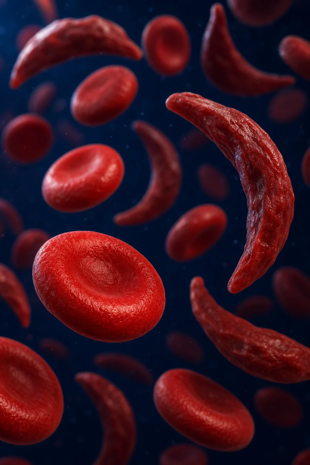
Sickle cell disease is caused by an autosomal recessive point mutation in the beta-globin (HBB) gene on chromosome 11p15.5. A single adenine-to-thymine substitution in the sixth codon changes glutamic acid to valine at position 6 of the beta-globin chain (Glu6Val, sometimes written Glu6Val or E6V), producing haemoglobin S (alpha-2 beta-S-2). Only one amino acid, out of 146 in each beta chain, is altered — yet this is enough to reshape the entire molecule.[1][2]
Inheritance is classically autosomal recessive. Homozygous HbSS (sickle cell anaemia) is the most common and most severe genotype. Compound heterozygotes — inheriting HbS from one parent and a different beta-globin variant from the other — produce the milder sickling syndromes. Heterozygous HbAS (sickle cell trait) is a carrier state: it is largely asymptomatic and, crucially, confers partial protection against severe falciparum malaria, which explains the high gene frequency across the malaria belt (sub-Saharan Africa, the Arabian peninsula, the Mediterranean, and parts of India). This is the textbook example of balanced polymorphism: the heterozygote advantage keeps a lethal recessive gene at high frequency in the population.[2]
HbSS (sickle cell anaemia)
- Homozygous — most common and most severe genotype
- Electrophoresis: HbS with NO HbA; HbF variable (5 to 15 percent)
- Severe chronic haemolysis, frequent crises, autosplenectomy by age five
- Functional asplenia — encapsulated sepsis risk
HbSC disease
- Compound heterozygote HbS plus HbC — moderate severity
- More target cells and crystal-shaped cells on film
- Less haemolysis than HbSS but HIGHER risk of proliferative retinopathy and osteonecrosis
- Splenomegaly often persists into adulthood
HbS-beta-thalassaemia
- HbS plus a beta-thalassaemia allele
- Beta-zero (no HbA): clinically severe, resembles HbSS
- Beta-plus (some HbA): moderate; raised HbA2 distinguishes it
- Electrophoresis shows reduced or absent HbA with raised HbA2
Sickle cell trait (HbAS)
- Heterozygous carrier — asymptomatic, normal haemoglobin and lifespan
- Electrophoresis: HbA exceeds HbS (about 55 to 60 percent HbA)
- Protects against severe falciparum malaria — balanced polymorphism
- Rare risks only at extreme stress: renal papillary necrosis, splenic infarct at high altitude, exertional collapse
Sickle cell disease — the numbers an examiner wants
Pathophysiology
The whole disease flows from a single molecular event. Normal adult haemoglobin (HbA) is soluble and flexible. When the Glu6Val substitution places a hydrophobic valine on the outer surface of deoxy-HbS, neighbouring beta chains lock together via a complementary hydrophobic pocket (the so-called donor-and-acceptor fit between valine-6 and phenylalanine-85 / leucine-88 on an adjacent molecule). Deoxy-HbS molecules then polymerise into rigid 14-strand fibres (tactoids) that align and stretch the red cell into the characteristic crescent.[1]
Polymerisation is kinetically delayed: it depends on deoxygenation, intracellular haemoglobin concentration (MCHC), temperature, pH and the proportion of non-S haemoglobins. This delay-time model explains why sickling is triggered by anything that lowers oxygen, raises acidity, or concentrates the cell: [1]
Polymerisation triggers
- Deoxygenation (the dominant trigger) — hence nocturnal or respiratory crises
- Acidosis — shifts the oxygen dissociation curve right, favours T-state (deoxy) HbS
- Dehydration and raised MCHC — concentrated HbS gels faster
- Fever, infection, cold, high altitude, hypoxia, pregnancy, surgery
Protective modifiers
- HbF (fetal haemoglobin) — cannot enter the HbS polymer; dilutes and inhibits gelation
- High HbF (hereditary persistence) ameliorates HbSS — basis of hydroxycarbamide
- HbA in compound heterozygotes lowers the effective HbS concentration
- Alpha-thalassaemia co-inheritance reduces MCHC and lessens haemolysis
Initially the sickling is reversible — on reoxygenation the cell recoils. But repeated cycles of polymerisation and depolymerisation damage the membrane irreversibly: calcium influx, potassium loss (via the Gardos channel), oxidative injury and dehydration produce the irreversibly sickled cell (ISC), which remains sickled even when fully oxygenated. These are the cells seen on the blood film and the ones that drive haemolysis.[1][2]
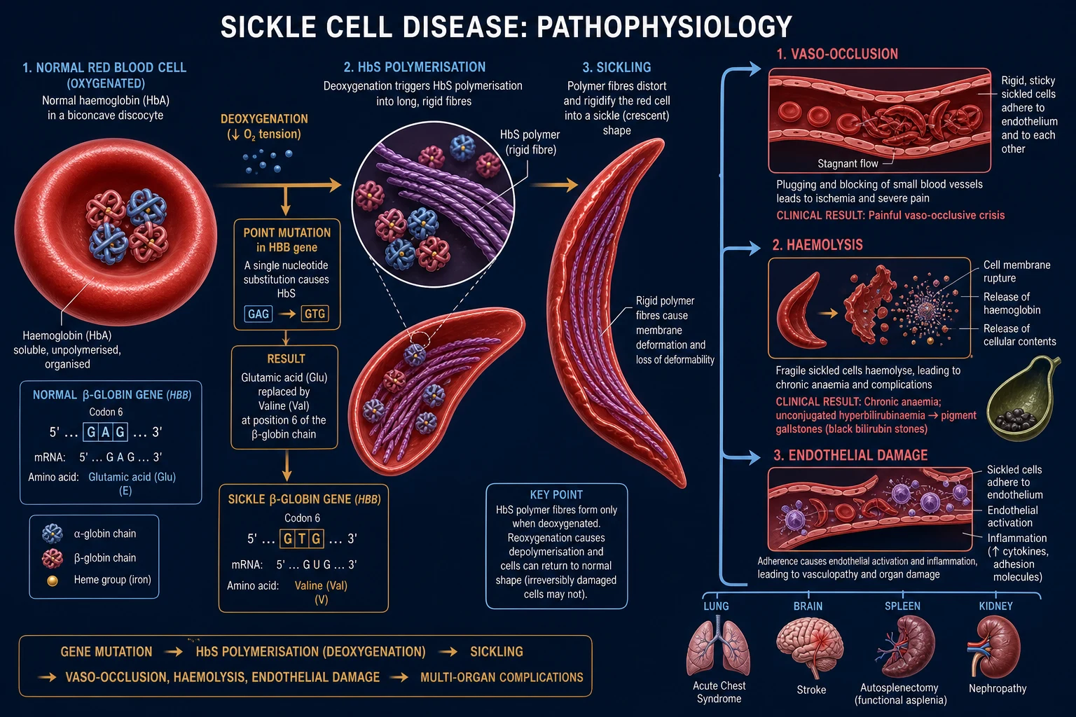
Three pathological consequences define the entire clinical phenotype: [1]
- Vaso-occlusion is not simple mechanical plugging. Sickled red cells, reticulocytes and neutrophils adhere to vascular endothelium (via VCAM-1, integrins and selectins), recruit platelets and coagulation factors, and form heterocellular aggregates in the microvasculature. The result is ischaemia, infarction and pain in bone, spleen, lung, brain, kidney and retina. The vaso-occlusive phenotype dominates the clinical picture and is the target of crizanlizumab (anti-P-selectin).[6]
- Chronic haemolysis destroys sickled cells within about 17 days (versus 120 for a normal cell), producing a steady-state haemoglobin of 60 to 90 g/L, compensatory reticulocytosis, jaundice and pigment (bilirubinate) gallstones. The bone marrow expands to compensate, causing frontal bossing and maxillary overgrowth in children. The haemolytic phenotype is the target of voxelotor (an HbS polymerisation inhibitor).[7]
- Endothelial dysfunction and haemolysis-associated vasculopathy — free plasma haemoglobin from intravascular haemolysis scavenges nitric oxide, while arginase released from ruptured red cells depletes the NO precursor arginine. The resulting NO depletion drives vasoconstriction, platelet aggregation, smooth-muscle proliferation and pulmonary hypertension, and contributes to priapism, leg ulcers, stroke and sickling nephropathy.[1]
Epidemiology & Risk Factors
Sickle cell disease is globally distributed but concentrated in the malaria belt. The HbS gene frequency tracks falciparum malaria across sub-Saharan Africa, the Mediterranean, the Middle East, and parts of India. About 300 million people worldwide carry the sickle gene, and an estimated 300,000 to 500,000 infants are born with HbSS each year — the majority in sub-Saharan Africa, where, without newborn screening and penicillin prophylaxis, the great majority die in early childhood from infection or undiagnosed crisis.[1]
In high-income settings the picture is transformed by screening. In the United States, about 1 in 365 African-American births has HbSS and about 1 in 12 African-Americans carries sickle cell trait. In the United Kingdom it is the most common serious inherited condition, included in the newborn blood-spot screening programme. India, Saudi Arabia and the eastern Mediterranean have substantial tribal and regional cohorts.[2]
Risk factors for crisis and severity include infection (the commonest trigger), dehydration, cold, high altitude, hypoxia, acidosis, pregnancy, surgery, emotional stress, and alcohol. Co-inheritance of alpha-thalassaemia tends to reduce haemolysis and stroke risk; high fetal haemoglobin (whether genetic or induced by hydroxycarbamide) is the single most important ameliorating factor. A high steady-state white-cell count predicts more frequent crises.[1]
Clinical Presentation
Because fetal haemoglobin (HbF) prevents HbS polymerisation, infants are protected in utero and for the first months of life. Symptoms appear only as HbF falls over the first six months, which is why the disease declares itself from around six months of age — and why the earliest manifestations (dactylitis) appear precisely then.[2]
The clinical picture is a mosaic of chronic haemolysis, recurrent acute crises, and progressive cumulative organ damage. A useful exam frame is to describe the presentation by age and organ. [1]
Sickle cell disease — presentation by age
Chronic haemolytic anaemia
A steady-state haemoglobin of 60 to 90 g/L with a reticulocytosis of 10 to 20 percent is tolerated remarkably well because 2,3-BPG shifts the oxygen-dissociation curve rightward. Patients look pale and mildly icteric (scleral icterus, jaundice), have a flow murmur, and develop pigment gallstones by adolescence. Growth retardation and delayed puberty are common, and frontal bossing and maxillary prominence reflect marrow expansion.[1]
Vaso-occlusive (painful) crisis
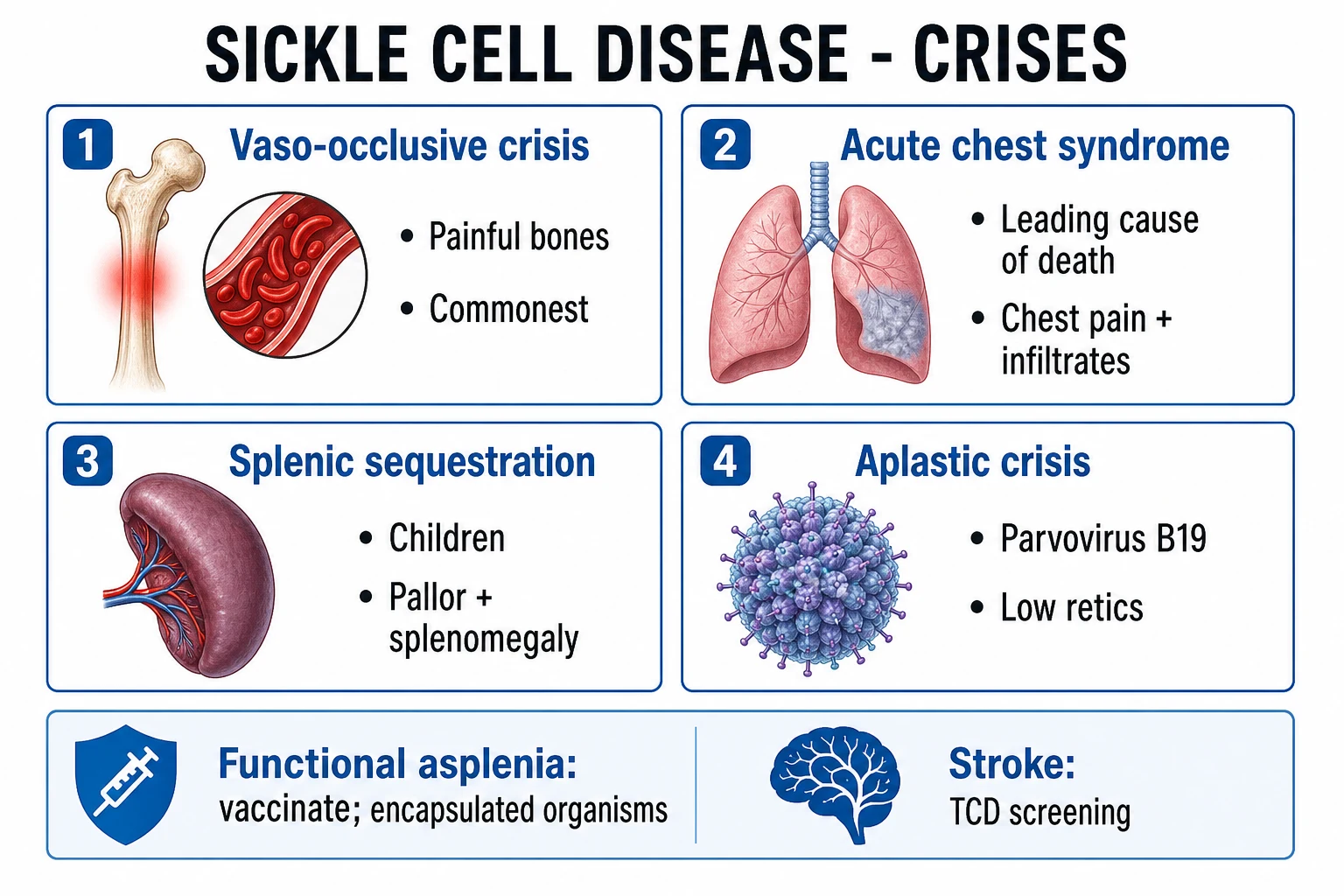
The commonest manifestation and the hallmark of the disease. Recurrent episodes of severe, deep, gnawing pain in the long bones, spine, ribs, sternum, chest and abdomen, often precipitated by infection, dehydration, cold or stress. Pain reflects bone-marrow and visceral micro-infarction. Infants present with dactylitis (hand-foot syndrome) — painful, symmetric, non-pitting swelling of the hands and feet from infarction of the small bones of the hands and feet, often the first presentation after HbF falls.[2]
Acute chest syndrome
The leading cause of death in SCD and the most common reason for intensive care. Defined as a new pulmonary infiltrate on chest imaging plus at least one of fever, chest pain, tachypnoea, wheeze, cough or hypoxia in a patient with SCD. Triggers include infection (viral, atypical and encapsulated bacteria), fat embolism from bone-marrow infarction, pulmonary vaso-occlusion, and atelectasis. It can complicate any painful crisis (especially one involving the ribs or thoracic spine, where splinting causes hypoventilation) and progresses rapidly to respiratory failure.[1]
Neurological — stroke
Without screening, about 11 percent of children with HbSS suffer a clinically evident stroke by age twenty, and another 20 percent have silent cerebral infarction detectable on MRI, which causes cognitive impairment. Ischaemic stroke predominates in children (from intimal hyperplasia and sickling in the large intracranial vessels, the circle of Willis); haemorrhagic stroke predominates in adults (from moyamoya-like collaterals). Any acute neurological deficit in a patient with SCD is a stroke until proven otherwise.[2][4]
Splenic sequestration and functional asplenia
Acute splenic sequestration is a childhood emergency: sickled cells trap in the splenic sinusoids, the spleen enlarges rapidly, and a large volume of blood sequesters, producing sudden severe anaemia, hypovolaemia and shock — classically in infants aged six months to two years with a still-palpable spleen. Recurrent episodes are common. Over the first years of life, repeated infarction produces autosplenectomy: the spleen scars and shrinks until it is impalpable, and functional asplenia is established by around age five. This removes splenic clearance of encapsulated bacteria, making invasive pneumococcal, Haemophilus influenzae type b and meningococcal infection a leading cause of death in young children.[5]
Aplastic crisis
A transient red-cell aplasia triggered almost always by parvovirus B19, which specifically infects and destroys erythroid precursors. Because the sickled red cell lives only about 17 days, shutting down erythropoiesis for even a week causes a precipitous fall in haemoglobin (often below 40 g/L) with a paradoxically low reticulocyte count. It is self-limiting (the virus is cleared in one to two weeks) but may require transfusion to bridge the patient.[1]
Other organ-specific manifestations
Priapism (prolonged, painful erection from sickling in the corpora cavernosa) is a urological emergency; stuttering episodes precede major events and recurrent priapism causes erectile dysfunction. Avascular (osteonecrosis) of the femoral head causes hip and groin pain, a limp, and eventually collapse of the joint — and the shoulder and other epiphyses are also affected. Lower-leg ulcers (over the malleoli, painful and slow to heal) reflect the haemolysis-NO-depletion vasculopathy. Renal disease spans hyposthenuria (inability to concentrate urine, causing nocturia and dehydration from early childhood), haematuria from papillary necrosis, proteinuria and focal segmental glomerulosclerosis, and progressive chronic kidney disease. Proliferative retinopathy (neovascularisation from retinal vaso-occlusion, especially in HbSC disease) threatens vision through vitreous haemorrhage and retinal detachment. Pulmonary hypertension (from haemolysis and chronic thromboembolism) is an independent predictor of mortality.[1][2]
Hyperhaemolytic crisis
A less common crisis in which haemolysis accelerates dramatically — haemoglobin drops with a RISING reticulocyte count, jaundice deepens, and there may be reticulocytopenia or a paradoxical fall after transfusion. It can be triggered by infection, transfusion, or drugs, and overlaps with delayed haemolytic transfusion reactions. It is distinguished from aplastic crisis (which has a LOW reticulocyte count) by the reticulocyte response.[1]
Differential Diagnosis
When a patient of the right ancestry presents with chronic haemolysis or an acute pain crisis, the differential splits into other haemolytic anaemias and the mimics of each acute complication. [1]
Other haemoglobinopathies
- Beta-thalassaemia major / intermedia — microcytic, electrophoresis shows raised HbA2 / HbF, no sickled cells
- HbSC, HbSD, HbSE — compound heterozygotes, distinguished only by electrophoresis
- Alpha-thalassaemia — microcytosis without sickling, often co-inherited and modifying SCD
Other haemolytic anaemias
- Autoimmune haemolytic anaemia — positive direct antiglobulin (Coombs) test, spherocytes
- Hereditary spherocytosis — family history, osmotic fragility, splenomegaly, no HbS
- G6PD deficiency — episodic haemolysis with triggers (fava beans, oxidant drugs), bite cells, X-linked
Abdominal pain crisis mimics
- Acute appendicitis, cholecystitis, pancreatitis, mesenteric ischaemia, torsion
- Abdominal crisis typically lacks peritonism and localising signs — but do not assume; image if in doubt
- Splenic sequestration — sudden pallor and enlarging spleen in an infant is a sequestration crisis until excluded
Acute chest mimics
- Pneumonia, pulmonary embolism, pulmonary oedema, asthma / bronchiolitis
- Acute chest syndrome is defined by the new infiltrate PLUS an SCD context; a sickle cell patient with a new infiltrate has ACS regardless of the pathogen
- Fat embolism from marrow infarction produces a rapidly progressive ARDS-like picture
Clinical & Bedside Assessment
The focused examination looks for anaemia and haemolysis, end-organ damage, and signs of acute complication. [1]
- General — pallor, scleral icterus, jaundice, growth and puberty (delayed), frontal bossing and maxillary prominence (marrow expansion in children).
- Abdomen — early splenomegaly that regresses as autosplenectomy supervenes (an absent spleen in an older child or adult is the rule in HbSS); hepatomegaly; gallbladder tenderness if cholecystitis.
- Cardiorespiratory — flow murmur of anaemia; tachypnoea, hypoxia, crackles and signs of right-heart strain (pulmonary hypertension) in acute chest syndrome or chronic lung disease.
- Musculoskeletal — dactylitis in infants; tenderness and swelling over long bones; a limp or limited hip rotation from avascular necrosis of the femoral head.
- Skin — malleolar leg ulcers, pallor.
- Neurological — focal deficit suggesting stroke; cognitive impairment from silent infarcts.
- Genitourinary — priapism; haematuria from papillary necrosis. [1]
Investigations
Diagnosis combines the blood film (suggestive) with haemoglobin electrophoresis or HPLC (definitive genotype). In the newborn, screening is by heel-prick HPLC or isoelectric focusing; in prenatal diagnosis, by chorionic villus sampling with DNA (PCR) analysis.[1][8]
Full blood count and film
- Steady-state Hb 60 to 90 g/L in HbSS; reticulocytes 10 to 25 percent
- Film: sickled cells, target cells, boat cells, polychromasia, Howell-Jolly bodies (asplenia)
- High white-cell and platelet counts are baseline, not necessarily infection
Haemoglobin electrophoresis / HPLC
- DEFINITIVE genotyping test
- HbSS: HbS with NO HbA; HbF variable 5 to 15 percent
- HbSC: HbS plus HbC; HbS-beta-thal: reduced/absent HbA with RAISED HbA2
- Trait (HbAS): HbA exceeds HbS — about 55 to 60 percent HbA
Sickledex / sodium metabisulphite
- Solubility (Sickledex) or metabisulphite sickling test SCREENS for HbS
- Confirms HbS is present but CANNOT distinguish trait from disease
- Never use alone for diagnosis — follow with electrophoresis
Newborn and prenatal screening
- Heel-prick HPLC or isoelectric focusing (electrophoresis) on the blood spot
- Enables early penicillin prophylaxis and parental counselling
- Prenatal: chorionic villus sampling (10 to 12 weeks) with PCR for the HBB mutation
Complication screening
- Transcranial Doppler (TCD) annually from age 1 to 2 to age 16
- LDH, bilirubin, AST (haemolysis); urinalysis and albumin/creatinine (nephropathy)
- Echocardiography for pulmonary hypertension (TRV above 2.5 m/s); retinal examination from age 10
In an acute crisis, send a full blood count and reticulocytes (the single most useful test — a low reticulocyte count in a falling haemoglobin points to aplastic crisis), blood film, group and screen, blood and urine cultures, lactate dehydrogenase, bilirubin, and a chest radiograph if respiratory. A reticulocyte count that is inappropriately low for the degree of anaemia, especially with a febrile prodrome, should prompt parvovirus B19 PCR.[1]
Management — the Four Pillars
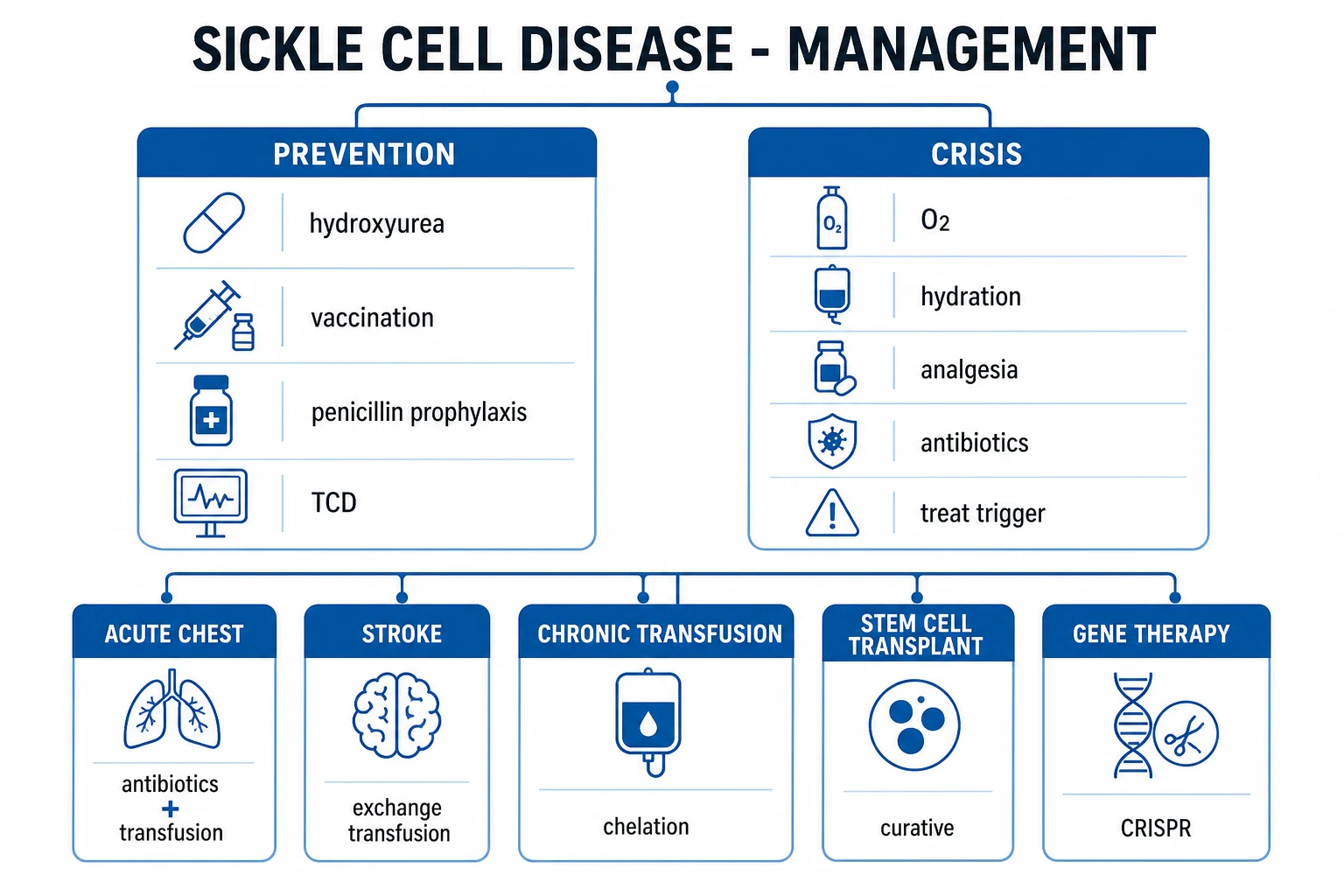
Management rests on four pillars: (1) prevent crises (hydroxycarbamide, vaccination, penicillin prophylaxis, trigger avoidance, folate); (2) screen for and prevent organ complications (transcranial Doppler for stroke, retinal and renal surveillance); (3) treat acute crises aggressively (analgesia, hydration, oxygen, transfusion); and (4) offer curative therapy (stem-cell transplant, gene therapy).[8]
Pillar one — disease-modifying prevention
Hydroxycarbamide (hydroxyurea) is the backbone of modern care. It raises fetal haemoglobin (HbF) by stimulating gamma-chain production (and has additional benefits: myelosuppression lowering the neutrophil count, improving red-cell hydration, and increasing NO). The landmark Multicenter Study of Hydroxyurea in Sickle Cell Anemia (MSH) showed it roughly halves the frequency of painful crises and reduces acute chest syndrome, transfusion need and mortality.[3] It is now recommended for nearly all patients with HbSS or HbS-beta-zero-thalassaemia from nine months of age, regardless of severity.[8]
Hydroxycarbamide dosing (UK / US / India consensus): start at 15 mg/kg once daily orally in adults (20 mg/kg/day in infants and children), escalate by 5 mg/kg every eight weeks to the maximum tolerated dose (about 25 to 35 mg/kg/day), monitoring the full blood count. Cytopenias are dose-limiting; the drug is teratogenic and must be stopped before conception and in pregnancy. Monitoring: FBC every four to eight weeks; renal and liver function; HbF response at three to six months. L-glutamine (Endari) 0.3 g/kg twice daily is an oral adjunct approved in the US for reducing crises.[3][8]
The newer disease-modifying drugs are added on top of (not instead of) hydroxycarbamide when control is inadequate. Crizanlizumab is a humanised monoclonal antibody against P-selectin that blocks the adhesion of sickled cells to the endothelium. The SUSTAIN trial showed it significantly reduces the rate of painful crises (and increased the proportion of patients with no crisis at all).[6] Voxelotor is a haemoglobin-S polymerisation inhibitor that stabilises the oxygenated (R-state) haemoglobin and prevents sickling; the HOPE trial showed it raises haemoglobin and reduces haemolysis markers.[7]
The disease-modifying drug ladder
Pillar two — functional asplenia prophylaxis
Because the spleen is lost to autosplenectomy, every infant with HbSS needs penicillin V prophylaxis from birth or diagnosis, which the PROPS (Prophylactic Penicillin Study) trial proved reduces mortality from pneumococcal septicaemia.[5] Any fever in a child with SCD is functional asplenia with possible overwhelming sepsis until proven otherwise and demands urgent empirical IV antibiotics within one hour.
Penicillin V prophylaxis (NHLBI / NHS consensus): 125 mg orally twice daily under age three, then 250 mg twice daily from age three to five. Prophylaxis is generally continued to age five in HbSS (longer in those with surgical splenectomy or prior invasive infection). Vaccination against encapsulated organisms is mandatory and lifelong: conjugate pneumococcal (PCV13 in infancy, then PPV23), Haemophilus influenzae type b, meningococcal ACWY and B, plus annual influenza. Give folic acid 5 mg once daily (lifelong) for the chronic haemolysis. Patients should carry an asplenia / sickle cell card.[5][8]
Pillar three — stroke screening and transfusion
Stroke is preventable. The STOP (Stroke Prevention Trial in Sickle Cell Anemia) trial showed that annual transcranial Doppler (TCD) ultrasonography identifies children at high risk: a timed-averaged mean velocity at or above 200 cm/s in the internal carotid or middle cerebral artery predicts high stroke risk, and instituting chronic transfusion in these children prevents about 90 percent of first strokes.[4] Children with HbSS should have annual TCD from age one to two until age sixteen. Chronic transfusion (aiming to keep HbS below 30 percent, with a top-up haemoglobin around 100 to 110 g/L) is also used for secondary stroke prevention after a first stroke (without it, recurrence is around 65 to 90 percent).[4][8]
The cost of chronic transfusion is iron overload, which damages the liver, heart and endocrine organs. Iron chelation is therefore essential once 20 to 25 units have been transfused or serum ferritin exceeds 1000 — options are deferasirox (oral, first-line), deferiprone (oral) and deferoxamine (subcutaneous infusion). [1]
Transfusion and exchange transfusion
Transfusion is the second pillar of SCD therapy after hydroxycarbamide, but it is not one intervention — it is a family of techniques chosen by indication. Simple (top-up) transfusion raises the haemoglobin and dilutes HbS; it is used for acute severe anaemia (aplastic or sequestration crisis, symptomatic anaemia in pregnancy), to raise a low preoperative haemoglobin, and as the chronic-modality in primary-stroke prevention after an abnormal TCD. Exchange transfusion (automated erythrocytapheresis, or manual two-volume exchange) removes the patient's HbS-rich blood and replaces it with donor cells, rapidly lowering the HbS fraction to below 30 percent without raising viscosity; it is the modality of choice for acute stroke, severe acute chest syndrome, and severe priapism refractory to first-line measures, and for perioperative cover in high-risk procedures. The risk of raising haemoglobin too high with simple transfusion (hyperviscosity, which can precipitate stroke) is why exchange is preferred when the starting haemoglobin is already near baseline.[4][8]
Simple (top-up) transfusion
- Raises Hb and dilutes HbS; easiest and cheapest
- For acute symptomatic anaemia, aplastic / sequestration crisis, low preop Hb
- Chronic top-up for primary stroke prevention (target HbS below 30 percent)
- Risk: hyperviscosity and stroke if Hb pushed above about 110 g/L; iron load per unit
Exchange transfusion
- Removes HbS-rich blood, replaces with donor cells; rapid HbS reduction
- First-line for acute stroke, severe acute chest, refractory priapism
- Target HbS below 30 percent; final Hb around 100 to 110 g/L, not above
- Lower net iron load per unit, but needs large-bore access and more donor exposure
Chronic transfusion programmes (every three to four weeks, to maintain HbS below 30 percent and pre-transfusion haemoglobin around 90 to 100 g/L) are indicated for secondary stroke prevention (where without transfusion the recurrence risk is 65 to 90 percent), for primary prevention after abnormal TCD, and sometimes for recurrent acute chest or severe refractory pain. Their two great burdens are iron overload (chelate with oral deferasirox first-line, aiming serum ferritin below 1000) and red-cell alloimmunisation (especially anti-C, anti-E and anti-Kell — minimise by using phenotype-matched or extended-matched blood, and always document antibodies). Delayed haemolytic transfusion reactions, and the rare hyperhaemolytic crisis (where the patient's own and donor cells both haemolyse), are the most feared transfusion complications in SCD.[1][8]
Pillar four — curative therapy
Haematopoietic stem-cell transplant (HSCT) from an HLA-matched sibling donor is the only widely established cure, with overall survival above 95 percent and event-free survival around 85 to 90 percent in well-selected children. It is offered to children with severe disease (stroke, recurrent acute chest, refractory pain) who have a matched sibling. Matched unrelated donor and haploidentical transplantation are expanding access.[1]
Gene therapy is the great advance of the past few years. Exagamglogene autotemcel (exa-cel / Casgevy) uses CRISPR-Cas9 to disrupt the BCL11A enhancer, re-activating fetal haemoglobin production in the patient's own autologous stem cells; the CLIMB-121 / Frangoul trial showed most treated patients became vaso-occlusive-crisis-free, and exa-cel was approved by the MHRA and FDA in 2023 to 2024. Betibeglogene autotemcel (beti-cel) uses lentiviral addition of a modified beta-globin gene (approved for beta-thalassaemia and developing for SCD).[9]
Management — Resuscitation of Acute Crises
The four acute crises — vaso-occlusive pain, acute chest, splenic sequestration, aplastic — share the same resuscitation bundle, tailored to severity. [1]
Acute vaso-occlusive (painful) crisis
A structured ABCDE assessment first, looking especially for a precipitant (infection, dehydration) and for chest involvement. The crisis bundle:[8]
- Analgesia within thirty minutes. Use titrated intravenous morphine (for example 0.1 mg/kg, repeat as needed) or subcutaneous diamorphine; combine with paracetamol and an NSAID where not contraindicated. Never under-treat pain — the historical fear of opioid dependence in SCD caused immense suffering. Reassess pain every 15 to 30 minutes and titrate; patient-controlled analgesia is appropriate for severe pain.
- Oxygen to maintain saturations at 94 to 98 percent (or the patient's baseline).
- Intravenous hydration with isotonic saline — maintenance plus replacement of deficit; avoid over-hydration, which precipitates pulmonary oedema and worsens acute chest.
- Treat the trigger — cultures and empirical antibiotics for infection; correct dehydration and hypoxia.
- Incentive spirometry every two hours — proven to prevent atelectasis and acute chest syndrome in patients with rib or thoracic pain. [1]
Acute chest syndrome
New infiltrate plus hypoxia or fever in an SCD patient = acute chest syndrome — escalate immediately:[1]
- Broad-spectrum antibiotics — a cephalosporin (ceftriaxone or cefuroxime) plus a macrolide (azithromycin) to cover typical, atypical and encapsulated organisms.
- Oxygen and respiratory support — escalate to CPAP, BiPAP or invasive ventilation for respiratory failure.
- Transfusion — simple top-up transfusion if the haemoglobin has fallen; exchange transfusion if severe (worsening hypoxia, multilobar infiltrate, rapid progression), aiming for HbS below 30 percent.
- Incentive spirometry, bronchodilators, and treat pain and the trigger. [1]
Acute stroke
Any new neurological deficit in SCD is a stroke until proven otherwise. Do NOT thrombolyse (as with adult atherosclerotic stroke) — the treatment is emergency exchange transfusion to reduce HbS below 30 percent. Confirm with urgent CT or MRI / MR angiography. After recovery, chronic transfusion for secondary prevention is mandatory.[4]
Splenic sequestration
An infant with sudden pallor and an enlarging spleen has acute splenic sequestration — emergency transfusion (and fluid resuscitation for shock). Parents are taught to palpate the spleen at home and to attend urgently if it enlarges or the child looks pale. Recurrent episodes may warrant splenectomy (which, after infancy, is safe given the trajectory toward autosplenectomy anyway). [1]
Aplastic crisis
A falling haemoglobin with a low reticulocyte count, classically after a febrile viral illness, is parvovirus B19 aplastic crisis. Confirm with B19 PCR. Transfuse to bridge until marrow recovery (typically one to two weeks). The patient is then infectious to pregnant women (congenital infection risk) until cleared. [1]
Specific Subtypes & Scenarios
The severity of disease is governed largely by which beta-globin variant is paired with HbS and by the level of HbF. [1]
HbSS is the most severe — the classic disease, with severe haemolysis, frequent crises, and autosplenectomy. HbSC disease is milder overall but carries a particular risk of proliferative retinopathy and osteonecrosis, and the spleen may remain enlarged (and sequester) into adulthood. HbS-beta-zero-thalassaemia (no normal HbA made) resembles HbSS in severity; HbS-beta-plus-thalassaemia retains some HbA and is milder, with a raised HbA2 on electrophoresis being the distinguishing clue. HbSD, HbSE and HbSO-Arab are rarer compound heterozygotes of variable severity.[2]
Sickle cell trait (HbAS) is a carrier state, not a disease: normal haemoglobin, normal life expectancy, and protection against severe falciparum malaria. Complications are rare and occur only at extreme physiological stress — renal papillary necrosis with haematuria, splenic infarction at high altitude, exertional rhabdomyolysis and sudden death in extreme exertion (e.g. elite sport, military training), and a small increase in venous thromboembolism. Genetic counselling is the main intervention for carriers.[2]
Complications & Pitfalls
The complications of SCD are the disease — every organ is eventually affected. They divide into acute and chronic, and into the three mechanistic streams (vaso-occlusion, haemolysis, vasculopathy). [1]
Acute complications
- Vaso-occlusive painful crisis, acute chest syndrome, splenic sequestration
- Aplastic crisis (parvovirus B19), hyperhaemolytic crisis
- Stroke (ischaemic in children), priapism (emergency if more than 4 hours)
- Overwhelming encapsulated sepsis (functional asplenia), hepatic sequestration
Chronic complications
- Functional asplenia and autosplenectomy; pigment gallstones
- Avascular necrosis (femoral and humeral head), chronic osteomyelitis
- Sickle nephropathy (hyposthenuria, papillary necrosis, FSGS, CKD)
- Pulmonary hypertension and chronic sickle lung disease; proliferative retinopathy; leg ulcers
Treatment-related complications
- Transfusional iron overload (cardiomyopathy, cirrhosis, diabetes) — chelate
- Alloimmunisation and delayed haemolytic transfusion reactions
- Hydroxycarbamide cytopenias and teratogenicity (stop in pregnancy)
- Transfusion-transmitted infection; venous-access complications
The classic pitfalls are: (1) under-treating pain out of fear of opioids; (2) missing acute chest syndrome by attributing a new infiltrate to simple pneumonia and not escalating to macrolide cover and transfusion; (3) over-hydrating and precipitating pulmonary oedema; (4) forgetting parvovirus B19 when the haemoglobin drops with a low reticulocyte count; (5) not screening with TCD; (6) assuming a fever is viral in a functionally asplenic child; (7) not chelating a chronically transfused patient until organ damage is done.[1]
Managing the chronic complications
Each chronic complication has its own evidence-based management, and examiners test them individually. Priapism lasting more than four hours is a urological emergency: first-line is intracavernosal aspiration plus phenylephrine injection, with concurrent hydration, analgesia and oxygen; exchange transfusion is reserved for refractory cases, and a surgical shunt is the last resort. Recurrent stuttering priapism is suppressed by an eta-blocker (e.g. etilefrine or terbutaline) at night and, where needed, hydroxycarbamide.[2]
Avascular necrosis of the femoral head is managed with analgesia, protected weight-bearing and physiotherapy early; core decompression for pre-collapse disease; and total hip replacement once the joint collapses (often needed bilaterally in young adults). Leg ulcers require meticulous wound care, compression, infection control, and transfusion or hydroxycarbamide to raise haemoglobin and reduce haemolysis; they are notoriously refractory. Proliferative sickle retinopathy (especially in HbSC) needs laser photocoagulation and, for vitreous haemorrhage or retinal detachment, vitrectomy — which is why annual retinal screening from around age ten matters. Sickle nephropathy is managed with ACE inhibitor or angiotensin-receptor blocker for proteinuria (the SCD analogue of diabetic nephropathy care), blood-pressure and volume control, and eventual renal replacement therapy or transplant in end-stage disease. Pulmonary hypertension is treated with optimal SCD care, oxygen, and (where appropriate and after echocardiographic and right-heart catheterisation confirmation) targeted pulmonary vasodilator therapy. Cholelithiasis is treated by elective laparoscopic cholecystectomy once symptomatic (with perioperative transfusion cover).[1][8]
Prognosis & Disposition
Historically, HbSS was fatal in childhood. With newborn screening, penicillin prophylaxis, hydroxycarbamide, TCD screening and transfusion, median survival in high-income settings is now about 45 to 55 years (somewhat longer for HbSC and HbS-beta-plus-thalassaemia). The leading causes of death are acute chest syndrome (in children and young adults) and organ failure, pulmonary hypertension and renal failure in older adults. In sub-Saharan Africa, without screening and prophylaxis, the great majority of children with SCD still die before age five — a stark global inequity.[1][2]
Hydroxycarbamide improves survival; curative transplant and gene therapy offer the prospect of a normal life expectancy for the minority who can access them. Adverse prognostic markers include a low steady-state haemoglobin, a high white-cell count, frequent crises, documented pulmonary hypertension, and proteinuria.[3]
Most acute crises are managed with admission for analgesia, hydration and observation; discharge is safe when pain is controlled on oral analgesia and acute chest is excluded. Patients with acute chest, stroke, sequestration, or aplastic crisis need high-dependency or intensive care and a haematology-led plan.[8]
Special Populations
Children
The paediatric priorities are newborn screening, penicillin prophylaxis to age five, full encapsulated-organism vaccination, hydroxycarbamide from nine months, and annual transcranial Doppler from age one to two until sixteen. Parents are taught to recognise splenic sequestration (pallor, enlarging spleen) and fever-as-an-emergency. Growth, puberty and schooling (silent cerebral infarcts impair cognition) require attention.[8]
Pregnancy
Pregnancy in SCD is high-risk: increased crises and acute chest, pre-eclampsia, fetal loss, intrauterine growth restriction and preterm birth. Care is multidisciplinary (obstetrics, haematology, anaesthetics) in a specialist unit. Folic acid 5 mg daily throughout; hydroxycarbamide is stopped before conception and during pregnancy and breastfeeding (teratogenic). Transfuse for severe anaemia (e.g. Hb below 60 to 70 g/L), symptomatic anaemia, or a prior stroke; prophylactic exchange transfusion is used in those with prior stroke or severe disease. Thromboprophylaxis is considered. Vaginal delivery is preferred; caesarean is for obstetric indications, with transfusion and avoidance of hypoxia, dehydration and hypothermia perioperatively.[8]
Anaesthesia and surgery
Perioperative sickling is a major hazard. Preoperative transfusion to a haemoglobin of about 100 g/L (and, for high-risk procedures, exchange transfusion to HbS below 30 percent), meticulous avoidance of hypoxia, dehydration, hypothermia and acidosis, regional techniques where possible, and early mobilisation and incentive spirometry reduce complications. Acute chest syndrome is the principal postoperative danger.[8]
Functional asplenia and the febrile child
Any fever above 38 degrees in a child with SCD is a medical emergency — take cultures and give empirical IV ceftriaxone within one hour (covering pneumococcus), observe, and look for a focus. The risk of overwhelming post-splenectomy infection persists for life. [1]
Evidence, Guidelines & Regional Differences
The evidence base for modern SCD care is built on four landmark randomised trials, each of which transformed practice:[1]
MSH — hydroxyurea (Charache, 1995)
NEJM 1995;332:1317
PMID 7715639
Key finding
Hydroxyurea roughly halved the median annual rate of painful crises and reduced acute chest syndrome and transfusions in adults with HbSS.
STOP — TCD and transfusion (Adams, 1998)
NEJM 1998;339:5
PMID 9647873
Key finding
In children with abnormal transcranial Doppler (velocity at or above 200 cm/s), chronic transfusion reduced first stroke by about 90 percent versus observation.
PROPS — penicillin prophylaxis (Gaston, 1986)
NEJM 1986;314:1593
PMID 3086721
Key finding
Oral penicillin prophylaxis reduced the rate of pneumococcal septicaemia in children with SCD by about 80 percent.
SUSTAIN — crizanlizumab (Ataga, 2017)
NEJM 2017;376:429
PMID 27959701
Key finding
Anti-P-selectin monoclonal antibody reduced the annual rate of sickle-cell pain crises and increased the proportion of patients crisis-free.
HOPE — voxelotor (Vichinsky, 2019)
NEJM 2019;381:509
PMID 31199090
Key finding
An oral HbS-polymerisation inhibitor raised haemoglobin and reduced haemolysis markers in patients with SCD.
CLIMB-121 — exa-cel (Frangoul, 2021)
NEJM 2021;384:252
PMID 33283989
Key finding
CRISPR-Cas9 editing of the BCL11A enhancer re-activated fetal haemoglobin; most treated patients became crisis-free.
Guideline deltas. The NHLBI 2014 evidence-based guidelines (Yawn, JAMA) underpin US and much of global practice; the British Society for Haematology (UKHSA) guidelines govern the UK, with universal newborn screening, 5 mg folic acid, and penicillin prophylaxis. India follows ICMR / Indian Society of Haematology guidance with regional newborn and tribal screening programmes; folic acid 5 mg daily is standard. Africa has no universal screening in most countries — the Sickle in Africa Consortium and Sickle Pan-African Research Consortium are building capacity. Doses (penicillin, folic acid, hydroxycarbamide titration) are essentially consensus worldwide, but folic acid 1 mg (US) versus 5 mg (UK/India) is the common regional delta. Voxelotor was withdrawn worldwide by Pfizer in September 2023 over a possible safety signal — know your local formulary.[8]
The main controversies and weak evidence areas are: the optimal duration of chronic transfusion (and whether it can ever be safely stopped for secondary stroke prevention, where recurrence risk remains high); whether and when to transition from transfusion to hydroxycarbamide after abnormal TCD (STOP II showed rebound risk on early switch); the long-term safety and accessibility of gene therapy; and the equitable global provision of screening, penicillin and hydroxycarbamide.[4]
Exam Pearls
Sickle cell disease — SICKLE mnemonic
SICKLE
on blood film plus target cells and Howell-Jolly bodies (asplenia) — diagnostic clue
autosomal recessive; Glu6Val mutation on HBB (chromosome 11p15.5) produces HbS
vaso-occlusive (commonest), splenic sequestration, aplastic (parvovirus B19), hyperhaemolytic
acute chest syndrome is the leading cause of death
functional asplenia by age five — vaccinate and penicillin prophylaxis
hydroxycarbamide; curative CRISPR gene therapy (exa-cel)
Exam application bank (NEET-PG / INICET)
One-line answer
Sickle cell disease (SCD) is an autosomal recessive haemoglobinopathy caused by a single glutamic-acid-to-valine substitution at position 6 of beta-globin (Glu6Val, HBB on chromosome 11p15.5) producing haemoglobin S (HbS), which polymerises under deoxygenation into rigid fibres that distort the red cell into a sickle. The consequences are vaso-occlusion, chronic haemolysis and endothelial dysfunction: chronic haemolytic anaemia punctuated by vaso-occlusive painful crises, acute chest syndrome (the leading cause of death), stroke, splenic sequestration and functional asplenia, aplastic crisis (parvovirus B19), priapism, avascular necrosis, leg ulcers, renal papillary necrosis and proliferative retinopathy. Diagnosis rests on haemoglobin electrophoresis / HPLC (HbS with no HbA in HbSS). Management rests on hydroxycarbamide (hydroxyurea) to raise HbF, transfusion and exchange transfusion fo
Worked stems (answer without another resource)
Stem 1 — Classic presentation. Map symptoms to mechanism; name the first investigation and first treatment step with dose/route if drug therapy is standard. [1]
Stem 2 — Unstable / complicated. List red flags that force immediate resuscitation, theatre, ICU, antidote, or reperfusion — and what you do in the first 15 minutes. [1]
Stem 3 — Atypical group. Elderly, pregnancy, child, or immunocompromised: how presentation and thresholds change. [1]
Stem 4 — Differential trap. Name the three closest mimics and one discriminator for each. [1]
Stem 5 — Disposition. Who goes home with safety-netting, who is admitted, who needs HDU/ICU/theatre, and what follow-up is mandatory. [1]
Rapid viva checklist
- Definition + classification
- Pathophysiology chain
- Bedside signs / criteria
- Score with exact components (if any)
- Emergency bundle
- Definitive therapy with doses
- Complications of disease and of treatment
- Special populations
- Guideline/trial name if classic
- Three exam traps
Coverage self-check
If you cannot answer any stem above from this page alone, re-read the matching section — the page is intended to be self-sufficient for final-prof and NEET-PG/INICET questions on Sickle Cell Disease.
References
- [1]Kato GJ, Piel FB, Reid CD, et al. Sickle cell disease Nat Rev Dis Primers, 2018.PMID 29542687
- [2]Rees DC, Williams TN, Gladwin MT. Sickle-cell disease Lancet, 2010.PMID 21131035
- [3]Charache S, Terrin ML, Moore RD, et al. Effect of hydroxyurea on the frequency of painful crises in sickle cell anemia. Investigators of the Multicenter Study of Hydroxyurea in Sickle Cell Anemia N Engl J Med, 1995.PMID 7715639
- [4]Adams RJ, McKie VC, Hsu L, et al. Prevention of a first stroke by transfusions in children with sickle cell anemia and abnormal results on transcranial Doppler ultrasonography N Engl J Med, 1998.PMID 9647873
- [5]Gaston MH, Verter JI, Woods G, et al. Prophylaxis with oral penicillin in children with sickle cell anemia. A randomized trial N Engl J Med, 1986.PMID 3086721
- [6]Ataga KI, Kutlar A, Kanter J, et al. Crizanlizumab for the Prevention of Pain Crises in Sickle Cell Disease N Engl J Med, 2017.PMID 27959701
- [7]Vichinsky E, Hoppe CC, Ataga KI, et al. A Phase 3 Randomized Trial of Voxelotor in Sickle Cell Disease N Engl J Med, 2019.PMID 31199090
- [8]Yawn BP, Buchanan GR, Afenyi-Annan AN, et al. Management of sickle cell disease: summary of the 2014 evidence-based report by expert panel members JAMA, 2014.PMID 25203083
- [9]Frangoul H, Altshuler D, Cappellini MD, et al. CRISPR-Cas9 Gene Editing for Sickle Cell Disease and β-Thalassemia N Engl J Med, 2021.PMID 33283989