Haematology · General Medicine
Thalassaemia (Alpha & Beta)
Also known as Thalassemia · Beta thalassaemia · Alpha thalassaemia · Mediterranean anaemia · Cooley anaemia
Thalassaemia is an inherited disorder of haemoglobin synthesis causing reduced (plus) or absent (zero) globin chain production, leading to microcytic hypochromic anaemia. Beta-thalassaemia (autosomal recessive, HBB on chromosome 11): major (Cooley anaemia; transfusion-dependent from 6 to 12 months with severe microcytic anaemia, failure to thrive, frontal bossing, hepatosplenomegaly, raised HbF and HbA2), intermedia (milder, transfusion-independent), minor/trait (asymptomatic, microcytosis out of proportion to anaemia, high RBC count, normal iron, raised HbA2). Alpha-thalassaemia (HBA on chromosome 16, four genes): one gene silent carrier, two trait (mild microcytosis, NORMAL electrophoresis), three HbH disease (moderate haemolytic anaemia, beta-4 tetramers), four Hb Bart hydrops fetalis (gamma-4 tetramers, incompatible with life). Diagnosis: FBC (microcytic hypochromic, high/normal RBC), Mentzer index over 13, Hb electrophoresis/HPLC (raised HbF and HbA2 in beta-thalassaemia), genetic testing for alpha trait. Treatment: lifelong transfusion every 2 to 4 weeks to keep pre-transfusion Hb 90 to 105 g/L plus iron chelation (deferasirox 20 to 40 mg/kg/day oral first-line; deferiprone 75 mg/kg/day; deferoxamine SC infusion), folic acid, splenectomy, stem cell transplant (curative), gene therapy (betibeglogene autotemcel, approved 2023/24), luspatercept.
On this page & tools
Your progress
Saved locally on this device.
Exam tags
Red flags
Overview & Definition
Thalassaemia — from the Greek thalassa (sea) and haima (blood), a nod to its Mediterranean recognition — is the commonest monogenic disorder in humans and the prototypical inherited quantitative defect of haemoglobin synthesis. Where sickle cell disease and the structural haemoglobin variants are qualitative disorders (a normal amount of an abnormal globin), thalassaemia produces a reduced or absent quantity of an otherwise normal globin chain, and the resulting imbalance between paired alpha and non-alpha chains is what damages the red cell. The disease is autosomal recessive in inheritance and clusters along the same tropical malaria belt as sickle cell disease, because heterozygous carriers enjoy partial protection against Plasmodium falciparum malaria.[1][2]
The clinical spectrum is exceptionally wide, ranging from the completely asymptomatic carrier identified on a routine full blood count, through moderate chronic haemolytic anaemia, to transfusion-dependent disease presenting in infancy, and — at the extreme — lethal hydrops fetalis. Which end of this spectrum a patient occupies is determined almost entirely by how many globin genes are affected and which chain is under-produced, so the genetics (chromosome 11 for beta, chromosome 16 for alpha) is not an academic detail but the direct explanation of the phenotype. A clinician who grasps the gene-dose logic can predict the clinical course from the genotype.[1]
Thomas Cooley's 1925 description of severe anaemia, splenomegaly, bony changes and "Mongoloid" facies in children of Mediterranean origin gave beta-thalassaemia major its eponym, Cooley anaemia. The defect was later localised to defective globin synthesis, and the molecular era — chromosome 11 beta-globin cloning in the late 1970s and alpha-globin mapping on chromosome 16 — turned a fatal disease of childhood into a chronic, manageable condition through transfusion and iron chelation, and most recently into a potentially curable disease through haematopoietic stem cell transplant and gene therapy.[2][4]
Classification
Thalassaemia is classified first by which globin chain is under-produced (alpha or beta), and then by genetic dose (how many alleles are affected) and clinical severity. The two axes give a clean, examinable structure.[1][2]
Beta-thalassaemia is caused by mutations in the single HBB gene on chromosome 11p15.4 (two alleles). Because there is one beta gene per chromosome, severity is graded by whether each allele produces no chains (beta-0), reduced chains (beta-plus), or only mildly reduced chains (beta-plus-plus). Three clinical syndromes result:
[1]- Beta-thalassaemia major — usually beta-0/beta-0, producing virtually no beta chains and therefore no normal HbA. Severe transfusion-dependent anaemia appears at 6 to 12 months as fetal haemoglobin (HbF) gives way to adult haemoglobin and the absent beta chain becomes apparent. Without transfusion the child dies in early childhood.
- Beta-thalassaemia intermedia (TI) — milder genotypes (two beta-plus alleles, or beta-0 with a mild partner, or co-inheritance of alpha-thalassaemia or determinants that raise HbF). Patients maintain Hb of 60 to 90 g/L without regular transfusion, present later in childhood or adulthood, and need transfusion only intermittently (infection, pregnancy, growth failure).
- Beta-thalassaemia minor (trait) — a single mutated allele (heterozygote). The patient is asymptomatic, with microcytosis disproportionate to a near-normal haemoglobin, a high red cell count, normal iron studies and HbA2 over 3.5 percent on electrophoresis.
Alpha-thalassaemia is caused by deletions or, less often, non-deletional mutations of the HBA1 and HBA2 genes on chromosome 16p13.3. Crucially there are four alpha alleles (two per chromosome, arranged in tandem), so severity rises in discrete steps as alleles are lost:
[1]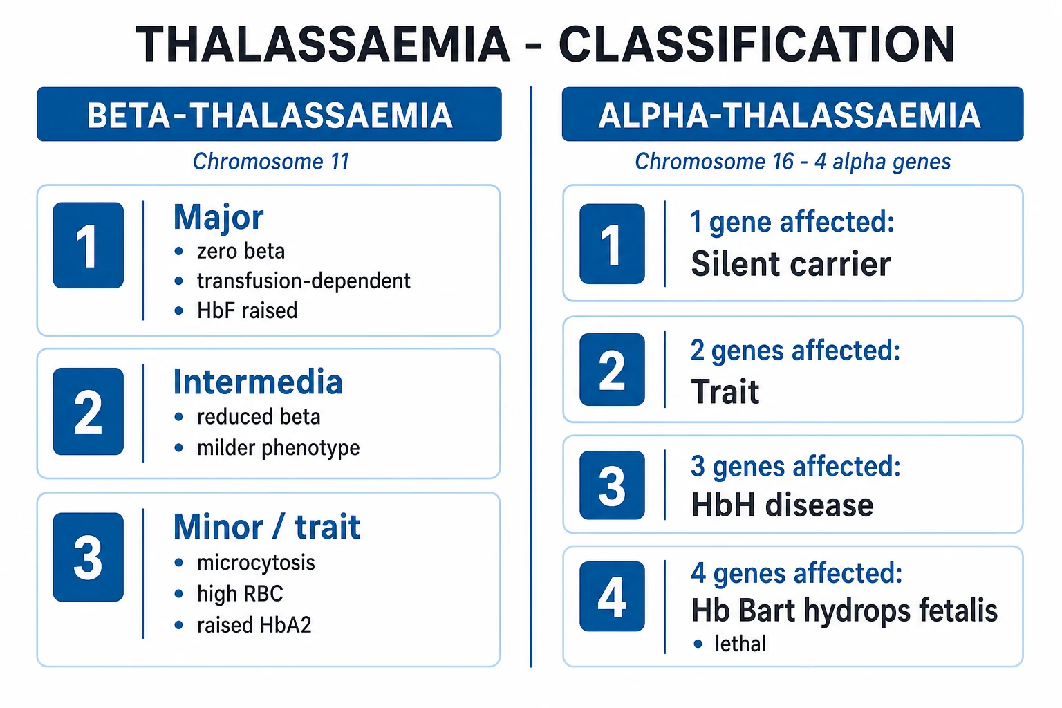
Alpha-thal (4 alleles, chr 16)
- 1 deletion (-alpha/alphaalpha): SILENT carrier — normal
- 2 deletions (-alpha/-alpha or --/alphaalpha): TRAIT — mild microcytosis, NORMAL electrophoresis
- 3 deletions (--/-alpha): HbH DISEASE — moderate haemolysis, beta-4 tetramers
- 4 deletions (--/--): Hb BART HYDROPS FETALIS — gamma-4 tetramers, LETHAL
Beta-thal (2 alleles, chr 11)
- Heterozygous (beta/beta-0 or beta/beta-plus): TRAIT — microcytosis, high RBC, raised HbA2
- beta-plus/beta-plus (milder): INTERMEDIA — transfusion-independent
- beta-0/beta-0 (severe): MAJOR (Cooley) — transfusion-dependent from infancy
- Modifiers: co-inheritance of alpha-thal or HbF-raising variants soften; extra alpha genes worsen
The cis versus trans arrangement of alpha deletions matters for reproductive risk. A cis deletion (both alpha genes lost from one chromosome, written --/alphaalpha, common in South-East Asians as the --SEA deletion) means a single parent can pass two deleted genes to a child; two cis-deletion carriers risk a four-gene Hb Bart hydrops fetus. A trans deletion (one gene lost from each chromosome, -alpha/-alpha, common in Africans) never produces hydrops in the offspring even when both parents are carriers. This distinction is the backbone of prenatal counselling.[1][5]
Thalassaemia — the numbers that decide a question
Epidemiology & Risk Factors
Thalassaemia is a global disease whose burden has shifted south and east. Carrier frequencies are highest along the historical malaria belt: 2 to 15 percent of the population in the Mediterranean (Greek, Italian, Cypriot, Sardinian), 3 to 17 percent across the Middle East, 3 to 17 percent in South Asia, up to 10 percent in South-East Asia, and substantial rates across sub-Saharan Africa. Roughly 5 to 7 percent of the world's population carries a clinically relevant haemoglobin variant, and an estimated 60 to 80 thousand children are born each year with a severe form of thalassaemia — the overwhelming majority in low- and middle-income countries where transfusion infrastructure and chelation access are limited.[2][5]
The geographic overlap with falciparum malaria is not coincidental: the carrier state confers a heterozygote advantage, and the high gene frequency in malarial regions is the evolutionary signature of that selection pressure. Within high-frequency populations, founder mutations and consanguinity concentrate disease. The mutation spectrum is ethnically distinctive: Mediterranean patients most often carry the IVS1-110 (G to A) splice mutation or codon 39 (C to T) nonsense mutation; South Asian (Indian) patients carry IVS1-5 (G to C) and the 619-base-pair deletion at the 3-prime end of HBB; Chinese patients carry codons 41/42 (-TCTT) and IVS2-654 (C to T); and South-East Asians characteristically carry the --SEA alpha-globin cis deletion that predisposes to Hb Bart hydrops.[2]
India — the regional delta that examiners test. India harbours an estimated 42 million beta-thalassaemia carriers (national average around 3 to 4 percent, but with regional peaks of 10 to 15 percent among Sindhis, Gujaratis, Punjabis, Bengalis and certain tribal populations), and approximately 10 to 15 thousand children with beta-thalassaemia major are born each year. The ICMR and the Indian Society of Haematology & Blood Transfusion drive universal antenatal screening in high-prevalence states, school-leaving carrier screening, and prenatal diagnosis through CVS or amniocentesis; the national goal is to prevent new major births through premarital and antenatal carrier detection. The ISHBT best-practice guidelines (2026) endorse deferasirox as first-line chelation and emphasise access to matched-sibling stem cell transplant in regional centres.[5][6]
Risk factors for clinically important disease are therefore ancestry in a high-frequency population, a family history of anaemia, splenectomy or "blood disease", and consanguinity. A second, iatrogenic risk factor dominates the natural history once disease is established: transfusion, which is life-saving but is the principal source of iron overload and its lethal cardiac consequences.[2]
Pathophysiology
The whole of thalassaemia follows from one principle: haemoglobin is a tetramer that requires balanced production of its alpha and non-alpha chains. Adult HbA is two alpha plus two beta chains; fetal HbF is two alpha plus two gamma chains. When one chain is under-produced, the partner chain is left in excess, and that excess is toxic.[1][2]
In beta-thalassaemia, alpha chains continue to be made at normal rate while beta chains are reduced or absent. Free alpha chains are unstable; they auto-oxidise, release haem, generate reactive oxygen species, and precipitate onto the inner red-cell membrane as insoluble inclusions (Heinz-body-like aggregates, attached to the membrane skeleton via the cytoskeletal protein alpha-haemoglobin-stabilising protein). The developing normoblast recognises this damage and undergoes apoptosis — this is ineffective erythropoiesis, the death of red-cell precursors inside the bone marrow before they ever circulate. In severe beta-thalassaemia major, over 80 percent of erythroid precursors die intramedullarily. The few cells that do reach the peripheral blood carry membrane-bound alpha-chain precipitates; the spleen identifies these mechanically damaged cells and removes them, producing chronic extravascular haemolysis.[2]
In alpha-thalassaemia, it is the non-alpha chains that are produced in excess. With three alpha genes deleted, the surplus beta chains self-assemble into beta-4 tetramers (haemoglobin H, HbH) that are relatively stable but functionally useless (no cooperativity, no Bohr effect) and slowly precipitate as the cell ages, producing a moderately severe chronic haemolytic anaemia. With all four alpha genes deleted, gamma-4 tetramers (haemoglobin Bart) form in the fetus. Hb Bart has such extreme oxygen affinity that it cannot release oxygen to tissues, causing severe tissue hypoxia, massive fluid transudation, and hydrops fetalis that is lethal in utero or within hours of birth unless an intrauterine transfusion programme is in place.[1]
Three downstream consequences follow from ineffective erythropoiesis and haemolysis:
[2]- Massive marrow expansion. The erythroid marrow proliferates up to twenty-fold trying to compensate for the cells dying within it. Expanding marrow erodes cortical bone from within, producing the classic frontal bossing, maxillary overgrowth ("rodent" or "chipmunk" facies), dental malocclusion, hair-on-end skull on radiograph, cortical thinning, pathological fractures and severe osteoporosis. Extramedullary haematopoiesis produces hepatosplenomegaly and, in older patients, paravertebral masses that may compress the spinal cord.
- Hepcidin suppression and iron overload. The expanded, ineffective erythroid mass secretes erythroferrone (ERFE) and other regulators that suppress hepatic hepcidin. With hepcidin low, the iron exporter ferroportin is unrestrained and dietary iron absorption rises, compounding the iron load from repeated transfusion. There is no physiological route for iron excretion, so every millilitre of transfused red cells adds roughly 1 mg of elemental iron that must be removed pharmacologically.
- Chronic hypoxia and end-organ damage. The anaemia, the expanded metabolically active marrow, and the high cardiac output state drive growth failure, endocrine failure, pigment gallstones (from chronic haemolysis), folate depletion (from chronic erythropoietic drive) and, ultimately, the iron-overload cardiomyopathy that dominates late mortality.[2]
Clinical Presentation
The clinical presentation is dictated by genotype, gene dose and — for transfusion-dependent patients — the adequacy of therapy. Examiners test the tempo and the triad of severe microcytic anaemia, bony changes and hepatosplenomegaly in the infant, and the incidental microcytosis in the adult.[1][2]
Beta-thalassaemia major declares itself at 6 to 12 months of age, the window in which fetal haemoglobin is physiologically replaced by adult haemoglobin and the absent beta chain becomes exposed. Parents describe a previously well infant who becomes progressively pale, listless and irritable, feeds poorly and stops gaining weight. Examination reveals severe pallor with icterus, failure to thrive, and the classic facial features of marrow expansion: frontal bossing, malar fullness, maxillary prominence producing malocclusion and the so-called rodent or chipmunk facies. The abdomen is distended by massive hepatosplenomegaly (from extramedullary haematopoiesis and removal of damaged red cells). There may be a systolic flow murmur of anaemia, a wide pulse pressure and a hyperdynamic precordium from the chronic high-output state. Untreated, the child develops recurrent infections, pathological fractures and heart failure, and dies within the first few years of life.[1][2]
Beta-thalassaemia intermedia presents later — typically between 2 and 7 years of age, or even in adulthood — with moderate anaemia (Hb 60 to 90 g/L), growth retardation, splenomegaly, and the skeletal and pigment-gallstone complications of chronic haemolysis. Because these patients are not regularly transfused, they are paradoxically more prone to thrombosis, pulmonary hypertension, leg ulcers and extramedullary haematopoietic masses than well-transfused major patients, while being spared the early transfusional iron load. Iron still accumulates, however, through increased gut absorption, and chelation becomes necessary later in life.[2]
Beta-thalassaemia minor (trait) is asymptomatic. It is discovered on a routine full blood count, an antenatal screen, or a preoperative assessment, as a microcytosis disproportionate to a haemoglobin that is normal or only mildly low. Occasionally a trait patient reports mild fatigue; the trait is never the cause of severe anaemia, and finding severe anaemia in a trait patient mandates a search for a second cause (iron deficiency, blood loss, another haemoglobinopathy). The essential bedside insight is that the red cell count is high or normal, the opposite of iron deficiency.[1]
HbH disease (three alpha deletions) presents as a moderate chronic haemolytic anaemia (Hb 70 to 100 g/L) with splenomegaly, intermittent jaundice and pigment gallstones, often recognised in childhood. Oxidant drugs (sulphonamides, dapsone, primaquine, nitrofurantoin) can precipitate an acute haemolytic crisis by destabilising the already fragile HbH tetramer.[1]
Hb Bart hydrops fetalis presents in utero or at birth with severe generalised oedema, massive hepatosplenomegaly, ascites and pleural effusions — the consequences of extreme tissue hypoxia from oxygen-trapped gamma-4 tetramers. The fetus is typically stillborn or dies within hours; the mother is at markedly increased risk of pre-eclampsia, antepartum haemorrhage and difficult delivery due to an enlarged placenta and fetus.[1][5]
Atypical and late presentations examiners probe deliberately: the adolescent with delayed puberty and short stature masking hypogonadotropic hypogonadism from transfusional iron overload; the young adult with new-onset diabetes or heart failure in a transfusion-dependent patient — iron-overload cardiomyopathy; the elderly patient with incidental microcytosis who is actually iron-deficient on top of long-standing trait; and the splenectomised patient with fever and rigors who may have overwhelming post-splenectomy infection within hours.[2]
Differential Diagnosis
Thalassaemia sits at the centre of the microcytic hypochromic anaemias. The complete differential, with the features that separate each, is essential — a single discriminator usually settles the diagnosis.[1][2]
Thalassaemia trait
- Microcytosis with HIGH or normal RBC count
- Normal ferritin, iron and transferrin saturation
- Raised HbA2 in BETA trait; NORMAL electrophoresis in ALPHA trait
- Mentzer index over 13; do NOT give iron
Iron deficiency
- Microcytosis with LOW RBC count
- LOW ferritin (under 30 mcg/L), low iron, HIGH TIBC / low transferrin saturation
- No HbA2 elevation; no splenomegaly
- Mentzer index under 13; treat with oral iron and find the cause of blood loss
Anaemia of chronic disease
- Normocytic or only mildly microcytic (MCV rarely under 75 fL)
- HIGH or normal ferritin with LOW transferrin saturation
- Raised CRP and ESR; underlying infection, inflammation, malignancy
- Treat the underlying cause; IV iron only if indicated
Sideroblastic anaemia
- Dimorphic film; ringed sideroblasts on Prussian-blue marrow stain
- HIGH ferritin and transferrin saturation
- Causes: hereditary (X-linked ALAS2), myelodysplasia, alcohol, isoniazid, lead, copper deficiency
- Pyridoxine response in some hereditary/drug forms
Lead poisoning
- Microcytosis with basophilic stippling; abdominal and neurological symptoms
- HIGH serum lead level; raised zinc protoporphyrin
- Risk: painters, battery/recycling workers, old paint ingestion in children
- Chelation with succimer / EDTA depending on level
Other haemoglobinopathies
- HbE trait/disease (SE Asia); HbE/beta-thal resembles thalassaemia major
- Hereditary persistence of fetal Hb: high HbF with no symptoms
- Congenital dyserythropoietic anaemia: multinucleate erythroblasts
- Distinguished on electrophoresis and DNA testing
The single highest-yield discriminator in the whole topic is the red cell count: thalassaemia trait keeps a high or normal RBC count because the marrow makes many small cells, whereas iron deficiency depletes both haemoglobin and cell number. Combined with the iron studies and the HbA2 level, this triad resolves most microcytic anaemias without recourse to DNA testing.[1]
Clinical & Bedside Assessment
There is no pathognomonic bedside sign for thalassaemia, but the focused examination builds a coherent picture that the full blood count then confirms. The clinician looks for evidence of anaemia, haemolysis, marrow expansion, iron overload and prior splenectomy, and — if the patient is transfusion-dependent — for the end-organ consequences of iron.[2]
General inspection reveals pallor, icterus, and — in the untreated child — growth failure with height and weight below the centiles and delayed puberty. The face shows frontal bossing, malar prominence, maxillary overgrowth and dental malocclusion (the rodent or chipmunk facies). The abdomen is distended by hepatosplenomegaly that can be massive in untreated major disease; in an intermedia patient look for a palpable gallbladder or Murphy's sign from pigment stones. The cardiovascular examination characteristically shows a systolic flow murmur, a hyperdynamic precordium and a wide pulse pressure from the chronic high-output state; in iron-overload cardiomyopathy, look instead for a gallop rhythm, raised jugular venous pressure, basal crackles and hepatomegaly suggesting biventricular failure. The skin may show bronze pigmentation from haemosiderin deposition. In the splenectomised patient inspect for the surgical scar and ask about vaccination and penicillin prophylaxis. A bedside trawl for endocrine failure checks for short stature, absent secondary sexual characteristics, polyuria (diabetes) and cold intolerance (hypothyroidism).[2]
The bedside reasoning is simple: a severe microcytic anaemia in an at-risk infant with hepatosplenomegaly and bony facies is beta-thalassaemia major until proven otherwise; an incidental microcytosis with a high red cell count in a well adult is trait until proven otherwise. Both are confirmed by the full blood count and electrophoresis, not at the bedside.[1]
Investigations
The investigation pathway is staged: a full blood count and film, iron studies and discriminant indices, then haemoglobin electrophoresis or HPLC, and — when alpha-thalassaemia is suspected — DNA testing. In established transfusion-dependent disease the focus shifts to iron-load monitoring and end-organ surveillance.[1][2]
Full blood count. Disease of any severity shows microcytic hypochromic anaemia with an MCV under 75 to 80 fL in major/intermedia disease and 70 to 80 fL in trait, an MCH under 27 pg, and a raised red cell distribution width (RDW) in disease. The red cell count is the discriminator: high or normal in trait (the marrow compensates by making many small cells), often low in major disease because of ineffective erythropoiesis. The reticulocyte count is inappropriately low for the degree of anaemia in beta-thal major (because most precursors die in the marrow) but raised in HbH disease and other haemolytic forms.[1]
Discriminant indices use the high RBC count of thalassaemia to separate it from iron deficiency at the index level. The most famous is the Mentzer index = MCV divided by RBC count (RBC expressed in millions per microlitre): a value over 13 supports thalassaemia trait, while under 13 supports iron deficiency. Other indices in the same family — Green and King, England and Fraser, Srivastava, Shine and Lal — perform similarly and are useful where iron studies are unavailable. None replaces iron studies and electrophoresis in definitive diagnosis.[2]
Peripheral blood film. Classic findings are microcytes, hypochromia, target cells, basophilic stippling, and — in severe disease — nucleated red cells, Howell-Jolly bodies (post-splenectomy) and poikilocytes. In HbH disease, supravital staining with brilliant cresyl blue produces the pathognomonic golf-ball inclusions (precipitated beta-4 tetramers).[1]
Haemoglobin electrophoresis / HPLC / capillary zone electrophoresis. This is the diagnostic centrepiece for beta-thalassaemia:
[1]Beta-thal TRAIT
- HbA2 OVER 3.5 percent (often 4 to 7 percent)
- HbF normal or mildly raised (under 2 percent)
- HbA reduced but present
- Normal iron studies confirm it is not combined deficiency
Beta-thal MAJOR
- HbF markedly raised — 70 to 90 percent
- HbA absent or markedly reduced (depends on beta-0 vs beta-plus)
- HbA2 raised or normal
- Severe microcytic anaemia, transfusion-dependent
Alpha-thal TRAIT
- Electrophoresis is NORMAL — HbA2 not raised
- Microcytosis with high RBC count and normal iron
- Diagnosis requires DNA testing (gap-PCR / MLPA)
- The single commonest thalassaemia trap
HbH disease
- HbH 5 to 30 percent on electrophoresis (fast-migrating band)
- Brilliant cresyl blue: golf-ball inclusions
- Hb Barts small fraction present at birth
- Three alpha-gene deletion on DNA testing
Iron studies show normal ferritin, normal transferrin saturation and normal soluble transferrin receptor in trait — the finding that excludes iron deficiency and prevents inappropriate iron prescription. In iron-overloaded transfusion-dependent patients ferritin rises (often over 1000 mcg/L), and serial measurement guides chelation.[1]
DNA testing is mandatory for alpha-thalassaemia (where electrophoresis is normal), for prenatal diagnosis, and for genetic counselling of at-risk couples. Gap-PCR detects the common deletions; multiplex ligation-dependent probe amplification (MLPA) detects rarer and atypical deletions; Sanger sequencing or ARMS-PCR identifies point mutations of HBB. Identifying the family's mutations allows first-trimester prenatal diagnosis by chorionic villus sampling.[5]
Skeletal survey (in untreated or late-presenting major) shows the hair-on-end skull, cortical thinning, metaphyseal expansion and generalised osteoporosis that reflect marrow expansion.[2]
Iron-load and end-organ monitoring is the backbone of long-term care in transfusion-dependent thalassaemia:
[2]Iron overload & surveillance — annual minimum
Cardiac T2-star MRI is the single most important prognostic test in transfusion-dependent thalassaemia: a T2-star over 20 ms is normal, 10 to 20 ms indicates mild-to-moderate iron loading, and under 10 ms indicates severe cardiac iron with a high one-year mortality from arrhythmia or heart failure. Hepatic iron and cardiac iron can be discordant, so a normal ferritin never excludes cardiac iron — both must be measured.[2]
Management — Resuscitation
Thalassaemia is a chronic disease, but two acute scenarios demand immediate, protocol-driven resuscitation: acute severe anaemia with heart failure, and overwhelming post-splenectomy infection (OPSI) in the splenectomised patient.[2][6]
Acute decompensated anaemia. A chronically anaemic patient has expanded their plasma volume to maintain perfusion and has a heart adapted to a low-viscosity, high-output state. Transfusion must therefore be slow and cautious, ideally with washed leucodepleted packed red cells, monitoring for volume overload and acute pulmonary oedema; a concomitant intravenous loop diuretic (furosemide 1 mg/kg, max 40 mg) is often given with the transfusion to off-load volume. Crossmatch carefully — alloimmunised patients need phenotypically matched units. In the acute haemolytic crisis of HbH disease (often triggered by an oxidant drug or infection), withdraw the trigger, support the patient with folate and cautious transfusion, and treat the underlying infection.[1][2]
Overwhelming post-splenectomy infection. In the asplenic patient, fever is a medical emergency. The classical pathogen is the pneumococcus, but meningococcus and Haemophilus influenzae type b are equally feared, and gram-negative sepsis (including Capnocytophaga canimorsus after dog bites) is recognised. The patient must be admonished that any fever is an emergency and must present immediately. The first-hour bundle is blood cultures followed immediately by empirical parenteral ceftriaxone 2 g (children 50 to 100 mg/kg) — do not wait for culture results. Where the organism is identified, narrow; add a macrolide if atypical infection is suspected. Aggressive supportive care (fluids, vasopressors, ICU) follows because OPSI carries a mortality that can approach 50 percent.[6]
Management — Definitive & Stepwise
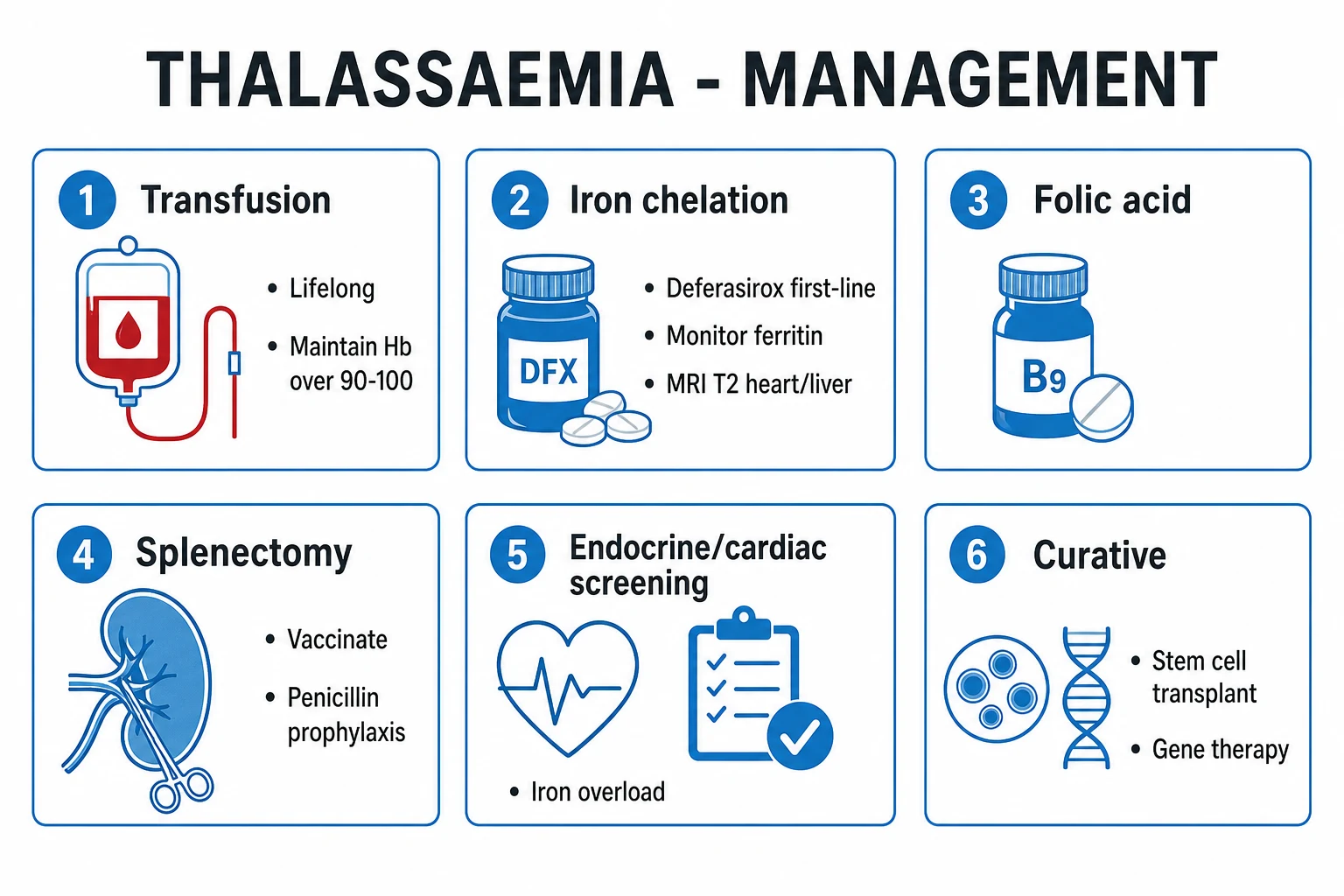
Definitive management is built around three pillars — transfusion, iron chelation and curative therapy — supported by folate, splenectomy when needed, and structured surveillance of iron-loaded organs. The goals are to suppress ineffective erythropoiesis, prevent bony deformity and allow normal growth in childhood, and to prevent or manage the iron-overload complications that determine survival.[2][6]
[2] [6]Pillar 1 — Regular transfusion. Patients with transfusion-dependent beta-thalassaemia major receive lifelong leucodepleted (and ideally washed, ABO/Rh- and extended-phenotype-matched) packed red cells every 2 to 4 weeks, titrated to keep the pre-transfusion haemoglobin at 90 to 105 g/L. This transfusion target is not arbitrary: it is the threshold that suppresses ineffective erythropoiesis, halts marrow expansion and bony deformity, allows normal growth and puberty in children, and limits the gastrointestinal iron absorption driven by expanded erythroid mass. Sub-target transfusion (below 90 g/L) allows marrow expansion to continue; over-transfusion (over 120 g/L) adds iron load without benefit. A child typically receives 10 to 15 mL/kg per transfusion, an adult 2 to 3 units. Alloimmunisation is minimised by extended phenotypic matching for the Rh and Kell antigens at first transfusion.[2][6]
Pillar 2 — Iron chelation (mandatory). Because there is no physiological route for iron excretion, every unit of transfused blood adds roughly 200 mg of elemental iron, and unchelated transfusion-dependent patients develop fatal iron overload within a decade. Chelation is started once ferritin rises or after 10 to 20 transfusions (around 100 mL/kg of red cells), and continued for life. Three agents are in routine use:
[2]Deferasirox (first-line)
- Oral, once-daily: 20 to 40 mg/kg/day (film-coated or dispersible)
- First-line for transfusion-dependent thalassaemia
- Effective on both hepatic and cardiac iron; convenient
- Monitor creatinine, LFTs and urine protein; rash, GI upset, rare renal tubular toxicity
Deferiprone
- Oral, 75 mg/kg/day in 3 divided doses
- Particularly effective at CARDIAC iron removal; used in combination for severe cardiac loading
- RISK OF AGRANULOCYTOSIS — weekly full blood count mandatory
- Other risks: arthropathy, zinc deficiency, hepatic dysfunction
Deferoxamine
- SC infusion 25 to 50 mg/kg over 8 to 12 hours, 5 to 7 nights/week via portable pump
- Original chelator — effective but highly burdensome
- Used IV in acute overload or when oral agents fail
- Risks: local infusion reactions, ototoxicity, retinopathy, growth retardation, Yersinia infection
Combination chelation (deferasirox plus deferiprone, or deferoxamine plus deferiprone) is used for severe or discordant iron loading, particularly when cardiac iron is high. Targets are a serum ferritin under 1000 mcg/L and a liver iron concentration of 3 to 7 mg/g dry weight with a cardiac T2-star over 20 ms. The chelation regimen is adjusted on the basis of ferritin trend, liver and cardiac MRI, and drug tolerability.[2][6]
Pillar 3 — Curative therapy. Two curative options exist:
[2]- Allogeneic haematopoietic stem cell transplant (HSCT) from an HLA-identical sibling donor, using myeloablative conditioning, is the standard of care in children with a matched sibling. Outcomes follow the Pesaro risk class: class 1 patients (no hepatomegaly, no portal fibrosis, regular chelation) achieve disease-free survival over 90 percent; results fall with age, hepatomegaly and iron loading. Matched unrelated donor and haploidentical transplants are increasingly used but carry higher graft-versus-host and rejection risks.[2]
- Gene therapy with betibeglogene autotemcel (beti-cel) — autologous CD34-positive haematopoietic stem cells transfused ex vivo with a lentiviral vector encoding a modified beta-globin (beta-T87Q) gene, then reinfused after myeloablation — achieved durable transfusion independence in most patients with a non-beta-0/beta-0 genotype, leading to regulatory approval in 2023/24. Exagamglogene autotemcel (exa-cel), which uses CRISPR-Cas9 editing of the BCL11A erythroid enhancer to reactivate fetal haemoglobin, is a complementary approach that has also received approval for transfusion-dependent thalassaemia. These therapies are transformative but currently expensive and accessible only in selected centres.[4][2]
Adjunctive therapy. Folic acid 5 mg daily compensates for the folate depleted by chronic erythropoietic drive. Vitamin C 2 to 3 mg/kg (max 100 to 200 mg) given on chelation days can enhance iron excretion with deferoxamine but increases iron availability and is used cautiously. Endocrine replacement — testosterone or oestrogen replacement for hypogonadism, growth hormone for growth failure, levothyroxine for hypothyroidism, insulin for diabetes — is tailored to the individual deficit. Bisphosphonates, calcium, vitamin D and weight-bearing exercise manage thalassaemia-associated osteoporosis.[2][6]
Splenectomy is reserved for hypersplenism — a transfusion requirement rising above 200 mL/kg/year of packed cells, symptomatic massive splenomegaly, or severe thrombocytopenia. It is delayed until after 4 to 6 years of age wherever possible to reduce the lifetime OPSI risk. Pre-operative preparation is non-negotiable: vaccinate against pneumococcus (PCV13 and PPSV23), meningococcus (ACWY conjugate plus MenB) and Haemophilus influenzae type b at least 2 weeks before splenectomy, and start lifelong oral penicillin V prophylaxis (children under 5: 125 mg twice daily; older children and adults: 250 to 500 mg twice daily), supplemented by a standby amoxicillin course and a fever-as-emergency patient action plan. Post-splenectomy thrombocytosis and venous thromboembolism risk require vigilance.[1][6]
Novel — luspatercept. For patients in whom curative therapy is not an option, the recombinant activin-receptor fusion protein luspatercept (a late-stage erythroid maturation agent, dosed at 1.0 to 1.25 mg/kg subcutaneously every 3 weeks) reduces transfusion burden by at least one-third in roughly one-third of transfusion-dependent beta-thalassaemia patients, with an acceptable safety profile, as established by the phase-3 BELIEVE trial.[3]
Specific Subtypes & Scenarios
Beta-thalassaemia minor (trait). Trait needs no treatment. The clinical task is to avoid the three errors: mistaking it for iron deficiency and prescribing iron (causes overload without benefit); missing a coexisting iron deficiency (treat the iron deficiency, not the trait); and failing to counsel carrier couples about reproductive risk. A trait patient with a haemoglobin below 100 g/L has something else going on — investigate.[1]
Beta-thalassaemia intermedia (non-transfusion-dependent thalassaemia, NTDT). Managed expectantly with folate, monitoring of growth and iron load, intermittent transfusion in pregnancy, infection, growth failure or acute complications, and chelation when liver iron concentration or ferritin rises even without regular transfusion (because gut iron absorption is increased by the expanded erythroid mass). NTDT patients carry a higher risk of venous thrombosis, pulmonary hypertension, leg ulcers, extramedullary haematopoietic masses and pigment gallstones than well-transfused major patients, and these complications drive the management plan as much as the anaemia.[2]
HbH disease. Folate supplementation, avoidance of oxidant drugs (sulphonamides, dapsone, primaquine, nitrofurantoin), transfusion in haemolytic crisis, and splenectomy for severe hypersplenism. The haemoglobin typically sits at 70 to 100 g/L and life expectancy is near-normal.[1]
Hb Bart hydrops fetalis. Incompatible with life; the parents (both carriers of a two-gene cis deletion, e.g. --SEA) must be counselled, the diagnosis confirmed prenatally, and termination offered. Rare survivors reach birth only through a programme of intrauterine transfusion and require lifelong transfusion or HSCT after birth. In every subsequent pregnancy the parents should be offered prenatal diagnosis by CVS at 10 to 12 weeks.[1][5]
Pregnancy. Trait usually tolerates pregnancy well; the priority is to screen the partner and, if both are carriers, offer prenatal diagnosis. In a woman with thalassaemia major, pregnancy is high-risk and demands a multidisciplinary team: optimise transfusion to a pre-transfusion Hb of 100 g/L, review chelation (deferasirox is discontinued pre-conception because of animal teratogenicity; deferiprone has been used in the second and third trimesters with specialist advice), assess cardiac function before conception (a cardiac T2-star over 20 ms is desirable), and give thromboprophylaxis in the splenectomised or immobilised patient. Outcomes in well-chelated women with preserved cardiac function are good.[2]
The splenectomised patient. Lifelong penicillin V prophylaxis, completion and boosting of the vaccination schedule, and a fever-as-emergency action plan with a standby course of amoxicillin. Thrombocytosis is monitored and venous thromboembolism prophylaxis given in high-risk situations. Pulmonary hypertension is screened for on echo.[6]
Complications & Pitfalls
Complications divide into those of the disease itself, those of transfusion and iron overload, and those of splenectomy — and examiners test the lethal one (iron-overload cardiomyopathy) repeatedly.[2]
Disease-related complications include growth retardation and delayed puberty from chronic anaemia and hypogonadism, bony deformity and osteoporosis with vertebral compression and long-bone fractures, extramedullary haematopoietic masses (paraspinal, pleural, intracranial), leg ulcers (especially in intermedia), pigment gallstones from chronic haemolysis, folate deficiency, splenomegaly with hypersplenism, and (in NTDT) a prothrombotic state with venous thromboembolism and pulmonary hypertension.[2]
Transfusion- and iron-overload-related complications dominate the adult course:
[2]Cardiac
- Iron-overload cardiomyopathy — dilated or restrictive, with arrhythmia and heart failure
- The LEADING CAUSE OF DEATH
- Detected by cardiac MRI T2-star (under 10 ms = severe)
- Reversed by intensive combination chelation (deferiprone-based)
Endocrine
- Diabetes mellitus (pancreatic iron)
- Hypogonadotropic hypogonadism, delayed puberty, infertility (pituitary iron)
- Hypothyroidism, hypoparathyroidism
- Growth hormone deficiency, short stature, osteoporosis
Hepatic
- Hepatic siderosis with fibrosis and eventual cirrhosis
- Increased hepatocellular carcinoma risk
- Transfusion-acquired viral hepatitis (HCV historically, HBV, HIV)
- Monitored by liver iron concentration and serology
Transfusion reactions
- Alloimmunisation (minimised by phenotypic matching)
- Febrile non-haemolytic and allergic reactions
- Delayed haemolytic transfusion reactions
- Volume overload in the high-output chronic anaemia patient
Splenectomy-related complications are overwhelming post-splenectomy infection (encapsulated organisms — pneumococcus, meningococcus, Haemophilus influenzae type b — and Capnocytophaga), post-splenectomy thrombocytosis with venous thromboembolism, and pulmonary hypertension.[6]
The classic pitfalls that cost marks (and lives) are: misdiagnosing trait as iron deficiency and giving iron; missing alpha-thal trait because the electrophoresis is normal (a normal electrophoresis in a microcytic patient of high-risk ancestry is alpha-thal trait until DNA testing shows otherwise); failing to chelate early so that cardiac iron accumulates silently; trusting a normal ferritin to exclude cardiac iron (cardiac and hepatic iron discordance is common — measure cardiac T2-star); and underestimating a fever in a splenectomised patient.[1][2]
Prognosis & Disposition
Untreated beta-thalassaemia major is fatal in early childhood from anaemia and high-output cardiac failure. With optimal transfusion and chelation, survival now extends into the fifth decade and beyond in well-resourced centres, and the determinants of long-term outcome are adherence to chelation, cardiac iron burden and endocrine reserve. The single strongest predictor of early mortality is the cardiac T2-star: a value under 10 ms carries a high one-year risk of arrhythmic death, and intensive chelation can reverse even severe cardiac loading if instituted in time.[2]
Haematopoietic stem cell transplant in childhood with a matched sibling donor yields disease-free survival over 90 percent and offers a definitive cure; outcome falls steadily with age and iron loading. Gene therapy with betibeglogene autotemcel has produced durable transfusion independence in most treated non-beta-0/beta-0 patients, with several years of follow-up; longer-term data are accruing.[4]
Patients require lifelong specialist haematology follow-up with a multidisciplinary team (cardiology for cardiac MRI and heart failure, endocrinology for hormone replacement and bone health, hepatology for iron-related liver disease, reproductive medicine for fertility, and dentistry for maxillary/dental complications). A structured transition from paediatric to adult services in adolescence prevents the loss-to-follow-up that drives late mortality. Patients are counselled about genetic risk to siblings and offspring, and offered carrier screening and prenatal diagnosis.[2][6]
Special Populations
Children. Transfusion is weight-based (10 to 15 mL/kg packed red cells); chelation is dosed per kilogram and deferred until ferritin or liver iron rises (typically after 10 to 20 transfusions, around 100 mL/kg). Growth, pubertal staging and bone age are monitored at every visit. HSCT is best performed early, while the child is in Pesaro class 1, before iron loading and hepatomegaly reduce survival. Deferoxamine can impair growth in young children and is now reserved for refractory cases.[2]
Pregnancy. Trait is well-tolerated; screen the partner and offer prenatal diagnosis if both are carriers. In thalassaemia major, plan conception in a multidisciplinary clinic: optimise transfusion to pre-transfusion Hb 100 g/L, discontinue deferasirox pre-conception, confirm cardiac fitness (cardiac T2-star over 20 ms, normal ejection fraction), and give thromboprophylaxis in the splenectomised or immobilised patient. Close fetal monitoring for growth restriction is warranted.[2]
The elderly trait patient. Incidental microcytosis in an older adult is often both trait and iron deficiency — iron studies still guide, and coexisting iron deficiency from occult blood loss must not be missed. Trait itself needs no treatment at any age.[1]
The immunocompromised and post-splenectomy patient. Aggressive fever management, completion of the vaccination schedule and penicillin prophylaxis are non-negotiable. Transfusion support in heavily pre-treated or immunosuppressed patients uses irradiated, CMV-matched components as locally indicated.[6]
At-risk ethnicities (Mediterranean, Middle Eastern, South Asian, South-East Asian) are the target of carrier screening programmes — school-leaving or premarital screening and universal antenatal screening — so that carrier couples are identified before an affected child is born.[5][6]
Evidence, Guidelines & Regional Differences
The international reference. The Thalassaemia International Federation (TIF) Guidelines set the global standard for transfusion practice (pre-transfusion Hb 90 to 100 g/L), chelation choice and monitoring, and end-organ surveillance. They form the basis of most national guidelines.[2]
Landmark trials.
[3] [4]Clinical evidence
Phase 3, randomised, double-blind, placebo-controlled
Population: Adults with transfusion-dependent beta-thalassaemia
Key finding
Reduction in transfusion burden of at least 33 percent from baseline over weeks 13 to 24 in roughly one-third of luspatercept-treated patients vs 0 percent on placebo
Clinical evidence
Phase 3, open-label, single-group
Population: Adults and children with non-beta-0/beta-0 genotype transfusion-dependent beta-thalassaemia
Key finding
Transfusion-free status achieved and sustained in most treated patients, with a clinically meaningful rise in total haemoglobin driven by vector-derived Hb
Regional deltas. In India, the ICMR and ISHBT drive universal antenatal haemoglobinopathy screening in high-prevalence states, with deferasirox first-line on cost-effectiveness grounds and a push to expand matched-sibling HSCT in regional centres; the ISHBT best-practice guidelines (2026) consolidate these recommendations.[5][6] In the United States and United Kingdom, the Sickle Cell and Thalassaemia Screening Programmes deliver newborn and antenatal screening, betibeglogene was approved by the FDA and MHRA in 2023/24, and exagamglogene autotemcel (Casgevy) was licensed for transfusion-dependent thalassaemia by the MHRA and FDA in 2024. In Europe, TIF guidelines dominate and access to gene therapy is expanding through selected centres. The central controversy across regions is equity of access to expensive curative therapies in the low- and middle-income countries where the disease burden actually lies.[2]
Exam Pearls
Alpha-thalassaemia gene-dose mnemonic — SILT
SILT
1 deletion — clinically silent, normal bloods
2 deletions — mild microcytosis, NORMAL electrophoresis
3 deletions — HbH disease, beta-4 tetramers, moderate haemolysis
4 deletions — Hb Bart hydrops fetalis, gamma-4 tetramers, lethal
Thalassaemia — THALS mnemonic
THALS
microcytosis with HIGH RBC count, normal iron; raised HbA2 in beta, NORMAL electrophoresis in alpha
four alpha-gene deletions; gamma-4 tetramers; incompatible with life — counsel and screen parents
inheritance; offer carrier screening and prenatal diagnosis to at-risk couples
iron-overload cardiomyopathy — prevented by chelation (deferasirox first-line)
curative with a matched sibling donor (DFS over 90 percent); gene therapy (betibeglogene) approved
The high-yield one-liners that win marks:
[1]- Beta = HBB on chromosome 11p15 (two alleles); alpha = HBA on chromosome 16p13 (four alleles). This pair of facts explains the entire clinical spectrum.
- Alpha-thal trait has a NORMAL Hb electrophoresis — raised HbA2 is beta trait only. The single commonest thalassaemia trap.
- Thalassaemia trait = microcytosis with a HIGH RBC count and normal iron/ferritin; do NOT give iron.
- Beta major presents at 6 to 12 months as HbF falls; HbF then re-rises to 70 to 90 percent; HbA absent.
- Hb Bart hydrops fetalis (four alpha deletions) is incompatible with life — counsel parents and offer prenatal diagnosis; the mother is at risk of pre-eclampsia and postpartum haemorrhage.
- Iron-overload cardiomyopathy is the leading cause of death — chelation is mandatory (deferasirox 20 to 40 mg/kg/day oral, first-line).
- Mentzer index = MCV / RBC count — over 13 thalassaemia trait, under 13 iron deficiency.
- Curative: matched-sibling HSCT in childhood (disease-free survival over 90 percent); gene therapy (betibeglogene autotemcel) approved 2023/24.
- Avoid oxidant drugs (sulphonamides, dapsone, primaquine, nitrofurantoin) in HbH disease.
- Splenectomy preparation is non-negotiable: vaccinate (pneumococcus, meningococcus, Hib) 2 weeks before, then lifelong penicillin V and a fever-as-emergency plan.
- Common Indian beta mutations: IVS1-5 (G to C) and the 619-base-pair deletion; common South-East Asian alpha deletion: --SEA.
- Cardiac T2-star is the prognostic test: over 20 ms normal, under 10 ms severe and high-risk — a normal ferritin does NOT exclude cardiac iron.[1][2]
Exam application bank (NEET-PG / INICET)
One-line answer
Thalassaemia is an inherited disorder of haemoglobin synthesis causing reduced (plus) or absent (zero) globin chain production, leading to microcytic hypochromic anaemia. Beta-thalassaemia (autosomal recessive, HBB on chromosome 11): major (Cooley anaemia; transfusion-dependent from 6 to 12 months with severe microcytic anaemia, failure to thrive, frontal bossing, hepatosplenomegaly, raised HbF and HbA2), intermedia (milder, transfusion-independent), minor/trait (asymptomatic, microcytosis out of proportion to anaemia, high RBC count, normal iron, raised HbA2). Alpha-thalassaemia (HBA on chromosome 16, four genes): one gene silent carrier, two trait (mild microcytosis, NORMAL electrophoresis), three HbH disease (moderate haemolytic anaemia, beta-4 tetramers), four Hb Bart hydrops fetalis (gamma-4 tetramers, incompatible with life). Diagnosis: FBC (microcytic hypochromic, high/normal RBC)
Worked stems (answer without another resource)
Stem 1 — Classic presentation. Map symptoms to mechanism; name the first investigation and first treatment step with dose/route if drug therapy is standard. [1]
Stem 2 — Unstable / complicated. List red flags that force immediate resuscitation, theatre, ICU, antidote, or reperfusion — and what you do in the first 15 minutes. [1]
Stem 3 — Atypical group. Elderly, pregnancy, child, or immunocompromised: how presentation and thresholds change. [1]
Stem 4 — Differential trap. Name the three closest mimics and one discriminator for each. [1]
Stem 5 — Disposition. Who goes home with safety-netting, who is admitted, who needs HDU/ICU/theatre, and what follow-up is mandatory. [1]
Rapid viva checklist
- Definition + classification
- Pathophysiology chain
- Bedside signs / criteria
- Score with exact components (if any)
- Emergency bundle
- Definitive therapy with doses
- Complications of disease and of treatment
- Special populations
- Guideline/trial name if classic
- Three exam traps
Coverage self-check
If you cannot answer any stem above from this page alone, re-read the matching section — the page is intended to be self-sufficient for final-prof and NEET-PG/INICET questions on Thalassaemia (Alpha & Beta).
References
- [1]Muncie HL Jr, Campbell J. Alpha and beta thalassemia Am Fam Physician, 2009.PMID 19678601
- [2]Kattamis A, Kwiatkowski JL, Aydinok Y, et al. Thalassaemia Lancet, 2022.PMID 35691301
- [3]Cappellini MD, Viprakasit V, Taher AT, et al. A Phase 3 Trial of Luspatercept in Patients with Transfusion-Dependent β-Thalassemia N Engl J Med, 2020.PMID 32212518
- [4]Locatelli F, Thompson AA, Kwiatkowski JL, et al. Betibeglogene Autotemcel Gene Therapy for Non-β(0)/β(0) Genotype β-Thalassemia N Engl J Med, 2022.PMID 34891223
- [5]Agarwal RK, et al. Prenatal hemoglobinopathy screening & prevention in India: A cross-sectional study Indian J Med Res, 2025.PMID 40844095
- [6]Dolai TK, et al. Best Practices for Thalassaemia Management: Recommendations by Indian Society of Haematology and Blood Transfusion Indian J Hematol Blood Transfus, 2026.PMID 42040714