Haematology · General Medicine
Thrombocytopenia & Immune Thrombocytopenia (ITP)
Also known as Thrombocytopenia · Immune thrombocytopenia · ITP · Idiopathic thrombocytopenic purpura · Low platelets
Thrombocytopenia (platelets under 150 x 10^9/L) is classified by MECHANISM: decreased production (marrow failure, leukaemia, chemo, B12/folate, alcohol, viruses), increased destruction (immune — ITP; microangiopathic — TTP/HUS; DIC; drugs; HIT) and sequestration (hypersplenism). Immune thrombocytopenia (ITP) is isolated thrombocytopenia (platelets under 100 x 10^9/L) with a normal marrow (normal or increased megakaryocytes) and no other cause. Presentation is mucocutaneous bleeding (petechiae, purpura, epistaxis, menorrhagia) or an asymptomatic incidental FBC finding. First-line: prednisolone 1 mg/kg (or dexamethasone 40 mg for 4 days); IVIg for rapid response, children, bleeding and pregnancy. Second-line: TPO receptor agonists (eltrombopag, romiplostim), rituximab, splenectomy, fostamatinib, rilzabrutinib. Red flags: TTP (thrombocytopenia plus neurology plus renal plus fever plus schistocytes) needs urgent plasma exchange and never platelets; HIT (platelet fall 5 to 10 days after heparin with thrombosis) needs all heparin stopped and argatroban; DIC needs the underlying cause treated.
On this page & tools
Your progress
Saved locally on this device.
Exam tags
Red flags
Overview & Definition
Thrombocytopenia — a platelet count under 150 x 10^9/L — is among the commonest abnormalities on a full blood count and the first step in evaluation is never the number itself but the mechanism behind it. Platelets are produced by marrow megakaryocytes, circulate for 7 to 10 days, and are removed mainly by the spleen. A low count therefore means production is too low, removal or consumption is too high, the platelets are pooled in an enlarged spleen, the blood has been diluted, or the count is simply spurious. The clinical skill is to classify by mechanism and, above all, to identify the dangerous causes (TTP, DIC, HIT, leukaemia, marrow failure) that demand immediate, specific action.[2]
Severity grading predicts bleeding risk and frames urgency: [1]
- Mild — 100 to 150 x 10^9/L: usually asymptomatic, often incidental.
- Moderate — 50 to 100 x 10^9/L: bruising, minor bleeding; safe for most procedures.
- Severe — under 50 x 10^9/L: spontaneous mucocutaneous bleeding.
- Critical — under 20 x 10^9/L: serious spontaneous bleeding and intracranial haemorrhage risk, which concentrates at platelets under 10. [1]
Immune thrombocytopenia (ITP) is an acquired autoimmune disorder defined by isolated thrombocytopenia (platelets under 100 x 10^9/L) with an otherwise normal full blood count, a normal bone marrow showing normal or increased megakaryocytes, and no other identifiable cause. It is the commonest cause of isolated severe thrombocytopenia in both adults and children, and its management has been transformed over the past decade by TPO receptor agonists and newer targeted agents that aim for a safe count rather than a cure.[1][3]
Classification
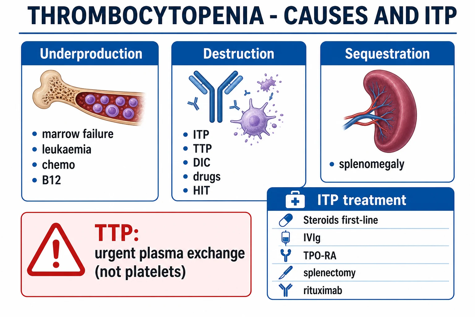
Thrombocytopenia is classified by MECHANISM — the framework that drives every differential and the single most rewarding organising principle in viva and long-case settings:[2]
- Decreased production — marrow failure (aplastic anaemia), marrow infiltration (acute leukaemia, myelodysplasia, myelofibrosis, metastatic carcinoma, lymphoma), chemotherapy and radiotherapy, megaloblastic anaemia (B12/folate deficiency), alcohol, and viral marrow suppression (HIV, parvovirus B19, dengue, rubella, mumps, EBV, CMV). The marrow is abnormal and there is often pancytopenia.
- Increased destruction — immune: primary ITP; secondary ITP (SLE, antiphospholipid syndrome, HIV, hepatitis C, common variable immunodeficiency, drugs, post-vaccination).
- Increased destruction — non-immune / consumptive (microangiopathic): TTP, haemolytic-uraemic syndrome (HUS), DIC, HELLP, pre-eclampsia, malignant hypertension, giant haemangioma (Kasabach-Merritt). The blood film shows schistocytes (fragmented red cells) — a finding that must never be missed.
- Sequestration — hypersplenism (portal hypertension, Gaucher disease, tropical splenomegaly, infiltration). Roughly a third of the platelet mass is normally pooled in the spleen; this rises to 90 percent when the spleen is massively enlarged.
- Dilutional — massive transfusion: each unit of stored whole blood carries few viable platelets, so bleeding replaced with packed cells and crystalloid without platelets dilutes the count.
- Spurious — EDTA-dependent platelet clumping (pseudothrombocytopenia); recheck in a citrate tube and confirm on the blood film. [1]
ITP is further classified by duration, which is a treatment-decision frame rather than mere taxonomy:[1]
- Newly diagnosed — under 3 months.
- Persistent — 3 to 12 months.
- Chronic — over 12 months (roughly 60 percent of adults; the chronic phase defines adult disease). [1]
And by aetiology: primary (idiopathic) versus secondary (driven by HIV, hepatitis C, H. pylori, SLE, drugs, common variable immunodeficiency, vaccination). The International Working Group standardised these terms in 2007 to replace the older "acute" and "chronic" labels, which implied aetiology that could not be inferred from duration alone.[1]
Epidemiology & Risk Factors
Adult ITP has an incidence of about 3 to 5 per 100,000 per year and a prevalence of 10 to 20 per 100,000. The age distribution is bimodal — a peak in young women (frequently associated with other autoimmunity) and a larger, more slowly rising peak in older adults, in whom the disease is more often chronic, harder to treat, and carries a higher bleeding risk at any count. In adults the disease is usually chronic.[1]
Childhood ITP affects about 5 per 100,000 per year, peaks at age 2 to 5, has an equal sex ratio, and is classically acute and post-viral, presenting one to four weeks after a respiratory or gastrointestinal infection or after MMR vaccination, with 70 to 80 percent recovering spontaneously within 6 months.[6]
Risk factors for SECONDARY ITP: HIV, hepatitis C, H. pylori (a higher-yield cause in high-prevalence regions such as India, Japan and Latin America, where eradication can produce lasting remission), SLE and antiphospholipid syndrome, certain drugs (quinine, quinidine, sulfonamides, vancomycin, rifampin, carbamazepine, GP IIb/IIIa inhibitors), and vaccination (MMR).[2]
Spurious (EDTA-dependent) thrombocytopenia affects 0.1 to 0.2 percent of samples and must be excluded before any treatment is given — treating pseudothrombocytopenia with steroids or splenectomy is a classic, avoidable catastrophe. Gestational thrombocytopenia complicates 5 to 8 percent of pregnancies and is the commonest cause of low platelets in pregnancy; it is benign for mother and baby. Neonatal alloimmune thrombocytopenia (NAIT), by contrast, affects about 1 in 1,000 to 2 in 1,000 births and can cause severe neonatal intracranial haemorrhage.[1]
Pathophysiology
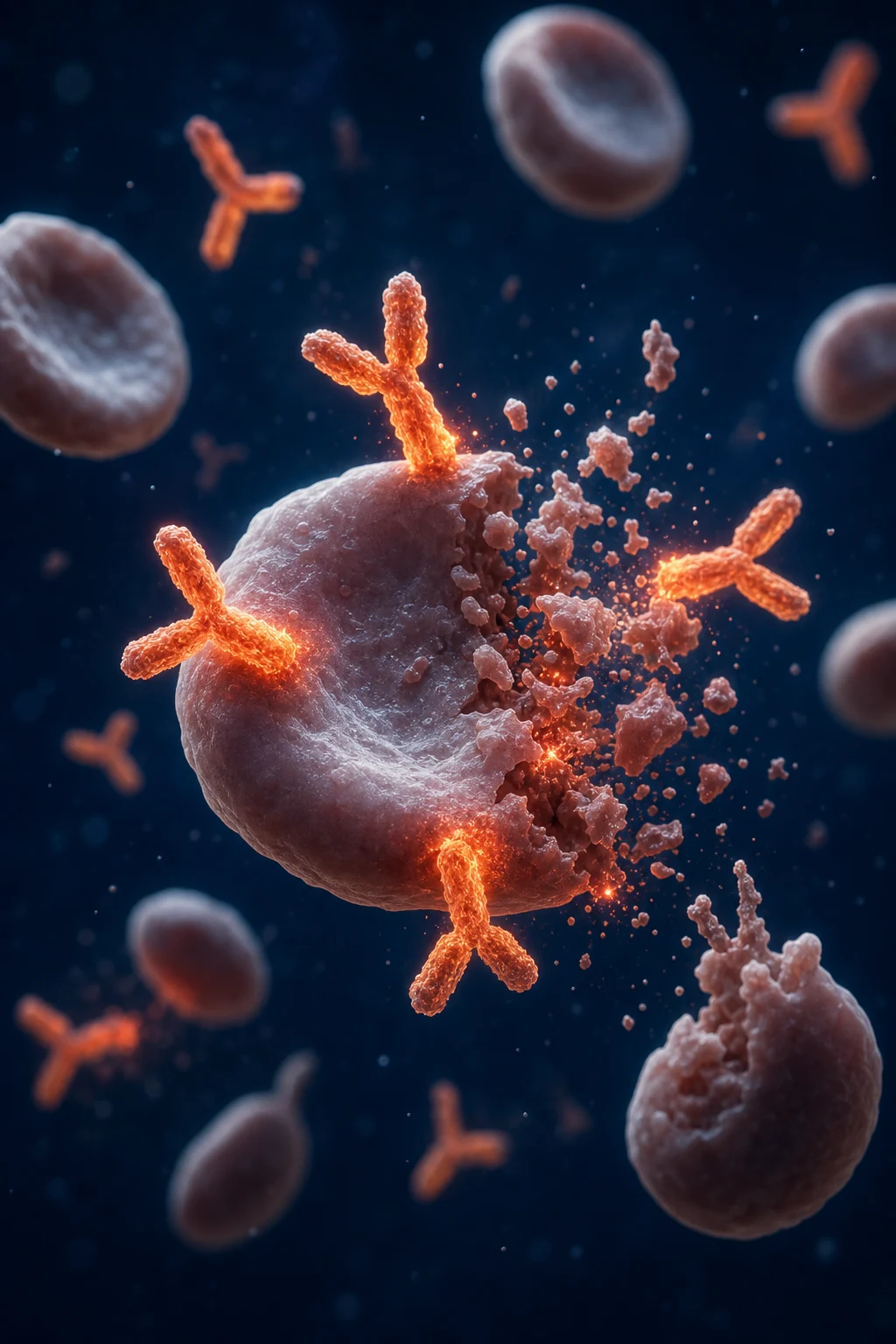
ITP is not simply a disorder of platelet destruction: it is a combined defect of autoantibody-mediated platelet destruction plus impaired platelet production. Understanding both limbs explains why different therapies work and why single-target treatment often disappoints.[1]
- Autoantibody formation. Polyclonal IgG autoantibodies are produced (largely in the spleen and lymph nodes) against platelet-surface glycoproteins, mainly GPIIb/IIIa (integrin alpha-IIb/beta-3) and GPIb/IX. The autoantibody response is T-cell driven: autoreactive CD4+ T-helper cells lose tolerance to these glycoprotein antigens and activate autoreactive B cells.
- Opsonisation and destruction. Antibody-coated platelets are recognised by splenic macrophage Fc-gamma receptors and phagocytosed, shortening platelet survival from the normal 7 to 10 days to hours. The liver can also clear platelets coated with anti-GPIb antibodies, which is why splenectomy is not universally curative.
- Impaired production. The same antibodies and cytotoxic CD8+ T cells damage megakaryocytes, so platelet production is inappropriately low for the degree of thrombocytopenia. This is why the marrow shows increased but dysfunctional megakaryocytes — they are present in number but cannot compensate.
- Inappropriately normal thrombopoietin (TPO). Unlike other cytopenias (where TPO or erythropoietin rise in response), TPO is not elevated in ITP, because TPO is produced constitutively by the liver and cleared by platelets. The surviving megakaryocyte pool is therefore not maximally stimulated — the rationale for TPO receptor agonists (eltrombopag, romiplostim, avatrombopag), which stimulate the surviving megakaryocytes. [1]
Why splenectomy works: the spleen is both the major site of platelet destruction and a major site of autoantibody production — removing it addresses both limbs. Even so, about a third of splenectomised patients relapse, reflecting extra-splenic (hepatic, marrow) clearance and antibody production.[1]
The dangerous destructive thrombocytopenias — distinct mechanisms (an exam favourite):[2]
- TTP — severe ADAMTS13 deficiency (an inherited or autoantibody-mediated loss of the von Willebrand factor-cleaving protease) leaves ultra-large von Willebrand factor multimers uncleaved on endothelial surfaces, generating platelet-rich microvascular thrombi across the arterioles and capillaries. The result is platelet consumption plus end-organ ischaemia (brain, kidney, heart) — microangiopathic haemolytic anaemia with schistocytes.
- HUS — Shiga-toxin-mediated endothelial injury (typically enterohaemorrhagic E. coli O157:H7) in children, producing a renal-predominant thrombotic microangiopathy.
- DIC — widespread, uncontrolled activation of coagulation (sepsis, malignancy, trauma, obstetric catastrophe) consumes platelets and clotting factors while generating microthrombi and bleeding simultaneously.
- HIT — IgG against the platelet factor 4 (PF4)-heparin complex cross-links platelet Fc receptors, causing platelet activation, consumption and a paradoxical prothrombotic state — the patient presents with thrombosis, not bleeding. [1]
Clinical Presentation
Typical ITP presents with mucocutaneous (platelet-type) bleeding, and the pattern is the key clue:[1]
- Petechiae — 1 to 2 mm, non-blanching pin-point haemorrhages, typically in dependent areas (lower legs, areas under a sphygmomanometer cuff) and pressure points. They are the hallmark of platelet-type bleeding.
- Purpura and ecchymoses — larger spontaneous bruises, often appearing overnight with no remembered trauma.
- Wet purpura — haemorrhagic blood blisters on the buccal mucosa; a high-risk marker for serious bleeding.
- Mucosal bleeding — epistaxis, gum bleeding, menorrhagia, gastrointestinal bleeding, haematuria, conjunctival haemorrhage. [1]
A critical, frequently-examined distinction: petechiae and mucocutaneous bleeding = a platelet problem (number or function); deep bleeding into joints and muscles (haemarthrosis, compartment bleeding) = a coagulation-factor problem (haemophilia, von Willebrand disease). The site of bleeding predicts the defect. [1]
Asymptomatic presentation is common in adults — many are detected on a routine full blood count showing isolated thrombocytopenia, or are picked up incidentally during evaluation for another problem. Fatigue, often disproportionate to the count, is increasingly recognised as part of ITP and significantly impairs quality of life.[1]
Intracranial haemorrhage is rare (well under 1 percent overall) but is the feared, potentially fatal complication, concentrated at platelets under 10 and more likely in elderly patients and those with wet purpura, active mucosal bleeding, or a previous bleed. [1]
Atypical presentations: elderly patients bleed more at any count and tolerate bleeding worse; pregnancy (gestational thrombocytopenia vs ITP, with neonatal risk); children present 1 to 4 weeks after a viral illness with abrupt isolated thrombocytopenia and are otherwise completely well; secondary ITP may carry the features of the underlying disease (malar rash and arthritis of SLE, lymphadenopathy of HIV or lymphoma). [1]
Key examination finding: in primary ITP the patient is otherwise well, with NO splenomegaly and NO lymphadenopathy. The presence of either — or of other cytopenias (anaemia, neutropenia), macrocytosis, or systemic features — points to another cause and mandates further investigation.[1]
ITP
- Isolated thrombocytopenia, otherwise well
- Normal PT/APTT, normal film (NO schistocytes)
- Normal marrow, increased megakaryocytes, no splenomegaly
- First-line prednisolone / IVIg; emergency only if platelets under 10 with bleeding
TTP
- Thrombocytopenia + neurology + renal + fever + schistocytes
- ADAMTS13 markedly low (under 10 percent)
- Urgent plasma exchange within 4 to 8 hours
- Do NOT give platelets (fuels microvascular thrombosis)
DIC
- Underlying severe illness (sepsis, malignancy, trauma, obstetric)
- PT and APTT prolonged, fibrinogen low, D-dimer high
- Consumes platelets AND factors; bleeds and clots together
- Treat the cause; supportive platelets/FFP/cryoprecipitate
HIT
- Platelet fall 5 to 10 days after heparin (faster if previously exposed)
- Thrombosis, not bleeding (paradoxical prothrombotic)
- IgG against PF4-heparin; positive ELISA and functional (serotonin-release) assay
- Stop ALL heparin; start argatroban or bivalirudin; warfarin only after platelet recovery
Differential Diagnosis
Every low platelet count must be run through the mechanism framework. The decisive investigations are the blood film (schistocytes, blasts, parasites), the coagulation screen, and, when indicated, the bone marrow. A common viva trap is anchoring on ITP before excluding the dangerous mimics.[2]
- Decreased production — aplastic anaemia, acute leukaemia, myelodysplasia, chemotherapy, megaloblastic anaemia (B12/folate), alcohol, viral marrow suppression. Marrow abnormal; often pancytopenia; macrocytosis may be present. The marrow examination is diagnostic.
- Increased destruction — immune: primary ITP; secondary ITP (SLE, HIV, HCV, antiphospholipid syndrome, common variable immunodeficiency); drugs — quinine, quinidine, sulfonamides, vancomycin, rifampin, GP IIb/IIIa inhibitors (abciximab, eptifibatide, tirofiban), carbamazepine, penicillin, vancomycin, rituximab, ranitidine. Drug-induced immune thrombocytopenia typically recovers 1 to 2 weeks after stopping the culprit.
- Increased destruction — microangiopathic (MAHA): TTP, HUS, DIC, HELLP, pre-eclampsia, malignant hypertension, disseminated malignancy. Blood film shows schistocytes (fragmented red cells, helmet cells) — a red flag that must not be missed, because the response (plasma exchange) is diametrically opposed to that of ITP.
- Sequestration: hypersplenism from portal hypertension, Gaucher disease, tropical splenomegaly, lymphoma. Splenomegaly is the clue and excludes primary ITP.
- Dilutional: massive transfusion — calculate roughly 1 platelet pool per unit of packed cells replaced when bleeding exceeds one blood volume.
- Pseudothrombocytopenia: EDTA-induced platelet clumping in the collection tube; recheck in citrate and on the film — the in vivo count is normal.
- Pregnancy-specific: gestational thrombocytopenia (mild, third trimester, benign), pre-eclampsia/HELLP, and rare TTP/HUS/AFLP that complicate the third trimester.
- Inherited thrombocytopenias (rare, but examine the family and the platelet size): Wiskott-Aldrich syndrome (small platelets, eczema, immunodeficiency, X-linked), Bernard-Soulier syndrome (giant platelets, GPIb deficiency, autosomal recessive), MYH9-related disease (giant platelets, Dohle-like bodies, nephritis, deafness, cataracts), and CAMT (congenital amegakaryocytic thrombocytopenia). The platelet volume (MPV) on the FBC is a cheap, high-yield clue: small platelets point to Wiskott-Aldrich; giant platelets point to Bernard-Soulier or MYH9-related disease.[1]
Gestational thrombocytopenia
- Commonest cause in pregnancy (5 to 8 percent)
- Mild, platelets usually over 75
- Third trimester onset, no bleeding
- Benign for mother and baby; resolves postpartum
ITP in pregnancy
- 1 to 2 percent of pregnancies
- Often predates pregnancy, lower counts
- Neonatal thrombocytopenia risk (transplacental IgG)
- Treat with IVIg +/- low-dose steroids; avoid TPO-RA and rituximab
Pre-eclampsia / HELLP
- After 20 weeks, with hypertension and proteinuria
- Platelets fall with haemolysis and abnormal LFTs (HELLP)
- Delivery is the treatment
- Coagulation may be deranged; watch for DIC
TTP / HUS in pregnancy
- Rare, often third trimester or postpartum
- Microangiopathic film with schistocytes, ADAMTS13 low (TTP)
- Plasma exchange (TTP); supportive (HUS)
- Can be life-threatening; do not deliver for thrombocytopenia alone
Key numbers in thrombocytopenia
Clinical & Bedside Assessment
A focused history defines the bleeding phenotype and hunts for a cause: bleeding pattern and sites, onset and tempo, drug history (prescription, over-the-counter, herbal, recent quinine in tonic water), recent infection or vaccination (MMR, COVID-19), pregnancy status, systemic features of SLE (malar rash, arthritis, photosensitivity, oral ulcers) or infection (fever, night sweats, weight loss), alcohol intake, and a family history of low platelets or bleeding (inherited thrombocytopenia).[2]
Quantify bleeding severity objectively with the ISTH Bleathing Assessment Tool (BAT) or the WHO bleeding scale (grades 0 to 4). A high BAT score predicts worse outcomes and supports treatment beyond the count alone — a patient with no bleeding and a count of 25 is managed differently from one with wet purpura and a count of 25. [1]
Examination is decisive:[1]
- Skin and mucous membranes — petechiae and purpura in dependent distribution, the oral cavity for wet purpura (blood blisters — a danger sign), gums, conjunctivae.
- Abdomen — palpate for splenomegaly (which excludes primary ITP and points to sequestration, infiltration, portal hypertension, or myeloproliferative disease) and hepatomegaly.
- Lymph nodes — lymphadenopathy points to lymphoma, HIV, or chronic infection.
- Systemic clues — malar rash and arthritis (SLE), jaundice and stigmata of chronic liver disease (hypersplenism, liver-synthesised factor deficiency), fever and infection focus (sepsis, DIC), signs of pregnancy, stigmata of malignancy. [1]
The cardinal bedside message: isolated thrombocytopenia in an otherwise well patient with no splenomegaly and no lymphadenopathy is ITP until proven otherwise — but splenomegaly or additional cytopenias demand a different diagnosis and a marrow examination. [1]
Investigations
ITP is a diagnosis of exclusion — investigations confirm the criteria and, more importantly, exclude the dangerous mimics. The goal is not to "prove" ITP but to exclude everything else.[1][3]
First-line tests (every patient): [1]
- Full blood count — isolated thrombocytopenia; haemoglobin, mean corpuscular volume, and white cell count (including differential) normal. Pancytopenia or macrocytosis demands another diagnosis (marrow failure, megaloblastic, leukaemia).
- Blood film (essential) — confirms true thrombocytopenia (excludes EDTA clumping); looks for large/giant platelets (young platelets, consistent with peripheral destruction), schistocytes (MAHA — do not miss TTP/HUS/DIC), blasts (leukaemia), malarial parasites, Dohle-like bodies (MYH9), and leucoerythroblastic features (marrow infiltration).
- Repeat platelet count in a citrate tube — to exclude EDTA-dependent pseudothrombocytopenia.
- Coagulation screen (PT, APTT, fibrinogen, D-dimer) — normal in ITP. Prolonged PT/APTT with low fibrinogen and high D-dimer = DIC; isolated APTT prolongation with thrombosis raises antiphospholipid syndrome.
- Blood group (ABO/Rh), renal and liver function, LDH, haptoglobin, direct antiglobulin test — to assess haemolysis and renal/hepatic contributors, and to plan anti-D therapy. [1]
Bone marrow aspirate and trephine — normal or increased megakaryocytes, otherwise normal marrow. It is indicated if there are atypical features (additional cytopenias, macrocytosis, abnormal film), age over 60, before splenectomy, or in refractory disease. It is not required in a typical young adult or child with isolated thrombocytopenia and a normal film — performing it routinely is over-investigation.[1]
Secondary-cause screen (in all newly diagnosed adults): HIV and hepatitis C serology (mandatory, because they change management), H. pylori testing (urea breath test or stool antigen; higher yield in high-prevalence regions such as India and Japan), antinuclear antibody and anti-dsDNA, beta-HCG, thyroid function (ITP can coexist with autoimmune thyroid disease), quantitative immunoglobulins (common variable immunodeficiency), and a pregnancy test in women of childbearing age.[2]
Tests NOT routinely useful: platelet-associated antibody (low specificity — present in many thrombocytopenias, not diagnostic); anti-GPIIb/IIIa antibody assays (research only); routine thrombopoietin level. ADAMTS13 assay is sent only when TTP is suspected (a level under 10 percent is diagnostic) — never as a blanket test. [1]
Management — Resuscitation
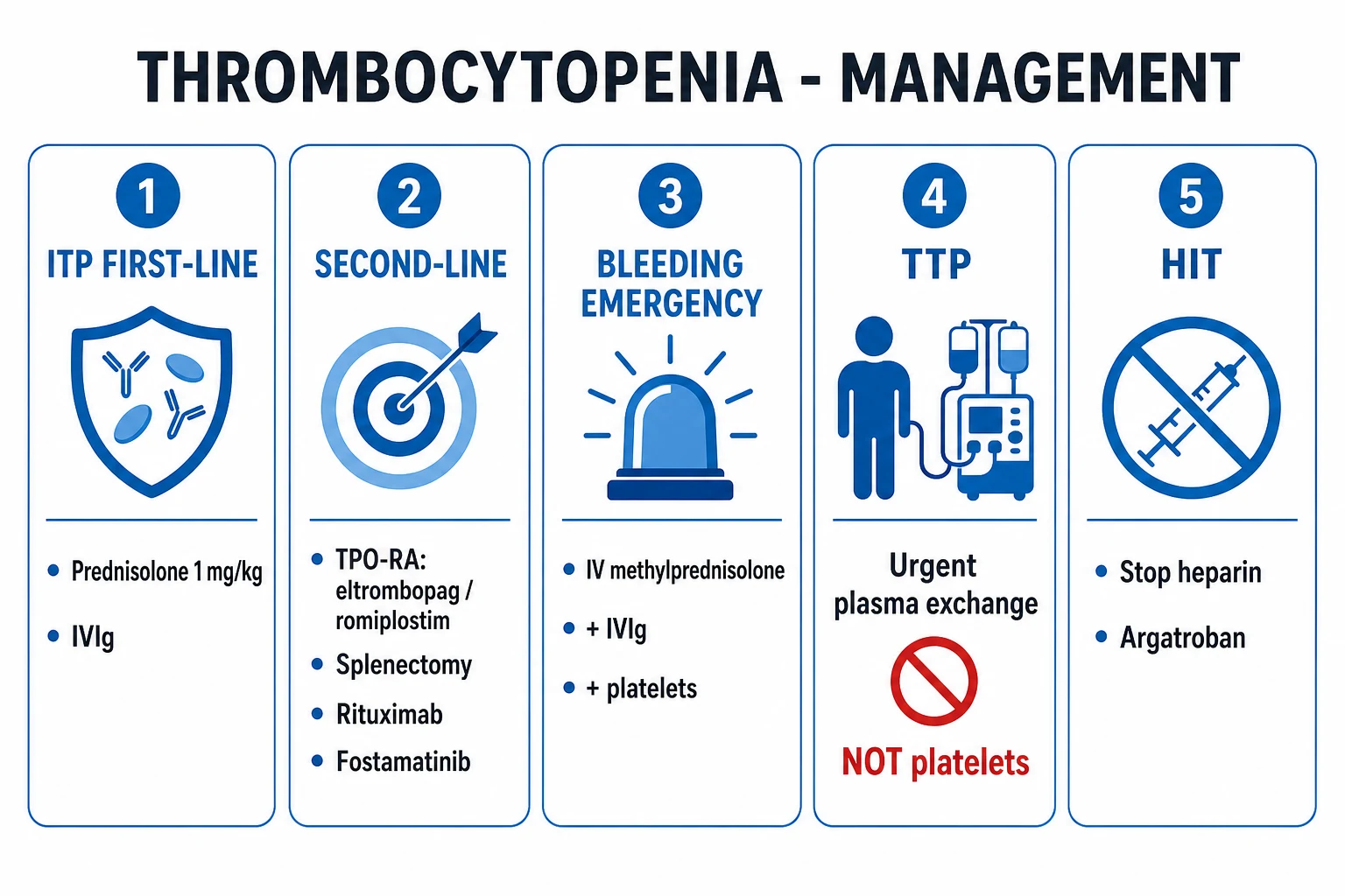
ABCDE assessment first. The single most important triage question is "is this ITP, or is this TTP/DIC/HIT?", because the treatments diverge completely. Life-threatening bleeding or suspected intracranial haemorrhage in ITP requires a simultaneous rescue bundle given together:[1][3]
- IV methylprednisolone 1 g daily for 1 to 3 days PLUS
- IVIg 0.4 to 1 g/kg/day PLUS
- Platelet transfusion (1 to 2 adult doses), all given concurrently, with tranexamic acid 1 g IV if there is no thrombotic contraindication. [1]
In ITP, transfused platelets are rapidly destroyed (survival measured in hours), so they are given with IVIg and steroids only for active life-threatening bleeding or before an emergency procedure, not prophylactically and not to raise the number alone. [1]
General haemostatic measures: avoid intramuscular injections, NSAIDs, antiplatelets (aspirin, clopidogrel), and invasive procedures; control hypertension; suppress menses; give tranexamic acid for mucosal bleeding when there is no thrombotic contraindication and no haematuria (clots can obstruct the renal tract). [1]
The dangerous mimics have diametrically different resuscitation — getting this wrong is fatal:[2]
- Suspected TTP — start urgent plasma exchange within 4 to 8 hours plus corticosteroids and caplacizumab if available; do NOT give platelet transfusions (they fuel microvascular thrombosis and worsen organ ischaemia); do NOT wait for the ADAMTS13 result — treat on clinical suspicion.
- Suspected HIT — stop ALL heparin (including flushes, heparin-coated lines, and LMWH) and start a non-heparin anticoagulant (argatroban or bivalirudin); warfarin is added only once the platelet count has recovered, to avoid warfarin-induced skin necrosis and protein C depletion.
- DIC — treat the underlying cause (sepsis source control, obstetric delivery, malignancy); give supportive blood-product support (platelets if bleeding and under 50, fresh frozen plasma if PT/APTT prolonged, cryoprecipitate if fibrinogen under 1.5 g/L) guided by counts and coagulation. [1]
Management — Definitive & Stepwise
The guiding principle, repeated until it is reflexive: aim for a SAFE platelet count (haemostatic, generally over 30), NOT a normal count. Asymptomatic patients with platelets over 20 to 30 may be observed without treatment. Treat the bleeding patient, not the number.[3]
- Prednisolone 1 mg/kg/day (range 0.5 to 2 mg/kg) orally, as a short course with taper over 4 to 8 weeks; about two-thirds respond within days to weeks, with the peak response at 1 to 2 weeks. Long, open-ended courses are avoided because of cumulative steroid toxicity.
- High-dose dexamethasone 40 mg daily for 4 days is an alternative first-line pulse — faster initial response and less cumulative steroid burden, though durable remission rates are similar to prednisolone.
- IVIg 0.4 g/kg/day for 5 days (or 1 g/kg for 1 to 2 days) — rapid response in 24 to 48 hours, lasting 2 to 4 weeks, in roughly 80 percent. Use for severe bleeding, before surgery or dental work, in pregnancy, in children with significant bleeding, and as a bridge to definitive therapy. It works by saturating splenic Fc receptors and blocking autoantibody-mediated clearance.
- IV anti-D 50 to 75 mcg/kg — an alternative for Rh(D)-positive, non-splenectomised patients; it works by coating the patient's own Rh(D)-positive red cells, diverting splenic macrophage clearance away from platelets. It causes a mild haemolysis (drop in haemoglobin of about 0.5 to 2 g/dL) — avoid if haemoglobin is already low, and watch for severe intravascular haemolysis, a rare but reported complication. [1]
Second-line therapy (chronic, persistent, or refractory disease, or steroid dependence):[1][3]
- TPO receptor agonists — eltrombopag 50 mg oral daily (start 25 mg in East-Asian ancestry or hepatic impairment; titrate to a maximum of 75 mg; take on an empty stomach, 2 hours apart from polyvalent cations — calcium, iron, antacids), romiplostim 1 to 10 mcg/kg subcutaneously weekly (titrate weekly), and avatrombopag. They stimulate megakaryocyte production; effective in roughly 80 percent and used long-term. The RAISE trial established eltrombopag as durable second-line therapy over 6 months in chronic ITP. Monitor liver function, platelet count (avoid overshoot thrombocytosis over 400), and reticulin fibrosis.[7]
- Rituximab 375 mg/m^2 IV weekly for 4 doses — anti-CD20 monoclonal antibody depleting B cells; a response rate of about 60 percent initially, with roughly a third maintaining a durable response at 1 to 5 years. Screen for hepatitis B before use (reactivation risk).
- Splenectomy — historically curative in about two-thirds; carries overwhelming post-splenectomy infection (OPSI) risk and portal vein thrombosis; vaccinate at least 14 days before surgery (pneumococcal, Haemophilus influenzae type b, and meningococcal ACWY vaccines), and give lifelong penicillin V 500 mg twice daily prophylaxis (or erythromycin if allergic). Preferred laparoscopically; once first-line for chronic ITP, now deferred and used far less because of TPO-RA efficacy.
- Fostamatinib 100 mg oral daily (titrate to 150 mg) — a SYK inhibitor approved for chronic ITP refractory to other treatments.
- Rilzabrutinib — an oral BTK inhibitor with demonstrated response in refractory ITP in a phase 1b/2 trial (NEJM 2022), expanding the targeted-therapy armamentarium.[5]
Escalation triggers: failure of, or dependence on, first-line therapy; recurrent bleeding; chronic disease with impaired quality of life. Second-line selection is individualised to side-effect profile, comorbidity, pregnancy intent, and patient preference — there is no rigid ladder, and combination therapy (e.g., TPO-RA plus rituximab) is increasingly used.[4]
Specific Subtypes & Scenarios
- Childhood ITP — acute, presenting 1 to 4 weeks after a viral illness or MMR vaccination; the child is otherwise completely well, with isolated thrombocytopenia and no splenomegaly. Management is observation first: a child with no or mild bleeding and platelets over 20 is observed with activity restriction and return precautions, because 70 to 80 percent recover spontaneously within 6 months. Treat only for significant bleeding (wet purpura, active mucosal bleeding) with IVIg 0.8 to 1 g/kg, anti-D 50 to 75 mcg/kg, or a short course of prednisolone 2 to 4 mg/kg/day for a few days (avoid prolonged steroids in children). Intracranial haemorrhage is rare (under 1 percent). Splenectomy is almost never performed in childhood — it is deferred at least until age 5 to 6 because of the high OPSI risk in young children and the high spontaneous remission rate.[6]
- Pregnancy — distinguish gestational thrombocytopenia (common, mild, platelets usually over 75, onset in the third trimester, no bleeding, resolves postpartum, no neonatal harm) from ITP (often earlier onset, lower counts, may predate pregnancy, and carries neonatal thrombocytopenia risk from transplacental IgG). Treat ITP in pregnancy with IVIg and/or low-dose steroids; TPO-RA and rituximab are not recommended in pregnancy. Aim for platelets over 50 for delivery and over 75 for epidural/spinal anaesthesia. Monitor the neonatal cord platelet count — neonatal thrombocytopenia peaks at 2 to 5 days postpartum. The mode of delivery is obstetric, not dictated by ITP.[1]
- Secondary ITP — treat the underlying cause (HIV with antiretrovirals; HCV with direct-acting antivirals; H. pylori with eradication, which can produce lasting remission in high-prevalence regions; SLE with immunosuppression), often alongside standard ITP therapy.[2]
- Drug-induced immune thrombocytopenia — stop the offending drug (quinine, quinidine, sulfonamides, vancomycin, rifampin, GP IIb/IIIa inhibitors — abciximab, eptifibatide, tirofiban, carbamazepine, ranitidine, penicillin, trimethoprim-sulfamethoxazole); recovery in 1 to 2 weeks after the drug clears. A careful drug history (including quinine in tonic water and over-the-counter remedies) is mandatory.
- Heparin-induced thrombocytopenia (HIT) — platelet fall 50 percent or more from baseline, 5 to 10 days after heparin (faster if previously sensitised within 100 days), with thrombosis (not bleeding). Diagnose with the 4Ts pretest probability (thrombocytopenia, timing, thrombosis, other cause) and confirm with a PF4 ELISA and a functional serotonin-release assay. Stop ALL heparin, start argatroban or bivalirudin, and bridge to warfarin only after platelet recovery; do not give platelet transfusions.
- Neonatal alloimmune thrombocytopenia (NAIT) and post-transfusion purpura are rare but important immune-mediated mechanisms. NAIT arises from maternal alloimmunisation against paternal HPA antigens (classically HPA-1a) and can cause severe neonatal intracranial haemorrhage; post-transfusion purpura causes profound thrombocytopenia 5 to 10 days after transfusion in previously sensitised patients.
Complications & Pitfalls
Disease complications: severe mucosal or gastrointestinal bleeding; menorrhagia (which can cause iron deficiency and require hormonal suppression); intracranial haemorrhage — rare overall (under 1 percent) but catastrophic when it occurs, concentrated at platelets under 10 and in elderly patients and those with active bleeding or wet purpura. Fatigue and impaired quality of life are common and independent of the count.[1]
Treatment-related complications: [1]
- Corticosteroids — diabetes, hypertension, osteoporosis, peptic ulcer, infection, mood disturbance, insomnia, weight gain, avascular necrosis, myopathy; the reason long courses are avoided and TPO-RA are deployed earlier in older patients.
- IVIg — infusion reactions, headache (including aseptic meningitis), thromboembolism (stroke, MI, DVT — important in older/vascular patients), renal impairment, haemolytic anaemia (especially in non-O blood groups, due to isohaemagglutinin load), and transfusion-related acute lung injury.
- Splenectomy — overwhelming post-splenectomy infection (OPSI) from encapsulated organisms (pneumococcus, meningococcus, H. influenzae), portal vein thrombosis, and the operative risks of a laparoscopic procedure. Requires pre-op vaccination, lifelong penicillin V, a medical alert, and a standing instruction to present with any febrile illness.
- TPO receptor agonists — thrombosis (when counts overshoot), hepatotoxicity (eltrombopag — monitor LFTs), rebound thrombocytopenia on abrupt cessation, and marrow reticulin fibrosis with long-term use (monitor with periodic marrow if used for years).
- Rituximab — infusion reactions, hepatitis B reactivation (screen first), late-onset neutropenia, hypogammaglobulinaemia with repeated courses, and rare progressive multifocal leukoencephalopathy. [1]
Classic pitfalls: missing TTP (fatal, never transfuse platelets); missing leukaemia or marrow failure (do a marrow if there are atypical features, age over 60, or refractory disease); over-treating the number (a well patient with platelets over 30 needs observation, not escalating immunosuppression); failing to vaccinate before splenectomy; missing secondary causes (HIV, HCV, H. pylori); diagnosing EDTA pseudothrombocytopenia as true disease (always check a citrate tube and the film); and confusing HIT (thrombosis, stop heparin) with other drug-induced thrombocytopenias (bleeding, stop the drug). [1]
Prognosis & Disposition
- Childhood ITP — 70 to 80 percent spontaneous remission within 6 months; only 20 to 25 percent become chronic; mortality from intracranial haemorrhage is very low (under 1 percent).[6]
- Adult ITP — usually chronic; only 10 to 30 percent achieve a lasting remission; overall mortality is low but intracranial haemorrhage is catastrophic when it occurs, concentrated in elderly patients with counts under 10 and active bleeding. Quality of life is significantly impaired by fatigue, bleeding worry, and treatment side-effects — treatment aims for a safe count and preserved quality of life, not cure.
- Disposition — outpatient management if there is no or mild bleeding and platelets are over 20 to 30; admit for active bleeding, wet purpura, platelets under 10 to 20, or to initiate urgent therapy.
- Splenectomy achieves a durable response in about two-thirds of operated patients, but is now used far less because of TPO-RA efficacy and the permanent OPSI risk.[1]
Bleeding risk by platelet count
Mucocutaneous bleeding risk; treat if symptomatic
Special Populations
- Children — weight-based dosing (prednisolone 1 to 2 mg/kg, IVIg 0.4 to 1 g/kg, anti-D 50 to 75 mcg/kg); observation-first for mild disease; splenectomy is avoided (deferred at least until age 5 to 6 due to OPSI risk). Activity restriction (no contact sports) while counts are low.[6]
- Pregnancy — prefer IVIg and low-dose steroids; avoid TPO-RA and rituximab; plan delivery with haematology and obstetrics; aim for platelets over 50 for delivery and over 75 for neuraxial anaesthesia; monitor the neonate for thrombocytopenia (transplacental IgG) with a cord platelet count.[1]
- Elderly — bleed more at any given platelet count, have a higher intracranial haemorrhage risk, and tolerate corticosteroid toxicity poorly (diabetes, osteoporosis, delirium, fluid retention), favouring earlier TPO-RA use and a lower threshold to treat.[4]
- Immunocompromised (HIV, post-transplant) — ITP is common; manage the underlying immunodeficiency and adjust immunosuppression; use rituximab cautiously (infection and reactivation risk) and watch for drug interactions.
- Anticoagulated patient with new thrombocytopenia — balance bleeding versus thrombosis; if HIT is possible, stop heparin first and switch to a non-heparin anticoagulant; temporarily interrupt anticoagulation if platelets fall under 30 to 50.
Evidence, Guidelines & Regional Differences
- The international consensus report (Provan 2019, Blood Advances) is the global reference standard for primary ITP, covering diagnosis, first- and second-line therapy, and emergency management.[1]
- The American Society of Hematology 2019 guidelines (Neunert), with a 2022 review update (Neunert 2024), formalise first- and second-line therapy, the role of TPO-RA, and the declining place of splenectomy, and explicitly endorse the "treat the patient, not the number" philosophy.[3][4]
- The RAISE trial (Cheng 2011, Lancet) established eltrombopag as durable 6-month second-line therapy for chronic ITP — a landmark that shifted TPO-RA from rescue to mainstream long-term therapy.[7]
- Romiplostim for paediatric ITP and TPO-RA broadly have transformed second-line therapy and reduced the need for splenectomy, which has fallen out of first favour.[6]
- Rilzabrutinib, an oral BTK inhibitor, demonstrated response in refractory ITP (NEJM 2022), expanding the targeted-therapy armamentarium alongside fostamatinib (SYK inhibitor).[5]
- Regional deltas. [2]
- ASH (US) guidelines explicitly prioritise TPO-RA early and de-emphasise splenectomy; they support either short-course prednisolone or high-dose dexamethasone as first-line.
- BCSH (UK) guidance is broadly aligned; in the UK, eltrombopag and romiplostim are commissioned for chronic ITP after failure of steroids/IVIg, and splenectomy requires pre-op vaccination and lifelong penicillin.
- ISTH / international consensus endorses individualised second-line selection and the safe-count target.
- H. pylori eradication has a higher yield in high-prevalence regions (India, Japan, Latin America) than in Western populations, where it is no longer routine.
- Pregnancy: TPO-RA are contraindicated across all three guidance sets; the epidural threshold of over 75 is widely adopted.
Across ASH (US), BCSH (UK) and ISTH (international) guidance, the safe-count (over 30) target, first-line prednisolone or dexamethasone + IVIg, and early TPO-RA for chronic disease are universal. Differences are narrow: ASH is most enthusiastic about early TPO-RA; UK commissioning is more stepwise; splenectomy is uniformly de-prioritised and always preceded by vaccination plus lifelong penicillin V 500 mg twice daily.
Exam Pearls
- ITP = isolated low platelets, NORMAL marrow with INCREASED megakaryocytes, no other cause, NO splenomegaly (and no lymphadenopathy).
- First-line: prednisolone 1 mg/kg (or dexamethasone 40 mg x 4 days); IVIg for rapid response, children, bleeding, pregnancy; anti-D for Rh(D)-positive non-splenectomised adults.
- Second-line: TPO-RA (eltrombopag oral, romiplostim SC) stimulate production, ~80 percent response; rituximab; splenectomy; fostamatinib (SYK); rilzabrutinib (BTK).
- TTP: thrombocytopenia + neurology + renal + fever + schistocytes = urgent plasma exchange; do NOT give platelets; ADAMTS13 under 10 percent.
- HIT: platelet fall 50 percent, 5 to 10 days after heparin, with thrombosis (not bleeding); stop ALL heparin, start argatroban or bivalirudin; bridge to warfarin only after platelet recovery.
- DIC: PT and APTT prolonged, fibrinogen low, D-dimer high; treat the cause.
- Spleen is NOT enlarged in ITP; splenomegaly = another cause (sequestration, infiltration, portal hypertension, myeloproliferative disease).
- Treat the patient, not the number: aim for a safe count (over 30); observe if asymptomatic and over 30.
- Childhood ITP: observe if well; usually self-limiting; 70 to 80 percent recover in 6 months; splenectomy is almost never done.
- Gestational thrombocytopenia: mild, third trimester, benign for mother and baby; aim over 50 for delivery, over 75 for epidural.
- Always exclude EDTA pseudothrombocytopenia (recheck in citrate) before treating.
- Before splenectomy, vaccinate against pneumococcus, meningococcus (ACWY) and Haemophilus influenzae type b at least 14 days pre-op, and give lifelong penicillin V 500 mg twice daily. [1]
Causes of thrombocytopenia — by MECHANISM
PRODS
marrow failure (aplastic), leukaemia, MDS, chemo/radiotherapy, B12/folate, alcohol, viruses (HIV, HCV, parvovirus B19, dengue)
immune (ITP, SLE, HIV, HCV, drugs) and consumptive — TTP, HUS, DIC, HELLP, HIT, malaria
hypersplenism (portal hypertension, Gaucher, tropical splenomegaly, infiltration)
massive transfusion (stored blood has few viable platelets)
EDTA-dependent clumping — recheck in citrate tube and on blood film
Exam application bank (NEET-PG / INICET)
One-line answer
Thrombocytopenia (platelets under 150 x 10^9/L) is classified by MECHANISM: decreased production (marrow failure, leukaemia, chemo, B12/folate, alcohol, viruses), increased destruction (immune — ITP; microangiopathic — TTP/HUS; DIC; drugs; HIT) and sequestration (hypersplenism). Immune thrombocytopenia (ITP) is isolated thrombocytopenia (platelets under 100 x 10^9/L) with a normal marrow (normal or increased megakaryocytes) and no other cause. Presentation is mucocutaneous bleeding (petechiae, purpura, epistaxis, menorrhagia) or an asymptomatic incidental FBC finding. First-line: prednisolone 1 mg/kg (or dexamethasone 40 mg for 4 days); IVIg for rapid response, children, bleeding and pregnancy. Second-line: TPO receptor agonists (eltrombopag, romiplostim), rituximab, splenectomy, fostamatinib, rilzabrutinib. Red flags: TTP (thrombocytopenia plus neurology plus renal plus fever plus schis [1]
Worked stems (answer without another resource)
Stem 1 — Classic presentation. Map symptoms to mechanism; name the first investigation and first treatment step with dose/route if drug therapy is standard. [1]
Stem 2 — Unstable / complicated. List red flags that force immediate resuscitation, theatre, ICU, antidote, or reperfusion — and what you do in the first 15 minutes. [1]
Stem 3 — Atypical group. Elderly, pregnancy, child, or immunocompromised: how presentation and thresholds change. [1]
Stem 4 — Differential trap. Name the three closest mimics and one discriminator for each. [1]
Stem 5 — Disposition. Who goes home with safety-netting, who is admitted, who needs HDU/ICU/theatre, and what follow-up is mandatory. [1]
Rapid viva checklist
- Definition + classification
- Pathophysiology chain
- Bedside signs / criteria
- Score with exact components (if any)
- Emergency bundle
- Definitive therapy with doses
- Complications of disease and of treatment
- Special populations
- Guideline/trial name if classic
- Three exam traps
Coverage self-check
If you cannot answer any stem above from this page alone, re-read the matching section — the page is intended to be self-sufficient for final-prof and NEET-PG/INICET questions on Thrombocytopenia & Immune Thrombocytopenia (ITP).
References
- [1]Provan D, Arnold DM, Bussel JB, et al. Updated international consensus report on the investigation and management of primary immune thrombocytopenia Blood Adv, 2019.PMID 31770441
- [2]Gafter-Gvili A. Current approaches for the diagnosis and management of immune thrombocytopenia Eur J Intern Med, 2023.PMID 36424271
- [3]Neunert C, Terrell DR, Arnold DM, et al. American Society of Hematology 2019 guidelines for immune thrombocytopenia Blood Adv, 2019.PMID 31794604
- [4]Neunert C, Kruse C, Crowther M, et al. The 2022 review of the 2019 American Society of Hematology guidelines on immune thrombocytopenia Blood Adv, 2024.PMID 38608258
- [5]Kuter DJ, Bussel JB, Aledort LM, et al. Rilzabrutinib, an Oral BTK Inhibitor, in Immune Thrombocytopenia N Engl J Med, 2022.PMID 35417637
- [6]Bussel JB, Hsieh L, Buchanan GR, et al. Romiplostim for the management of pediatric immune thrombocytopenia: drug development and current practice Blood Adv, 2019.PMID 31239245
- [7]Cheng G, Saleh MN, Marcher C, et al. Eltrombopag for management of chronic immune thrombocytopenia (RAISE): a 6-month, randomised, phase 3 study Lancet, 2011.PMID 20739054